Article

Structural Transformations in Duplex Stainless Steel CF8 Under Intensive Cold Plastic Deformation

by
Mikhail Vereshchak
,
Irina Manakova
*,
Gaukhar Yeshmanova
and
Zhandos Tleubergenov
Institute of Nuclear Physics, Ibragimov St. 1, Almaty 050032, Kazakhstan
*
Author to whom correspondence should be addressed.
Metals 2024, 14(12), 1449; https://doi.org/10.3390/met14121449
Submission received: 31 October 2024 / Revised: 10 December 2024 / Accepted: 13 December 2024 / Published: 17 December 2024

Abstract

:
The austenitic–martensitic transformation in austenitic–ferritic duplex stainless steel CF8 subjected to cold plastic deformation with a deformation degree ε = 10–95% is studied here using transmission Mössbauer spectroscopy (MS), conversion electron Mössbauer spectroscopy (CEMS), and X-ray diffraction (XRD) methods. It is assumed that the α′-martensite phase appeared at ε > 10%. The CEMS results showed that the formation of α′-martensite occurred most intensively in the near-surface layers of the steel, distributing in depth with the growth of the deformation degree. The volume fraction of the α′-martensite was determined based on the results of calculations carried out via the MS and XRD methods, and a good correlation was observed. A modified Olson–Cohen model was proposed to determine the dependence of the amount of α′-martensite on the deformation degree ε. The coefficients included in the Olson–Cohen expression were found.

1. Introduction

Austenitic–ferritic stainless steels are assumed to be advanced structural materials for nuclear reactor components due to their high corrosion resistance, strength, ductility, and low cost [1,2,3]. Steels of this class, which include CF8 stainless steel as a representative, are characterized by a duplex microstructure consisting of both austenite and ferrite. The ferrite phase plays a decisive role in the formation of mechanical properties and corrosion resistance of these steels [3]. However, since the δ-ferrite phase is in nonequilibrium, it develops and becomes embrittled during long-term operation in the temperature range of 300–500 °C [4,5,6]. In addition, ferrite is subject to embrittlement during thermal aging. Previous studies [3,7,8] have shown that the ferrite phase of duplex stainless steels undergoes spinodal decomposition during aging, which leads to a change in the lattice parameter of the ferrite phase and a change in elastic properties. During spinodal decomposition, ferrite disintegrates into α and α′ domains enriched in Fe and Cr, respectively [3,8,9].
As is widely known, the mechanical properties of austenitic steels depend on the microstructural characteristics, which include chemical composition, grain size, and the quantity of precipitated phases [1]. These steels have a high hardening coefficient, so plastic deformation is one of the methods for increasing their yield strength, toughness, durability, and hardness [1,10,11]. An acceptable way to improve mechanical properties is to create an α′-martensite (bcc) phase from the γ-austenite phase transformation during cold plastic deformation [1,7,12,13,14,15]. During the deformation of these steels, austenite (retained austenite) transforms into martensite, resulting in the formation of a martensitic–ferritic structure [15,16]. Residual austenite is a by-product of the martensitic transformation, in which its small part remains unchanged [16]. Based on the above, the study of the processes of residual austenite stabilization in steels is important for predicting the long-term properties of steel. The identification of the patterns of martensitic transformations is also of considerable practical importance. The amount of γ-austenite and α′-martensite can be controlled using Mössbauer spectroscopy in the transmission geometry (MS) mode and in the electron channel backscattering mode (CEMS). Mössbauer spectroscopy provides the registration of atomic redistribution in the immediate environment of iron atoms, the study of local properties of magnetic materials, and the analysis of structural and phase transformations of deformed samples [17,18,19,20,21,22,23]. Atomic redistribution of alloying elements resulting from plastic deformation is explained by the diffusion of the point defects into the sinks (grain or subgrain boundaries, phase interfaces, etc.), so that sink areas have a higher or lower concentration of elements with different atomic radii [24]. The results of studying the processes of short-range atomic ordering in alloys and steels are presented in many papers [25,26,27,28,29]. Despite this, information on CF8 stainless steel is still insufficient.
The purpose of this paper was to study the austenitic–martensitic transformation and quantitatively determine the α′-martensite content in the cast duplex CF8 stainless steel subjected to cold plastic deformation.

2. Materials and Methods

The Argonne National Laboratory (USA) provided samples of two types of CF8 steel (unaged and aged) in the form of plates 10 mm × 10 mm in size and 600 μm thick for this study. Aging was performed according to the following scheme: heating to 350 °C and exposure at the specified temperature for 10,000 h. Then, the aged steel samples were rolled to thicknesses of 50 and 100 μm and annealed at a temperature of 900 °C in a vacuum of 1 × 10⁻⁶ mm Hg for 4 h. To produce the samples with a deformation degree ε = 10–40% (Table 1), the annealed samples of 50 μm thickness (ε = 0%) were rolled on the rollers to 45, 40, and 30 μm thicknesses. To produce the samples with ε = 50–80%, the annealed samples of 100 μm thickness were rolled to 50, 40, 30, and 20 μm thicknesses. The samples with ε > 90% were prepared by rolling the initial samples of aged stainless steel of 600 μm thickness to 50 and 30 μm. Thus, 10 steel samples with a thickness of 20–50 μm were prepared for the studies. The obtained thicknesses made the samples suitable for the study by Mössbauer spectroscopy in transmission geometry, which is widely used to study the chemical state of iron in the structure of iron-containing materials, including stainless steels [13,26,30]. The CEMS method was used to study plastic deformation in the near-surface layer of the material, providing information about a layer up to 100 nm thick [14,22,31]. For this purpose, the samples were rolled from a thickness of 600 μm to 480, 240, 200, and 50 μm, which amounted to a degree of deformation in the measured layers of 20, 60, 67, and 92%, respectively. The MS and CEMS spectra were measured at room temperature in the constant acceleration mode on an MS-1104Em spectrometer (Research Institute of Southern Federal University, Rostov-on-Don, Russia). The source of γ-quanta was 57Co in a chromium matrix. The obtained spectra were analyzed and processed using the SpectrRelax software package (Version 2.4, Lomonosov Moscow State University, Moscow, Russia) [32]. Partial spectra (subspectra) were characterized by the parameters of hyperfine interaction at the 57Fe nuclei, in particular by the isomer shift relative to metallic iron Is, the quadrupole splitting Δ, the quadrupole shift Qs for magnetically split spectra, the magnetic field H, and the integrated intensity of the subspectrum S. The error in determining the integrated intensity of the subspectra of the MS and CEMS spectra did not exceed 0.4 and 1%, respectively. The reconstruction of the hyperfine magnetic field distribution p(H) was used for the analysis of the hyperfine magnetic field.
X-ray diffraction analysis (XRD) was used to determine the crystal structure of the studied samples. The measurements were carried out on a D8 ADVANCE diffractometer (Bruker, Karlsruhe, Germany) with a Cu anode. The operating parameters were 40 kV, 40 mA, increment 0.02°, and scanspeed 1.0 s/stp. The phase analysis was carried out in the EVA program with an integrated ICDD database. The elemental analysis of the near-surface layer of the samples was determined by scanning electron microscopy (SEM) using a JEOL JSM-06610 microscope (Tokyo, Japan) equipped with an IncaX-act energy dispersive analyzer (Oxford Instruments, Abingdon, UK) with an energy resolution on the characteristic radiation line Mn Kα1,2 of 123 eV at an accelerating voltage of 0.3–30 kV and an accumulation time of five min. The composition was determined as the average value of measurements from six areas.
The elemental composition of the CF8 stainless duplex steel is given in Table 2.
Based on the results of the XRD studies, it was found that the main phase of the unaged and aged steels is austenite with an fcc lattice. CF-8 unaged is an unstable steel with a content of δ-ferrite phase with a bcc lattice of ~6 wt.%. The structure of CF-8 aged steel generally corresponds to the structure of CF-8 unaged steel, but aging led to the fact that the amount of ferrite phase decreased to ~1 wt.%. CEMS spectra were obtained for the samples of both steels (Figure 1). It can be seen that the unaged steel, unlike aged steel, contained a certain amount of ferromagnetic phase—ferrite. Apparently, during aging, small inclusions dissolve in the solid solution of austenite and larger inclusions decrease in size. However, due to the low ferrite content, it seems problematic to determine its amount by the СEMS method.

3. Results

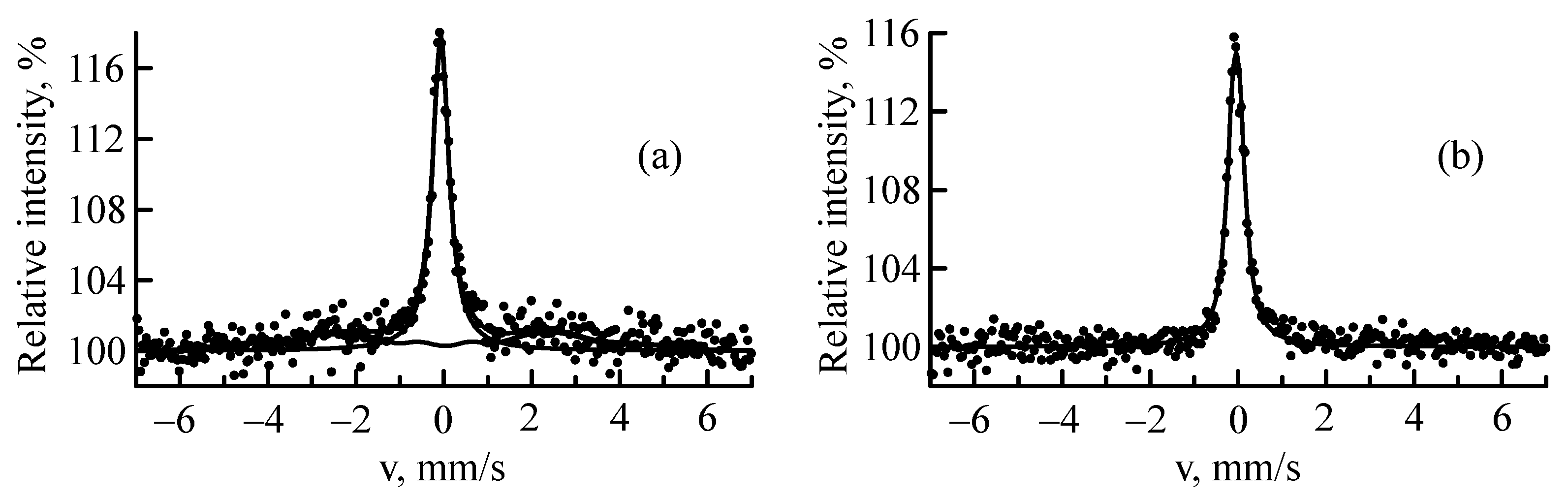
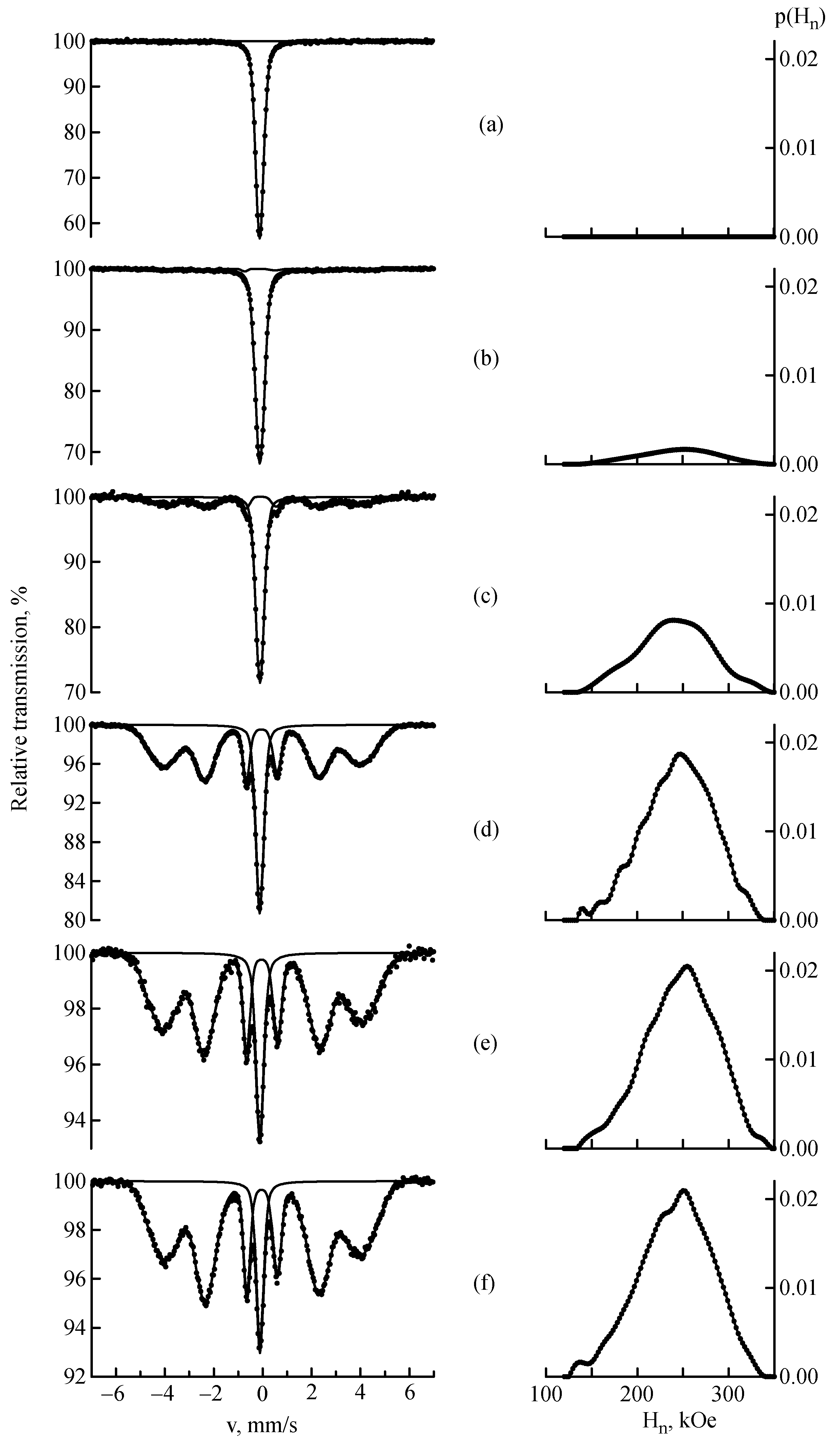
3.1. Mössbauer Spectroscopy

Figure 2 shows the MS spectra and reconstructed distributions of the hyperfine magnetic field p(Hn) at the 57Fe nuclei of the CF8 stainless steel samples before deformation (Figure 2a) and with a deformation degree ε = 20–95% (Figure 2b–f). The spectrum of the sample before deformation showed a single line with an isomer shift Is = −0.10 ± 0.03 mm/s, which characterizes the paramagnetic structure of γ-austenite. The MS spectrum of γ-austenite was characterized by one peak, but the substitution and interstitial atoms (mainly Cr and Ni) in the austenitic stainless steel caused a broadening of the spectrum; therefore, the MS spectrum was fitted with a doublet with a small quadrupole splitting Δ = 0.17 ± 0.04 mm/s, similar to [31,33]. The deformation with ε = 10% was not reflected in the MS spectrum. After rolling the sample with ε ≥ 20%, the MS spectra were superpositions of two partial subspectra. In addition to the paramagnetic doublet, a magnetic sextet was observed, which corresponded to α′-martensite. The dynamics of changes in the relative intensity of the MS spectrum of α′-martensite indicated the presence of a certain value of the deformation degree (in our case ε~10%), above which the process of α′-martensite formation can be followed by the MS method. The growth of the deformation degree caused an increase in the relative intensity of the sextet, reaching ~88% at ε = 95% (Figure 2f). The content of γ-austenite decreased accordingly. In this case, the isomer shift of the doublet did not change, which indicated the invariability of the electronic structure of austenite. No noticeable change was observed in the value of the average hyperfine magnetic field <Hn> at ε ≤ 80%; <Hn> was ~245 ± 3 kOe for such samples. In the MS spectra of more deformed samples, a slight decrease in <Hn> occurred (241 ± 1 kOe). When the fraction of γ-austenite/α′-martensite was calculated from the areas of the Mössbauer subspectra, the possible differences between the Mössbauer–Lamb factors were not taken into account. It is known that the ratio of iron-containing phases in the same material also depends on the thickness of the absorber due to different saturation effects [34]. It was difficult to take into account the saturation effect in this experiment, since plastic deformation is accompanied by the processes that lead to broadening of the resonance lines. These are γ → α′ transformations, the segregation of atoms (Cr, Ni) in the near-surface layer of the sample, and the ordering–disordering process.
According to Wanders et al. [20,29,34], the theoretical hyperfine field of the magnetic phase in steel can be calculated using the following equation:
<Hn> = 330.1 − 296.1·c (kOe)
where c is the concentration of chromium (+ nickel).
In our case, Equation (1) yields the value <Hn> = 247.1 kOe, which agreed quite well with the experimental data.
The change in the average magnetic field <Hn> in the spectra of deformed samples was revealed as a result of CEMS studies. The structural distortion of the nearest environment in the first and second coordination spheres of 57Fe atoms was studied using the reconstructed distribution of hyperfine magnetic field p(Hn). Figure 3 shows the CEMS spectra and the distribution of hyperfine field p(Hn) of the stainless steel samples with deformation degrees of 20, 60, 67, and 92%. The ferromagnetic phase was ~21% at ε = 20% and ~81% at ε = 60%. At ε = 20 and 60%, the content of α′-martensite in the MS spectra was ~6 and 76%, accordingly. At ε = 67 and 92%, the content of α′-martensite in the CEMS and MS spectra was approximately equal. From the comparative analysis of the MS and CEMS spectra, it was evident that the formation of α′-martensite during plastic deformation occurred most intensively in the near-surface layers of the steel, distributing in depth with the growth of the deformation degree. This behavior did not agree with all the predictive models. However, the authors of [14] also showed that the highest content of α′-martensite occurred at the surface and decreased with depth.
The average field <Hn> decreased from 245 ± 2 to 234 ± 1 kOe when the deformation degree increased. This was associated with the fact that plastic deformation caused an increase in the number of defects, such as dislocations, vacancies, and micro-cracks. These defects disrupted the orientation of the magnetic moments of the atoms. In addition, the efficiency of the interaction of magnetic domains decreased due to changes in their shape and size caused by deformation. It should also be noted that plastic deformation affected the magnetic anisotropy and the distribution of magnetic fields inside the material, which contributed to a decrease in magnetic susceptibility. When restoring the distribution of the hyperfine magnetic field p(Hn) of the CEMS spectrum of the sample with ε = 92%, the contribution from Hn~170–190 kOe increased (Figure 3c), which was probably caused by the presence of more than three chromium atoms in the immediate vicinity of an iron atom [18,19,28,29]. This indicated an increase in the concentration of impurity atoms in the α′-martensite.

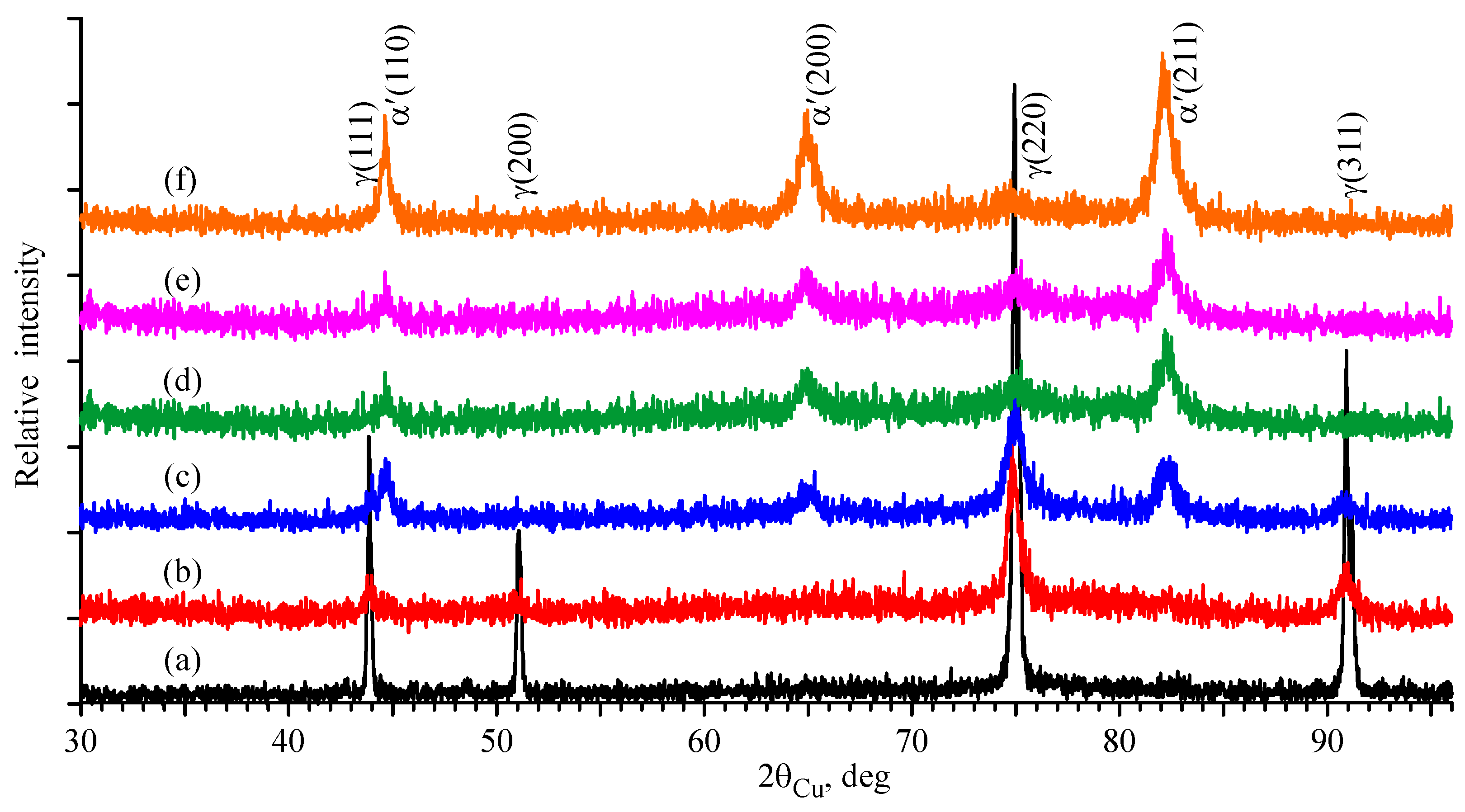
3.2. X-Ray Diffraction

Changes in the crystal structure of the studied steel before and after deformation were identified using XRD (Figure 4). The X-ray diffraction pattern of the sample before deformation was represented by a single phase with an fcc lattice corresponding to γ-austenite. With the increase in the deformation degree, the amount of this phase decreased as a result of the appearance and growth of the bcc phase—α′-martensite. The ε-martensite formation is possible at the beginning of the deformation or for small deformation rates [35,36]; however, the XRD results did not confirm the presence of this phase. It should be noted that the ε-martensite consists of overlapping stacking faults and a heavily faulted crystal structure. Therefore, the XRD peaks of ε-martensite are wide, and their intensities are low [36,37]. The diffraction peaks of ε-martensite cannot be found so easily for this reason. A comparison of the diffraction patterns of the deformed samples showed a broadening of the γ-austenite peaks after deformation. This could be related to a decrease in the grain size and the presence of internal stress [35]. The intensity ratios of the γ-austenite peaks remained virtually unchanged, indicating the constancy of the crystallographic structure. At ε = 10–20%, the appearance of α′-martensite in the X-ray diffraction patterns was weakly noticeable. The sample subjected to deformation ε = 40% showed the presence of α′-martensite in the reflections (110)α, (200)α, and (211)α. At high deformation degrees, this phase was particularly clearly visible; γ-austenite showed only traces in the reflection (220)γ.
Using the reflection (211)α, the volume fraction of α′-martensite was calculated using the following equation [35,38]:
f α = 1 0.65 ( I 311 γ + I 220 γ ) I 211 α + 0.65 ( I 311 γ + I 220 γ )
The following actions were used to determine the peak areas: the model background was subtracted from the data and the peaks were integrated. This approach reduced the errors during area determination. According to Equation (2), the volume fraction of the α′-martensite in the studied steel at different deformation degrees was estimated from 2 to 93%.

3.3. Olson–Cohen Model

Figure 5 shows the dependence of the α′-martensite content in CF8 stainless steel on the deformation degree according to the results of a quantitative analysis carried out by the MS and XRD methods. The integral intensities of the subspectrum of the ferromagnetic part of S were used to determine the amount of α′-martensite by the MS method. A quantitative phase analysis by XRD was carried out using the intensities of the main diffraction peaks of the α′- and γ-phases.
Olson et al. [39] stated that the intersections of shear bands in the austenitic phases were effective areas for the formation of martensite nuclei caused by deformation. Shear bands can be in the form of ε-martensite, mechanical twins, or dense bundles of stacking faults [39]. As the strain level increases, α′-martensite grows, consuming the ε-martensite and austenite phases through the γ → ε → α′ transformations. Based on the above, Olson and Cohen proposed a model to express the volume fraction of martensite as a function of plastic strain. This model has been successfully used in previous studies of steels [36,37,40,41,42,43]. The modification of this model and the development of alternative models are still relevant topics [40,41,42,43,44,45].
The Olson–Cohen model [39] is described by the following expression:
f α = 1 exp β [ 1 exp ( α ε ) ] n
where the exponent n is a fixed value, which is usually taken to be 4.5 for austenitic stainless steels [36,40]. The parameter α describes the rate of shear band formation depending on the deformation, and the parameter β is associated with the probability of martensite nucleus formation when crossing the shear band.
The present experimental data were approximated using Equation (3). As a result, the parameters characterizing the kinetics of martensitic transformation in CF8 stainless steel were determined. The selected coefficients α = 2.9 and β = 3.8 were in agreement with the data in [36,41,43]. For example, in [41], the value α was ~3 for AISI 304 steels, which have a similar composition to CF8. The curve obtained using Formula (3) had a sigmoidal shape (Figure 5). In addition, the calculation results obtained by the MS and XRD methods were in good agreement with the Olson–Cohen model. Figure 5 shows that, according to the XRD results, at a deformation degree starting from 10 to 70%, there was a rapid growth of α′-martensite nucleation, and then at ε = 70%, the process stabilized, indicating almost complete recrystallization of the steel structure. The curve obtained by the MS method shows that the process stabilized already at a deformation degree of 60%, which may be caused by a more sensitive measurement using the MS method compared to XRD. It is assumed that the formation of martensite (nucleation) (~2%) occurred at a deformation degree of more than 10%. The occurrence of the martensitic transformation at a low deformation degree can be related to the energetically favorable nucleation sites generated under the deformation action [40]. At a strain level of ~95%, maximum martensite contents of 88 and 93% were achieved according to the MS and XRD data, respectively.

4. Conclusions

The austenitic–martensitic transformation in the austenitic–ferritic duplex stainless steel CF8 subjected to cold plastic deformation at ε = 10–95% was studied using MS, CEMS, and XRD methods. It is assumed that the process of α′-martensite formation started at a deformation of ε > 10%, which can be related to the energetically favorable nucleation sites generated under the deformation effect. The CEMS results showed that the formation of α′-martensite occurred most intensively in the near-surface layers of the steel, distributing in depth with the growth of the deformation degree. The analysis of the MS spectra made it possible to obtain the distribution of hyperfine magnetic fields depending on the plastic deformation degree. The CEMS method revealed a change in the average hyperfine magnetic field <Hn>. As the deformation degree increased, <Hn> decreased from 245 ± 2 to 234 ± 1 kOe. The volume fraction of the α′-martensite was determined based on the results of calculations carried out via the MS and XRD methods, and a good correlation was observed. At the maximum deformation degree ε = 95%, the martensite content was 88% according to the MS results and 93% according to the XRD results. A modified Olson–Cohen model was proposed to determine the dependence of the α′-martensite amount on the deformation degree ε. It was found that the values α = 2.9 and β = 3.8 provide the best agreement between the experimental results and the Olson–Cohen model.

Author Contributions

Conceptualization and project administration, M.V.; methodology, formal analysis, and writing—original draft preparation, M.V. and I.M.; investigation, I.M. and Z.T.; writing—review and editing, M.V., I.M. and G.Y.; visualization, I.M. and G.Y.; resources, M.V. and Z.T. All authors have read and agreed to the published version of the manuscript.

Funding

This research was funded by the Committee of Science of the Ministry of Science and Higher Education of the Republic of Kazakhstan (Grant No. АР19679693).

Data Availability Statement

The data presented in this study are available on request from the corresponding author. The data are not publicly available due to privacy.

Conflicts of Interest

The authors declare no conflicts of interest.

References

  1. Meric de Bellefon, G.; van Duysen, J.C. Tailoring plasticity of austenitic stainless steels for nuclear applications: Review of mechanisms controlling plasticity of austenitic steels below 400 °C. J. Nucl. Mater. 2016, 475, 168–191. [Google Scholar] [CrossRef]
  2. Li, S.; Wang, Y.; Wang, H.; Xin, C.; Wang, X. Effects of long-term thermal aging on the stress corrosion cracking behavior of cast austenitic stainless steels in simulated PWR primary water. J. Nucl. Mater. 2016, 469, 262–268. [Google Scholar] [CrossRef]
  3. Collins, D.A.; Barkley, E.L.; Lach, T.G.; Thak, S.B. Effects of thermal aging on the fracture toughness of cast stainless steel CF8. Int. J. Press. Vessels Pip. 2019, 173, 45–54. [Google Scholar] [CrossRef]
  4. Saillet, S.; Le Delliou, P. Prediction of J-R curves and thermoelectric power evolution of cast austenitic stainless steels after very long-term aging (200,000 h) at temperatures below 350 °C. J. Nucl. Mater. 2020, 540, 152328. [Google Scholar] [CrossRef]
  5. Lu, W.-F.; Huang, J.-Y.; Yung, T.-Y.; Chen, T.-C.; Tsai, K.-C. Effects of thermal aging on the stress corrosion cracking behavior of cast stainless steel with different δ-ferrite levels in high temperature water environment. J. Nucl. Mater. 2022, 568, 153900. [Google Scholar] [CrossRef]
  6. Chen, Y.; Alexandreanu, B.; Natesan, K.; Rao, A.S. Cracking of irradiated cast stainless steels in low corrosion potential environments. In Proceedings of the The 16th International Conference on Environmental Degradation of Materials in Nuclear Power Systems-Water Reactors, Asheville, NC, USA, 11–15 August 2013; NACE International: Houston, TX, USA, 2013. [Google Scholar]
  7. Lo, K.H.; Shek, C.H.; Lai, J.K.L. Recent developments in stainless steels. Mater. Sci. Eng. R Rep. 2009, 65, 39–104. [Google Scholar] [CrossRef]
  8. Li, M.; Miller, M.K.; Chen, W.-Y. Phase stability in thermally-aged CASS CF8 under heavy ion irradiation. J. Nucl. Mater. 2015, 462, 214–220. [Google Scholar] [CrossRef]
  9. Kwon, J.-D.; Park, J.-C.; Lee, Y.-S.; Lee, W.-H.; Park, Y.-W. An investigation of the degradation characteristics for casting stainless steel, CF8M, under high temperatures. Nucl. Eng. Des. 2000, 198, 227–240. [Google Scholar] [CrossRef]
  10. Zheng, C.; Liu, C.; Ren, M.; Jiang, H.; Li, L. Microstructure and mechanical behavior of an AISI 304 austenitic stainless steel prepared by cold- or cryogenic-rolling and annealing. Mater. Sci. Eng. A 2018, 724, 260–268. [Google Scholar] [CrossRef]
  11. Lia, J.; Wang, Y.; Chen, X.; Yan, X.; Xiang, L. Microstructure evolution in stress relaxation behavior of austenite AISI 304 stainless steel spring. Mater. Charact. 2019, 148, 266–271. [Google Scholar] [CrossRef]
  12. Eid, E.A.; Sadawy, M.M. Role of effective strain during cold rolling deformation on mechanical characteristics of AISI 304 steel. Met. Mater. Int. 2021, 27, 4536–4549. [Google Scholar] [CrossRef]
  13. Mijovilovich, A.; Goncalves Viera, A.; Paniago, R.; Pfannes, H.-D.; Mendonca Gonzales, B. Mössbauer study of the retained austenitic phase in multiple steels. Mater. Sci. Eng. A 2000, 283, 65–69. [Google Scholar] [CrossRef]
  14. Oliveira, D.A.; Brito, P.P.; Magalhaes, F.C.; Azzi, P.C.; Ardisson, J.D.; Abrao, A.M. Influence of low plasticity burnishing on the formation of strain induced martensite in the surface layer. J. Mater. Res. Technol. 2023, 27, 4573–4594. [Google Scholar] [CrossRef]
  15. Cabanillas, E.D.; Terminiello, L.; Cantalejos, N.A.; Versaci, R.; Mercader, R.C. Mössbauer studies of strain-induced transformation of retained austenite in dual-phase steels. Mater. Sci. Eng. A 1992, 150, 113–116. [Google Scholar] [CrossRef]
  16. Pechousek, J.; Kouril, L.; Novak, P.; Kaslik, J.; Navarik, J. Austenitemeter—Mössbauer spectrometer for rapid determination of residual austenite in steels. Measurement 2019, 131, 671–676. [Google Scholar] [CrossRef]
  17. Goldanskii, V.; Herber, R. Chemical Applications of Mössbauer Spectroscopy; Academic: New York, NY, USA, 1968; 701p. [Google Scholar]
  18. Kozlov, K.; Shabashov, V.; Kozlov, A.; Sagaradze, V.; Semyonkin, V.; Panchenko, V.; Zamatovskii, A.; Kataeva, N.; Nikitina, A. Mössbauer spectroscopy investigation of the effect of a high-dose neutron irradiation on the atomic redistribution in the industrial steel EP823. J. Nucl. Mater. 2022, 558, 153384. [Google Scholar] [CrossRef]
  19. Dubiel, S.M.; Cies´lak, J.; Zukrowski, J. Distribution of Cr atoms in the surface zone of Fe-rich Fe–Cr alloys quenched into various media: Mössbauer spectroscopic study. Appl. Surf. Sci. 2015, 359, 526–532. [Google Scholar] [CrossRef]
  20. Waanders, F.; Vorster, S.; Engelbrecht, A. Mössbauer and SEM characterisation of the scale on type 304 stainless steel. Scr. Mater. 2000, 42, 997–1000. [Google Scholar] [CrossRef]
  21. Andrianov, V.A.; Bedelbekova, K.A.; Ozernoy, A.N.; Vereshchak, M.F.; Manakova, I.A. Mössbauer studies of 57Fe implantation in metal Ta and Mo. Nucl. Instrum. Methods Phys. Res. Sect. B 2020, 475, 71–76. [Google Scholar] [CrossRef]
  22. Vereshchak, M.; Manakova, I.; Shokanov, A. Mössbauer and X-ray Studies of Radiation-Induced Processes in Nb–Zr Alloys Implanted with 57Fe Ions. Materials 2023, 16, 3813. [Google Scholar] [CrossRef]
  23. Kislitsin, S.B.; Vereschak, M.F.; Manakova, I.A.; Ozernoi, A.N.; Satpaev, D.A.; Tuleushev, Y.Z. Blistering and α ⇆ γ phase transitions at annealing of stainless steel C12Cr18Ni10Ti irradiated by low energy alpha-particles. Probl. At. Sci. Tech. 2013, 2, 17–22. [Google Scholar]
  24. Deryagin, A.I.; Zavalishin, V.A.; Sagaradze, V.V. Atomic redistribution of alloying elements in nanocrystalline austenitic chromium-nickel steels obtained by strong plastic deformation. Nanostructured Mater. 1998, 10, 411–418. [Google Scholar] [CrossRef]
  25. Shabashov, V.; Sagaradze, V.; Zamatovskii, A.; Kozlov, K.; Kataeva, N.; Danilov, S. Regulation of the concentration heterogeneity and thermal expansion coefficient in the metastable invar FeNi31.1 alloy. Materials 2022, 15, 8627. [Google Scholar] [CrossRef]
  26. Shabashov, V.; Sagaradze, V.; Kozlov, K.; Ustyugov, Y. Atomic order and submicrostructure in iron alloys at megaplastic deformation. Metals 2018, 8, 995. [Google Scholar] [CrossRef]
  27. Shabashov, V.; Kozlov, K.; Ustyugov, Y.; Zamatovskii, A.; Tolmachev, T.; Novikov, E. Mössbauer analysis of deformation–induced acceleration of short-range concentration separation in Fe-Cr alloys—Effect of the substitution impurity: Sb and Au. Metals 2020, 10, 725. [Google Scholar] [CrossRef]
  28. Kozlov, K.; Shabashov, V.; Zamatovskii, A.; Novikov, E.; Ustyugov, Y. Inversion of the sign of the short-range order as a function of the composition of Fe–Cr alloys at warm severe plastic deformation and electron irradiation. Metals 2020, 10, 659. [Google Scholar] [CrossRef]
  29. Eymerya, J.-P.; Merakebb, N.; Goudeaua, P.; Fnidikic, A.; Bouzabatab, B. A Mossbauer comparative study of the local environment in metastable 304 stainless steel films depending on the preparation mode. J. Magn. Magn. Mater. 2003, 256, 227–236. [Google Scholar] [CrossRef]
  30. Principi, G. The Mössbauer effect: A romantic scientific page. Metals 2020, 10, 992. [Google Scholar] [CrossRef]
  31. Linderhof, F.; Mashlan, M.; Doláková, H.; Ingr, T.; Ivanova, T. Surface micromorphology and structure of stainless and maraging steel obtained via selective laser melting: A Mössbauer spectroscopy study. Metals 2021, 11, 1028. [Google Scholar] [CrossRef]
  32. Matsnev, M.E.; Rusakov, V.S. SpectrRelax: An application for Mössbauer spectra modeling and fitting. AIP Conf. Proc. 2012, 1489, 178–185. [Google Scholar]
  33. Cook, D.C. Strain induced martensite formation in stainless steel. Met. Mater. Trans. A 1987, 18, 201–210. [Google Scholar] [CrossRef]
  34. Gavrilyuk, V.G.; Nadutov, V.M.; Gladun, O.V. Distribution of nitrogen in Fe-N austenite. Phys. Met. Metallogr. 1990, 69, 129–135. [Google Scholar]
  35. Etienne, A.; Radiguet, B.; Genevois, C.; Le Breton, J.-M.; Valiev, R.Z.; Pareige, P. Thermal stability of ultrafine-grained austenitic stainless steels. Mater. Sci. Eng. A 2010, 527, 5805–5810. [Google Scholar] [CrossRef]
  36. Hedayati, A.; Najafizadeh, A.; Kermanpur, A.; Forouzan, F. The effect of cold rolling regime on microstructure and mechanical properties of AISI 304L stainless steel. J. Mater. Process. Technol. 2010, 210, 1017–1022. [Google Scholar] [CrossRef]
  37. Talonen, J.; Hänninen, H.; Nenonen, P.; Pape, G. Effect of strain rate on the strain-induced γ– α′martensite transformation and mechanical properties of austenitic stainless steels. Metall. Mater. Trans. A 2005, 36, 421–432. [Google Scholar] [CrossRef]
  38. Fultz, B.; Howe, J.M. Transmission Electron Microscopy and Diffractometry of Materials, 2nd ed.; Springer: Berlin/Heidelberg, Germany, 2002; 748p. [Google Scholar]
  39. Olson, G.B.; Cohen, M. Kinetics of strain-induced martensitic nucleation. Metall. Trans. A 1975, 6, 791–795. [Google Scholar] [CrossRef]
  40. Soleimani, M.; Kalhor, A.; Mirzadeh, H. Transformation-induced plasticity (TRIP) in advanced steels: A review. Mater. Sci. Eng. A 2020, 795, 140023. [Google Scholar] [CrossRef]
  41. Naghizadeh, M.; Mirzadeh, H. Modeling the kinetics of deformation-induced martensitic transformation in AISI 316 metastable austenitic stainless steel. Vacuum 2018, 157, 243–248. [Google Scholar] [CrossRef]
  42. Zheng, C.; Jiang, H.; Hao, X.; Ye, J.; Li, L.; Li, D. Tailoring mechanical behavior of a fine-grained metastable austenitic stainless steel by pre-straining. Mater. Sci. Eng. A 2019, 746, 332–340. [Google Scholar] [CrossRef]
  43. Naghizadeh, M.; Mirzadeh, H. Processing of fine grained AISI 304L austenitic stainless steel by cold rolling and high-temperature short-term annealing. Mater. Res. Express 2018, 5, 056529. [Google Scholar] [CrossRef]
  44. Mirzadeh, H.; Najafizadeh, A. ANN modeling of strain-induced martensite and its applications in metastable austenitic stainless steels. J. Alloys Compd. 2009, 476, 352–355. [Google Scholar] [CrossRef]
  45. Ahmedabadi, P.M.; Kain, V.; Agrawal, A. Modelling kinetics of strain-induced martensite transformation during plastic deformation of austenitic stainless steel. Mater. Des. 2016, 109, 466–475. [Google Scholar] [CrossRef]
Figure 1. CEMS spectra at the 57Fe nuclei in the unaged (a) and aged (b) CF8 steel samples of 600 µm thickness.
Metals 14 01449 g001
Figure 2. MS spectra and distributions of the hyperfine magnetic field p(Hn) at the 57Fe nuclei in the CF8 stainless steel samples before deformation (a) and with deformation degrees of 20 (b), 40 (c), 60 (d), 80 (e), and 95% (f).
Metals 14 01449 g002
Figure 3. CEMS spectra and distributions of the hyperfine magnetic field p(Hn) at the 57Fe nuclei in the CF8 stainless steel samples with deformation degrees of 20 (a), 60 (b), 67 (c), and 92% (d).
Metals 14 01449 g003
Figure 4. X-ray diffraction patterns of the CF8 stainless steel samples before deformation (a) and with deformation degrees of 20 (b), 40 (c), 60 (d), 80 (e), and 95% (f).
Metals 14 01449 g004
Figure 5. The content of the α’-martensite in CF8 stainless steel as a function of the deformation degree.
Metals 14 01449 g005
Table 1. Samples of the CF8 austenitic stainless steel for MS.
Degree of Deformation, %Thickness, µm
050
1045
2040
4030
5050
6040
7030
8020
9250
9530
Table 2. Chemical composition of the CF8 austenitic stainless steel, wt. %.
ElementsASTM A743Measured ValueAbsolute Error
C<0.080.070.01
Si<21.60.1
Mn<1.51.00.1
P<0.04--
S<0.04--
Cr18.0–21.019.40.6
Ni8.0–11.08.60.4
Febalance69.32.1
Disclaimer/Publisher’s Note: The statements, opinions and data contained in all publications are solely those of the individual author(s) and contributor(s) and not of MDPI and/or the editor(s). MDPI and/or the editor(s) disclaim responsibility for any injury to people or property resulting from any ideas, methods, instructions or products referred to in the content.

