Abstract
The main disadvantage of the implants is the associated infections. Therefore, in the long term, the possibility of improving the antibacterial capacity of different types of implants (dental, orthopedic) is being researched. The severity of the problem lies in the increasing bacterial resistance and finding appropriate alternative treatments for infectious diseases, which is an important research field nowadays. The purpose of this review is to draw a parallel between different studies analyzing the antibacterial activity and mechanism of silver nanoparticles (NP Ag) deposited on the titanium nanotubes (NTT), as well as the analysis of the NP Ag toxicity. This review also provides an overview of the synthesis and characterization of TiO2-derived nanotubes (NT). Thus, the analysis aims to present the existing knowledge to better understand the NP Ag implants benefits and their antibacterial activity.
1. Introduction
Titanium, together with its alloys, is one of the most used elements in orthopedic and dental applications due to its biocompatibility, mechanical properties, and corrosion resistance [1]. It is known that titanium-based alloys are used to manufacture medical appliances, such as artificial blood vessels and orthopedic/dental implants [2]. The causes of implant rejection may still be unknown and impossible to predict preoperatively. Usually, it is not due to the body’s intolerance to titanium but rather because of a local cause. One of the leading causes of implant rejection is an immune system imbalance and a combination of bacterial infection with adverse reactions of the immune system [3,4]. Some of the situations that can lead to infections are non-compliance after surgery, prolonged operation time and post-surgical tissue contamination. Therefore, because of the increasing bacterial resistance to antimicrobial agents, infections are often difficult to treat even with specific antibiotics, thus increasing the risk of mortality and morbidity [5]. However, several types of microorganisms (bacteria, viruses, parasites, and fungi) can cause bone infections [6].
Osteomyelitis is a bone infection induced by microorganisms such as Staphylococcus spp., which leads to progressive bone loss and tissue damage [7]. Because Staphylococcus spp., are ubiquity bacteria, they can contaminate medical devices. Out of all the cases of pyogenic osteomyelitis, 80–90% are caused by Staphylococcus aureus. Although it is part of human skin flora, Staphylococcus epidermidis can colonize medical devices, including orthopedic implants and catheters [6]. One method of preventing these infections is to add antimicrobial agents to the surface of titanium implants [8]. During infection, respectively, colonization can create biofilms, where bacteria are protected from the immune system and antibacterial agents. [9]. While forming these biofilms, bacteria are rigidly attached to the medical devices’ active or inert surface. Bacterial colonies can reproduce, differentiate, and secretion of extracellular polymeric substances such as proteins, polysaccharides, and lipids [10]. Thus, when biofilms are formed on medical devices (catheters or implants), the patients can develop complications such as chronic, difficult to treat infections [11]. However, biofilms on human surfaces are not always harmful, such as dental plaque composition that can determine the presence or absence of disease [12].
A promising approach to restrict the microbial adhesion and the colonization of medical devices is coating implant surfaces with thin antimicrobial bioactive films [2]. Previous studies demonstrated that Ag ions could infiltrate the microbial cell wall and bind to cellular DNA, interrupting the bacterial replication, exhibiting antimicrobial and antifungal properties [1,2,8].
In this review, the perspectives related to the characterization and synthesis of nanotubes (NT) derived from TiO2 are followed. Moreover, the activity and antibacterial mechanism of silver nanoparticles (NP Ag) incorporated in titanium nanotubes (NTT) are presented and the toxicological aspects of NP Ag.
2. Methodology
This review’s content is based on data obtained by PubMed, Science Direct, MDPI, Springer, and other databases. The most relevant studies regarding NP Ag and NTT were selected. Search terms were: “silver nanoparticles” (title) and “silver” (topic), “titanium dioxide” (title) and “nanotubes” (topic), in different combinations (topic) with “biological”, “implants”, “dentistry”, “orthopedics”, and “antibacterial” (MeSH browser keywords). Depending on the references obtained, other associations of keywords were used.
3. Characterization of NT Derived from TiO2
To use nanostructured materials for medical purposes, they must meet specific requirements. The first requirement is biocompatibility; this is the capacity of the material to induce an appropriate host response, in the case of a particular application [13], accepted definition by International Standards Organization (ISO), Food and Drug Administration (FDA) and American Society for Testing and Materials (ASTM). Titanium (Ti) surfaces modified with TiO2 nanotubes (TNT) are material with excellent biocompatibility and integrate well into bone tissue. Ti surfaces have chemical/thermal stability, controllable size, generous contact surface, and pores with adjustable sizes, surface chemistry, and good surface/volume ratio, due to their long and well-aligned structure. These features support the differentiation and proliferation of bone cells [14]. The bioactivity (the ability to interact with body tissue) is another mandatory requirement that nanostructures must meet [13].
Antibacterial studies focused on the manufacture of TiO2 nanotubes, acting as carriers for Ag, which has a proven and well-known antibacterial effect. Various methods can be used to attach Ag particles to the surface of TiO2 nanotubes: Ultrasonication [15], soaking in AgNO3 solution [16], electrodeposition [17], or sputter deposition techniques [18].
There are three polymorphic forms of TiO2 known in nature: Anatase (tetragonal), brookite (orthorhombic), and rutile (tetragonal) [12]. Anatase has a pyramidal crystalline structure (used as a photocatalyst under UV irradiation), rutile has a tetragonal structure with a prismatic habit, and brookite has an orthorhombic crystalline structure. The most common forms used to manufacture Ti nanotubes are anatase and rutile because brookite is less stable [19]. TiO2 is also a monoclinic mineral and is relatively new to the TiO2 family [20]. Of all, rutile is the most abundant in nature. It constitutes almost two-thirds of the available and known pigments. It is also considered to play a crucial role as a catalyst, mainly due to its high refractive index [21].
4. Synthesis of NT Derived from TiO2
The physicochemical properties (size, shape, morphology, and composition of titanium oxide) are essential for photocatalytic control and performance. A synthetic procedure is necessary for the development of desired properties for photocatalytic activity [22].
There are numerous methods to synthesize NT derived from TiO2, the most important of which are: (a) deposition (by physical or chemical processes) in templates of nanoporous materials, (b) directed growth, starting from the germination centers of nanostructures, (c) electrochemical anodizing, which uses self-organizing processes at the oxide-electrolyte interface, (d) hydrothermal methods, and (e) sol-gel method. Of all these methods, the most used were: electrochemical anodizing, the sol-gel method, the hydrothermal method, and the solvothermal method [23]. Electrochemical anodizing is a known process of growing oxide layers on metal substrates. Nanotubes are structures of great interest due to the combination of morphology and functionality. Additionally, the diameter, wall thickness, length, and uniform density of NTT can be controlled by different parameters: electrolyte composition, applied voltage, anodizing time, temperature, and pH of the electrolyte [24]. Ti in contact with air can form on its surface a layer of titanium oxide (TiO2—thickness about 10 nm) having excellent biocompatibility. Furthermore, the formation of the biofilm in the human body is due to the implantation of Ti. Thus, the human tissues directly contact the TiO2 layer on the implant surfaces [2].
4.1. Sol-gel Method
One of the most widely used NT synthesis methods derived from TiO2 is the sol-gel technique, as it is an efficient and versatile method that can be used to obtain both films and powders [25]. The sol-gel technique is a chemical method of preparing materials. A colloidal suspension of solid particles in a liquid (sol) is transformed into a gel by hydrolysis, condensation, and polymerization. The hydrolysis reaction is usually acidic (nitric or sulfuric acid) or basic (sodium hydroxide) catalyzed in a sequential condensation reaction. Thus, from a chemical point of view, the sol-gel method is mainly based on hydrolysis reactions and condensation/polymerization reactions of the precursors, both for the inorganic pathway and the metal-organic path [24,26]. Thus, this method takes place by dissolving a precursor of metal oxide in a low molecular weight solvent medium using a catalyst (acid, base, or ion) followed by a hydrolysis (water) and polycondensation step [27].
Thus, during the sol-gel reaction, dense three-dimensional crosslinked structures are formed, which turn into gels. These, through suitable heat treatment, lead to amorphous or crystalline nanostructures (films, powders). Therefore, the obtained TiO2 is generally found only in the amorphous phase. The advantages of the sol-gel process are numerous and among them are: Obtaining nanomaterials with high purity, relatively low processing temperature, the possibility of stoichiometry control, obtaining materials with different shapes and/or with predetermined structure. Disadvantages of the sol-gel process are long synthesis time and high calcination temperature [28].
4.2. Hydrothermal/Solvothermal Method
The hydrothermal/solvothermal method is frequently used in the powder synthesis of nanostructures. TiO2 synthesis is usually performed in small Morey type autoclaves, fitted with Teflon gaskets. The conditions selected for the synthesis of TiO2 particles are: T ≤ 200 °C and pressure (p) < 100 bar [29,30].
The hydrothermal method refers to a process of preparing materials in a closed pressure vessel. The precursors can be dissolved (in water), reacted and crystallized, this being done similarly to the traditional aqueous solution method [30]. Ultrafine TiO2 powders can also be prepared by hydrothermal oxidation of H2O2 starting from metallic Ti. Thus, the hydrothermal treatment of the gel can be performed under different conditions: Oxidation, heating, and hydrothermal treatment [30].
In comparison to the hydrothermal method, a non-aqueous solvent is used for the solvothermal. Besides, this method showed improved control over the properties of TiO2. A distinctive feature of the solvothermal synthesis is the possibility of the reaction taking place at a temperature higher than the used solvent’s boiling point in the synthesis. The hydrothermal method’s essential characteristics are ionic viscosity, density, thermal capacity/conductivity, and dielectric constant of the product. This method primarily depends on pressure and temperature. The specific properties of the solvent can be obtained by adjusting the synthesis parameters [31].
4.3. Electrochemical Anodizing
The anodizing method is widely used due to its controllable and reproducible results, easy management of the anodizing parameters (applied voltage, electrolyte, electrodes, annealing time, and temperature) and also, it is considered as a simple process [32]. TiO2 nanotubes may undergo specific changes by changing the anodizing process conditions, such as pH, temperature, applied potential, current density, anodizing time, acid concentration, and different morphological parameters (height, surface, and diameter) [33]. Usually, the anodizing of titanium is done by introducing it in an aqueous or organic electrolytic medium (in the component of which fluorine ions are found), applying a constant voltage [34]. Therefore, due to the incorporation of fluoride into the anodizing electrolyte, the increased surface roughness and the ability to stimulate mechanically, NTT cells are compatible with bone cell function, even in the absence of drugs [35]. In a typical process, the conductive part (being anodized) is positioned in an electrolytic bath and serves as an anode [36]. In general, the diameter of the nanotube is adjusted according to the applied voltage. The length of the tube is controlled according to the anodizing time [37]. Regarding the clinical success rate, anodized surface of implants, in compromised bone cases, has shown about a 95% success rate [38].
4.4. Electrospinning
Electrospinning is defined by simplicity, flexibility, and adaptability. Electrospinning can manufacture various nanofibers or microfibers using a wide range of materials such as (ceramics, polymers, and semiconductors) and their nanocomposites [39,40]. Electrospinning consists of four main components: (1) a capillary tube containing a polymer solution (or melt), (2) a tab or nozzle, (3) a collector, and (4) a high voltage source. By this method, fiber production uses electric force to pull yarns loaded with polymer solutions or molten polymers up to fiber diameters on the order of about 100 nm. Many of the research studied solution-based electrospinning [41]. Thus, during the fiber manufacturing process (diameter between sub-micrometer and nanometer), the polymer solution or melt uses a high voltage power supply to accelerate the injection on a collection plate with opposite polarity [42]. Because the incorporation of nanoparticles into the polymer changes the solution’s characteristics (solvent volatility, surface tension, and polymer viscosity), they present difficulties in handling existing needle electrospinning systems in nanofibers’ production. In needle electrospinning, a sphere-shaped drop with the semi-vertical angle (α) is formed on the nozzle’s tip, helping to create a Taylor cone with a minimum tension [42,43].
In conclusion, the solution’s main parameters that strongly influence the fibers are the polymer’s molecular weight, polymer chain entanglements, solution concentration, viscosity, conductivity, surface tension, and dielectric effect solvent. All these features can be modified or adjusted [44].
5. The Obtaining of NP Ag Deposited into NTT
The NP Ag can be easily incorporated into TiO2 nanotubes using simple procedures. These procedures involved AgNO3 immersion and UV irradiation [45]. In addition to NP silver and NP copper, zinc, fluorine can be incorporated into TiO2 nanotubes for biomedical applications. For example, zinc can be easily incorporated hydrothermally into nanotubes [46]. Moreover, in situ, other anodizing possibilities were made for gold (TiAu) [47] and platinum (TiPt) alloys [48].
6. Antibacterial Activity of NP Ag Incorporated into NTT
6.1. The Antibacterial Mechanism of NP Ag Deposited into NTT
The antibacterial mechanism (Figure 1) of silver that leads to bacterial death is still not completely understood. One essential parameter of the antibacterial activity of NP Ag is the surface area of the nanomaterial. The antibacterial activity does not rely only on the release of silver ions from NP Ag. Depending on the bacterial species, NP Ag penetrates bacteria more or less than silver ions [49]. Two antibacterial mechanisms of NP Ag-deposited on the NTT are described: Contact mode and non-contact mode. NP Ag are attached to the bacteria in contact mode and cause several lethal reactions [50]. The non-contact way is by preventing microorganisms’ adhesion to the surface using non-sticking agents [51]. Although the antibacterial mechanism is still fully described, various studies involving the antibacterial effect of the modified TiO2 on bacteria (Gram-positive/negative) have been performed [52]. It has been shown that by binding and infiltrating the bacterial cell wall, NP Ag cause physical changes and damage the cell wall, leading to leakage of cell contents and subsequent destruction of bacteria [53].
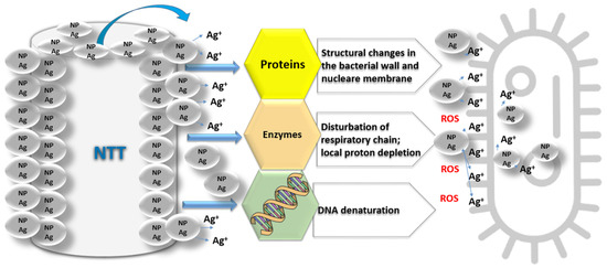
Figure 1.
The proposed mechanism of action of silver nanoparticles (NP Ag) released by titanium nanotubes (NTT).
By attaching NP Ag to NTT, it was shown that the antibacterial effect increased from 20% (for NT) to 100% (for Ag/NTT) [54]. Because of their capacity to generate reactive oxygen species (ROS) in contact with UV light, NP TiO2 are often used as photocatalytic agents exhibiting antibacterial activity. However, because of the UV toxicity towards human cells, the utility of NP TiO2 under UV light is restricted [55].
TiO2 NPs inactivate microorganisms by generating ROS in the presence of light. Newly formed hydroxyl radicals, superoxide anion, and hydrogen peroxide, interact with the sulfhydryl (-SH) groups of proteins and DNA’s nucleotide base pairs causing DNA destruction [56].
Silver ions can inhibit several enzymes, the passage of the succinate into the cell, disrupt respiratory chain (in E. coli), accelerate the generation of ROS, inhibit cell signaling, inhibit protein synthesis, and inhibit the production metabolites efflux [57,58,59]. TiO2 nanoparticles can create more ROS, and the energy created on the surface is higher than NTT’s. ROS can trigger the peroxidation of membrane lipids causing bacterial death [60]. They also generate intracellular oxidative stress, damage to the cell membrane, and irreversible DNA structure changes [61].
It is believed that silver ions react better with side groups negatively charged with bacterial membrane proteins such as phosphate, sulfate, and nitrate compounds, which results in cell structure changes and the formation of irregular pits in the cell wall. These reactions lead to the destruction of the cell membrane that promotes Ag ions’ entry into the cell, reacts with vital enzyme’s thiol groups, and by damaging the DNA, it stops bacteria’s ability to replicate. Furthermore, silver ions trigger free radicals producing ROS with high toxicity that cause damage to cellular structures (Figure 1) [62,63].
The photocatalytic process’s efficiency varies, depending on the used material, as well as the environmental factors in which the process takes place. The photocatalytic process’s optimization depends on obtaining materials with characteristic properties, such as crystal structure, particle size, specific surface area, catalyst concentration, and solution pH [19].
As mentioned, the increasing amount of silver enhances the photocatalytic activity is most likely due to increased silver concentration. It results in one of the following effects: Acts as a barrier that prevents the absorption of light by TiO2; prevents the organic substrate from coming into contact with the TiO2 surface; and silver can become an essential center for the recombination of electron holes [64].
6.2. The Antibacterial Activity of NP Ag-Deposited into NTT Used in the Field of Orthopedics
One of an essential issue in clinical cases is infection frequency of oral and orthopedic implants. The primary reason for secondary implant failure is the immune system imbalance and bacterial infection combined with the immune system’s adverse reactions [3]. Modern oral implants have demonstrated survival rates of 95–99% (over ten years) and have an infection rate of 0.2% [65,66].
On the other hand, revision hips and knees have more than five times the infection rate of primary hip arthroplasties (1–5% infection rate) and osseointegrated femoral fixtures placed in high amputation levels may have 20% infection rate over ten years [3]. These figures have led many researchers to look for solutions to lower the infection rate. One of the answers, discussed in this review, is the treatment with silver nanoparticles deposited on TiO2.
In general, most Ti alloys provide suitable mechanical properties for orthopedic and dental applications [67]. A critical aspect for implantable biomaterials is the osteogenesis and osseointegration. Osseointegration does not occur with orthopedic implants that display distance osteogenesis; infection is most rare in oral and orthopedic implants [3]. The effective prevention of implant-centered infections is essential [68]. During surgery, metal orthopedic implants can become a substrate for bacterial infections, which can cause severe microbial infections or implant failure [69]. Staphylococcus spp. and Escherichia coli are pathogens that are frequently involved in implant-associated infections [68]. Implant infections attributed to the adhesion of antibiotic-resistant bacteria to medical device surfaces have a low cure rate (even when treated with specific antibiotics) and often exclude the implant to eradicate the infection. Therefore, there is a growing interest for complementary methods to prevent or even cure medical-related infections avoiding antibiotic therapy. A change in the surface structure decreases the bacterial activity on the implant [70].
Most Ti alloys provide suitable mechanical properties for orthopedic applications. These Ti-based alloys have excellent corrosion resistance and good biocompatibility, in vivo and in vitro, because their surface is covered with a passive TiO2 layer [67]. An important advantage of NTT is the possibility of incorporating antibiotics and other materials with similar potential, in a structure that organic chemicals such as cytokines, special proteins, and growth factors can also attach. Another advantage of NTT is modifying properties such as surface roughness, energy, wettability, and tube diameter [71].
TiO2 nanotubes have a similarity with natural bones due to the structure and the open-ended tube’s elasticity. These NT are covered with thin layers of materials with excellent biocompatibility. Besides the anticorrosive effect, the thin layers have an essential infection control role through antimicrobial agents [8]. The antibacterial activity of NP Ag results from the interaction between positive silver ions (positively charged) and the bacterial cell wall (negatively charged). Thus, NP Ag increases the permeability of the cell wall and inhibits bacterial growth [54].
Wei et al. confirmed the existence of increased antibacterial activity of long-term Ag-based antibacterial agents for the open-ended tubular nanostructure of TiO2 nanotubes under the concomitant action of NP Ag and silver ions [72]. Because the toxicity of silver to microorganisms is exceptionally high, but relatively low to human tissues, being used in appropriate doses, it is non-cytotoxic, providing antimicrobial activity in implants for long-term applications [73,74,75]. Its biological effect as an antimicrobial agent is well known and proven against various types of bacteria (Gram-positive and Gram-negative), as well as drug-resistant bacterial strains (MRSA) [76]. Pathogenic bacteria from infected oral implant sites, including Streptococcus mutans [77], Streptococcus oralis [78], Staphylococcus aureus [79], and Staphylococcus epidermidid [79] were entirely or significantly reduced in contact with Ag-implanted devices.
Ruparelia et al., 2008 concluded that using medium-sized NP Ag (3 nm and 9 nm, respectively), a more effective antibacterial activity was observed for E. coli (Gram-negative bacteria) than for Bacillus subtilis (Gram-positive bacteria) [80]. Instead, Yoon et al., using large NP Ag (40 nm) showed better efficacy for Bacillus subtilis (Gram-positive bacteria) than for E. coli (Gram-negative bacteria). In conclusion, the antibacterial activity of NP Ag is influenced by their size [81]. Such differences between the studies’ results are frequently found in the literature, most often coming from the fact that different bacterial strains were tested, or other methods were used. The biological effects of NP Ag are influenced by their size, shape, and concentration, their use in orthopedic and dental implants being limited due to its cytotoxicity [75,82].
Li et al., 2016 [62] demonstrated that untreated Ti has no antibacterial activity. Compared to pure Ti, NTT inhibited many bacterial colonies’ growth, implying that NTT have valuable antibacterial properties [62]. Many researchers have shown that the Ag/NTT group significantly inhibited several bacterial strains, suggesting that Ag ions released from Ag/NTT exhibit a significant bactericidal effect [75,81,82].
Pham Van Viet demonstrated that oxygen from the air could react with the electrons from the sample surface forming superoxide and hydroxyl radicals. X-ray photoelectron spectroscopy proved that the sample contained Ti3+, Ag+, and Ag0 on the NTT surface. Hence, the presence of Ti3+ ion can lead to the generation of ROS [83].
Vu et al., 2017 demonstrated better photocatalytic performance for Ag/NTT than NTT due to the separation efficiency of electrons and induced photo voids, respectively, due to the resonance of surface plasmon for Ag. For an Ag content with a percentage of 5% by weight, the best photocatalytic performance was obtained [84].
Qiao et al., 2015 hypothesized that the silver plasma immersion ion-implantation technique (Ag-PIII) could decrease NP Ag mobility embedded in the sandblasted and acid-etched implants surface. The micro/nanostructure surface can have enhanced bioactive properties and may further improve, in vivo, the osseointegration of the implant. Therefore, they demonstrated that the distribution and NP Ag size played a crucial role in promoting osteoconductivity [85].
Several researchers have shown that when the NP Ag diameter exceeds 10 nm, the antibacterial effect is conditioned by the released Ag+ ions. If the NP Ag is smaller than 10 nm, the NP may interact more efficiently with the bacterial cell (Table 1) [8,75].

Table 1.
Antibacterial activity of NP Ag incorporated into NTT.
6.3. The Antibacterial Activity of NP Ag-Deposited into NTT Used in the Field of Dentistry
Streptococcus oralis, Streptococcus mitis, Streptococcus sanguis (Gram-positive bacteria), Eikenella corrodens, Prevotella loescheii, and Veillonella atypica (Gram-negative bacteria) are among the first bacterial colonizers that can adhere to tooth surfaces, as well as to implant surfaces. These early colonizers frequently form coaggregation bridges with Fusobacterium nucleatum, bridges that continue to coaggregates with many Gram-negative and anaerobic bacteria, including Porphyromonas gingivalis, Aggregatibacter actinomycetemcomitans, Prevotella intermedia, Treponema denticola, or Tannerella forsythia [97,98,99].
There is a risk for early infections for dental implants at the time of implant placement where there is a significant reduction of clinical problems if prophylactic antibiotics are prescribed [100]. After osseointegration, the oral implant has a known defense against bacteria [66]. For the long-term survival and the success of the dental implants and the bone-implant osteointegration, the prevention of after placement infections is also an important condition. However, the protection against infection is a part of the incorporation of implants [3]. Nanomaterials are current innovation with a significant impact on dentistry field [101]. To prevent the dental implants’ failure, coating the implant with NP Ag may benefit by preventing biofilms’ formation on the implant surfaces [99]. An essential advantage of the NP Ag (compared with the traditional antibiotic treatment) is the recognized antibacterial activity, without the bacterial resistance as a side effect. A combination of these two therapies may be beneficial. It was demonstrated that NP Ag enhance the bacterial activity of some traditional antibiotics (ceftazidime, cefotaxime, ciprofloxacin, and meropenem) [102]. Another benefit may come from the fact that NP Ag can restore the antibacterial activity of some inactive substances against multi-resistant bacterial strains. When NP Ag are synthesized with a proper capping agent, it can improve the antibacterial effect against the Gram-negative bacteria frequently involved in periodontal infections. The antibacterial activity of the NP Ag against anaerobic oral pathogens is increased at the smaller sized particles.
There are some side-effects which can consist of adverse tissue reactions and depend on the size and type of the NP Ag [88,103].
NP Ag can be incorporated in Ti coating in dental implants, into acrylic resins used for removable dentures, in material for endodontic obturation, in guided bone/tissue regeneration (GBR/GTR) membrane [104]. There are several coating procedures of the dental implants with steadily releasing the silver ion, which has an excellent long-term antibacterial effect [105]. Medical grade Ti alloys are frequently used for bone or dental implants. Still, the Ti alloy alone does not have an antimicrobial activity that could reduce infection risk after surgery. Gunputh studied the growth of NP Ag on TiO2 nanotubes on Ti-6Al-4V discs. He also used S. aureus that was cultured on the composite coatings. Using the Live/Dead assay, the nano-sized micron-sized clusters of the NP Ag proved to exhibit antibacterial activity [87].
By immobilizing NP Ag on implant surfaces, the antibacterial capacity is prolonged. The embedded NP Ag inhibit bacterial cells without consumption, through direct, continuous, contact with it. Even if NP Ag with smaller diameter usually present better antimicrobial activity, NP Ag with larger size embedded in Ti had an increased antibacterial effect, probably because of the higher consumption of protons [106].
Ti embedded with NP Ag can enhance bone formation, bone mineral density, and trabecular pattern, while the adjacent dental implants tissues are not affected. NP Ag can have biocidal activity against S. aureus and P. aeruginosa at a small concentration, with no significant cytotoxicity to the osteoblastic cells [107].
Implants are partly exposed to the oral cavity but are also in permanent contact with gingival tissues and bones. The rapid establishment of a seal of firm soft tissue that can firmly seal around the dental implant can prevent bacterial invasion. By modifying the surface of NTT, Ma et al. [17] created a surface with preserved antibacterial activity, but with lower cytotoxicity. Immobilizing the NP Ag/fibroblast growth factor 2 (FGF-2) compound’s bioactive factors on a NTT surface they obtained a sample with a great cytocompatibility, lower cytotoxicity, and improved hFGF (human fibroblast growth factor) functions such as cell proliferation, cell attachment, and extracellular-matrix-related gene expression [17].
Pokrowiecki [108] measured (over different periods) the wettability, the silver release profile, and the surface roughness, of the discs incorporated with NP Ag, by Tollens reaction. The surface roughness increased and decreased wettability in a dose-dependent manner [108]. The antibacterial activity for a concentration of 0.05 ppm of silver ions was present for Streptococcus mutans, Streptococcus mitis, Streptococcus sanguis, Streptococcus oralis, S. aureus, E. coli, and Porphyromonas gingivalis.
There are some side-effects which can consist of adverse tissue reactions and depend on the size and type of the NP Ag [103]. As a side effect, Pokrowiecki noticed a significant decrease in the viability of the cells after exposing human osteoblasts to 0.1 ppm of silver ions by using ToxiLight™ BioAssay Kit after 72 h. The cytotoxic and antibacterial activity can be observed at similar concentration [108]. Although the NP Ag present some cytotoxicity, this could be contained by controlling the silver ions release rate, so that both long-term antibacterial activity and bio integration can be achieved [16].
7. Toxicity of NP Ag
Implant infection prevention remains an essential goal in surgery. Using NP Ag, among other prophylactic measures, could be an alternative tool to prevent microbial infections [109]. Thus, it has been observed that catheters impregnated with NP Ag were effective in surgical interventions for the infection’s prophylaxis [110]. It is also imperative to accurately distinguish the rate of toxicity related to NP Ag or ionic silver [111]. Therefore, it was proved that the surface of NP Ag attached to NTT is cytotoxic at an increased concentration of Ag because of the high content of Ag ions released [54]. Particle dimensions and surface area are important factors that directly and significantly influence the toxicity of the nanoparticles. Thus, smaller nanoparticles show more significant toxicity due to higher surface area [112]. NP Ag can lead to apoptotic or necrotic phenomena due to oxidative damage, both in the cell membrane and other cell components (nucleus, mitochondria, and lysosomes). Moreover, by causing oxidative stress, NP Ag may lead to inflammatory reactions, including the activation of the natural innate immunity and increased endothelial cells’ permeability [111,113]. Thus, metallic or inorganic nanomaterials’ toxicity has been observed mainly due to apoptotic pathways in the cell [114].
An often-overlooked aspect refers to the adverse effects of TiO2 nanostructures after local and systemic exposure. Toxicological data should be considered, as well as its benign and well-tolerated tissue potential [115]. However, in vitro studies have shown a level of toxicity to TiO2, which depends on the cell type. TiO2 affects cellular functions (differentiation, cell proliferation, mobility, and apoptosis). These negative effects could not be demonstrated in vivo [116].
The induction of ROS influences the cytotoxic mechanism of NP Ag. Therefore, ROS have many essential roles in physiological and pathological processes. Silver is stable in water and needs an oxidizing element to achieve oxidative dissolution. Furthermore, when oxidizing agents are present (oxygen or hydrogen peroxide), they dissolve NP Ag to release Ag+, so the release of Ag+ leads to the generation of ROS inside cells, which may further dissolve nanoparticles leading to DNA damage (apoptosis and necrosis) [76]. Therefore, ROS generation is a consequence of aerobic metabolism. Oxidative stress is defined as a situation in which there is an inequality between the generation of ROS, reactive nitrogen species and antioxidant defense. ROS include oxygen-free radicals such as superoxide, peroxyl, hydroxyl, hydroxyperoxyl, and alkoxyl, but also non-radicals derived from oxygen: Hydrogen peroxide (H2O2) and hypochlorous acid (HOCl) [115].
Albers CE et al., 2011 demonstrated the cytotoxic effects of silver microparticles (3 μm) and NP Ag on osteoblasts, respectively, on osteoclasts. Thus, the cytotoxic effects on osteoblasts and osteoclasts are strong for NP Ag (50 nm) and weak for silver microparticles (3 μm). This effect is due to dose-dependent cell differentiation [109]. Thus, reported experimental results are insufficient to establish the toxic effects and cytotoxic mechanisms of NP Ag [116].
8. Conclusions
This review provides an overview of TiO2-derived NT synthesized by various methods (electrochemical anodizing, sol-gel and hydrothermal/solvothermal methods), and its antimicrobial activity, obtained by incorporating NTT with suitable agents such as ions of Ag. This antibacterial agents’ essential advantages are the broad spectrum of activity, respectively deficient levels of antibacterial resistance, creating a significant challenge for future research directions in finding an optimal balance between antimicrobial activity and regenerative capacity.
The antibacterial mechanism of NP Ag issued by the literature consists of: NP Ag acts on the membrane pores, inactivates them, destroying the bacteria due to the ease of antibiotics penetration; NP Ag reaches the inside of the cell and affects the DNA, destroying the cell; NP Ag penetrates the mitochondria and affects the process of respiration; NP Ag reaches the ribosomal level and affects the synthesis of ribosomes, that results into the synthesis of protein membranes.
Further research will focus on improving long-term antibacterial capacity and reducing toxicity for NTT. Given that all implanted biomaterials are exposed to the danger of infection, thrombosis and fibrosis, which can lead to implant failure, adequate biological integrity needs to be improved.
Therefore, new research directions are opened: The mechanism of action of NP Ag in bacteria; NP Ag effect on inhibition or formation of biofilm; and the influence of NTT on antibacterial activities if NP Ag has been added and toxicity nanoparticles and nanotubes released over time.
Author Contributions
Conceptualization, C.T. and A.R.; investigation, A.N.C. and A.M.; resources, A.M.; writing—original draft preparation, A.N.C., A.M. and E.B.; writing—review and editing, C.T. and A.R.; supervision, A.R.; project administration, A.M. All authors have read and agreed to the published version of the manuscript.
Funding
This work was supported by the “George Emil Palade” University of Medicine, Pharmacy, Sciences and Technology of Târgu Mure Research Grant number 293/1/14.01.2020.
Informed Consent Statement
Not applicable.
Conflicts of Interest
The authors declare no conflict of interest.
References
- Guo, Z.; Chen, C.; Gao, Q.; Li, Y.; Zhang, L. Fabrication of Silver-Incorporated TiO2 Nanotubes and Evaluation on Its Antibacterial Activity. Mater. Lett. 2014, 137, 464–467. [Google Scholar] [CrossRef]
- Lan, M.-Y.; Liu, C.-P.; Huang, H.-H.; Lee, S.-W. Both Enhanced Biocompatibility and Antibacterial Activity in Ag-Decorated TiO2 Nanotubes. PLoS ONE 2013, 8, e75364. [Google Scholar] [CrossRef] [PubMed]
- Albrektsson, T.; Becker, W.; Coli, P.; Jemt, T.; Mölne, J.; Sennerby, L. Bone Loss around Oral and Orthopedic Implants: An Immunologically Based Condition. Clin. Implant Dent. Relat. Res. 2019, 21, 786–795. [Google Scholar] [CrossRef] [PubMed]
- Li, Y.; Yang, Y.; Li, R.; Tang, X.; Guo, D.; Qing, Y.; Qin, Y. Enhanced Antibacterial Properties of Orthopedic Implants by Titanium Nanotube Surface Modification: A Review of Current Techniques. Int. J. Nanomed. 2019, 14, 7217. [Google Scholar] [CrossRef]
- Li, B.; Webster, T.J. Bacteria Antibiotic Resistance: New Challenges and Opportunities for Implant-associated Orthopedic Infections. J. Orthop. Res. 2018, 36, 22–32. [Google Scholar] [CrossRef] [PubMed]
- Birt, M.C.; Anderson, D.W.; Toby, E.B.; Wang, J. Osteomyelitis: Recent Advances in Pathophysiology and Therapeutic Strategies. J. Orthop. 2017, 14, 45–52. [Google Scholar] [CrossRef] [PubMed]
- Collignon, P.J.; McEwen, S.A. One Health—Its Importance in Helping to Better Control Antimicrobial Resistance. Trop. Med. Infect. Dis. 2019, 4, 22. [Google Scholar] [CrossRef]
- Ivask, A.; Kurvet, I.; Kasemets, K.; Blinova, I.; Aruoja, V.; Suppi, S.; Vija, H.; Käkinen, A.; Titma, T.; Heinlaan, M. Size-Dependent Toxicity of Silver Nanoparticles to Bacteria, Yeast, Algae, Crustaceans and Mammalian Cells in Vitro. PLoS ONE 2014, 9, e102108. [Google Scholar] [CrossRef]
- Abad, C.; Haleem, A. Prosthetic Joint Infections: An Update. Curr. Infect. Dis. Rep. 2018, 20, 15. [Google Scholar] [CrossRef]
- Li, X.; Wu, B.; Chen, H.; Nan, K.; Jin, Y.; Sun, L.; Wang, B. Recent Developments in Smart Antibacterial Surfaces to Inhibit Biofilm Formation and Bacterial Infections. J. Mater. Chem. B 2018, 6, 4274–4292. [Google Scholar] [CrossRef]
- López, D.; Vlamakis, H.; Kolter, R. Biofilms. Cold Spring Harb. Perspect. Biol. 2010, 2, a000398. [Google Scholar] [CrossRef] [PubMed]
- Bisht, K.; Wakeman, C.A. Discovery and Therapeutic Targeting of Differentiated Biofilm Subpopulations. Front. Microbiol. 2019, 10, 1908. [Google Scholar] [CrossRef]
- Zemtsova, E.G.; Arbenin, A.Y.; Valiev, R.Z.; Orekhov, E.V.; Semenov, V.G.; Smirnov, V.M. Two-Level Micro-to-Nanoscale Hierarchical TiO2 Nanolayers on Titanium Surface. Materials 2016, 9, 1010. [Google Scholar] [CrossRef] [PubMed]
- Fu, Y.; Mo, A. A Review on the Electrochemically Self-Organized Titania Nanotube Arrays: Synthesis, Modifications, and Biomedical Applications. Nanoscale Res. Lett. 2018, 13, 187. [Google Scholar] [CrossRef] [PubMed]
- Sun, L.; Li, J.; Wang, C.; Li, S.; Lai, Y.; Chen, H.; Lin, C. Ultrasound Aided Photochemical Synthesis of Ag Loaded TiO2 Nanotube Arrays to Enhance Photocatalytic Activity. J. Hazard. Mater. 2009, 171, 1045–1050. [Google Scholar] [CrossRef]
- Zhao, L.; Wang, H.; Huo, K.; Cui, L.; Zhang, W.; Ni, H.; Zhang, Y.; Wu, Z.; Chu, P.K. Antibacterial Nano-Structured Titania Coating Incorporated with Silver Nanoparticles. Biomaterials 2011, 32, 5706–5716. [Google Scholar] [CrossRef]
- Ma, Q.; Mei, S.; Ji, K.; Zhang, Y.; Chu, P.K. Immobilization of Ag Nanoparticles/FGF-2 on a Modified Titanium Implant Surface and Improved Human Gingival Fibroblasts Behavior. J. Biomed. Mater. Res. Part A 2011, 98, 274–286. [Google Scholar] [CrossRef]
- Roguska, A.; Pisarek, M.; Andrzejczuk, M.; Lewandowska, M.; Kurzydlowski, K.J.; Janik-Czachor, M. Surface Characterization of Ca-P/Ag/TiO2 Nanotube Composite Layers on Ti Intended for Biomedical Applications. J. Biomed. Mater. Res. Part A 2012, 100, 1954–1962. [Google Scholar] [CrossRef]
- Yadav, H.M.; Kim, J.-S.; Pawar, S.H. Developments in Photocatalytic Antibacterial Activity of Nano TiO2: A Review. Korean J. Chem. Eng. 2016, 33, 1989–1998. [Google Scholar] [CrossRef]
- Akpan, U.G.; Hameed, B.H. Parameters Affecting the Photocatalytic Degradation of Dyes Using TiO2-Based Photocatalysts: A Review. J. Hazard. Mater. 2009, 170, 520–529. [Google Scholar] [CrossRef]
- Stride, J.A.; Tuong, N.T. Controlled Synthesis of Titanium Dioxide Nanostructures. In Solid State Phenomena; Trans Tech Publications Ltd.: Stafa-Zurich, Switzerland, 2010; Volume 162, pp. 261–294. [Google Scholar]
- Sharon, M.; Modi, F.; Sharon, M. Titania Based Nanocomposites as a Photocatalyst: A Review. Mater. Sci 2016, 3, 1236–1254. [Google Scholar] [CrossRef]
- Haggerty, J.E.; Schelhas, L.T.; Kitchaev, D.A.; Mangum, J.S.; Garten, L.M.; Sun, W.; Stone, K.H.; Perkins, J.D.; Toney, M.F.; Ceder, G. High-Fraction Brookite Films from Amorphous Precursors. Sci. Rep. 2017, 7, 15232. [Google Scholar] [CrossRef] [PubMed]
- Huo, K.; Gao, B.; Fu, J.; Zhao, L.; Chu, P.K. Fabrication, Modification, and Biomedical Applications of Anodized TiO2 Nanotube Arrays. RSC Adv. 2014, 4, 17300–17324. [Google Scholar] [CrossRef]
- Gaur, A.; Joshi, R.; Kumar, M. Deposition of Doped TiO2 Thin Film by Sol Gel Technique and Its Characterization: A Review. In Proceedings of the World Congress on Engineering, London, UK, 6–8 July 2011; Volume 2, pp. 6–8. [Google Scholar]
- Prasad, K.; Pinjari, D.; Pandit, A.; Mhaske, S. Phase Transformation of Nanostructured Titanium Dioxide from Anatase-to-Rutile via Combined Ultrasound Assisted Sol–Gel Technique. Ultrason. Sonochem. 2010, 17, 409–415. [Google Scholar] [CrossRef] [PubMed]
- Moein, M.M.; Abdel-Rehim, A.; Abdel-Rehim, M. Recent Applications of Molecularly Imprinted Sol-Gel Methodology in Sample Preparation. Molecules 2019, 24, 2889. [Google Scholar] [CrossRef]
- Singh, R.; Dutta, S. A Review on H2 Production through Photocatalytic Reactions Using TiO2/TiO2-Assisted Catalysts. Fuel 2018, 220, 607–620. [Google Scholar] [CrossRef]
- Wang, Y.; Wu, T.; Zhou, Y.; Meng, C.; Zhu, W.; Liu, L. TiO2-Based Nanoheterostructures for Promoting Gas Sensitivity Performance: Designs, Developments, and Prospects. Sensors 2017, 17, 1971. [Google Scholar] [CrossRef]
- Garg, D.; Sarkar, A.; Chand, P.; Bansal, P.; Gola, D.; Sharma, S.; Khantwal, S.; Mehrotra, R.; Chauhan, N.; Bharti, R.K. Synthesis of Silver Nanoparticles Utilizing Various Biological Systems: Mechanisms and Applications—A Review. Prog. Biomater. 2020, 9, 81–95. [Google Scholar] [CrossRef]
- Nyamukamba, P.; Okoh, O.; Mungondori, H.; Taziwa, R.; Zinya, S. Synthetic Methods for Titanium Dioxide Nanoparticles: A Review. In Titanium Dioxide—Material for a Sustainable Environment; Yang, D., Ed.; BoD–Books on Demand: Norderstedt, Germany, 2018; pp. 151–175. [Google Scholar]
- Abd Aziz, A. Synthesis and Characterization of TiO2 Nanotube Using Electrochemical Anodization Method. Int. J. Eng. Technol. Sci. 2018, 5, 132–139. [Google Scholar]
- Khrunyk, Y.Y.; Belikov, S.V.; Tsurkan, M.V.; Vyalykh, I.V.; Markaryan, A.Y.; Karabanalov, M.S.; Popov, A.A.; Wysokowski, M. Surface-Dependent Osteoblasts Response to TiO2 Nanotubes of Different Crystallinity. Nanomaterials 2020, 10, 320. [Google Scholar] [CrossRef]
- Marenzi, G.; Spagnuolo, G.; Sammartino, J.C.; Gasparro, R.; Rebaudi, A.; Salerno, M. Micro-Scale Surface Patterning of Titanium Dental Implants by Anodization in the Presence of Modifying Salts. Materials 2019, 12, 1753. [Google Scholar] [CrossRef] [PubMed]
- Gulati, K.; Ivanovski, S. Dental Implants Modified with Drug Releasing Titania Nanotubes: Therapeutic Potential and Developmental Challenges. Expert Opin. Drug Deliv. 2017, 14, 1009–1024. [Google Scholar] [CrossRef] [PubMed]
- Ali, I.; Suhail, M.; Alothman, Z.A.; Alwarthan, A. Recent Advances in Syntheses, Properties and Applications of TiO2 Nanostructures. RSC Adv. 2018, 8, 30125–30147. [Google Scholar] [CrossRef]
- Liao, C.; Li, Y.; Tjong, S.C. Visible-Light Active Titanium Dioxide Nanomaterials with Bactericidal Properties. Nanomaterials 2020, 10, 124. [Google Scholar] [CrossRef] [PubMed]
- Mishra, S.K.; Kumar, M.A.; Chowdhary, R. Anodized Dental Implant Surface. Indian J. Dent. Res. 2017, 28, 76. [Google Scholar] [PubMed]
- Ibrahim, Y.S.; Hussein, E.A.; Zagho, M.M.; Abdo, G.G.; Elzatahry, A.A. Melt Electrospinning Designs for Nanofiber Fabrication for Different Applications. Int. J. Mol. Sci. 2019, 20, 2455. [Google Scholar] [CrossRef]
- Teo, W.E.; Ramakrishna, S. A Review on Electrospinning Design and Nanofibre Assemblies. Nanotechnology 2006, 17, R89. [Google Scholar] [CrossRef]
- Pant, B.; Park, M.; Park, S.-J. Drug Delivery Applications of Core-Sheath Nanofibers Prepared by Coaxial Electrospinning: A Review. Pharmaceutics 2019, 11, 305. [Google Scholar] [CrossRef]
- Zhao, L.; Duan, G.; Zhang, G.; Yang, H.; He, S.; Jiang, S. Electrospun Functional Materials toward Food Packaging Applications: A Review. Nanomaterials 2020, 10, 150. [Google Scholar] [CrossRef]
- Prabu, G.; Dhurai, B. A Novel Profiled Multi-Pin Electrospinning System for Nanofiber Production and Encapsulation of Nanoparticles into Nanofibers. Sci. Rep. 2020, 10, 4302. [Google Scholar] [CrossRef]
- Aliheidari, N.; Aliahmad, N.; Agarwal, M.; Dalir, H. Electrospun Nanofibers for Label-Free Sensor Applications. Sensors 2019, 19, 3587. [Google Scholar] [CrossRef] [PubMed]
- Zhang, Y.; Dong, C.; Yang, S.; Chiu, T.-W.; Wu, J.; Xiao, K.; Huang, Y.; Li, X. Enhanced Silver Loaded Antibacterial Titanium Implant Coating with Novel Hierarchical Effect. J. Biomater. Appl. 2018, 32, 1289–1299. [Google Scholar] [CrossRef] [PubMed]
- Chen, B.; You, Y.; Ma, A.; Song, Y.; Jiao, J.; Song, L.; Shi, E.; Zhong, X.; Li, Y.; Li, C. Zn-Incorporated TiO2 Nanotube Surface Improves Osteogenesis Ability Through Influencing Immunomodulatory Function of Macrophages. Int. J. Nanomed. 2020, 15, 2095. [Google Scholar] [CrossRef] [PubMed]
- Noothongkaew, S.; Han, J.K.; Lee, Y.B.; Thumthan, O.; An, K.-S. Au NPs Decorated TiO2 Nanotubes Array Candidate for UV Photodetectors. Prog. Nat. Sci. Mater. Int. 2017, 27, 641–646. [Google Scholar] [CrossRef]
- Anitha, V.; Zazpe, R.; Krbal, M.; Yoo, J.; Sopha, H.; Prikryl, J.; Cha, G.; Slang, S.; Schmuki, P.; Macak, J.M. Anodic TiO2 Nanotubes Decorated by Pt Nanoparticles Using ALD: An Efficient Electrocatalyst for Methanol Oxidation. J. Catal. 2018, 365, 86–93. [Google Scholar] [CrossRef]
- Durán, N.; Durán, M.; De Jesus, M.B.; Seabra, A.B.; Fávaro, W.J.; Nakazato, G. Silver Nanoparticles: A New View on Mechanistic Aspects on Antimicrobial Activity. Nanomed. Nanotechnol. Biol. Med. 2016, 12, 789–799. [Google Scholar] [CrossRef]
- Yin, L.; Fu, Z.; Li, Y.; Liu, B.; Lin, Z.; Lu, J.; Chen, X.; Han, X.; Deng, Y.; Hu, W. Enhanced Antibacterial Properties of Biocompatible Titanium via Electrochemically Deposited Ag/TiO2 Nanotubes and Chitosan–Gelatin–Ag–ZnO Complex Coating. RSC Adv. 2019, 9, 4521–4529. [Google Scholar] [CrossRef]
- Zhang, L.; Ning, C.; Zhou, T.; Liu, X.; Yeung, K.; Zhang, T.; Xu, Z.; Wang, X.; Wu, S.; Chu, P.K. Polymeric Nanoarchitectures on Ti-Based Implants for Antibacterial Applications. ACS Appl. Mater. Interfaces 2014, 6, 17323–17345. [Google Scholar] [CrossRef]
- Barkema, H.W.; Green, M.; Bradley, A.J.; Zadoks, R. Invited Review: The Role of Contagious Disease in Udder Health. J. Dairy Sci. 2009, 92, 4717–4729. [Google Scholar] [CrossRef]
- Yun’an Qing, L.C.; Li, R.; Liu, G.; Zhang, Y.; Tang, X.; Wang, J.; Liu, H.; Qin, Y. Potential Antibacterial Mechanism of Silver Nanoparticles and the Optimization of Orthopedic Implants by Advanced Modification Technologies. Int. J. Nanomed. 2018, 13, 3311. [Google Scholar] [CrossRef]
- Awad, N.K.; Edwards, S.L.; Morsi, Y.S. A Review of TiO2 NTs on Ti Metal: Electrochemical Synthesis, Functionalization and Potential Use as Bone Implants. Mater. Sci. Eng. C 2017, 76, 1401–1412. [Google Scholar] [CrossRef] [PubMed]
- Naskar, A.; Kim, K. Nanomaterials as Delivery Vehicles and Components of New Strategies to Combat Bacterial Infections: Advantages and Limitations. Microorganisms 2019, 7, 356. [Google Scholar] [CrossRef] [PubMed]
- Joshi, B.; Regmi, C.; Dhakal, D.; Gyawali, G.; Lee, S.W. Efficient Inactivation of Staphylococcus aureus by Silver and Copper Loaded Photocatalytic Titanate Nanotubes. Prog. Nat. Sci. Mater. Int. 2018, 28, 15–23. [Google Scholar] [CrossRef]
- Wang, X.; Hou, X.; Luan, W.; Li, D.; Yao, K. The Antibacterial and Hydrophilic Properties of Silver-Doped TiO2 Thin Films Using Sol–Gel Method. Appl. Surf. Sci. 2012, 258, 8241–8246. [Google Scholar] [CrossRef]
- Pan, X.; Medina-Ramirez, I.; Mernaugh, R.; Liu, J. Nanocharacterization and Bactericidal Performance of Silver Modified Titania Photocatalyst. Colloids Surf. B Biointerfaces 2010, 77, 82–89. [Google Scholar] [CrossRef]
- Rusu, A.; Hancu, G.; Munteanu, A.C.; Uivarosi, V. Development Perspectives of Silver Complexes with Antibacterial Quinolones: Successful or Not? J. Organomet. Chem. 2017, 839, 19–30. [Google Scholar] [CrossRef]
- Dutta, R.; Nenavathu, B.P.; Gangishetty, M.K.; Reddy, A. Studies on Antibacterial Activity of ZnO Nanoparticles by ROS Induced Lipid Peroxidation. Colloids Surf. B Biointerfaces 2012, 94, 143–150. [Google Scholar] [CrossRef]
- Liu, W.; Su, P.; Chen, S.; Wang, N.; Wang, J.; Liu, Y.; Ma, Y.; Li, H.; Zhang, Z.; Webster, T.J. Antibacterial and Osteogenic Stem Cell Differentiation Properties of Photoinduced TiO2 Nanoparticle-Decorated TiO2 Nanotubes. Nanomedicine 2015, 10, 713–723. [Google Scholar] [CrossRef]
- Li, G.; Zhao, Q.; Yang, H.; Cheng, L. Antibacterial and Microstructure Properties of Titanium Surfaces Modified with Ag-Incorporated Nanotube Arrays. Mater. Res. 2016, 19, 735–740. [Google Scholar] [CrossRef]
- Li, H.; Cui, Q.; Feng, B.; Wang, J.; Lu, X.; Weng, J. Antibacterial Activity of TiO2 Nanotubes: Influence of Crystal Phase, Morphology and Ag Deposition. Appl. Surf. Sci. 2013, 284, 179–183. [Google Scholar] [CrossRef]
- Seery, M.K.; George, R.; Floris, P.; Pillai, S.C. Silver Doped Titanium Dioxide Nanomaterials for Enhanced Visible Light Photocatalysis. J. Photochem. Photobiol. A Chem. 2007, 189, 258–263. [Google Scholar] [CrossRef]
- Wennerberg, A.; Albrektsson, T.; Chrcanovic, B. Long-Term Clinical Outcome of Implants with Different Surface Modifications. Eur. J. Oral. Implant. 2018, 11, S123–S136. [Google Scholar]
- Albrektsson, T.; Jemt, T.; Mölne, J.; Tengvall, P.; Wennerberg, A. On Inflammation-immunological Balance Theory—A Critical Apprehension of Disease Concepts around Implants: Mucositis and Marginal Bone Loss May Represent Normal Conditions and Not Necessarily a State of Disease. Clin. Implant Dent. Relat. Res. 2019, 21, 183–189. [Google Scholar] [CrossRef] [PubMed]
- Yuan, B.; Zhu, M.; Chung, C.Y. Biomedical Porous Shape Memory Alloys for Hard-Tissue Replacement Materials. Materials 2018, 11, 1716. [Google Scholar] [CrossRef]
- Pan, C.; Liu, T.; Yang, Y.; Liu, T.; Gong, Z.; Wei, Y.; Quan, L.; Yang, Z.; Liu, S. Incorporation of Sr2+ and Ag Nanoparticles into TiO2 Nanotubes to Synergistically Enhance Osteogenic and Antibacterial Activities for Bone Repair. Mater. Des. 2020, 196, 109086. [Google Scholar] [CrossRef]
- Xu, Z.; Li, M.; Li, X.; Liu, X.; Ma, F.; Wu, S.; Yeung, K.; Han, Y.; Chu, P.K. Antibacterial Activity of Silver Doped Titanate Nanowires on Ti Implants. ACS Appl. Mater. Interfaces 2016, 8, 16584–16594. [Google Scholar] [CrossRef]
- Sarraf, M.; Nasiri-Tabrizi, B.; Yeong, C.H.; Hosseini, H.R.M.; Saber-Samandari, S.; Basirun, W.J. Mixed Oxide Nanotubes in Nanomedicine: A Dead-End or a Bridge to the Future? Ceram. Int. 2020, 47, 2917–2948. [Google Scholar] [CrossRef]
- Kunrath, M.F.; Leal, B.F.; Hubler, R.; de Oliveira, S.D.; Teixeira, E.R. Antibacterial Potential Associated with Drug-Delivery Built TiO2 Nanotubes in Biomedical Implants. AMB Express 2019, 9, 51. [Google Scholar] [CrossRef]
- Wei, L.; Wang, H.; Wang, Z.; Yu, M.; Chen, S. Preparation and Long-Term Antibacterial Activity of TiO2 Nanotubes Loaded with Ag Nanoparticles and Ag Ions. RSC Adv. 2015, 5, 74347–74352. [Google Scholar] [CrossRef]
- Shivaram, A.; Bose, S.; Bandyopadhyay, A. Mechanical Degradation of TiO2 Nanotubes with and without Nanoparticulate Silver Coating. J. Mech. Behav. Biomed. Mater. 2016, 59, 508–518. [Google Scholar] [CrossRef]
- Huang, Y.; Song, G.; Chang, X.; Wang, Z.; Zhang, X.; Han, S.; Su, Z.; Yang, H.; Yang, D.; Zhang, X. Nanostructured Ag+-Substituted Fluorhydroxyapatite-TiO2 Coatings for Enhanced Bactericidal Effects and Osteoinductivity of Ti for Biomedical Applications. Int. J. Nanomed. 2018, 13, 2665. [Google Scholar] [CrossRef] [PubMed]
- Martínez-Castañon, G.-A.; Nino-Martinez, N.; Martinez-Gutierrez, F.; Martinez-Mendoza, J.; Ruiz, F. Synthesis and Antibacterial Activity of Silver Nanoparticles with Different Sizes. J. Nanopart. Res. 2008, 10, 1343–1348. [Google Scholar] [CrossRef]
- Firdhouse, M.J.; Lalitha, P. Biosynthesis of Silver Nanoparticles and Its Applications. J. Nanotechnol. 2015, 2015, 829526. [Google Scholar] [CrossRef]
- Narendrakumar, K.; Kulkarni, M.; Addison, O.; Mazare, A.; Junkar, I.; Schmuki, P.; Sammons, R.; Iglič, A. Adherence of Oral Streptococci to Nanostructured Titanium Surfaces. Dent. Mater. 2015, 31, 1460–1468. [Google Scholar] [CrossRef] [PubMed]
- Lee, J.-H.; El-Fiqi, A.; Mandakhbayar, N.; Lee, H.-H.; Kim, H.-W. Drug/Ion Co-Delivery Multi-Functional Nanocarrier to Regenerate Infected Tissue Defect. Biomaterials 2017, 142, 62–76. [Google Scholar] [CrossRef]
- Oliveira, W.; Silva, P.; Silva, R.; Silva, G.; Machado, G.; Coelho, L.; Correia, M. Staphylococcus Aureus and Staphylococcus Epidermidis Infections on Implants. J. Hosp. Infect. 2018, 98, 111–117. [Google Scholar] [CrossRef]
- Ruparelia, J.P.; Chatterjee, A.K.; Duttagupta, S.P.; Mukherji, S. Strain Specificity in Antimicrobial Activity of Silver and Copper Nanoparticles. Acta Biomater. 2008, 4, 707–716. [Google Scholar] [CrossRef]
- Yoon, K.-Y.; Byeon, J.H.; Park, J.-H.; Hwang, J. Susceptibility Constants of Escherichia coli and Bacillus subtilis to Silver and Copper Nanoparticles. Sci. Total Environ. 2007, 373, 572–575. [Google Scholar] [CrossRef]
- Ion, R.; Necula, M.G.; Mazare, A.; Mitran, V.; Neacsu, P.; Schmuki, P.; Cimpean, A. Drug Delivery Systems Based on Titania Nanotubes and Active Agents for Enhanced Osseointegration of Bone Implants. Curr. Med. Chem. 2020, 27, 854–902. [Google Scholar] [CrossRef]
- Van Viet, P.; Trung, N.C.; Nhut, P.M.; Van Hieu, L.; Thi, C.M. The Fabrication of the Antibacterial Paste Based on TiO2 Nanotubes and Ag Nanoparticles-Loaded TiO2 Nanotubes Powders. J. Exp. Nanosci. 2017, 12, 220–231. [Google Scholar] [CrossRef]
- Le, T.N.T.; Ton, N.Q.T.; Tran, V.M.; Dang Nam, N.; Vu, T.H.T. TiO2 Nanotubes with Different Ag Loading to Enhance Visible-Light Photocatalytic Activity. J. Nanomater. 2017, 2017, 6092195. [Google Scholar] [CrossRef]
- Qiao, S.; Cao, H.; Zhao, X.; Lo, H.; Zhuang, L.; Gu, Y.; Shi, J.; Liu, X.; Lai, H. Ag-Plasma Modification Enhances Bone Apposition around Titanium Dental Implants: An Animal Study in Labrador Dogs. Int. J. Nanomed. 2015, 10, 653. [Google Scholar]
- Gao, A.; Hang, R.; Huang, X.; Zhao, L.; Zhang, X.; Wang, L.; Tang, B.; Ma, S.; Chu, P.K. The Effects of Titania Nanotubes with Embedded Silver Oxide Nanoparticles on Bacteria and Osteoblasts. Biomaterials 2014, 35, 4223–4235. [Google Scholar] [CrossRef]
- Gunputh, U.F.; Le, H.; Handy, R.D.; Tredwin, C. Anodised TiO2 Nanotubes as a Scaffold for Antibacterial Silver Nanoparticles on Titanium Implants. Mater. Sci. Eng. C 2018, 91, 638–644. [Google Scholar] [CrossRef]
- Gunputh, U.F.; Le, H.; Lawton, K.; Besinis, A.; Tredwin, C.; Handy, R.D. Antibacterial Properties of Silver Nanoparticles Grown in Situ and Anchored to Titanium Dioxide Nanotubes on Titanium Implant against Staphylococcus Aureus. Nanotoxicology 2020, 14, 97–110. [Google Scholar] [CrossRef] [PubMed]
- Jose, M.; Sienkiewicz, P.; Szymańska, K.; Darowna, D.; Moszyński, D.; Lendzion-Bieluń, Z.; Szymański, K.; Mozia, S. Influence of Preparation Procedure on Physicochemical and Antibacterial Properties of Titanate Nanotubes Modified with Silver. Nanomaterials 2019, 9, 795. [Google Scholar] [CrossRef]
- Hajjaji, A.; Elabidi, M.; Trabelsi, K.; Assadi, A.; Bessais, B.; Rtimi, S. Bacterial Adhesion and Inactivation on Ag Decorated TiO2-Nanotubes under Visible Light: Effect of the Nanotubes Geometry on the Photocatalytic Activity. Colloids Surf. B Biointerfaces 2018, 170, 92–98. [Google Scholar] [CrossRef]
- Mangayayam, M.; Kiwi, J.; Giannakis, S.; Pulgarin, C.; Zivkovic, I.; Magrez, A.; Rtimi, S. FeOx Magnetization Enhancing E. coli Inactivation by Orders of Magnitude on Ag-TiO2 Nanotubes under Sunlight. Appl. Catal. B Environ. 2017, 202, 438–445. [Google Scholar] [CrossRef]
- Song, Y.-Y.; Yang, T.; Cao, J.; Gao, Z.; Lynch, R.P. Protein-Mediated Synthesis of Antibacterial Silver Nanoparticles Deposited on Titanium Dioxide Nanotube Arrays. Microchim. Acta 2012, 177, 129–135. [Google Scholar] [CrossRef]
- Guin, D.; Manorama, S.V.; Latha, J.N.L.; Singh, S. Photoreduction of Silver on Bare and Colloidal TiO2 Nanoparticles/Nanotubes: Synthesis, Characterization, and Tested for Antibacterial Outcome. J. Phys. Chem. C 2007, 111, 13393–13397. [Google Scholar] [CrossRef]
- Yan, Y.; Zhang, X.; Huang, Y.; Ding, Q.; Pang, X. Antibacterial and Bioactivity of Silver Substituted Hydroxyapatite/TiO2 Nanotube Composite Coatings on Titanium. Appl. Surf. Sci. 2014, 314, 348–357. [Google Scholar] [CrossRef]
- Wang, Z.; Sun, Y.; Wang, D.; Liu, H.; Boughton, R.I. In Situ Fabrication of Silver Nanoparticle-Filled Hydrogen Titanate Nanotube Layer on Metallic Titanium Surface for Bacteriostatic and Biocompatible Implantation. Int. J. Nanomed. 2013, 8, 2903. [Google Scholar]
- Kudhier, M.; Sabry, R.; Al-Haidarie, Y.; Al-Marjani, M. Significantly Enhanced Antibacterial Activity of Ag-Doped TiO2 Nanofibers Synthesized by Electrospinning. Mater. Technol. 2018, 33, 220–226. [Google Scholar] [CrossRef]
- Socransky, S.S.; Smith, C.; Haffajee, A.D. Subgingival Microbial Profiles in Refractory Periodontal Disease. J. Clin. Periodontol. 2002, 29, 260–268. [Google Scholar] [CrossRef] [PubMed]
- Kolenbrander, P.E.; Palmer, R.J., Jr.; Rickard, A.H.; Jakubovics, N.S.; Chalmers, N.I.; Diaz, P.I. Bacterial Interactions and Successions during Plaque Development. Periodontology 2000 2006, 42, 47–79. [Google Scholar] [CrossRef]
- Sivolella, S.; Stellini, E.; Brunello, G.; Gardin, C.; Ferroni, L.; Bressan, E.; Zavan, B. Silver Nanoparticles in Alveolar Bone Surgery Devices. J. Nanomater. 2012, 2012, 1–12. [Google Scholar] [CrossRef]
- Kashani, H.; Hilon, J.; Rasoul, M.H.; Friberg, B. Influence of a Single Preoperative Dose of Antibiotics on the Early Implant Failure Rate. A Randomized Clinical Trial. Clin. Implant Dent. Relat. Res. 2019, 21, 278–283. [Google Scholar] [CrossRef]
- Foong, L.K.; Foroughi, M.M.; Mirhosseini, A.F.; Safaei, M.; Jahani, S.; Mostafavi, M.; Ebrahimpoor, N.; Sharifi, M.; Varma, R.S.; Khatami, M. Applications of Nano-Materials in Diverse Dentistry Regimes. RSC Adv. 2020, 10, 15430–15460. [Google Scholar] [CrossRef]
- Panáček, A.; Smékalová, M.; Večeřová, R.; Bogdanová, K.; Röderová, M.; Kolář, M.; Kilianová, M.; Hradilová, Š.; Froning, J.P.; Havrdová, M. Silver Nanoparticles Strongly Enhance and Restore Bactericidal Activity of Inactive Antibiotics against Multiresistant Enterobacteriaceae. Colloids Surf. B Biointerfaces 2016, 142, 392–399. [Google Scholar] [CrossRef]
- Prasetyo, B.C.; Sugiharti, R.J.; Mahendra, I.; Halimah, I.; Widyasar, E.M.; Rusminah, N.; Mustika, I. Evaluation of Silver Nanoparticles Addition in Periodontal Dressing for Wound Tissue Healing by 99mTc-Ciprofloxacin. J. Young Pharm. 2019, 11, 17. [Google Scholar] [CrossRef]
- Yin, I.X.; Zhang, J.; Zhao, I.S.; Mei, M.L.; Li, Q.; Chu, C.H. The Antibacterial Mechanism of Silver Nanoparticles and Its Application in Dentistry. Int. J. Nanomed. 2020, 15, 2555. [Google Scholar] [CrossRef]
- Lampé, I.; Beke, D.; Biri, S.; Csarnovics, I.; Csik, A.; Dombrádi, Z.; Hajdu, P.; Hegedűs, V.; Rácz, R.; Varga, I. Investigation of Silver Nanoparticles on Titanium Surface Created by Ion Implantation Technology. Int. J. Nanomed. 2019, 14, 4709. [Google Scholar] [CrossRef]
- Zhou, W.; Jia, Z.; Xiong, P.; Yan, J.; Li, Y.; Li, M.; Cheng, Y.; Zheng, Y. Bioinspired and Biomimetic AgNPs/Gentamicin-Embedded Silk Fibroin Coatings for Robust Antibacterial and Osteogenetic Applications. ACS Appl. Mater. Interfaces 2017, 9, 25830–25846. [Google Scholar] [CrossRef] [PubMed]
- Salaie, R.N.; Besinis, A.; Le, H.; Tredwin, C.; Handy, R.D. The Biocompatibility of Silver and Nanohydroxyapatite Coatings on Titanium Dental Implants with Human Primary Osteoblast Cells. Mater. Sci. Eng. C 2020, 107, 110210. [Google Scholar] [CrossRef] [PubMed]
- Pokrowiecki, R.; Zaręba, T.; Szaraniec, B.; Pałka, K.; Mielczarek, A.; Menaszek, E.; Tyski, S. In vitro Studies of Nanosilver-Doped Titanium Implants for Oral and Maxillofacial Surgery. Int. J. Nanomed. 2017, 12, 4285. [Google Scholar] [CrossRef] [PubMed]
- Albers, C.E.; Hofstetter, W.; Siebenrock, K.A.; Landmann, R.; Klenke, F.M. In vitro Cytotoxicity of Silver Nanoparticles on Osteoblasts and Osteoclasts at Antibacterial Concentrations. Nanotoxicology 2013, 7, 30–36. [Google Scholar] [CrossRef]
- Haider, A.; Kang, I.-K. Preparation of Silver Nanoparticles and Their Industrial and Biomedical Applications: A Comprehensive Review. Adv. Mater. Sci. Eng. 2015, 2015, 165257. [Google Scholar] [CrossRef]
- Burdușel, A.-C.; Gherasim, O.; Grumezescu, A.M.; Mogoantă, L.; Ficai, A.; Andronescu, E. Biomedical Applications of Silver Nanoparticles: An up-to-Date Overview. Nanomaterials 2018, 8, 681. [Google Scholar] [CrossRef]
- Khanna, P.; Ong, C.; Bay, B.H.; Baeg, G.H. Nanotoxicity: An Interplay of Oxidative Stress, Inflammation and Cell Death. Nanomaterials 2015, 5, 1163–1180. [Google Scholar] [CrossRef]
- You, C.; Han, C.; Wang, X.; Zheng, Y.; Li, Q.; Hu, X.; Sun, H. The Progress of Silver Nanoparticles in the Antibacterial Mechanism, Clinical Application and Cytotoxicity. Mol. Biol. Rep. 2012, 39, 9193–9201. [Google Scholar] [CrossRef]
- Park, H.-G.; Yeo, M.-K. Comparison of Gene Expression Changes Induced by Exposure to Ag, Cu-TiO2, and TiO2 Nanoparticles in Zebrafish Embryos. Mol. Cell. Toxicol. 2013, 9, 129–139. [Google Scholar] [CrossRef]
- Kulkarni, M.; Mazare, A.; Gongadze, E.; Perutkova, Š.; Kralj-Iglič, V.; Milošev, I.; Schmuki, P.; Iglič, A.; Mozetič, M. Titanium Nanostructures for Biomedical Applications. Nanotechnology 2015, 26, 062002. [Google Scholar] [CrossRef] [PubMed]
- Yildirimer, L.; Thanh, N.T.; Loizidou, M.; Seifalian, A.M. Toxicology and Clinical Potential of Nanoparticles. Nano Today 2011, 6, 585–607. [Google Scholar] [CrossRef] [PubMed]

Publisher’s Note: MDPI stays neutral with regard to jurisdictional claims in published maps and institutional affiliations. |
© 2021 by the authors. Licensee MDPI, Basel, Switzerland. This article is an open access article distributed under the terms and conditions of the Creative Commons Attribution (CC BY) license (http://creativecommons.org/licenses/by/4.0/).