Article

Diversity and Phylogenetic Analyses of Bacterial Symbionts in Three Whitefly Species from Southeast Europe

1
Department of Applied Sciences, Institute for Adriatic Crops, Put Duilova 11, Split 21000, Croatia
2
Department of Entomology, Agricultural Research Organization, Institute of Plant Protection, the Volcani Center, Bet Dagan 50250, Israel
3
Fraunhofer Institute for Molecular Biology and Applied Ecology (IME), Bioresources Project Group, Winchesterstrasse 2, 35394 Giessen, Germany
4
Faculty of Science, University of Split, Rudera Boskovica 33, Split 21000, Croatia
5
Laboratory for Aquaculture, Institute of Oceanography and Fisheries, Setaliste Ivana Mestrovica 62, Split 21000, Croatia
*
Author to whom correspondence should be addressed.
These two authors contributed equally to this work.
Insects 2017, 8(4), 113; https://doi.org/10.3390/insects8040113
Submission received: 7 August 2017 / Revised: 17 September 2017 / Accepted: 15 October 2017 / Published: 20 October 2017
(This article belongs to the Special Issue Parasite-Insect Interactions)

Abstract

:
Bemisia tabaci (Gennadius), Trialeurodes vaporariorum (Westwood), and Siphoninus phillyreae (Haliday) are whitefly species that harm agricultural crops in many regions of the world. These insects live in close association with bacterial symbionts that affect host fitness and adaptation to the environment. In the current study, we surveyed the infection of whitefly populations in Southeast Europe by various bacterial symbionts and performed phylogenetic analyses on the different symbionts detected. Arsenophonus and Hamiltonella were the most prevalent symbionts in all three whitefly species. Rickettsia was found to infect mainly B. tabaci, while Wolbachia mainly infected both B. tabaci and S. phillyreae. Furthermore, Cardinium was rarely found in the investigated whitefly populations, while Fritschea was never found in any of the whitefly species tested. Phylogenetic analyses revealed a diversity of several symbionts (e.g., Hamiltonella, Arsenophonus, Rickettsia), which appeared in several clades. Reproductively isolated B. tabaci and T. vaporariorum shared the same (or highly similar) Hamiltonella and Arsenophonus, while these symbionts were distinctive in S. phillyreae. Interestingly, Arsenophonus from S. phillyreae did not cluster with any of the reported sequences, which could indicate the presence of Arsenophonus, not previously associated with whiteflies. In this study, symbionts (Wolbachia, Rickettsia, and Cardinium) known to infect a wide range of insects each clustered in the same clades independently of the whitefly species. These results indicate horizontal transmission of bacterial symbionts between reproductively isolated whitefly species, a mechanism that can establish new infections that did not previously exist in whiteflies.

1. Introduction

Insects share a long-term and intimate association with bacterial symbionts, which has helped their hosts to become well adapted to numerous challenges present in their environment [1,2,3,4]. Primary bacterial symbionts are essential for insects to live on nutritionally poor diets, while secondary bacterial symbionts can additionally affect traits such as development, reproduction, thermal tolerance, protection from natural enemies, and immunity [2,5,6,7,8,9].
Whitefly species such as Bemisia tabaci (Gennadius), Trialeurodes vaporariorum (Westwood), and Siphoninus phillyreae (Haliday) are phloem-feeding pests that damage numerous agricultural crops [10,11,12,13]. In particular, B. tabaci and T. vaporariorum are globally distributed, extremely polyphagous, and cause indirect damage by transmitting important plant viruses [14,15,16]. All three whitefly species are invasive and have a long history of resistance to insecticides from different chemical classes [13,17,18]. B. tabaci presents a species complex, composed of at least 40 morphologically indistinguishable species [10,15,19,20,21,22,23,24,25,26,27]. B. tabaci species Middle East-Asia Minor 1 (MEAM1) and Mediterranean (MED) are globally distributed and the most challenging to control [15,28,29,30]. The high diversity in mitochondrial cytochrome oxidase I (mtCOI) gene sequences revealed differentiation within B. tabaci (MED) into four groups: Q1 (western Mediterranean populations), Q2 (Middle Eastern populations), Q3 (Burkina Faso populations), and Africa Silver Leafing (ASL) (Ivory Coast, Burkina Faso, and Cameroon) [31,32,33].
All whitefly species harbor a primary symbiont, Portiera aleyrodidarum, which supplements the insects’ diet with essential amino acids and carotenoids [34]. In B. tabaci, several bacterial symbionts have been described, including Hamiltonella, Rickettsia, Wolbachia, Cardinium, Arsenophonus, and Fritschea [35,36,37,38]. Several studies have attempted to link infection with the secondary symbiotic community with B. tabaci speciation and performance [33,38]. However, due to horizontal transmission, this is difficult to prove [33,39,40].
Diverse secondary symbionts have been found in T. vaporariorum and S. phillyreae populations as well [39,41]. T. vaporariorum was infected with Hamiltonella, Rickettsia, Wolbachia, Cardinium, and Arsenophonus, while S. phillyreae contained the same symbiotic composition as T. vaporariorum with no detected presence of Rickettsia. Wolbachia, Rickettsia, Cardinium, and Arsenophonus are known to manipulate host reproduction in many insect species [2,42,43,44]. In B. tabaci, Rickettsia induces the expression of genes required for thermotolerance [45], provides the host with fitness benefits [46], increases its susceptibility to insecticides [47], and is involved in hosts’ response to parasitism [48]. Arsenophonus was additionally found to act as an obligatory mutualist in lice [49,50]. The GroEL proteins produced by Arsenophonus in B. tabaci (Asia II species) and Hamiltonella in Israeli populations of B. tabaci (MEAM1) were found to interact with the coat protein of begomovirus and therefore facilitate virus transmission [51,52]. The GroEL produced by other symbionts of B. tabaci (MEAM1 and MED) did not interact with the virus and therefore were not involved in virus transmission [52]. Furthermore, in the pea aphid, Hamiltonella confers resistance to parasitoids and increases thermotolerance [53,54]. Recently, the genome of Hamiltonella from B. tabaci (MED) was sequenced, and this dataset provided insights into nutritional benefits this symbiont may provide to the whitefly host [55,56]. Fritschea has been reported only in the New World B. tabaci species, and at present, nothing is known concerning its function [57,58].
The focus of this and our previous studies were whiteflies from Southeast Europe (Croatia, Montenegro, Bosnia and Herzegovina, Macedonia, and Serbia) and their associated bacterial symbionts. In this region, B. tabaci, T. vaporariorum, and S. phillyreae have been identified and possibly predominate [39,41]. T. vaporariorum has been reported as the predominant whitefly species in the surveyed region, while B. tabaci and S. phillyreae have never been reported from Serbia [41] (J. Bocanski, pers. comm.). Based on molecular markers, two invasive species of B. tabaci were detected; in Montenegro, both the MEAM1 and MED species were detected, whereas in Croatia and Bosnia and Herzegovina, only the MED species was confirmed [39,41]. Q1 and Q2 groups of B. tabaci (MED) were reported from Croatia and Montenegro [19,41]. All three whitefly species had unique co-infections with bacterial symbionts compared to previously reported studies [28,37,58]. Our aim was to add further details concerning the global phylogenetic diversity of bacterial symbionts between the three whitefly species. This may give further insights about the functions these symbionts may have in whitefly biology.

2. Materials and Methods

2.1. Whitefly Populations

Populations of the three whitefly species were collected in the summers from 2009 to 2013 in regions with intense agricultural activity in Croatia, Montenegro, Macedonia, and Serbia (Figure 1, Table S1). Whitefly specimens were collected from open fields or greenhouses when high population densities were available. Sampling locations, whitefly species, and host plants are shown in Table S1 and Table 3. Whitefly populations from Macedonia and Serbia were newly examined; two laboratory populations of B. tabaci from Israel were used for comparison, whereas other whitefly populations from Croatia and Montenegro were previously screened in Skaljac et al. (2010, 2013) [39,41] and used in this study as well. Adults of the three whitefly species were collected using a Pasteur pipette attached to a hand-held aspirator and then moved into absolute ethanol and kept at −20 °C until processing.

2.2. Screening and Sequencing of Secondary Bacterial Symbionts and Molecular Identification of B. tabaci Species

Adults (n = 10–20 per population) from the whitefly populations (Table S1) were tested for the presence of secondary symbionts and B. tabaci species (MED or MEAM1) identification. Genomic DNA of each individual was extracted in lysis buffer as previously described in Chiel et al. (2007) [37] and used for further screening. Bemisia tabaci MED or MEAM1 groups were identified using microsatellite markers by polymerase chain reaction (PCR) amplification using Bem 23 primers and fragment sizes. The product size obtained from MEAM1 was 200 bp, and from the MED, it was 400 bp. In addition, we amplified mtCOI gene fragment for phylogenetic analyses of B. tabaci species (Table 1). Secondary bacterial symbionts were tested by PCR using genus-specific primers amplifying 16S or 23S rDNA (Table 1). We did not detect Fritschea in our previous studies, and due to the lack of a positive control, Fritschea was not considered in this study.
PCR was carried out in 25 μL volumes containing 4 μL DNA lysate, 10 pmol of each primer, 10 mM dNTP mix, 10× PCR Rxn Buffer, 2 mM MgCl2, and 5 units/μL of Platinum Taq DNA polymerase (Invitrogen). PCR products were visualized on a 1.5% agarose gel containing ethidium bromide. The PCR products were eluted, and DNA was ligated into a pGEM-T Easy Vector (Promega, Madison, WI, USA) following the manufacturer’s instructions. After transfection into RapidTrans TAM1 competent E. coli (Active Motif, Carlsbad, CA, USA), cells were grown on LB agar (Invitrogen) at 37 °C overnight. Clones were then screened for PCR inserts using the primers listed in Table 1. Colonies were then grown overnight in 5 mL LB medium containing ampicillin (100 µg/mL) in a shaking incubator at 200 rpm and 37 °C. Plasmid DNA from independent colonies was purified using a QIAprep Spin Miniprep kit (Qiagen, Hilden, Germany) and sent for sequencing (3730xl DNA analyzer, Macrogen Europe, Amsterdam, The Netherlands). The sequences were obtained from 10–20 individual specimens per primer set and compared with those in the databases using the BLAST algorithm at the NCBI [63].

2.3. Phylogenetic Analyses

Mitochondrial COI and secondary symbiont sequences for all B. tabaci, T. vaporarium, and S. phillyreae obtained during this study, together with reference sequences from GenBank, were aligned using the program MAFFT 7 [64] followed by visual inspection and manual adjustment. The appropriate model of evolution was estimated using the Bayesian Information Criterion (BIC) with Jmodeltest 2.1.6 on CIPRES Science Gateway [65], and selected models were the same for ML inferences. Analyses were initiated from random starting trees. The models selected were GTR+G for mtCOI, HKY for Hamiltonella, GTR+G for Arsenophonus and Wolbachia, and HKY+I for Cardinium and Rickettsia. Using these models, phylogenetic reconstruction was carried out using Bayesian inference (BI). For BI analysis, two independent runs of Markov Chain Monte Carlo (MCMC) were done for 10 million generations using eight chains and a sampling frequency of 1000 generations by MrBayes ver. 3.2 [66]. The first 25% of the samples were discarded by setting the burn-in fraction to 0.25. This burn-in setting was shared across the SUMT command to discard 25% of sampled trees. These phylogenetic trees were viewed by FigTree V. 1.4.2, available at http://tree.bio.ed.ac.uk/software/figtree/. Pairwise genetic distances for whitefly mtCOI sequences were analyzed using the p-distance model of evolution in MEGA 6.0 [67]. The whitefly sequences characterized in this study were assigned to different species on the basis of the rule of >3.5% pairwise sequence divergence [19].

2.4. Nucleotide Sequence Accession Numbers

The nucleotide sequences of the genes identified in this study have been deposited in GenBank under the accession numbers KY623456 to KY623475 for Arsenophonus, from KY621483 to KY621519 for Hamiltonella, from KY994554 to KY994569 for Rickettsia, from KY994545 to KY994553 for Wolbachia, from KY994541 to KY994544 for Cardinium, and from KY628431 to KY628448 for mtCOI from B. tabaci.

3. Results

3.1. Whitefly Infection with Secondary Bacterial Symbionts

Ten to 20 individuals from each population of the three whitefly species were tested for the presence of the secondary bacterial symbionts. Infection frequencies with multiple secondary symbionts present in 24 whitefly populations used in this and our previous studies are given in Table 2 and Table 3, and Figure S1. All the tested individuals were positive for the primary symbiont of whiteflies, P. aleyrodidarum, and this was considered to control the quality of extracted DNA.
We could not find a correlation between infection with bacterial symbionts and host plants that whiteflies infested, as was suggested in our previous studies (Table 2) [39,41].

3.1.1. Bemisia tabaci Infection with Secondary Bacterial Symbionts

B. tabaci populations were collected in Croatia, Montenegro, and Macedonia (Figure 1; Table S1). All populations used in this study were identified as MED species and they belonged to groups Q1 and Q2 (Table 2 and Table 3). These populations had high prevalence of mixed infections with Hamiltonella, Arsenophonus, Rickettsia, Wolbachia, and Cardinium (Table 2). Hamiltonella was highly prevalent or fixed in B. tabaci MED (Q1) populations from Macedonia (Doiran, Gevgelija) and Montenegro (Darza). Arsenophonus was prevalent in populations from Montenegro (Bar—MED Q2, Lastva Grbaljska—MED Q1), whereas Rickettsia was fixed in population collected in Bar (MED Q2). Individuals of both Q1 and Q2 groups of B. tabaci MED were mainly infected with one (5–75%) or two symbionts (15–35%), whereas infections with three and four symbionts were only common in Q2 B. tabaci MED populations from Zadar (Croatia) and Bar (Montenegro) (Table 3). The two populations of B. tabaci MED from Croatia (Turanj-Q2, Split-Q1) had 35% of individuals that did not contain any of the tested secondary symbionts. Rickettsia infected around 70–75% of individuals of both Israeli B. tabaci MED (Q2) and MEAM1 populations. Additionally, Arsenophonus was fixed in Israeli MED (Q2), whereas Hamiltonella was fixed in MEAM1 population. Beside Rickettsia and Arsenophonus, around 20% of individuals of the Israeli MED (Q2) population were infected with Wolbachia. All individuals of both Israeli MED (Q2) and MEAM1 populations contained at least one secondary symbiont. In Israeli MED (Q2), 25% of individuals were infected with one, 55% with two symbionts, and 20% of individuals with three symbionts. Israeli MEAM1 population contained individuals infected with one (30%) and two (70%) secondary symbionts.

3.1.2. Trialeurodes vaporariorum Infection with Secondary Bacterial Symbionts

T. vaporariorum populations were collected in Croatia, Montenegro, Macedonia, and Serbia (Figure 1 and Table S1). All populations used in this study were predominantly infected with Hamiltonella and Arsenophonus (Table 2). The only exception was T. vaporariorum population collected in Montenegro (Podgorica), which was additionally infected with Rickettsia, Wolbachia, and Cardinium. Arsenophonus was fixed or close to fixation in the populations collected in Montenegro, Macedonia, and Serbia, while the infection rate for Hamiltonella varied between 5% and 90% independently of the location (Table 2). Individuals of T. vaporariorum were mainly infected with one (10–100%) or two symbionts (5–90%), whereas there were no infections detected with three or four symbionts. The populations of T. vaporariorum from Croatia (Split) and Serbia (Opovo, Zorka Subotica) had 5–25% of individuals that did not contain any of the tested secondary symbionts.

3.1.3. Siphoninus phillyreae Infection with Secondary Bacterial Symbionts

S. phillyreae populations were collected in Croatia and Montenegro (Figure 1 and Table S1). All populations used in this study had high prevalence of mixed infections with Hamiltonella, Arsenophonus, Wolbachia, and Cardinium (Table 2). Interestingly, Rickettsia was not found in any of the tested populations. Both Arsenophonus and Hamiltonella were highly prevalent in all the tested S. phillyreae populations (25–100% and 65–100%, respectively). All the tested S. phillyreae populations were infected with Wolbachia (15–55%), while Cardinium was detected only in populations from Croatia (Supetar, Pucisca, Ljuta). Individuals of S. phillyreae were commonly infected with one (5–60%), two (20–50%), and three symbionts (5–55%) (Table 3). Infections with four symbionts were rare and appeared only in individuals from Croatian S. phillyreae populations from Opuzen and Ljuta in 5% and 10%, respectively. Overall, all S. phillyreae individuals contained at least one secondary symbiont.

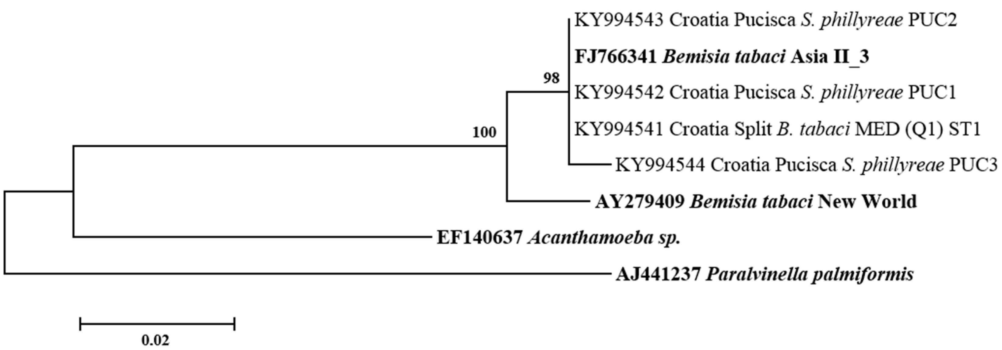
3.2. Phylogenetic Relationships of B. tabaci Sequences

A total of 18 B. tabaci sequences were characterized from Croatia, Montenegro, and Macedonia. In this study, B. tabaci sequences from Israeli populations served as a reference. A phylogenetic tree was constructed from the mtCOI sequences of B. tabaci and divergence analyses were compared with 40 putative species previously reported [10,15,19,20,21,22,23,24,25,26,27] (Figure 2). Our results showed that B. tabaci populations from Croatia, Montenegro, Macedonia, and Israel were identified as MED (Q1 and Q2) species. Populations of B. tabaci MED from Macedonia (Doiran, Gevgelija), Montenegro (Darza, Lastva Grbaljska), and Croatia (Split) belonged to the Q1 group, whereas populations from Croatia (Turanj) and Montenegro (Bar) belonged to the Q2 group (Figure 2). The exception was a B. tabaci MED population from Croatia (Zadar), which contained individuals from both Q1 and Q2 groups (Figure 2). In addition, the B. tabaci MEAM1 species was identified from the Israeli population only.

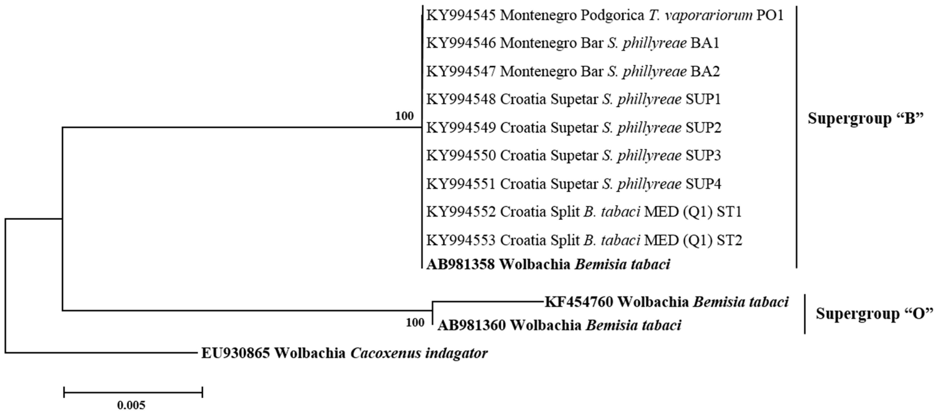
3.3. Phylogenetic Analysis of Secondary Symbionts in Whiteflies

All secondary symbionts sequenced in this study were compared with bacterial reference sequences from GenBank (https://www.ncbi.nlm.nih.gov/genbank/). Sequence analysis revealed that, in addition to species diversity, several bacterial subgroups/strains may exist within the different bacterial genera tested. For Hamiltonella, the sequences from B. tabaci, T. vaporarium, and S. phillyreae formed two groups (H1 and H2) (Figure 3). Clade H1 contains Hamiltonella from B. tabaci and T. vaporarium from Croatia, Montenegro, Serbia, and Macedonia. Group H2 contains Hamiltonella from S. phillyreae and T. vaporarium from Croatia and Montenegro, respectively. 23rDNA sequences of Arsenophonus cluster into two major groups A1 and A2 (Figure 4). The A1 group contains strains from B. tabaci and T. vaporarium. Group A2 contains a distinct Arsenophonus strain from S. phillyreae from Croatia and T. vaporarium from Montenegro, which differs from the A1 group. Rickettsia was grouped into two major clusters, R1 and R2 (Figure 5). The top clade (R1) consists of Rickettsia from both B. tabaci and T. vaporarium from Croatia and Montenegro. Clade R2 consists of Rickettsia from only T. vaporarium from Montenegro. Interestingly, none of the S. phillyreae individuals tested positive for Rickettsia. In the case of Wolbachia, to date, 16 supergroups successively named A–Q infecting a wide range of arthropods and filarial nematodes have been reported [68]. In our analysis, the B supergroup was observed in all three species tested from Croatia, Montenegro, and Macedonia (Figure 6). 16S rDNA sequences of Cardinium from B. tabaci and S. phillyreae are grouped under a single clade (Figure 7).

4. Discussion

Numerous studies have shown that whitefly-associated secondary bacterial symbionts (Arsenophonus, Hamiltonella, Rickettsia, Wolbachia, and Cardinium) play major roles in whitefly biology and have developed through evolutionary history numerous mechanisms to invade and persist in the insect host [1,45,46,52,69,70]. Besides common secondary symbionts, several studies reported the presence of other bacteria in B. tabaci (e.g., Bacillus, Enterobacter, Paracoccus, and Acinetobacter) [71,72]. However, so far none of those bacteria have established a relationship with whiteflies as secondary symbionts investigated in this study. All secondary symbionts are vertically transmitted with high fidelity, but most of them are frequently transferred via horizontal transmission routes, allowing them to spread successfully within and between the species [73].
Our recent studies have extensively investigated infection and localization of secondary symbionts in the three whitefly species (B. tabaci, T. vaporariorum, S. phillyreae) in Southeast Europe [39,41]. We found unique co-infection patterns, revealing the presence of similar secondary symbionts in reproductively isolated whiteflies. This occurrence is likely to be due to highly dynamic processes of horizontal transfer of symbionts between insect hosts, possibly via sharing host plants, mating, or parasitoids [38,73,74]. These events are necessary for the symbiont to spread to novel hosts [75]. Such acquisition of bacterial symbionts can directly impact host biology [46]. For example, the spread of Rickettsia in B. tabaci from the United States resulted in large fitness benefits and female bias in the tested whiteflies [46].
Intensive plant trade occurs between Mediterranean countries and this provides opportunities for introducing new whitefly species and symbionts in regions where they have not existed before [76]. Several studies reported the invasion of B. tabaci MED (Q2), previously associated only with the Middle East countries of the Mediterranean basin, into Spain, France, and Italy, where it coexisted with the native B. tabaci MED (Q1) [76,77]. Our study revealed the presence of both Q1 and Q2 groups of B. tabaci MED in the investigated region (Table 2, Figure 2). Two populations identified as Q2 were collected in Croatia (Turanj) and Montenegro (Bar). Interestingly, a B. tabaci MED population (Croatia-Zadar) collected on plants imported from Italy consisted of both Q1 and Q2 groups and they carried Arsenophonus, which was not previously detected in existing B. tabaci MED (Q1) populations (Table 2, Figure 2) [39].
Variations in the diversity and infection frequencies of bacterial symbionts between the three whitefly species in this study were substantial (Table 2 and Table 3, Figure S1).
Our results revealed that Arsenophonus and Hamiltonella from both B. tabaci and T. vaporariorum were phylogenetically grouped in the same clades (with several exceptions; e.g., Hamiltonella from T. vaporariorum Podgorica), compared with those symbionts from S. phillyreae, which were placed in more distant clades (Figure 3 and Figure 4). B. tabaci and T. vaporariorum frequently share host plants, therefore the possibility of horizontal transmission of bacterial symbionts between these two whitefly species is high [39]. S. phillyreae was only found on pomegranate plants, which was not a host for the other two whitefly species [40,42]. Placement of Hamiltonella in different clades was also reflected on its localization pattern in the whitefly hosts. Hamiltonella found in B. tabaci and T. vaporariorum was localized exclusively in bacteriosomes, while in S. phillyreae it was found to be scattered throughout the entire bodies of nymphs and adults [39,41].
Hamiltonella was present at high frequencies in the investigated species of B. tabaci MED (Q1) and MEAM1 (Table 2), suggesting it had a major impact on the host biology. The infection frequencies of Hamiltonella were in accordance with the global screening reports of bacterial symbionts in B. tabaci MED (Q1) and MEAM1 [33,38,40,78,79]. However, these studies showed absence of Hamiltonella in B. tabaci species such as MED (Q2, ASL), Sub-Saharan Africa 1–5, Asia II, and China 1 [33,78,80]. This was contrary to our results, where Hamiltonella infected 25% and 65% of individuals from MED (Q2) populations from Croatia (Turanj) and Montenegro (Bar), respectively (Table 2). Infection of Hamiltonella varied in T. vaporariorum populations (from absence of infection to high prevalence) in this study, whereas infection with this symbiont was highly prevalent in S. phillyreae. Surprisingly, a large study by Kapantaidaki et al. (2014) [81] did not report infection of Hamiltonella in T. vaporariorum populations from different global locations.
In general, the infection frequencies of Arsenophonus in B. tabaci varied greatly and sometimes depended on the genetic group. B. tabaci MEAM1 had low infection rates with Arsenophonus [33,38,39,40]. This was also similar to several B. tabaci (MED) populations from Southeast Europe (Q1) and China, but some MED genetic groups from Montenegro (Q1), Israel (Q2), and Burkina Faso (Q3) were highly infected with Arsenophonus [33,39,41,78]. This variable occurrence of Arsenophonus in B. tabaci is in agreement with our results. For example, Arsenophonus was fixed in B. tabaci MED (Q2) from Israel and prevalent in MED (Q1 and Q2) from Montenegro (Lastva Grbaljska, Bar) (85% infection rate). However, this symbiont was absent in other MED (Q1 and Q2) populations (Table 2). Arsenophonus was constantly present at high frequencies in T. vaporariorum and S. phillyreae in our and other studies [39,40,41,81].
In this study, Rickettsia was present in nearly all populations of B. tabaci MED. However, infection rates were much lower independently of Q1 and Q2 groups compared to Israeli MED and MEAM1 populations (Table 2). This suggests recent introduction of Rickettsia in B. tabaci populations in the investigated region. Other studies have shown that Rickettsia was present at high infection rates in the MEAM1, MED (Q2), and China 1 groups of B. tabaci, while lower infection rates were observed in Asia II and the MED groups of B. tabaci from Burkina Faso (Q3) and China [33,38,40,78,79]. This symbiont was rarely detected in T. vaporariorum, and no infection was found in S. phillyreae populations (Table 2) [41,81].
Wolbachia is one of the most commonly present symbionts in insects [82]. In this study, nearly all B. tabaci and S. phillyreae populations were infected with Wolbachia (10–60% and 15–50%, respectively), while it was rarely found in T. vaporariorum (Table 2). Several studies reported highly diverse infection rates of Wolbachia in B. tabaci MED (Q1, Q2, ASL), while it was not observed in other genetic groups [33,40]. Bing et al. (2013) [78] found that native genetic groups of B. tabaci (Asia II, China 1) were highly infected with Wolbachia, whereas this symbiont was not detected in invasive MED and MEAM1 groups. Cardinium, another known reproductive manipulator, was rarely detected among populations of the three whitefly species in this study (Table 2) [39,41,43]. Other studies have revealed large differences in rates of Cardinium infection in populations from the B. tabaci species complex [33,38,40,78,79]. Phylogenetically, Wolbachia and Cardinium present in the three whitefly species from this study appeared in the same clades, together with other reported sequences of the same symbionts (Figure 6 and Figure 7).
Mixed infections and heterogeneity among populations in our study could be a result of competition for space and resources among the symbionts or the tolerance of the host to maintain diverse bacterial community [39,83]. This can contribute to multiple events of horizontal transmissions of secondary symbionts within the same whitefly species and reproductively isolated B. tabaci and T. vaporariorum (Table 2 and Table 3). In our study, horizontal transmission seems likely for Hamiltonella, Arsenophonus, Wolbachia, and Cardinium between B. tabaci and T. vaporariorum. Rickettsia was mostly associated with B. tabaci, and there were rare individuals from T. vaporariorum that were positive for this symbiont, which could reflect isolated events of horizontal transmission [41].
Chiel et al. (2009) [75] demonstrated horizontal transmission of Rickettsia in B. tabaci using parasitoid wasps, while transmission of Hamiltonella failed in this system. In addition, Rickettsia was successfully shown in B. tabaci to be transmitted via host plants [73]. There are evidences for horizontal transmission of bacterial symbionts such as Arsenophonus, Cardinium, and Wolbachia either among whiteflies, between whiteflies and arthropods present in the environment, or, as previously mentioned, via host plants [62,84,85,86]. Phylogenies of secondary symbionts are largely incongruent with those of their hosts, which is not the case with the phylogeny of primary symbionts [86]. This suggests an ancient infection of a host with primary symbiont followed by vertical transmission and consequent cospeciation with the insect host. In contrast, incongruent phylogenies suggest multiple infections and horizontal transfer between different hosts [86,87,88].
In the investigated bacterial community of the three whiteflies, we have found more than 20 different symbiotic combinations in a range from no infected individuals to those that were infected with four symbionts (Table 3). In populations of B. tabaci and T. vaporariorum single infections dominated (43% and 56%, respectively), while in S. phillyreae single (25%), double (38%), and triple (31%) infections were present in similar range. Double infections were present in 25% and 38% of individuals of B. tabaci and T. vaporariorum, respectively. Interestingly, T. vaporariorum in this study had no infections with three or four symbionts, while those infections existed in B. tabaci in 14% and 7.5%, respectively. In B. tabaci, T. vaporariorum, and S. phillyreae, individuals with no infections were present in 11%, 5%, and 3%, respectively. Our results were slightly different from study of Gueguen et al. (2010) [33], where infections of global populations of B. tabaci with two or more symbionts were common and double infections dominated (59%). Furthermore, several studies on sub-Saharan populations of B. tabaci report increasing number of secondary symbiont-free whiteflies [88,89]. It is likely to conclude that heterogeneity of symbiotic community of the whiteflies depends on location, population, and time [88].

5. Conclusions

The present study found substantial diversity of bacterial symbionts in the three whitefly species, especially between those that shared habitat (B. tabaci and T. vaporariorum) and in S. phillyreae, which was exclusively found on pomegranate plants. Dynamic processes of horizontal transmission could bring phylogenetically distant symbionts together in previously uninfected whiteflies (e.g., via parasitoids). This finding can contribute to the discovery of new functions that symbionts such as Hamiltonella or Arsenophonus may fulfill in their whitefly hosts.

Supplementary Materials

The following are available online at www.mdpi.com/2075-4450/8/4/113/s1, Figure S1: Individual and multiple infections by secondary bacterial symbionts in five B. tabaci and six T. vaporariorum populations from Croatia, Montenegro, Macedonia, and Serbia. Each square represents one population and each column represents one type of symbiont; the 20 rows per table represent the 20 individuals tested per population. Gray fields indicate positive infection for the tested symbiont. Population number, country and geographical location, species, and number of tested individuals are indicated at the top of each table. Symbionts: R—Rickettsia, H—Hamiltonella, A—Arsenophonus, W—Wolbachia, C—Cardinium; Table S1. The whitefly populations used in this study.

Acknowledgments

The authors would like to thank Snjezana Hrncic and Milorad Raicevic (Biotechnical Faculty, Podgorica, Montenegro), Stanislava Lazarevska and Slobodan Bandzo (Faculty of Agriculture, Skopje, Macedonia), Mirjana Prijovic (Institute of Pesticides and Environmental Protection, Belgrade, Serbia), Ivona Mladineo (Institute of Oceanography and Fisheries, Split, Croatia), and Ivica Samanic (Faculty of Science, University of Split, Split, Croatia) for their valuable help. This study was funded by the Ministry of Science, Education, and Sports of the Republic of Croatia (grant nos. 091-0910468-0281 and 177-11911196-0829).

Author Contributions

Marisa Skaljac and Murad Ghanim conceived and designed the experiments. Marisa Skaljac and Ivana Lepen Pleic performed the experiments. Surapathrudu Kanakala, Marisa Skaljac and Jasna Puizina analyzed the data. Katja Zanic, Jasna Puizina, and Murad Ghanim contributed reagents/materials/analysis tools. Marisa Skaljac and Murad Ghanim wrote the paper.

Conflicts of Interest

The authors declare no conflict of interest.

References

  1. Moran, N.A.; McCutcheon, J.P.; Nakabachi, A. Genomics and evolution of heritable bacterial symbionts. Annu. Rev. Genet. 2008, 42, 165–190. [Google Scholar] [CrossRef] [PubMed]
  2. Dale, C.; Moran, N.A. Molecular interactions between bacterial symbionts and their hosts. Cell 2006, 126, 453–465. [Google Scholar] [CrossRef] [PubMed]
  3. Brownlie, J.C.; Johnson, K.N. Symbiont-mediated protection in insect hosts. Trends Microbiol. 2009, 17, 348–354. [Google Scholar] [CrossRef] [PubMed]
  4. Sanchez-Contreras, M.; Vlisidou, I. The diversity of insect-bacteria interactions and its applications for disease control. Biotechnol. Genet. Eng. Rev. 2008, 25, 203–244. [Google Scholar] [CrossRef]
  5. Kikuchi, Y.; Hosokawa, T.; Nikoh, N.; Meng, X.Y.; Kamagata, Y.; Fukatsu, T. Host-symbiont co-speciation and reductive genome evolution in gut symbiotic bacteria of acanthosomatid stinkbugs. BMC Biol. 2009, 7, 2. [Google Scholar] [CrossRef] [PubMed]
  6. Feldhaar, H. Bacterial symbionts as mediators of ecologically important traits of insect hosts. Ecol. Entomol. 2011, 36, 533–543. [Google Scholar] [CrossRef]
  7. Dunbar, H.E.; Wilson, A.C.C.; Ferguson, N.R.; Moran, N.A. Aphid thermal tolerance is governed by a point mutation in bacterial symbionts. PLoS Biol. 2007, 5. [Google Scholar] [CrossRef] [PubMed]
  8. Tsuchida, T.; Koga, R.; Matsumoto, S.; Fukatsu, T. Interspecific symbiont transfection confers a novel ecological trait to the recipient insect. Biol. Lett. 2011, 7, 245–248. [Google Scholar] [CrossRef] [PubMed]
  9. Ferrari, J.; Vavre, F. Bacterial symbionts in insects or the story of communities affecting communities. Phil. Trans. R. Soc. B Biol. Sci. 2011, 366, 1389–1400. [Google Scholar] [CrossRef] [PubMed]
  10. De Barro, P.J.; Liu, S.S.; Boykin, L.M.; Dinsdale, A.B. Bemisia tabaci: A statement of species status. Annu. Rev. Entomol. 2011, 56, 1–19. [Google Scholar] [CrossRef] [PubMed]
  11. Ash Whitefly, Siphoninus Phillyreae (Haliday) (Insecta: Aleyrodidae: Aleyrodinae). Available online: http://www.doacs. state.fl.us/pi/enpp/ento/entcirc/ent337.pdf (accessed on 18 May 2012).
  12. Brown, J.K. The bemisia tabaci complex: Genetic and phenotypic variation and relevance to tylcv-vector interactions. In Tomato Yellow Leaf Curl Virus Disease (Managment, Molecular Biology, Breeding for Resistance); Czosnek, H., Ed.; Springer: Houten, The Netherlands, 2007; pp. 25–57. [Google Scholar]
  13. CABI. Improving Lives by Solving Problems in Agriculture and the Environment. Available online: http://www.cabi.org (accessed on 1 September 2017).
  14. Jones, D.R. Plant viruses transmitted by whiteflies. Eur. J. Plant Pathol. 2003, 109, 195–219. [Google Scholar] [CrossRef]
  15. Hu, J.; De Barro, P.; Zhao, H.; Wang, J.; Nardi, F.; Liu, S.-S. An extensive field survey combined with a phylogenetic analysis reveals rapid and widespread invasion of two alien whiteflies in china. PLoS ONE 2011, 6. [Google Scholar] [CrossRef] [PubMed]
  16. Gao, R.R.; Zhang, W.P.; Wu, H.T.; Zhang, R.M.; Zhou, H.X.; Pan, H.P.; Zhang, Y.J.; Brown, J.K.; Chu, D. Population structure of the greenhouse whitefly, trialeurodes vaporariorum (westwood), an invasive species from the americas, 60 years after invading china. Int. J. Mol. Sci. 2014, 15, 13514–13528. [Google Scholar] [CrossRef] [PubMed]
  17. Sparks, T.C.; Nauen, R. Irac: Mode of action classification and insecticide resistance management. Pestic. Biochem. Physiol. 2015, 121, 122–128. [Google Scholar] [CrossRef] [PubMed]
  18. Kapantaidaki, D.E.; Sadikoglou, E.; Tsakireli, D.; Kampanis, V.; Stavrakaki, M.; Schorn, C.; Ilias, A.; Riga, M.; Tsiamis, G.; Nauen, R.; et al. Insecticide resistance in trialeurodes vaporariorum populations and novel diagnostics for kdr mutations. Pest. Manag. Sci. 2017. [Google Scholar] [CrossRef] [PubMed]
  19. Dinsdale, A.; Cook, L.; Riginos, C.; Buckley, Y.; Barro, P.D. Refined global analysis of bemisia tabaci (hemiptera: Sternorrhyncha: Aleyrodoidea: Aleyrodidae) mitochondrial cytochrome oxidase 1 to identify species level genetic boundaries. Ann. Entomol. Soc. Am. 2010, 103, 196–208. [Google Scholar] [CrossRef]
  20. Firdaus, S.; Vosman, B.; Hidayati, N.; Jaya Supena, E.D.; Visser, R.G.F.; van Heusden, A.W. The bemisia tabaci species complex: Additions from different parts of the world. Insect Sci. 2013, 20, 723–733. [Google Scholar] [CrossRef] [PubMed]
  21. Chowda-Reddy, R.; Kirankumar, M.; Seal, S.E.; Muniyappa, V.; Valand, G.B.; Govindappa, M.; Colvin, J. Bemisia tabaci phylogenetic groups in india and the relative transmission efficacy of tomato leaf curl bangalore virus by an indigenous and an exotic population. J. Integr. Agric. 2012, 11, 235–248. [Google Scholar] [CrossRef]
  22. Hu, J.; Jiang, Z.L.; Nardi, F.; Liu, Y.Y.; Luo, X.R.; Li, H.X.; Zhang, Z.K. Members of bemisia tabaci (hemiptera: Aleyrodidae) cryptic species and the status of two invasive alien species in the Yunnan province (China). J. Insect Sci. Online 2014, 14. [Google Scholar] [CrossRef] [PubMed]
  23. Alemandri, V.; De Barro, P.; Bejerman, N.; Arguello Caro, E.B.; Dumon, A.D.; Mattio, M.F.; Rodriguez, S.M.; Truoli, G. Species within the bemisia tabaci (hemiptera: Aleyrodidae) complex in soybean and bean crops in argentina. J. Econ. Entomol. 2012, 105, 48–53. [Google Scholar] [CrossRef] [PubMed]
  24. Ueda, S.; Kitamura, T.; Kijima, K.; Honda, K.I.; Kanmiya, K. Distribution and molecular characterization of distinct asian populations of bemisia tabaci (hemiptera: Aleyrodidae) in Japan. J. Appl. Entomol. 2009, 133, 355–366. [Google Scholar] [CrossRef]
  25. Parrella, G.; Scassillo, L.; Giorgini, M. Evidence for a new genetic variant in the bemisia tabaci species complex and the prevalence of the biotype q in Southern Italy. J. Pest Sci. 2012, 85, 227–238. [Google Scholar] [CrossRef]
  26. Esterhuizen, L.L.; Mabasa, K.G.; van Heerden, S.W.; Czosnek, H.; Brown, J.K.; van Heerden, H.; Rey, M.E.C. Genetic identification of members of the bemisia tabaci cryptic species complex from south africa reveals native and introduced haplotypes. J. Appl. Entomol. 2013, 137, 122–135. [Google Scholar] [CrossRef]
  27. Hu, J.; Zhang, X.; Jiang, Z.; Zhang, F.; Liu, Y.; Li, Z.; Zhang, Z. New putative cryptic species detection and genetic network analysis of bemisia tabaci (hempitera: Aleyrodidae) in China based on mitochondrial coi sequences. Mitochondrial DNA Part A DNA Mapp. Seq. Anal. 2017, 1–11. [Google Scholar] [CrossRef] [PubMed]
  28. Tsagkarakou, A.; Mouton, L.; Kristoffersen, J.B.; Dokianakis, E.; Grispou, M.; Bourtzis, K. Population genetic structure and secondary endosymbionts of q bemisia tabaci (hemiptera: Aleyrodidae) from greece. Bull. Entomol. Res. 2012, 102, 353–365. [Google Scholar] [CrossRef] [PubMed]
  29. Gauthier, N.; Clouet, C.; Perrakis, A.; Kapantaidaki, D.; Peterschmitt, M.; Tsagkarakou, A. Genetic structure of bemisia tabaci med populations from home-range countries, inferred by nuclear and cytoplasmic markers: Impact on the distribution of the insecticide resistance genes. Pest Manag. Sci. 2014, 70, 1477–1491. [Google Scholar] [CrossRef] [PubMed]
  30. Chen, W.; Hasegawa, D.K.; Kaur, N.; Kliot, A.; Pinheiro, P.V.; Luan, J.; Stensmyr, M.C.; Zheng, Y.; Liu, W.; Sun, H.; et al. The draft genome of whitefly bemisia tabaci meam1, a global crop pest, provides novel insights into virus transmission, host adaptation, and insecticide resistance. BMC Biol. 2016, 14, 110. [Google Scholar] [CrossRef] [PubMed]
  31. Boykin, L.M.; Shatters, R.G., Jr.; Rosell, R.C.; McKenzie, C.L.; Bagnall, R.A.; De Barro, P.; Frohlich, D.R. Global relationships of bemisia tabaci (hemiptera: Aleyrodidae) revealed using bayesian analysis of mitochondrial coi DNA sequences. Mol. Phylogenet. Evol. 2007, 44, 1306–1319. [Google Scholar] [CrossRef] [PubMed]
  32. Chu, D.; Wan, F.-H.; Tao, Y.-L.; Liu, G.-X.; Fan, Z.-X.; Bi, Y.-P. Genetic differentiation of bemisia tabaci (gennadius) (hemiptera: Aleyrodidae) biotype q based on mitochondrial DNA markers. Insect Sci. 2008, 15, 115–123. [Google Scholar] [CrossRef]
  33. Gueguen, G.; Vavre, F.; Gnankine, O.; Peterschmitt, M.; Charif, D.; Chiel, E.; Gottlieb, Y.; Ghanim, M.; Zchori-Fein, E.; Fleury, F. Endosymbiont metacommunities, mtdna diversity and the evolution of the bemisia tabaci (hemiptera: Aleyrodidae) species complex. Mol. Ecol. 2010, 19, 4365–4376. [Google Scholar] [CrossRef] [PubMed]
  34. Sloan, D.B.; Moran, N.A. Endosymbiotic bacteria as a source of carotenoids in whiteflies. Biol. Lett. 2012, 8, 986–989. [Google Scholar] [CrossRef] [PubMed]
  35. Zchori-Fein, E.; Brown, J.K. Diversity of prokaryotes associated with bemisia tabaci (gennadius) (hemiptera: Aleyrodidae). Ann. Entomol. Soc. Am. 2002, 95, 711–718. [Google Scholar] [CrossRef]
  36. Gottlieb, Y.; Ghanim, M.; Chiel, E.; Gerling, D.; Portnoy, V.; Steinberg, S.; Tzuri, G.; Horowitz, A.R.; Belausov, E.; Mozes-Daube, N.; et al. Identification and localization of a rickettsia sp. In bemisia tabaci (homoptera: Aleyrodidae). Appl. Environ. Microbiol. 2006, 72, 3646–3652. [Google Scholar] [CrossRef] [PubMed]
  37. Chiel, E.; Gottlieb, Y.; Zchori-Fein, E.; Mozes Daube, N.; Katzir, N.; Inbar, M. Biotype-dependent secondary symbiont communities in sympatric populations of bemisia tabaci. Bull. Entomol. Res. 2007, 97, 407. [Google Scholar] [CrossRef] [PubMed]
  38. Gottlieb, Y.; Ghanim, M.; Gueguen, G.; Kontsedalov, S.; Vavre, F.; Fleury, F.; Zchori-Fein, E. Inherited intracellular ecosystem: Symbiotic bacteria share bacteriocytes in whiteflies. FASEB J. Off. Publ. Fed. Am. Soc. Exp. Biol. 2008, 22, 2591–2599. [Google Scholar] [CrossRef] [PubMed]
  39. Skaljac, M.; Zanic, K.; Ban, S.G.; Kontsedalov, S.; Ghanim, M. Co-infection and localization of secondary symbionts in two whitefly species. BMC Microbiol. 2010, 10, 142. [Google Scholar] [CrossRef] [PubMed]
  40. Marubayashi, J.M.; Kliot, A.; Yuki, V.A.; Rezende, J.A.M.; Krause-Sakate, R.; Pavan, M.A.; Ghanim, M. Diversity and localization of bacterial endosymbionts from whitefly species collected in brazil. PLoS ONE 2014, 9, e108363. [Google Scholar] [CrossRef] [PubMed]
  41. Skaljac, M.; Zanic, K.; Hrncic, S.; Radonjic, S.; Perovic, T.; Ghanim, M. Diversity and localization of bacterial symbionts in three whitefly species (hemiptera: Aleyrodidae) from the east coast of the adriatic sea. Bull. Entomol. Res. 2013, 103, 48–59. [Google Scholar] [CrossRef] [PubMed]
  42. Gherna, R.L.; Werren, J.H.; Weisburg, W.; Cote, R.; Woese, C.R.; Mandelco, L.; Brenner, D.J. Arsenophonus nasoniae gen. Nov., sp. Nov., the causative agent of the son-killer trait in the parasitic wasp nasonia vitripennis. Int. J. Syst. Evol. Microbiol. 1991, 41, 563–565. [Google Scholar] [CrossRef]
  43. Zchori-Fein, E.; Perlman, S.J. Distribution of the bacterial symbiont cardinium in arthropods. Mol. Ecol. 2004, 13, 2009–2016. [Google Scholar] [CrossRef] [PubMed]
  44. Werren, J.H.; Baldo, L.; Clark, M.E. Wolbachia: Master manipulators of invertebrate biology. Nat. Rev. Microbiol. 2008, 6, 741–751. [Google Scholar] [CrossRef] [PubMed]
  45. Brumin, M.; Kontsedalov, S.; Ghanim, M. Rickettsia influences thermotolerance in the whitefly bemisia tabaci b biotype. Insect Sci. 2011, 18, 57–66. [Google Scholar] [CrossRef]
  46. Himler, A.G.; Adachi-Hagimori, T.; Bergen, J.E.; Kozuch, A.; Kelly, S.E.; Tabashnik, B.E. Rapid spread of a bacterial symbiont in an invasive whitefly is driven by fitness benefits and female bias. Science 2011, 332, 254–256. [Google Scholar] [CrossRef] [PubMed]
  47. Kontsedalov, S.; Zchori-Fein, E.; Chiel, E.; Gottlieb, Y.; Inbar, M.; Ghanim, M. The presence of rickettsia is associated with increased susceptibility of bemisia tabaci (homoptera: Aleyrodidae) to insecticides. Pest. Manag. Sci. 2008, 64, 789–792. [Google Scholar] [CrossRef] [PubMed]
  48. Mahadav, A.; Gerling, D.; Gottlieb, Y.; Czosnek, H.; Ghanim, M. Parasitization by the wasp eretmocerus mundus induces transcription of genes related to immune response and symbiotic bacteria proliferation in the whitefly bemisia tabaci. BMC Genom. 2008, 9, 342. [Google Scholar] [CrossRef] [PubMed]
  49. Allen, J.M.; Reed, D.L.; Perotti, M.A.; Braig, H.R. Evolutionary relationships of “candidatus riesia spp.,” endosymbiotic enterobacteriaceae living within hematophagous primate lice. Appl. Environ. Microbiol. 2007, 73, 1659–1664. [Google Scholar] [CrossRef] [PubMed]
  50. Perotti, M.A.; Allen, J.M.; Reed, D.L.; Braig, H.R. Host-symbiont interactions of the primary endosymbiont of human head and body lice. FASEB J. Off. Publ. Fed. Am. Soc. Exp. Biol. 2007, 21, 1058–1066. [Google Scholar] [CrossRef] [PubMed]
  51. Rana, V.S.; Singh, S.T.; Priya, N.G.; Kumar, J.; Rajagopal, R. Arsenophonus groel interacts with clcuv and is localized in midgut and salivary gland of whitefly b. Tabaci. PLoS ONE 2012, 7, e42168. [Google Scholar] [CrossRef] [PubMed]
  52. Gottlieb, Y.; Zchori-Fein, E.; Mozes-Daube, N.; Kontsedalov, S.; Skaljac, M.; Brumin, M.; Sobol, I.; Czosnek, H.; Vavre, F.; Fleury, F.; et al. The transmission efficiency of tomato yellow leaf curl virus by the whitefly bemisia tabaci is correlated with the presence of a specific symbiotic bacterium species. J. Virol. 2010, 84, 9310–9317. [Google Scholar] [CrossRef] [PubMed]
  53. Oliver, K.M.; Russell, J.A.; Moran, N.A.; Hunter, M.S. Facultative bacterial symbionts in aphids confer resistance to parasitic wasps. Proc. Natl. Acad. Sci. USA 2003, 100, 1803–1807. [Google Scholar] [CrossRef] [PubMed]
  54. Russell, J.A.; Moran, N.A. Horizontal transfer of bacterial symbionts: Heritability and fitness effects in a novel aphid host. Appl. Environ. Microbiol. 2005, 71, 7987–7994. [Google Scholar] [CrossRef] [PubMed]
  55. Rao, Q.; Wang, S.; Su, Y.-L.; Bing, X.-L.; Liu, S.-S.; Wang, X.-W. Draft genome sequence of “candidatus hamiltonella defensa,” an endosymbiont of the whitefly bemisia tabaci. J. Bacteriol. 2012, 194. [Google Scholar] [CrossRef] [PubMed]
  56. Su, Q.; Xie, W.; Wang, S.; Wu, Q.; Liu, B.; Fang, Y.; Xu, B.; Zhang, Y. The endosymbiont hamiltonella increases the growth rate of its host bemisia tabaci during periods of nutritional stress. PLoS ONE 2014, 9. [Google Scholar] [CrossRef] [PubMed]
  57. Thao, M.L.; Baumann, L.; Hess, J.M.; Falk, B.W.; Ng, J.C.; Gullan, P.J.; Baumann, P. Phylogenetic evidence for two new insect-associated chlamydia of the family simkaniaceae. Curr. Microbiol. 2003, 47, 46–50. [Google Scholar] [CrossRef] [PubMed]
  58. Everett, K.D.E.; Thao, M.; Horn, M.; Dyszynski, G.E.; Baumann, P. Novel chlamydiae in whiteflies and scale insects: Endosymbionts “candidatus fritschea bemisiae” strain falk and “candidatus fritschea eriococci” strain elm. Int. J. Syst. Evol. Microbiol. 2005, 55, 1581–1587. [Google Scholar] [CrossRef] [PubMed]
  59. De Barro, P.J.; Scott, K.D.; Graham, G.C.; Lange, C.L.; Schutze, M.K. Isolation and characterization of microsatellite loci in bemisia tabaci. Mol. Ecol. Notes 2003, 3, 40–43. [Google Scholar] [CrossRef]
  60. Frohlich, D.R.; Torres-Jerez, I.I.; Bedford, I.D.; Markham, P.G.; Brown, J.K. A phylogeographical analysis of the bemisia tabaci species complex based on mitochondrial DNA markers. Mol. Ecol. 1999, 8, 1683–1691. [Google Scholar] [CrossRef] [PubMed]
  61. Thao, M.L.; Baumann, P. Evidence for multiple acquisition of arsenophonus by whitefly species (sternorrhyncha: Aleyrodidae). Curr. Microbiol. 2004, 48, 140–144. [Google Scholar] [CrossRef] [PubMed]
  62. Weeks, A.; Breeuwer, J. A new bacterium from the cytophaga-flavobacterium—bacteroides phylum that causes sex-ratio distortion. In Insect Symbiosis; CRC Press: Boca Raton, FL, USA, 2003; pp. 165–176. [Google Scholar]
  63. Altschul, S.F.; Gish, W.; Miller, W.; Myers, E.W.; Lipman, D.J. Basic local alignment search tool. J. Mol. Biol. 1990, 215, 403–410. [Google Scholar] [CrossRef]
  64. Katoh, K.; Standley, D.M. Mafft multiple sequence alignment software version 7: Improvements in performance and usability. Mol. Biol. Evol. 2013, 30, 772–780. [Google Scholar] [CrossRef] [PubMed]
  65. Miller, M.A.; Pfeiffer, W.; Schwartz, T. Creating the cipres science gateway for inference of large phylogenetic trees. In Gateway Computing Environments Workshop (GCE), 2010; IEEE: New Orleans, LA, USA, 2010; pp. 1–8. [Google Scholar]
  66. Ronquist, F.; Huelsenbeck, J.P. Mrbayes 3: Bayesian phylogenetic inference under mixed models. Bioinformatics 2003, 19, 1572–1574. [Google Scholar] [CrossRef] [PubMed]
  67. Tamura, K.; Stecher, G.; Peterson, D.; Filipski, A.; Kumar, S. Mega6: Molecular evolutionary genetics analysis version 6.0. Mol. Biol. Evol. 2013, 30, 2725–2729. [Google Scholar] [CrossRef] [PubMed]
  68. Glowska, E.; Dragun-Damian, A.; Dabert, M.; Gerth, M. New wolbachia supergroups detected in quill mites (acari: Syringophilidae). Infect. Genet. Evol. 2015, 30, 140–146. [Google Scholar] [CrossRef] [PubMed]
  69. Ghanim, M.; Kontsedalov, S. Susceptibility to insecticides in the q biotype of bemisia tabaci is correlated with bacterial symbiont densities. Pest. Manag. Sci. 2009, 65, 939–942. [Google Scholar] [CrossRef] [PubMed]
  70. Degnan, P.H.; Yu, Y.; Sisneros, N.; Wing, R.A.; Moran, N.A. Hamiltonella defensa, genome evolution of protective bacterial endosymbiont from pathogenic ancestors. Proc. Natl. Acad. Sci. USA 2009, 106, 9063–9068. [Google Scholar] [CrossRef] [PubMed]
  71. Singh, S.T.; Priya, N.G.; Kumar, J.; Rana, V.S.; Ellango, R.; Joshi, A. Diversity and phylogenetic analysis of endosymbiotic bacteria from field caught bemisia tabaci from different locations of north india based on 16s rdna library screening. Infect. Genet. Evol. 2012, 12, 411–419. [Google Scholar] [CrossRef] [PubMed]
  72. Xie, W.; Meng, Q.S.; Wu, Q.J.; Wang, S.L.; Yang, X.; Yang, N.N.; Li, R.M.; Jiao, X.G.; Pan, H.P.; Liu, B.M.; et al. Pyrosequencing the bemisia tabaci transcriptome reveals a highly diverse bacterial community and a robust system for insecticide resistance. PLoS ONE 2012, 7. [Google Scholar] [CrossRef] [PubMed]
  73. Caspi-Fluger, A.; Inbar, M.; Mozes-Daube, N.; Katzir, N.; Portnoy, V.; Belausov, E.; Hunter, M.S.; Zchori-Fein, E. Horizontal transmission of the insect symbiont rickettsia is plant-mediated. Proc. R. Soc. B Biol. Sci. 2012, 279, 1791–1796. [Google Scholar] [CrossRef] [PubMed]
  74. Gonella, E.; Pajoro, M.; Marzorati, M.; Crotti, E.; Mandrioli, M.; Pontini, M.; Bulgari, D.; Negri, I.; Sacchi, L.; Chouaia, B.; et al. Plant-mediated interspecific horizontal transmission of an intracellular symbiont in insects. Sci. Rep. 2015, 5. [Google Scholar] [CrossRef] [PubMed] [Green Version]
  75. Chiel, E.; Zchori-Fein, E.; Inbar, M.; Gottlieb, Y.; Adachi-Hagimori, T.; Kelly, S.E.; Asplen, M.K.; Hunter, M.S. Almost there: Transmission routes of bacterial symbionts between trophic levels. PLoS ONE 2009, 4. [Google Scholar] [CrossRef] [PubMed]
  76. Terraz, G.; Gueguen, G.; Arno, J.; Fleury, F.; Mouton, L. Nuclear and cytoplasmic differentiation among mediterranean populations of bemisia tabaci: Testing the biological relevance of cytotypes. Pest. Manag. Sci. 2014, 70, 1503–1513. [Google Scholar] [CrossRef] [PubMed]
  77. Parrella, G.; Nappo, A.G.; Manco, E.; Greco, B.; Giorgini, M. Invasion of the q2 mitochondrial variant of mediterranean bemisia tabaci in southern italy: Possible role of bacterial endosymbionts. Pest. Manag. Sci. 2014, 70, 1514–1523. [Google Scholar] [CrossRef] [PubMed]
  78. Bing, X.; Ruan, Y.; Rao, Q.; Wang, X.; Liu, S. Diversity of secondary endosymbionts among different putative species of the whitefly bemisia tabaci. Insect Sci. 2013, 20, 194–206. [Google Scholar] [CrossRef] [PubMed]
  79. Chu, D.; Gao, C.S.; De Barro, P.; Zhang, Y.J.; Wan, F.H.; Khan, I.A. Further insights into the strange role of bacterial endosymbionts in whitefly, bemisia tabaci: Comparison of secondary symbionts from biotypes b and q in china. Bull. Entomol. Res. 2011, 101, 477–486. [Google Scholar] [CrossRef] [PubMed]
  80. Ghosh, S.; Bouvaine, S.; Maruthi, M. Prevalence and genetic diversity of endosymbiotic bacteria infecting cassava whiteflies in africa. BMC Microbiol. 2015, 15. [Google Scholar] [CrossRef] [PubMed]
  81. Kapantaidaki, D.E.; Ovcarenko, I.; Fytrou, N.; Knott, K.E.; Bourtzis, K.; Tsagkarakou, A. Low levels of mitochondrial DNA and symbiont diversity in the worldwide agricultural pest, the greenhouse whitefly trialeurodes vaporariorum (hemiptera: Aleyrodidae). J. Hered. 2014, 106, 80–92. [Google Scholar] [CrossRef] [PubMed]
  82. Correa, C.C.; Ballard, J. Wolbachia associations with insects: Winning or losing against a master manipulator. Front. Ecol. Evol. 2016, 3. [Google Scholar] [CrossRef]
  83. Vautrin, E.; Vavre, F. Interactions between vertically transmitted symbionts: Coperation or conflict? Trends Microbiol. 2009, 17, 95–99. [Google Scholar] [CrossRef] [PubMed]
  84. Sintupachee, S.; Milne, J.R.; Poonchaisri, S.; Baimai, V.; Kittayapong, P. Closely related wolbachia strains within the pumpkin arthropod community and the potential for horizontal transmission via the plant. Microb. Ecol. 2006, 51, 294–301. [Google Scholar] [CrossRef] [PubMed]
  85. Mouton, L.; Thierry, M.; Henri, H.; Baudin, R.; Gnankine, O.; Reynaud, B.; Zchori-Fein, E.; Becker, N.; Fleury, F.; Delatte, H. Evidence of diversity and recombination in arsenophonus symbionts of the bemisia tabaci species complex. BMC Microbiol. 2012, 12 (Suppl. 1). [Google Scholar] [CrossRef] [PubMed] [Green Version]
  86. Vavre, F.; Fleury, F.; Lepetit, D.; Fouillet, P.; Bouletreau, M. Phylogenetic evidence for horizontal transmission of wolbachia in host-parasitoid associations. Mol. Biol. Evol. 1999, 16, 1711–1723. [Google Scholar] [CrossRef] [PubMed]
  87. Ahmed, M.Z.; De Barro, P.J.; Ren, S.X.; Greeff, J.M.; Qiu, B.L. Evidence for horizontal transmission of secondary endosymbionts in the bemisia tabaci cryptic species complex. PLoS ONE 2013, 8. [Google Scholar] [CrossRef] [PubMed]
  88. Tajebe, L.S.; Guastella, D.; Cavalieri, V.; Kelly, S.E.; Hunter, M.S.; Lund, O.S.; Legg, J.P.; Rapisarda, C. Diversity of symbiotic bacteria associated with bemisia tabaci (homoptera: Aleyrodidae) in cassava mosaic disease pandemic areas of tanzania. Ann. Appl. Biol. 2015, 166, 297–310. [Google Scholar] [CrossRef]
  89. Ghosh, S.; Bouvaine, S.; Richardson, S.C.W.; Ghanim, M.; Maruthi, M.N. Fitness costs associated with infections of secondary endosymbionts in the cassava whitefly species bemisia tabaci. J. Pest Sci. 2017. [Google Scholar] [CrossRef]
Figure 1. Locations of collected whitefly populations (according to population numbers in Table S1) with overview of overall infection frequencies of secondary bacterial symbionts (according to Table 2) in the three whitefly species in Southeast Europe. Each chart is marked with a code (B, T, and S) in order to distinguish between the whitefly species. B, Bemisia tabaci; T, Trialeurodes vaporariorum; S, Siphoninus phillyreae.
Insects 08 00113 g001
Figure 2. Phylogenetic tree based on the Bayesian analysis of 657 bp of mtCOI gene sequences of Bemisia tabaci populations. Sequences from GenBank are indicated in bold, while those obtained in this study are rearranged based on the geographical origin of B. tabaci individuals. Posterior probabilities are indicated at nodes.
Insects 08 00113 g002
Figure 3. Molecular phylogenetic placements of Hamiltonella from whitefly species. The tree was constructed via Bayesian inference (BI) using an HKY substitution model. Sequences from GenBank are indicated in bold, while those obtained in this study are rearranged based on geographical origin and associated whitefly hosts, B. tabaci, T. vaporariorum, and S. phillyreae.
Insects 08 00113 g003
Figure 4. Molecular phylogenetic placements of Arsenophonus from whitefly species. The tree was constructed via Bayesian inference (BI) using a TPM1 µf substitution model. Sequences from GenBank are indicated in bold, while those obtained in this study are rearranged based on geographical origin and associated whitefly hosts, B. tabaci, T. vaporariorum, and S. phillyreae.
Insects 08 00113 g004
Figure 5. Molecular phylogenetic placements of Rickettsia from whitefly species. The tree was constructed via Bayesian inference (BI) using a GTR+G substitution model. Sequences from GenBank are indicated in bold, while those obtained in this study are rearranged based on geographical origin and associated whitefly hosts, B. tabaci and T. vaporariorum.
Insects 08 00113 g005
Figure 6. Molecular phylogenetic placements of Wolbachia from whitefly species. The tree was constructed via Bayesian inference (BI) using a GTR+G substitution model. Sequences from GenBank are indicated in bold, while those obtained in this study are rearranged based on geographical origin and associated whitefly hosts, B. tabaci, T. vaporariorum, and S. phillyreae.
Insects 08 00113 g006
Figure 7. Molecular phylogenetic placements of Cardinium from whitefly species. The tree was constructed via Bayesian inference (BI) using an HKY+I substitution model. Sequences from GenBank are indicated in bold, while those obtained in this study are rearranged based on geographical origin and associated whitefly hosts, B. tabaci and S. phillyreae.
Insects 08 00113 g007
Table 1. The primers used in this study.
Primer NameSequence (5′→3′)Annealing (°C)/Size (bp)GeneReference
Bem 23 F
Bem 23 R
CGGAGCTTGCGCCTTAGTC
CGGCTTTATCATAGCTCTCGT
55/MEAM1 = 200; MED = 400Microsatellite[59]
C1-J-2195
L2-N-3014
TTGATTTTT TGGTCATCCAGAAGT
TCCAATGCACTAATCTGCCATATTA
51/850Cytochrome oxidase I (mtCOI)[60]
Por-F
Por-R
TGCAAGTCGAGCGGCATCAT
AAAGTTCCCGCCTTATGCGT
59/1000Portiera 16S rDNA [35]
Rb F
Rb R
GCTCAGAACGAACGCTATC
GAAGGAAAGCATCTCTGC
59/900Rickettsia 16S rDNA[36]
92 F
Hb R
TGAGTAAAGTCTGGGAATCTGG
AGTTCAAGACCGCAACCTC
62/700Hamiltonella 16S rDNA[35]
Ars23S-1
Ars23S-2
CGTTTGATGAATTCATAGTCAAA
GGTCCTCCAGTTAGTGTTACCCAAC
59/600Arsenophonus 23S rDNA[61]
Wol16S F
Wol16S R
CGG GGGAAAAATTTATTGCT
AGCTGTAATACAGAAAGTAAA
55/650Wolbachia 16S rDNA[38]
CFB F
CFB R
GCGGTGTAAAATGAGCGTG
ACCTMTTCTTAACTCAAGCCT
59/500Cardinium 16S rDNA[62]
Table 2. Overview of overall infection frequencies of secondary bacterial symbionts in the three whitefly species in Southeast Europe with associated host plants.
Population Number, Location and SpeciesHost PlantnInfection of Bacterial Symbiont (%)Reference
HARWC
1Croatia/Turanj MED (Q2)Cucumis sativus2025-302030[39]
2Croatia/SplitMED (Q1)Euphorbia pulcherrima2035-2010-This study
3Croatia/Zadar MED (Q1 and Q2)Hibiscus sp.2095356050-[41]
4Montenegro/Bar MED (Q2)Dipladenia sanderi20658510040-
5Montenegro/Lastva GrbaljskaMED (Q1)Cucumis sativus2030854030-This study
6Montenegro/DarzaMED (Q1)Cucumis melo2095-1010-
7Macedonia/DoiranMED (Q1)Lycopersicon esculentum20100--4020
8Macedonia/GevgelijaMED (Q1)Cucumis melo20100-560-
9IsraelMED (Q2)Gossypium hirsutum20-1007520-[39]
10IsraelMEAM1 Gossypium hirsutum20100-70--
11Croatia/SplitT. vaporariorumSonchus oleraceus20575---
12Croatia/Split§T. vaporariorumEuphorbia pulcherrima105080---[41]; This study
13Montenegro/PodgoricaT. vaporariorumSonchus oleraceus20209551035
14Macedonia/DoiranT. vaporariorumLycopersicon esculentum2025100---This study
15Macedonia/GrcisteT. vaporariorumLycopersicon esculentum20-100---
16Serbia/TrbusaniT. vaporariorumCucurbita pepo2090100---
17Serbia/Vranjska BanjaT. vaporariorumGerbera sp.2045100---
18Serbia/OpovoT. vaporariorumLycopersicon esculentum207090---
19Serbia/Zorka SuboticaT. vaporariorumChrysanthemum sp.20-95---
20Croatia/Brac-SupetarS. phillyreaePunica granatum206535-405[41]
21Croatia/Brac-PuciscaS. phillyreaePunica granatum2010025-20-
22Croatia/OpuzenS. phillyreaePunica granatum208595-555
23Croatia/LjutaS. phillyreaePunica granatum208585-1570
24Montenegro/BarS. phillyreaePunica granatum2090100-50-
n, total number of individual tested per population; H, Hamiltonella; A, Arsenophonus; R, Rickettsia; W, Wolbachia; C, Cardinium; -, zero. Population imported by trade from Italy (Rome, region Lazio). § Population imported by trade from Slovenia (Catez ob Savi). For additional information on imported whitefly populations, please refer to Skaljac et al. (2013) [41].
Table 3. Infection frequencies of different secondary bacterial symbiont combinations in the three whitefly species in this study.
Population Number, Location, and SpeciesInfection Frequencies of Bacterial Symbiont Combination (%)
RHAWCRHRAHAHWHCAWACWCRHARHWRAWRWCHAWHACHWCAWCRHWCRHAWHAWCNo Infection
1Croatia/Turanj
MED (Q2)
1010-51515------5---5----5--35
2Croatia/Split
MED (Q1)
2035-10--------------------35
3Croatia/Zadar
MED (Q1 and Q2)
-20---15-155-----255------15--
4Montenegro/Bar
MED (Q2)
5----520------30510------25--
5Montenegro/L. Grbaljska MED (Q1) --355--15---5--10---5----15-10
6Montenegro/Darza MED (Q1) -75---10--10---------------5
7Macedonia/Doiran MED (Q1)-55------255---------15-----
8Macedonia/Gevgelija MED (Q1) -40------55-----5----------
9Israel MED (Q2)--25---55--------20---------
10Israel MEAM1 -30---70-------------------
11Croatia/Split§
T. vaporariorum
--70----5----------------25
12Croatia§/Split
T. vaporariorum
--30----50----------------20
13Montenegro/Podgorica
T. vaporariorum
--355---20--535-------------
14Macedonia/Doiran
T. vaporariorum
--75----25-----------------
15Macedonia/Grciste
T. vaporariorum
--100----------------------
16Serbia/Trbusani
T. vaporariorum
--10----90-----------------
17Serbia/Vranjska Banja
T. vaporariorum
--55----45-----------------
18Serbia/Opovo
T. vaporariorum
-525----65----------------5
19Serbia/Z. Subotica
T. vaporariorum
--95---------------------5
20Croatia/Brac-Supetar
S. phillyreae
-25-10---2010-5-5----10------15
21Croatia/Brac-Pucisca
S. phillyreae
-60-----2015--------5-------
22Croatia/Opuzen
S. phillyreae
-55----35--10------40-----5-
23Croatia/Ljuta
S. phillyreae
-105----15---5------50-5--10-
24Montenegro/Bar
S. phillyreae
--5----45--5------45-------
H, Hamiltonella; A, Arsenophonus; R, Rickettsia; W, Wolbachia; C, Cardinium; -, zero. Population imported by trade from Italy (Rome, region Lazio). § Population imported by trade from Slovenia (Catez ob Savi). For additional information on imported whitefly populations, please refer to Skaljac et al. (2013) [41].

