Article

Host Specificity of Snodgrassella in Eastern and Western Honeybees and Its Effects on Naturally Occurring Deformed Wing Virus Titers

1
College of Animal Sciences, Zhejiang University, Hangzhou 310058, China
2
Institute of Agricultural Research, Xizang Academy of Agriculture and Animal Husbandry Sciences, Lasa 850032, China
*
Author to whom correspondence should be addressed.
Insects 2025, 16(5), 478; https://doi.org/10.3390/insects16050478
Submission received: 2 March 2025 / Revised: 26 April 2025 / Accepted: 28 April 2025 / Published: 1 May 2025
(This article belongs to the Section Social Insects and Apiculture)

Simple Summary

The composition of the honeybee gut microbiota is relatively simple, making it an ideal model for studying host-microbe interactions. While the host-specific evolution of gut symbionts in Apis mellifera and bumblebees has been well studied, similar research within the Apis genus remains limited. This study focuses on the core gut symbiont Snodgrassella from two closely related species, Apis cerana and A. mellifera, to explore how host-specific evolutionary adaptations of the gut microbiota influence the functional development of honeybees.

Abstract

Honeybee gut microbiota undergoes long-term adaptive evolution with the host, resulting in host-specific genomic and functional characteristics. However, the specific role of host-specialized strains in shaping host physiological functions remains understudied. This study investigates the host specificity of the core gut symbiont Snodgrassella in A. cerana and A. mellifera, exploring its effects on immune response and natural virus suppression through genomic analysis and colonization experiments. Genomic analysis revealed that strain from A. mellifera, exhibited a larger genome and greater gene content compared to strain from A. cerana. Competitive colonization experiments showed that although strains from different origins had similar colonization efficiency in the host, host-specific strain displayed a clear home-field advantage in the competitive colonization process. Moreover, Inoculation of A. mellifera with its native Snodgrassella strain significantly reduced Deformed Wing Virus (DWV) titers, whereas a non-native strain had no effect. In contrast, neither strain altered DWV or Sacbrood Virus (SBV) levels in A. cerana. Immune gene analysis revealed that only the native Snodgrassella strain upregulated defensin 2 in A. mellifera, while no significant changes occurred in A. cerana with either strain. These results suggest that Snodgrassella exhibits host specificity at the strain level, influencing both host immune response and virus suppression, with non-native strains showing reduced efficacy in these functional roles, especially in A. mellifera.

1. Introduction

The digestive tracts of animals harbor complex microbial communities, including bacteria, archaea, and fungi [1]. These gut microbiota provide key functions for the host, such as energy acquisition, metabolism, and immune regulation [2,3]. Additionally, gut microbiota reflects the evolutionary history of the host [4]. Typically, gut microbiota within the same species were more similar to each other than between different species, and interspecies differences in microbial communities are closely linked to the host’s evolutionary divergence. Host-specific microbiota plays a critical role in maintaining host health and facilitating interspecies adaptation [5]. For instance, in rodents, host-specific microbiota is involved in important processes such as pathogen resistance, nutrient utilization, and growth and development [5,6,7]. Compared to mammals, the gut microbiota of eusocial bees is relatively simple and stable [8]. The core microbiota of adult worker bees consists of 8 to 10 bacterial types [9,10,11]. Within these social bee species, the diversity and host specificity of gut microbiota have been demonstrated at the strain level. For example, Snodgrassella exhibits high species diversity and broad host adaptability [12]. These clusters exhibit functional differentiation and ecological niche specialization through gene recombination or single nucleotide polymorphisms, forming a nutritional network with other symbiotic bacteria and establishing a close symbiotic relationship with the host [13,14]. The diversity and composition of gut microbiota not only influence the host’s social behavior and task allocation but also drive the adaptive evolution of host species [13,15,16,17]. However, the extent to which gut microbiota from closely related honeybee species contribute differently to host function, particularly in terms of native and non-native strains, remains largely unresolved.
The core lineages of eusocial bees began co-evolving with their hosts around 80 million years ago, diverging from corbiculate bees and gradually developing host-specific structures [18,19,20]. A. mellifera and bumblebee share similar distribution patterns and core gut microbiota. Genomic analysis and colonization experiments based on the core gut members Snodgrassella, Gilliamella, and Lactobacillus of these two bee species show that the core bacterial types shared by eusocial bees have evolved into different sister lineages and hold a home-field advantage in host competition [12,20,21,22,23,24]. The colonization advantage of core strains such as Snodgrassella alvi, Gilliamella apicola, and Frischella perrara is closely related to their encoding of the Type VI Secretion System (T6SS) and associated effectors [25,26]. In S. alvi, two T6SS systems are present, with T6SS-1 playing a crucial role in regulating competition between same-species bacteria [27]. At the transcriptional level, the host A. mellifera activates gut immune pathways to reject colonization by non-native strains (such as those derived from bumblebees), further supporting the adaptive evolution between the host and its gut microbiota, further supporting the adaptive evolution between the host and its gut microbiota [24]. As two closely related species within the genus Apis, A. cerana and A. mellifera show significant strain-level variation in their gut microbiota based on metagenomic sequencing, and functional differentiation is also observed [28,29]. Recent studies have shown that genomic differences between Gilliamella strains from A. cerana and A. mellifera affect functional divergence and influence host transcriptional responses [30]. However, research on the colonization of Snodgrassella strains in the gut of A. cerana and A. mellifera, and the influence of Snodgrassella strains from different sources on the physiological processes of A. cerana, remains limited. As a native species of Asia, A. cerana is a critical pollinator and honey producer in the region. Therefore, an in-depth study of its gut microbiota’s function and evolution, particularly exploring strain variation and host interaction patterns among closely related species, will provide important insights for formulating targeted and sustainable management strategies.
Honeybee health is threatened by various viruses, with DWV and SBV being common asymptomatic infections in A. mellifera and A. cerana, respectively [31]. DWV is the most widely distributed honeybee virus globally and is transmitted by the Varroa destructor mite, making it a major contributor to the increased overwintering mortality of A. mellifera colonies [32]. In contrast, SBV poses a significant threat to A. cerana, primarily infecting 1-2-day-old larvae and spreading rapidly within colonies [33]. Its impact is particularly pronounced during periods of low nectar and pollen availability, such as early spring and late autumn [34]. Antimicrobial peptide, including apidaecin, abaecin, hymenoptaecin, defensin, royalisin and melittin, play key roles in the immune response to pathogens in honeybee hemolymph [35]. For instance, honeybees upregulate AMP-related gene expression through the modulation of the Toll/Imd signaling pathway to resist viral infections such as DWV and Israeli Acute Paralysis Virus (IAPV) [36,37]. The gut microbiota is crucial in regulating host immune responses and antiviral defenses. Inoculation with a complete gut microbiota has been shown to significantly reduce A. cerana mortality from Nosema ceranae infection and upregulate the expression of apidaecin, abaecin, and hymenoptaecin genes [38]. Furthermore, core symbionts, such as S. alvi, have been shown to stimulate the expression of apidaecin and hymenoptaecin genes, thereby enhancing honeybee resistance to DWV infection [39]. However, studies on whether the regulation of viral titers by gut microbiota varies due to the host origin remain limited. Therefore, further investigation of the regulation of viral titers by honeybee gut microbiota from different host origins will provide important insights into host-microbe co-evolution and its impact on honeybee health.
In this study, genomic analysis, and colonization experiments were conducted using gut Snodgrassella isolates from A. cerana and A. mellifera to validate the host-specific evolution of gut microbiota between closely related hosts. The study also evaluated the impact of strain differences on naturally occurring viral titers and immune gene expression, with the aim of revealing the effects of gut microbiota strain variation on host physiology and further elucidating the adaptive evolution pattern between hosts and their gut microbiota.

2. Materials and Methods

2.1. Strain Cultivation and DNA Extraction

At the experimental apiary of Zhejiang University, 10 forager bees were collected from each of the two honeybee species, A. cerana and A. mellifera. Both honeybee colonies were kept in the same apiary and had not been treated with antibiotics. Specifically, no pharmaceutical interventions were used in the management of A. cerana, while A. mellifera colonies were only treated with routine acaricides for mite control. The honeybees were surface-sterilized with 75% ethanol for 30 s, followed by three rinses with sterile deionized water to remove any ethanol residue. The honeybees were then placed on ice for dissection. Using sterile tweezers, the gut was carefully extracted by gently pulling from the base of the abdomen near the sting, ensuring the entire gut tissue was collected. The tissue was then placed in a 1.5 mL sterile centrifuge tube containing 1 × PBS. The gut tissues were then homogenized using a tissue grinder. The resulting homogenate was serially diluted, and 200 μL of the 10−5 and 10−6 dilutions were plated onto Tryptic Soy Agar (TSA, T8650, Solarbio, Beijing, China). The plates were incubated at 37 °C with 5% CO2 for 48 h. Single colonies were picked and transferred to Tryptic Soy Broth (TSB, LA0110, Solarbio, Beijing, China) for enrichment culture under the same conditions for enrichment. The resulting culture was then mixed with 50% glycerol at a 1:1 ratio and stored at −80 °C for future use.
For DNA extraction, 5 mL of bacterial culture in the logarithmic growth phase was collected, and bacterial pellets were harvested by centrifugation. Genomic DNA was extracted using a bacterial DNA extraction kit (D3350, Omega Bio-Tek, Norcross, GA, USA). DNA concentration and quality were assessed using a fluorescence detector (TBS-380, Turner BioSystems, Sunnyvale, CA, USA) and 0.8% agarose gel electrophoresis. Qualified DNA samples were used for subsequent sequencing library construction.

2.2. Library Construction and Illumina HiSeq Sequencing

Sequencing libraries were constructed using the genomic DNA of qualified strains, and paired-end sequencing (2 × 150 bp) was performed on the Illumina platform. The paired-end library, with an insert size of approximately 400 bp, was constructed following the standard protocol. Genomic DNA was first fragmented to the target size using a Covaris sonicator (M220, Woburn, MA, USA), and then T4 DNA polymerase was used to generate blunt ends. The 3′ ends of the phosphorylated blunt-ended fragments were added with an “A” base, and adapters were ligated to the DNA ends. The target fragments were purified by gel electrophoresis and selectively enriched and amplified by PCR. During PCR amplification, index tags were incorporated into the adapters, and the library quality was assessed. Finally, qualified paired-end libraries were sequenced using the Illumina Hiseq platform (PE150 mode, San Diego, CA, USA).

2.3. Genome Assembly

Raw sequencing data was generated by Illumina’s base calling software CASAVA v1.8.2 as outlined in the corresponding manuscript “https://support.illumina.com/sequencing/sequencing_software/casava.ilmn (accessed on 22 January 2025)”. Contamination reads, such as those containing adaptors or primers, were identified and removed by Trimmomatic [40] with default settings. The clean data, obtained after this quality control step, were then used for subsequent analyses. Genome assembly was conducted with ABySS, utilizing multiple-Kmer values to optimize the results [41]. To address remaining gaps and correct single base polymorphisms, GapCloser was applied to refine the final assembly [42].

2.4. Genome Annotation and Phylogenetic Construction

The Bakta command with default parameters was used to predict the structural and functional annotations [43]. tRNA were identified with tRNAscan-SE (v1.23) [44] while rRNA genes were detected using RNAmmer (v1.2) [45]. Mobile Genetic Elements (MGEs) were identified with mobileOG-db Beatrix-1.6, setting the E-value threshold to 1.0 × 10⁻¹⁰ [46]. Genome comparison was conducted using the OrthoFinder tool, with parameters set as Identity 70, Ratio (core) 0.95, and Support −1 [47]. KEGG was used for genome annotation, with a similarity threshold of 0.5 [48]. Phylogenetic analysis was performed using FastTree from VBCG software (v 1.3), with the max missing genes parameter set to 2 [49].

2.5. Colonization Experiments

Healthy brood combs used for rearing Germ-free worker bees were obtained from the experimental apiary at Zhejiang University. The method for obtaining germ-free worker bees was followed as previously described [28]. The procedure was as follows: Germ-free worker bee pupae were carefully extracted using sterile forceps and placed in a sterile 48-well cell culture plate. The plates were then incubated in an incubator at 32 ± 1 °C with 80 ± 5% relative humidity until the bees emerged (0 days). Newly emerged worker bees (0–48 h old) were subjected to a 3 h fasting period before being fed a sucrose solution containing bacterial suspension at OD600 = 1. In the cross-colonization experiment, worker bees were inoculated with strain sourced either from A. mellifera or A. cerana in two treatment groups, while the control group was fed sterile sucrose solution. For the competitive colonization experiment, strains from both A. mellifera and A. cerana were mixed in a 1:1 ratio and inoculated into the gut of worker bees from both species. Each group consisted of three honeybee cages, with each cage containing 30 worker bees. To meet the protein requirements of the worker bees, sterile rapeseed pollen was provided to all groups. Throughout the experiment, the sugar water was replaced daily, and honeybee samples were collected 7 days post-inoculation. The honeybees were then dissected using sterile forceps to remove the gut, which was stored at −80 °C for further analysis.

2.6. DNA Extraction from the Gut and Bacterial Quantification

Genomic DNA was extracted from honeybee gut tissues using the TIANamp Stool DNA Kit (DP328, Tiangen, Beijing, China) following the manufacturer’s instructions. DNA quality and integrity were assessed by 1% agarose gel electrophoresis. DNA concentration and purity were measured using a NanoDrop 2000 spectrophotometer (Thermo Fisher Scientific, Waltham, MA, USA). To quantify the abundance of bacteria originating from different honeybee species in the gut, specific primers were designed to target unique sequences associated with Snodgrassella strains from A. cerana and A. mellifera. The primers used for quantification were: AC-SNO (5′~3′): [F: TGTGGTAATGGTGGTATGAA], [R: CCGCTGTACGAATATAGAAC] for A. cerana derived Snodgrassella, and AM-SNO (5′~3′): [F: CTGGGCGTACATGCTATT], [R: CAATCTTGCCAGCCATATC] for A. mellifera derived Snodgrassella. These primers were used to assess the abundance of different Snodgrassella strains in the gut of both honeybee species. Bacterial copy numbers in the honeybee gut were quantified using a StepOne real-time quantitative PCR system (4376357, Thermo Fisher Scientific, Foster City, CA, USA). A standard curve between DNA concentration and CT value was established based on the method of Zhou et al. [30]. The qPCR amplification reaction was performed in a 10 μL volume, which included: 5 μL of 2 × SYBR Green Pro Taq HS (AG11701, Accurate Biology, Changsha, China), 1 μL of each 10 μM primer (Sangon, Shanghai, China), 2 μL of H2O, and 1 μL of template DNA. The qPCR cycling conditions were as follows: an initial denaturation at 95 °C for 30 s, followed by 40 cycles of denaturation at 95 °C for 30 s, and final annealing at 60 °C for 30 s. A melting curve analysis was conducted at the end of the amplification to verify primer specificity.

2.7. RNA Extraction, Immune Gene Expression, and Virus Titer Measurement

To investigate the effect of Snodgrassella strain colonization from different sources on honeybee transcriptional levels, RNA was isolated from worker bee samples 7 days after bacterial inoculation. Total RNA was extracted using the RNA extraction kit (AG11707, Accurate Biology, Changsha, China) according to the manufacturer’s protocol. Subsequently, cDNA was synthesized using a reverse transcription kit (AG11728, Accurate Biology, Changsha, China) and was used for subsequent qPCR analysis. The RT-qPCR procedure and reaction setup were consistent with the standard qPCR protocol outlined in Section 2.6. The relative expression levels of immune genes were calculated using the 2^(-ΔΔCt) method [38]. Primer sequences are provided in Table S2. Virus titers were determined by establishing standard curves for DWV (5′~3′): [F: ACCTGGAACATCAGGTAAGCG], [R: TTGAATCTCGAGTTCGGGACG] [32] and SBV (5′~3′): [F: AACGTCCACTACACCGAAATGTC], [R: ACACTGCGCGTCTAACATTCC] [50], and the absolute abundance of the viruses in the samples were calculated based on the Ct values.

2.8. Statistical Analysis

The phylogenetic tree of the genomic sequences was visualized using the tvBOT tool [51]. Statistical analysis was performed using GraphPad Prism software (8.0.2). One-way analysis of variance (ANOVA) followed by post-hoc multiple t-tests was used to assess differences in strain colonization levels, immune gene expression, and naturally occurring virus titers between groups. For each measurement, 6 samples per group were used to ensure the reproducibility and reliability of the experiment. A p-value of less than 0.05 was considered statistically significant.

3. Results

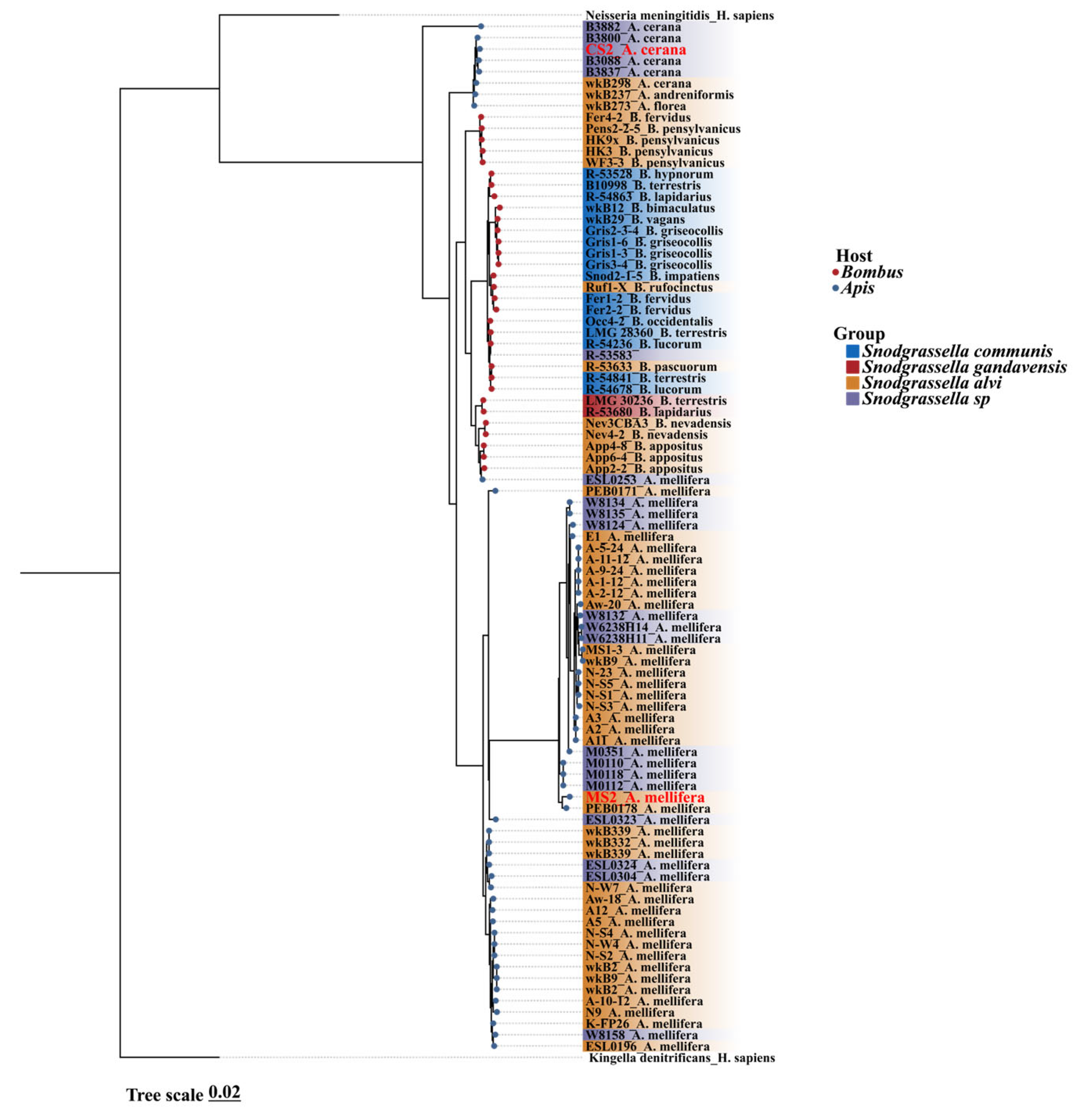
3.1. Snodgrassella Phylogenetic Trees Based on Core Gene Sequences Were Clustered by Host Type

We constructed a phylogenetic tree using 89 Snodgrassella genomes sourced from the NCBI database, along with two strains isolated in this study. Additionally, we selected Kingella denitrificans (GCA_016127355.1) and Neisseria meningitidis (GCA_022869645.1) as outgroups for tree construction. Of these, 59 strains were from the Apis genus, with 51 originating from A. mellifera, 6 from A. cerana, and two from other Apis species. Additionally, 31 strains were derived from the Bombus genus, and one strain, R-53583, was of unknown origin. The inclusion criteria for genome selection were genome completeness greater than 90% and contamination levels below 5%. The phylogenetic inference for Snodgrassella was constructed based on core genes. The strains in the phylogenetic tree predominantly clustered according to their host species. Our A. mellifera derived isolate, strain MS2, clustered within the S. alvi clade, whereas the A. cerana derived strain, CS2, grouped within an unclassified Snodgrassella clade (Figure 1). Notably, within the Snodgrassella phylogeny, strains from the same host species typically clustered together. Strains derived from A. cerana and Bombus species formed two sister clades, which were in turn sister to the Snodgrassella strains of A. mellifera.

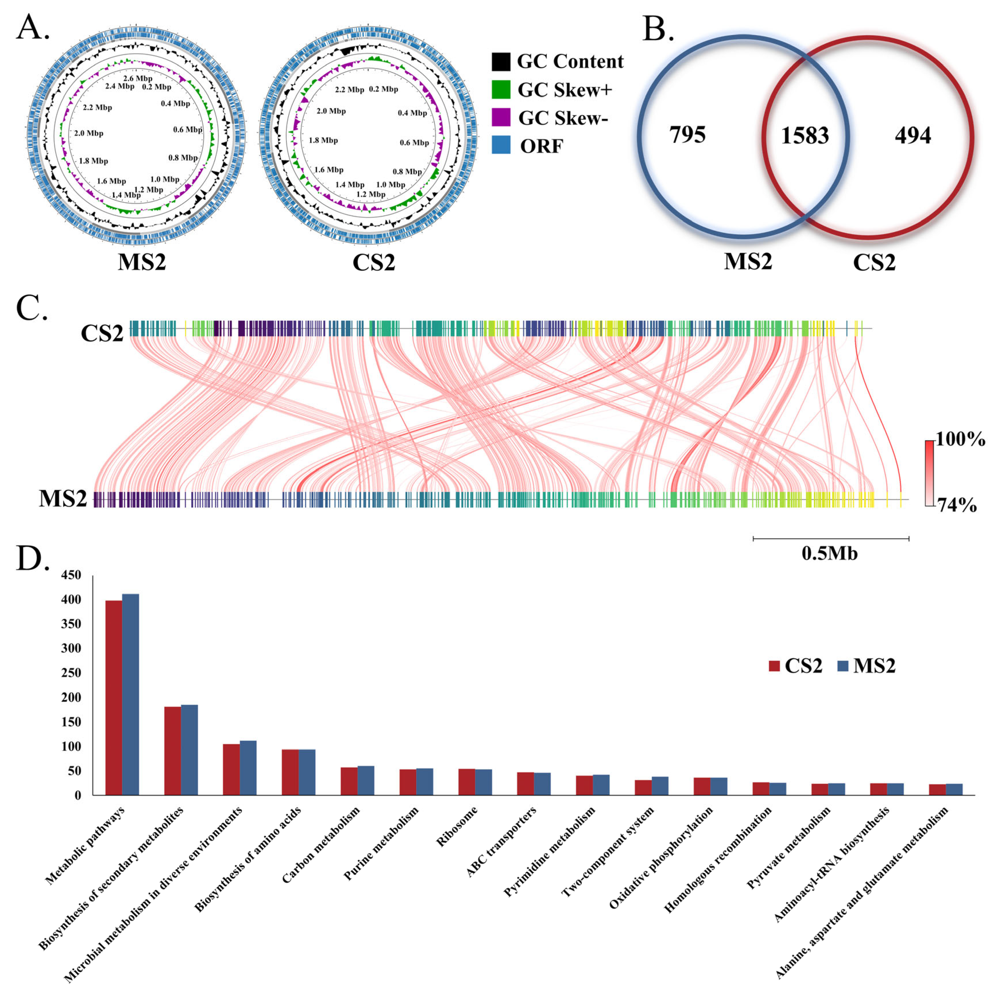
3.2. Genome Characteristics of Snodgrassella

The genomes of the isolated strains CS2 and MS2 were assembled into 34 and 39 scaffolds, respectively, resulting in genome sizes of 2,386,043 bp and 2,621,985 bp (Figure 2A). The CS2 genome encodes 2077 proteins, 55 tRNAs, and 3 rRNA operons, while the MS2 genome encodes 2378 proteins, 57 tRNAs, and 4 rRNA operons (Figure 2B and Table S1). The MS2 genome is larger than that of CS2, with a higher number of protein-coding genes, non-coding RNAs (ncRNAs), and mobile genetic elements (Table S1). The average nucleotide identity (ANI) values among the 91 Snodgrassella genomes ranged from 73.58% to 99.99% (Figure S1), with a ANI of 79.83% between the genomes of CS2 and MS2 (Figure 2C). KEGG annotation revealed that the two strains share a similar functional profile, with genes primarily enriched in categories such as metabolic pathways, biosynthesis of secondary metabolites, microbial metabolism in diverse environments, biosynthesis of amino acids, and carbon metabolism (Figure 2D). Notably, MS2 exhibited a higher number of genes enriched in these major functional categories compared to CS2 (Figure 2D).

3.3. Gut Colonization of Snodgrassella Strains

To explore the physiological basis of host-specific evolution, Snodgrassella strains from A. cerana and A. mellifera were inoculated into the gut of germ-free worker bees of the corresponding species, which were reared in the laboratory. The bacterial load in the germ-free worker bees was less than 105, confirming the successful establishment of the germ-free honeybee model (Figure 3A,B). The results from the cross-colonization experiment demonstrated that both A. cerana and A. mellifera strains were able to successfully colonize the gut of honeybees from different host species, with colonization levels reaching 108 copies (Figure 3A,B). Although no significant difference in colonization levels was observed between native and non-native strain in both A. cerana and A. mellifera gut (Figure 3A,B), the competitive colonization experiment showed that native strain exhibited a stronger colonization advantage in the honeybee gut (Figure 3C).

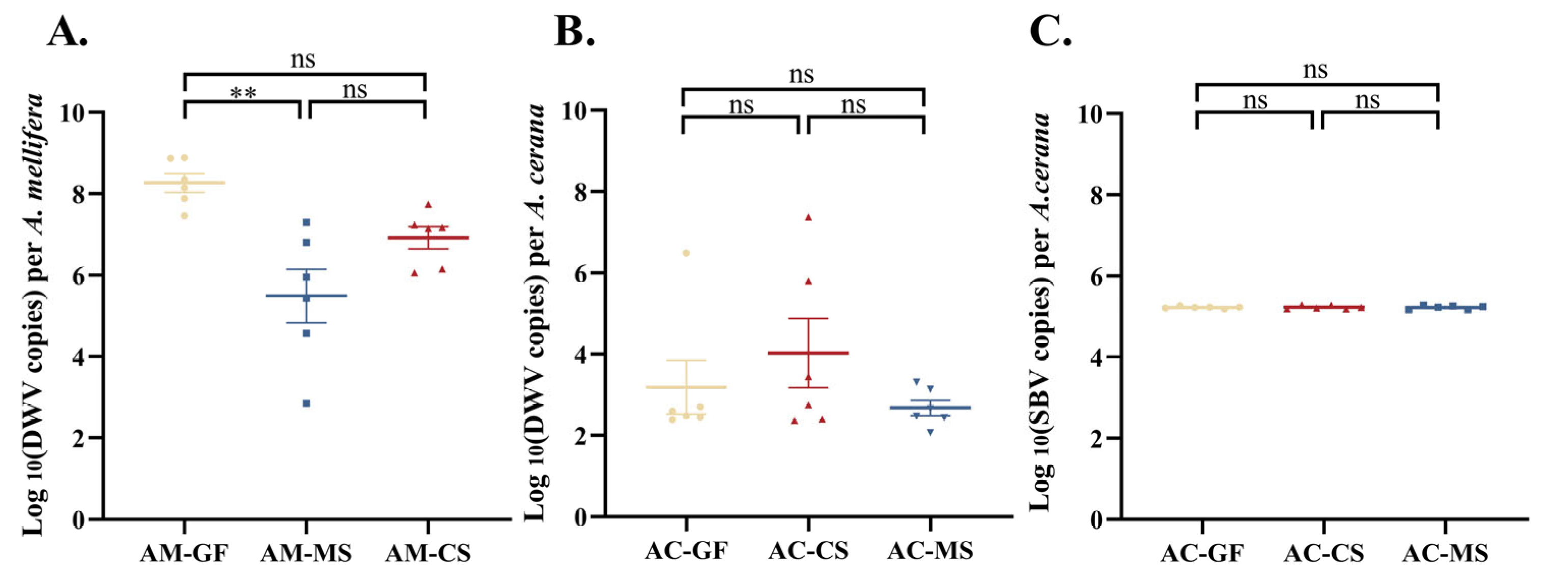
3.4. Colonization of Native Snodgrassella Strain Reduces Naturally Occurring Virus Titers in A. mellifera

Viral titers naturally produced in the host following colonization with native and non-native Snodgrassella strains were measured. Compared to the germ-free control group, the MS2 treatment significantly reduced DWV titers in A. mellifera, whereas the CS2 treatment had no significant effect on DWV titers (Figure 4A). In contrast, for A. cerana, inoculation with either MS2 or CS2 did not result in significant changes in the titers of SBV or DWV (Figure 4B,C). Notably, regardless of treatment or the origin of the Snodgrassella strain, the natural DWV titers in A. cerana were consistently lower than those in A. mellifera under the same experimental conditions. (Figure 4B,C).

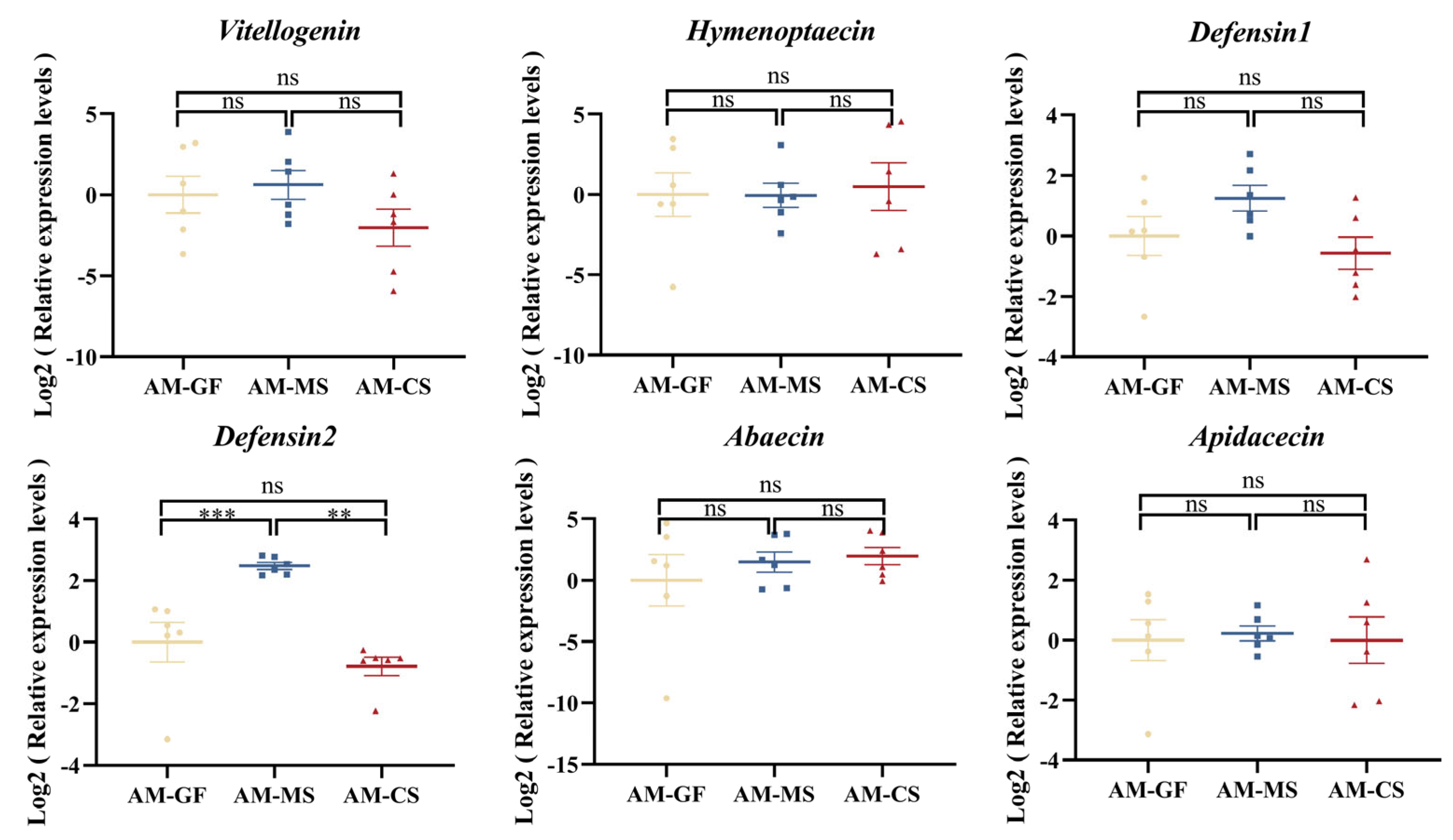
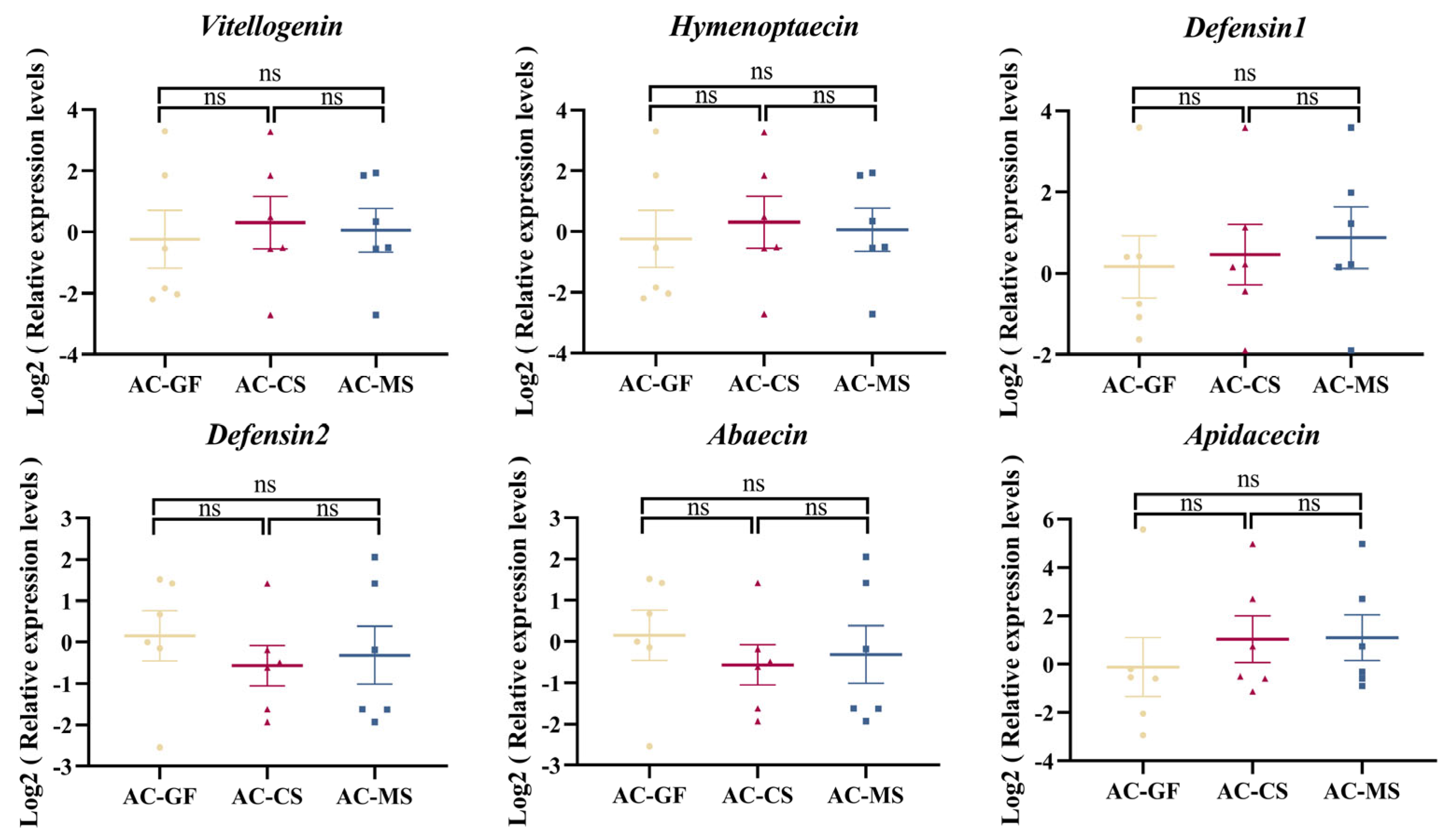
3.5. Colonization of Native Snodgrassella Strain Promotes the Expression of the Defensin 2 Gene in A. mellifera

To investigate whether the effect of Snodgrassella colonization on host immunity varies depending on the strain origin, we measured the expression levels of immune-related genes in the host after stable colonization of the gut with Snodgrassella strains from different sources. The results showed that, in A. mellifera, inoculation with MS2 significantly increased the expression of defensin 2 gene compared to both the control group and the CS2 group (Figure 5). However, in A. cerana, no significant changes in immune gene expression were observed, regardless of whether the strain was native or non-native (Figure 6).

4. Discussion

Revealing the interactions between animal hosts and their symbiotic microbiota in different animal models helps enhance our understanding of the diversity of host-symbiont relationships and the evolutionary history of these interactions. The geographic isolation of A. cerana and A. mellifera for millions of years has limited the potential interactions between their gut microbiota, allowing for the development of specialized gut microbiomes that co-evolved with their respective hosts [52]. In this study, we isolated Snodgrassella strains from the gut microbiota of both honeybee species and conducted colonization experiments to explore the host specificity of Snodgrassella strains and their underlying physiological and functional basis. Our results confirm the evolution of gut microbiome host specificity within species and reveal the impact of strain-level variation on differences in host physiological functions.
Phylogenetic analysis of existing Snodgrassella strains based on core genes revealed that Snodgrassella strains from A. cerana and A. mellifera formed distinct branches, indicating extensive genomic differences between species. Interestingly, the phylogeny of Snodgrassella strains did not align with host phylogeny, which is consistent with previous studies based on gene flow trees [13]. However, recent metagenomic sequencing based on the core genome of Snodgrassella showed a phylogenetic pattern that is consistent with the host phylogeny [53]. This discrepancy may be attributed to occasional host-switching events, disrupting the co-diversification between symbionts and their hosts [21,54]. The ANI values between the genomes of Snodgrassella strains still exceed 95%, suggesting that finer taxonomic distinctions are needed for classifying bee gut microbiota. Consistent with previous metagenomic sequencing results of A. cerana and A. mellifera [28,29], our genomic analysis of individual strains reveals that the A. mellifera isolates have larger genomes, higher gene content, and a greater number of MGEs. This may be related to the translocation and expansion of A. mellifera populations outside their native range, which requires larger genomes and functional genes to rapidly adapt to changing environments. Variations in the genes of gut microbiota and their functional differentiation are key drivers of host adaptive evolution and provide the physiological and functional basis for host-specific colonization [13]. Previous studies on the host specificity of gut microbiota in A. mellifera and bumblebees have shown that native strains exhibit a distinct competitive advantage in colonization experiments [12,20,21,22,23,24]. In our study, we similarly observed a competitive advantage of native strain in the gut of both A. cerana and A. mellifera. These results suggest that even among closely related species within the same genus, gut microbiota exhibit host specificity, forming the physiological and functional foundation for host-specific colonization.
The gut microbiota plays a crucial role in defending honeybees against pathogen invasion, including resistance to opportunistic bacteria, fungal pathogens, and RNA viruses [55,56,57,58]. When honeybees are exposed to DWV, those lacking a microbiome exhibit significantly lower survival rates compared to normal honeybees [56]. Antibiotic-induced disruption of the gut microbiota reduces honeybee resistance to IAPV, increasing susceptibility [59,60]. Additionally, pathogen-induced dysbiosis in the gut promotes the proliferation of Chronic Bee Paralysis Virus in the honeybee gut, leading to higher mortality [61]. Studies have shown that S. alvi inoculation can improve honeybee survival under DWV infection [39]. Based on the protective role of S. alvi against host pathogens, plasmid-modified S. alvi has been developed to combat honeybee pathogens such as Microsporidia and DWV through RNA interference [57,62]. In this study, we found that inoculation with native Snodgrassella strain significantly reduced the naturally occurring DWV titers in A. mellifera, whereas inoculation with non-native strain did not significantly alter the virus titers. This highlighting the importance of host specificity in symbiotic systems. These results further confirm the protective role of Snodgrassella in defending A. mellifera from pathogen invasion and underscore the impact of strain-level variation on host physiological functions. However, regardless of whether native or non-native Snodgrassella strains were inoculated, there was no significant change in naturally occurring DWV titers in A. cerana. This indicates a more complex interaction between host populations, microbiota, and virus adaptability. In A. cerana, DWV typically exhibits lower virulence, and the virus-host relationship appears to be more stable [63]. In contrast, in A. mellifera, the virus is more virulent due to cross-species transmission via V. destructor [64]. Initially, the DWV-A virus, originating in the ancestral host A. cerana, is transmitted to A. mellifera by V. destructor, leading to an increase in viral virulence and posing a significant threat to A. mellifera populations [65]. Notably, using universal primers for DWV, we observed that naturally occurring DWV titers in A. mellifera were higher than in A. cerana under the same experimental conditions. Under symptomless viral load limits, the presence of the core gut microbiota Snodgrassella in honeybees seemed to play a role in reducing higher viral titers of naturally occurring DWV. In contrast, Snodgrassella had minimal effect on lower viral titers. Similarly, inoculation with Snodgrassella did not significantly affect the naturally occurring SBV levels in honeybees, which exhibited lower viral titers. Therefore, the beneficial effects of Snodgrassella colonization in reducing naturally occurring DWV titers may be more pronounced in A. mellifera than in A. cerana. These findings suggest that, during seasons with high V. destructor infection rates, inoculation with native Snodgrassella strains could be a potential strategy to control sudden DWV outbreaks by reducing the viral load in asymptomatically infected honeybees. This could help mitigate the synergistic effects of DWV and V. destructor on colony health. These findings align with previously established roles of probiotics in honeybee colonies. For instance, supplementing colonies with the probiotic LX3 (a mixture of Lactiplantibacillus plantarum Lp39, Lacticaseibacillus rhamnosus GR-1, and Apilactobacillus kunkeei BR-1), derived from honeybee gut isolates, has been shown to more effectively reduce pathogen loads compared to oxytetracycline treatment [66]. Furthermore, host-derived strains, such as Lactobacillus kunkeei and Lactiplantibacillus plantarum, provide protective effects against chalkbrood infections [67,68]. Collectively, these results underscore the potential of host-derived bacterial strains as probiotics, enhancing the colony’s resilience to multiple stressors.
The gut microbiota may influence honeybee health by modulating host immune response [69,70,71]. Colonization by the microbiota or its individual members can upregulate the expression of host immune genes, providing protection against opportunistic bacteria, virus, fungi, and parasites [72,73,74,75]. S. alvi colonization can promote the upregulation of host genes encoding antimicrobial peptides [27]. In this study, we also observed that inoculation with native Snodgrassella strain significantly upregulated the expression of the defensin 2 gene in A. mellifera, while inoculation with non-native strain did not induce significant changes in immune gene expression. In contrast, in A. cerana, no significant changes in immune gene expression were observed, regardless of whether the strain was native or non-native. This suggests that the immunomodulatory effects of Snodgrassella strains are host-specific in A. mellifera. The expression of immune genes in A. cerana further confirmed that the host or gut microbiota response is limited when the viral titers are at low copy numbers. The upregulation of defensin 2 gene level in A. mellifera following Snodgrassella inoculation may be one of the mechanisms through which Snodgrassella reduces naturally occurring DWV titers. However, the molecular mechanisms underlying these effects require further investigation. Although A. cerana and A. mellifera are closely related species, we observed differences in their gut Snodgrassella strains roles in immune regulation and virus defense. These findings highlight the species-specific nature of the gut microbiome’s effects on host immunity and underscore the importance of further investigating the interactions between A. cerana gut microbiota and its host.

5. Conclusions

Our study demonstrates that Snodgrassella strains in A. cerana and A. mellifera exhibit host-specific genomic adaptations and colonization advantages, which directly influence host health. In A. mellifera, native Snodgrassella inoculation reduced DWV loads and upregulated the expression of defensin 2, suggesting a protective role against viral proliferation. These findings propose a new biological control strategy: applying native Snodgrassella to asymptomatically DWV-infected colonies can reduce sudden outbreaks and spread of DWV. This suggests that strain specificity should be carefully considered when developing honeybee probiotic formulations. Future work should optimize strain delivery methods and evaluate field efficacy, offering a sustainable tool to enhance pollinator health and ecosystem resilience.

Supplementary Materials

The following supporting information can be downloaded at: https://www.mdpi.com/article/10.3390/insects16050478/s1. Figure S1: The ANI values between the genomes of 91 strains under the Snodgrassella genus; Table S1: Genomic characteristics of 2 Snodgrassella strains; Table S2: Immune gene primers.

Author Contributions

Conceptualization, N.Z., D.L., F.H. and H.Z.; investigation, N.Z., S.Y. and R.W.; data curation, N.Z., S.Y. and R.W.; writing-original draft preparation, N.Z. and D.L.; writing-review and editing, N.Z., F.H., D.L. and H.Z.; supervision, D.L., F.H. and H.Z.; funding acquisition, F.H. and H.Z. Author order was determined on the basis of their contribution. All authors have read and agreed to the published version of the manuscript.

Funding

This study was supported by China Agriculture Research System of MOF and MARA (grant number CARS-44).

Data Availability Statement

The original contributions presented in this study are included in the article/Supplementary Materials. The genomes of the isolates are deposited in the NCBI under BioProject PRJNA1256677. Further inquiries can be directed to the corresponding author.

Acknowledgments

We are very grateful to the Experimental Teaching Centre, College of Animal Sciences, Zhejiang University, for providing the quantitative fluorescence instrument to conduct experiments and for supplying the raw data feedback.

Conflicts of Interest

The authors declare no conflicts of interest.

References

  1. Bäckhed, F.; Ley, R.E.; Sonnenburg, J.L.; Peterson, D.A.; Gordon, J.I. Host-Bacterial Mutualism in the Human Intestine. Science 2005, 307, 1915–1920. [Google Scholar] [CrossRef]
  2. Brestoff, J.R.; Artis, D. Commensal Bacteria at the Interface of Host Metabolism and the Immune System. Nat. Immunol. 2013, 14, 676–684. [Google Scholar] [CrossRef]
  3. Turnbaugh, P.J.; Ley, R.E.; Mahowald, M.A.; Magrini, V.; Mardis, E.R.; Gordon, J.I. An Obesity-Associated Gut Microbiome with Increased Capacity for Energy Harvest. Nature 2006, 444, 1027–1031. [Google Scholar] [CrossRef] [PubMed]
  4. Sprockett, D.D.; Price, J.D.; Juritsch, A.F.; Schmaltz, R.J.; Real, M.V.F.; Goldman, S.L.; Sheehan, M.; Ramer-Tait, A.E.; Moeller, A.H. Home-Site Advantage for Host Species–Specific Gut Microbiota. Sci. Adv. 2023, 9, eadf5499. [Google Scholar] [CrossRef]
  5. Moeller, A.H.; Gomes-Neto, J.C.; Mantz, S.; Kittana, H.; Segura Munoz, R.R.; Schmaltz, R.J.; Ramer-Tait, A.E.; Nachman, M.W. Experimental Evidence for Adaptation to Species-Specific Gut Microbiota in House Mice. mSphere 2019, 4, e00387-19. [Google Scholar] [CrossRef] [PubMed]
  6. Zmora, N.; Zilberman-Schapira, G.; Suez, J.; Mor, U.; Dori-Bachash, M.; Bashiardes, S.; Kotler, E.; Zur, M.; Regev-Lehavi, D.; Brik, R.B.-Z.; et al. Personalized Gut Mucosal Colonization Resistance to Empiric Probiotics Is Associated with Unique Host and Microbiome Features. Cell 2018, 174, 1388–1405.e21. [Google Scholar] [CrossRef]
  7. Brooks, A.W.; Kohl, K.D.; Brucker, R.M.; van Opstal, E.J.; Bordenstein, S.R. Correction: Phylosymbiosis: Relationships and Functional Effects of Microbial Communities across Host Evolutionary History. PLoS Biol. 2017, 15, e1002587. [Google Scholar] [CrossRef]
  8. Zheng, H.; Steele, M.I.; Leonard, S.P.; Motta, E.V.S.; Moran, N.A. Honey Bees as Models for Gut Microbiota Research. Lab. Anim. 2018, 47, 317–325. [Google Scholar] [CrossRef]
  9. Martinson, V.G.; Danforth, B.N.; Minckley, R.L.; Rueppell, O.; Tingek, S.; Moran, N.A. A Simple and Distinctive Microbiota Associated with Honey Bees and Bumble Bees. Mol. Ecol. 2011, 20, 619–628. [Google Scholar] [CrossRef]
  10. Corby-Harris, V.; Maes, P.; Anderson, K.E. The Bacterial Communities Associated with Honey Bee (Apis mellifera) Foragers. PLoS ONE 2014, 9, e95056. [Google Scholar] [CrossRef]
  11. Jeyaprakash, A.; Hoy, M.A.; Allsopp, M.H. Bacterial Diversity in Worker Adults of Apis mellifera capensis and Apis mellifera scutellata (Insecta: Hymenoptera) Assessed Using 16S rRNA Sequences. J. Invertebr. Pathol. 2003, 84, 96–103. [Google Scholar] [CrossRef] [PubMed]
  12. Kwong, W.K.; Engel, P.; Koch, H.; Moran, N.A. Genomics and Host Specialization of Honey Bee and Bumble Bee Gut Symbionts. Proc. Natl. Acad. Sci. USA 2014, 111, 11509–11514. [Google Scholar] [CrossRef] [PubMed]
  13. Li, Y.; Leonard, S.P.; Powell, J.E.; Moran, N.A. Species Divergence in Gut-Restricted Bacteria of Social Bees. Proc. Natl. Acad. Sci. USA 2022, 119, e2115013119. [Google Scholar] [CrossRef]
  14. Zhang, Z.; Guo, Y.; Yang, F.; Li, J. Pan-Genome Analysis Reveals Functional Divergences in Gut-Restricted Gilliamella and Snodgrassella. Bioengineering 2022, 9, 544. [Google Scholar] [CrossRef]
  15. Zhang, Z.; Mu, X.; Cao, Q.; Shi, Y.; Hu, X.; Zheng, H. Honeybee Gut Lactobacillus Modulates Host Learning and Memory Behaviors via Regulating Tryptophan Metabolism. Nat. Commun. 2022, 13, 2037. [Google Scholar] [CrossRef] [PubMed]
  16. Liberti, J.; Kay, T.; Quinn, A.; Kesner, L.; Frank, E.T.; Cabirol, A.; Richardson, T.O.; Engel, P.; Keller, L. The Gut Microbiota Affects the Social Network of Honeybees. Nat. Ecol. Evol. 2022, 6, 1471–1479. [Google Scholar] [CrossRef]
  17. Jones, J.C.; Fruciano, C.; Marchant, J.; Hildebrand, F.; Forslund, S.; Bork, P.; Engel, P.; Hughes, W.O.H. The Gut Microbiome Is Associated with Behavioural Task in Honey Bees. Insect. Soc. 2018, 65, 419–429. [Google Scholar] [CrossRef]
  18. Sauers, L.A.; Sadd, B.M. An Interaction between Host and Microbe Genotypes Determines Colonization Success of a Key Bumble Bee Gut Microbiota Member. Evolution 2019, 73, 2333–2342. [Google Scholar] [CrossRef]
  19. Koch, H.; Abrol, D.P.; Li, J.; Schmid-Hempel, P. Diversity and Evolutionary Patterns of Bacterial Gut Associates of Corbiculate Bees. Mol. Ecol. 2013, 22, 2028–2044. [Google Scholar] [CrossRef]
  20. Kwong, W.K.; Moran, N.A. Evolution of Host Specialization in Gut Microbes: The Bee Gut as a Model. Gut Microbes 2015, 6, 214–220. [Google Scholar] [CrossRef]
  21. Powell, E.; Ratnayeke, N.; Moran, N.A. Strain Diversity and Host Specificity in a Specialized Gut Symbiont of Honeybees and Bumblebees. Mol. Ecol. 2016, 25, 4461–4471. [Google Scholar] [CrossRef] [PubMed]
  22. Oh, P.L.; Benson, A.K.; Peterson, D.A.; Patil, P.B.; Moriyama, E.N.; Roos, S.; Walter, J. Diversification of the Gut Symbiont Lactobacillus reuteri as a Result of Host-Driven Evolution. ISME J. 2010, 4, 377–387. [Google Scholar] [CrossRef] [PubMed]
  23. Amiri, N.; Keady, M.M.; Lim, H.C. Honey Bees and Bumble Bees Occupying the Same Landscape Have Distinct Gut Microbiomes and Amplicon Sequence Variant-Level Responses to Infections. PeerJ 2023, 11, e15501. [Google Scholar] [CrossRef] [PubMed]
  24. Guo, L.; Tang, J.; Tang, M.; Luo, S.; Zhou, X. Reactive Oxygen Species Are Regulated by Immune Deficiency and Toll Pathways in Determining the Host Specificity of Honeybee Gut Bacteria. Proc. Natl. Acad. Sci. USA 2023, 120, e2219634120. [Google Scholar] [CrossRef]
  25. Steele, M.I.; Kwong, W.K.; Whiteley, M.; Moran, N.A. Diversification of Type VI Secretion System Toxins Reveals Ancient Antagonism among Bee Gut Microbes. mBio 2017, 8, e01630-17. [Google Scholar] [CrossRef]
  26. Steele, M.I.; Moran, N.A. Evolution of Interbacterial Antagonism in Bee Gut Microbiota Reflects Host and Symbiont Diversification. mSystems 2021, 6, e00063-21. [Google Scholar] [CrossRef]
  27. Motta, E.V.S.; Lariviere, P.J.; Jones, K.R.; Song, Y.; Moran, N.A. Type VI Secretion Systems Promote Intraspecific Competition and Host Interactions in a Bee Gut Symbiont. Proc. Natl. Acad. Sci. USA 2024, 121, e2414882121. [Google Scholar] [CrossRef] [PubMed]
  28. Wu, Y.; Zheng, Y.; Wang, S.; Chen, Y.; Tao, J.; Chen, Y.; Chen, G.; Zhao, H.; Wang, K.; Dong, K.; et al. Genetic Divergence and Functional Convergence of Gut Bacteria between the Eastern Honey Bee Apis cerana and the Western Honey Bee Apis mellifera. J. Adv. Res. 2022, 37, 19–31. [Google Scholar] [CrossRef]
  29. Ellegaard, K.M.; Suenami, S.; Miyazaki, R.; Engel, P. Vast Differences in Strain-Level Diversity in the Gut Microbiota of Two Closely Related Honey Bee Species. Curr. Biol. 2020, 30, 2520–2531.e7. [Google Scholar] [CrossRef]
  30. Zhou, N.; Zheng, Q.; Liu, Y.; Huang, Z.; Feng, Y.; Chen, Y.; Hu, F.; Zheng, H. Strain Diversity and Host Specificity of the Gut Symbiont Gilliamella in Apis mellifera, Apis cerana and Bombus terrestris. Microbiol. Res. 2025, 293, 128048. [Google Scholar] [CrossRef]
  31. Grozinger, C.M.; Flenniken, M.L. Bee Viruses: Ecology, Pathogenicity, and Impacts. Annu. Rev. Entomol. 2019, 64, 205–226. [Google Scholar] [CrossRef] [PubMed]
  32. Highfield, A.C.; El Nagar, A.; Mackinder, L.C.M.; Noël, L.M.-L.J.; Hall, M.J.; Martin, S.J.; Schroeder, D.C. Deformed Wing Virus Implicated in Overwintering Honeybee Colony Losses. Appl. Environ. Microbiol. 2009, 75, 7212–7220. [Google Scholar] [CrossRef] [PubMed]
  33. Wei, R.; Cao, L.; Feng, Y.; Chen, Y.; Chen, G.; Zheng, H. Sacbrood Virus: A Growing Threat to Honeybees and Wild Pollinators. Viruses 2022, 14, 1871. [Google Scholar] [CrossRef] [PubMed]
  34. Chen, G.; Wu, Y.; Deng, J.; Wen, Z.; Wang, S.; Chen, Y.; Hu, F.; Zheng, H. Seasonal Variation of Viral Infections between the Eastern Honey Bee (Apis cerana) and the Western Honey Bee (Apis mellifera). Microbiol. Open 2021, 10, e1162. [Google Scholar] [CrossRef]
  35. Evans, J.D.; Aronstein, K.; Chen, Y.P.; Hetru, C.; Imler, J.-L.; Jiang, H.; Kanost, M.; Thompson, G.J.; Zou, Z.; Hultmark, D. Immune Pathways and Defence Mechanisms in Honey Bees Apis mellifera. Insect Mol. Biol. 2006, 15, 645–656. [Google Scholar] [CrossRef]
  36. Chen, Y.P.; Pettis, J.S.; Corona, M.; Chen, W.P.; Li, C.J.; Spivak, M.; Visscher, P.K.; DeGrandi-Hoffman, G.; Boncristiani, H.; Zhao, Y.; et al. Israeli Acute Paralysis Virus: Epidemiology, Pathogenesis and Implications for Honey Bee Health. PLoS Pathog. 2014, 10, e1004261. [Google Scholar] [CrossRef]
  37. Mookhploy, W.; Krongdang, S.; Chantawannakul, P. Effects of Deformed Wing Virus Infection on Expressions of Immune- and Apoptosis-Related Genes in Western Honeybees (Apis mellifera). Insects 2021, 12, 82. [Google Scholar] [CrossRef]
  38. Wu, Y.; Zheng, Y.; Chen, Y.; Chen, G.; Zheng, H.; Hu, F. Apis cerana Gut Microbiota Contribute to Host Health Though Stimulating Host Immune System and Strengthening Host Resistance to Nosema ceranae. R. Soc. Open Sci. 2020, 7, 192100. [Google Scholar] [CrossRef]
  39. Leonard, S.P.; Powell, J.E.; Perutka, J.; Geng, P.; Heckmann, L.C.; Horak, R.D.; Davies, B.W.; Ellington, A.D.; Barrick, J.E.; Moran, N.A. Engineered Symbionts Activate Honey Bee Immunity and Limit Pathogens. Science 2020, 367, 573–576. [Google Scholar] [CrossRef]
  40. Bolger, A.M.; Lohse, M.; Usadel, B. Trimmomatic: A Flexible Trimmer for Illumina Sequence Data. Bioinformatics 2014, 30, 2114–2120. [Google Scholar] [CrossRef] [PubMed]
  41. Simpson, J.T.; Wong, K.; Jackman, S.D.; Schein, J.E.; Jones, S.J.M.; Birol, I. ABySS: A Parallel Assembler for Short Read Sequence Data. Genome Res. 2009, 19, 1117–1123. [Google Scholar] [CrossRef]
  42. Xu, M.; Guo, L.; Gu, S.; Wang, O.; Zhang, R.; Peters, B.A.; Fan, G.; Liu, X.; Xu, X.; Deng, L.; et al. TGS-GapCloser: A Fast and Accurate Gap Closer for Large Genomes with Low Coverage of Error-Prone Long Reads. GigaScience 2020, 9, giaa094. [Google Scholar] [CrossRef] [PubMed]
  43. Schwengers, O.; Jelonek, L.; Dieckmann, M.A.; Beyvers, S.; Blom, J.; Goesmann, A. Bakta: Rapid and Standardized Annotation of Bacterial Genomes via Alignment-Free Sequence Identification. Microb. Genom. 2021, 7, 685. [Google Scholar] [CrossRef]
  44. Lowe, T.M.; Eddy, S.R. tRNAscan-SE: A Program for Improved Detection of Transfer RNA Genes in Genomic Sequence. Nucleic Acids Res. 1997, 25, 955–964. [Google Scholar] [CrossRef] [PubMed]
  45. Lagesen, K.; Hallin, P.; Rødland, E.A.; Stærfeldt, H.-H.; Rognes, T.; Ussery, D.W. RNAmmer: Consistent and Rapid Annotation of Ribosomal RNA Genes. Nucleic Acids Res. 2007, 35, 3100–3108. [Google Scholar] [CrossRef]
  46. Brown, C.L.; Mullet, J.; Hindi, F.; Stoll, J.E.; Gupta, S.; Choi, M.; Keenum, I.; Vikesland, P.; Pruden, A.; Zhang, L. mobileOG-Db: A Manually Curated Database of Protein Families Mediating the Life Cycle of Bacterial Mobile Genetic Elements. Appl. Environ. Microbiol. 2022, 88, e00991-22. [Google Scholar] [CrossRef]
  47. Emms, D.M.; Kelly, S. OrthoFinder: Phylogenetic Orthology Inference for Comparative Genomics. Genome Biol. 2019, 20, 238. [Google Scholar] [CrossRef]
  48. Kanehisa, M.; Goto, S.; Sato, Y.; Furumichi, M.; Tanabe, M. KEGG for Integration and Interpretation of Large-Scale Molecular Data Sets. Nucleic Acids Res. 2012, 40, D109–D114. [Google Scholar] [CrossRef]
  49. Tian, R.; Imanian, B. VBCG: 20 Validated Bacterial Core Genes for Phylogenomic Analysis with High Fidelity and Resolution. Microbiome 2023, 11, 247. [Google Scholar] [CrossRef]
  50. Blanchard, P.; Guillot, S.; Antùnez, K.; Köglberger, H.; Kryger, P.; de Miranda, J.R.; Franco, S.; Chauzat, M.-P.; Thiéry, R.; Ribière, M. Development and Validation of a Real-Time Two-Step RT-qPCR TaqMan® Assay for Quantitation of Sacbrood Virus (SBV) and Its Application to a Field Survey of Symptomatic Honey Bee Colonies. J. Virol. Methods 2014, 197, 7–13. [Google Scholar] [CrossRef]
  51. Xie, J.; Chen, Y.; Cai, G.; Cai, R.; Hu, Z.; Wang, H. Tree Visualization By One Table (tvBOT): A Web Application for Visualizing, Modifying and Annotating Phylogenetic Trees. Nucleic Acids Res. 2023, 51, W587–W592. [Google Scholar] [CrossRef] [PubMed]
  52. Nannan, L.; Huamiao, L.; Yan, J.; Xingan, L.; Yang, L.; Tianjiao, W.; Jinming, H.; Qingsheng, N.; Xiumei, X. Geometric Morphology and Population Genomics Provide Insights into the Adaptive Evolution of Apis cerana in Changbai Mountain. BMC Genom. 2022, 23, 64. [Google Scholar] [CrossRef] [PubMed]
  53. Feng, Y.; Wei, R.; Chen, Q.; Shang, T.; Zhou, N.; Wang, Z.; Chen, Y.; Chen, G.; Zhang, G.; Dong, K.; et al. Host Specificity and Cophylogeny in the “Animal-Gut Bacteria-Phage” Tripartite System. npj Biofilms Microbiomes 2024, 10, 72. [Google Scholar] [CrossRef]
  54. Kwong, W.K.; Medina, L.A.; Koch, H.; Sing, K.-W.; Soh, E.J.Y.; Ascher, J.S.; Jaffé, R.; Moran, N.A. Dynamic Microbiome Evolution in Social Bees. Sci. Adv. 2017, 3, e1600513. [Google Scholar] [CrossRef] [PubMed]
  55. Raymann, K.; Moran, N.A. The Role of the Gut Microbiome in Health and Disease of Adult Honey Bee Workers. Curr. Opin. Insect Sci. 2018, 26, 97–104. [Google Scholar] [CrossRef] [PubMed]
  56. Dosch, C.; Manigk, A.; Streicher, T.; Tehel, A.; Paxton, R.J.; Tragust, S. The Gut Microbiota Can Provide Viral Tolerance in the Honey Bee. Microorganisms 2021, 9, 871. [Google Scholar] [CrossRef]
  57. Lang, H.; Wang, H.; Wang, H.; Zhong, Z.; Xie, X.; Zhang, W.; Guo, J.; Meng, L.; Hu, X.; Zhang, X.; et al. Engineered Symbiotic Bacteria Interfering Nosema Redox System Inhibit Microsporidia Parasitism in Honeybees. Nat. Commun. 2023, 14, 2778. [Google Scholar] [CrossRef]
  58. Miller, D.L.; Smith, E.A.; Newton, I.L.G. A Bacterial Symbiont Protects Honey Bees from Fungal Disease. Mbio 2021, 12, e00503-21. [Google Scholar] [CrossRef]
  59. Deng, Y.; Yang, S.; Zhao, H.; Diao, Q.; Hou, C. IAPV-Induced Paralytic Symptoms Associated with Tachypnea via Impaired Tracheal System Function. Int. J. Mol. Sci. 2021, 22, 10078. [Google Scholar] [CrossRef]
  60. Liu, Y.; Jia, S.; Wu, Y.; Zhou, N.; Xie, Y.; Wei, R.; Huang, Z.; Chen, Y.; Hu, F.; Zheng, H. Tetracycline-Induced Gut Community Dysbiosis and Israeli Acute Paralysis Virus Infection Synergistically Negatively Affect Honeybees. Ecotoxicol. Environ. Saf. 2024, 282, 116706. [Google Scholar] [CrossRef]
  61. Deng, Y.; Yang, S.; Zhang, L.; Chen, C.; Cheng, X.; Hou, C. Chronic Bee Paralysis Virus Exploits Host Antimicrobial Peptides and Alters Gut Microbiota Composition to Facilitate Viral Infection. ISME J. 2024, 18, wrae051. [Google Scholar] [CrossRef] [PubMed]
  62. Huang, Q.; Lariviere, P.J.; Powell, J.E.; Moran, N.A. Engineered Gut Symbiont Inhibits Microsporidian Parasite and Improves Honey Bee Survival. Proc. Natl. Acad. Sci. USA 2023, 120, e2220922120. [Google Scholar] [CrossRef] [PubMed]
  63. Wang, S.; Chen, G.; Lin, Z.; Wu, Y.; Hu, F.; Zheng, H. Occurrence of Multiple Honeybee Viruses in the Ectoparasitic Mites Varroa Spp. in Apis cerana Colonies. J. Invertebr. Pathol. 2019, 166, 107225. [Google Scholar] [CrossRef] [PubMed]
  64. McMahon, D.P.; Natsopoulou, M.E.; Doublet, V.; Fürst, M.; Weging, S.; Brown, M.J.F.; Gogol-Döring, A.; Paxton, R.J. Elevated Virulence of an Emerging Viral Genotype as a Driver of Honeybee Loss. Proc. R. Soc. B Biol. Sci. 2016, 283, 20160811. [Google Scholar] [CrossRef]
  65. Hasegawa, N.; Techer, M.A.; Adjlane, N.; al-Hissnawi, M.S.; Antúnez, K.; Beaurepaire, A.; Christmon, K.; Delatte, H.; Dukku, U.H.; Eliash, N.; et al. Evolutionarily Diverse Origins of Deformed Wing Viruses in Western Honey Bees. Proc. Natl. Acad. Sci. USA 2023, 120, e2301258120. [Google Scholar] [CrossRef]
  66. Li, J.H.; Evans, J.D.; Li, W.F.; Zhao, Y.Z.; DeGrandi-Hoffman, G.; Huang, S.K.; Li, Z.G.; Hamilton, M.; Chen, Y.P. Lactobacillus Spp. Attenuate Antibiotic-Induced Immune and Microbiota Dysregulation in Honey Bees. PLoS ONE 2017, 12, e0187505. [Google Scholar] [CrossRef]
  67. Iorizzo, M.; Lombardi, S.J.; Ganassi, S.; Testa, B.; Ianiro, M.; Letizia, F.; Succi, M.; Tremonte, P.; Vergalito, F.; Cozzolino, A.; et al. Antagonistic Activity against Ascosphaera apis and Functional Properties of Lactobacillus Kunkeei Strains. Antibiotics 2020, 9, 262. [Google Scholar] [CrossRef]
  68. Iorizzo, M.; Testa, B.; Ganassi, S.; Lombardi, S.J.; Ianiro, M.; Letizia, F.; Succi, M.; Tremonte, P.; Vergalito, F.; Cozzolino, A.; et al. Probiotic Properties and Potentiality of Lactiplantibacillus plantarum Strains for the Biological Control of Chalkbrood Disease. J. Fungi 2021, 7, 379. [Google Scholar] [CrossRef]
  69. Sattayawat, P.; Inwongwan, S.; Noirungsee, N.; Li, J.; Guo, J.; Disayathanoowat, T. Engineering Gut Symbionts: A Way to Promote Bee Growth? Insects 2024, 15, 369. [Google Scholar] [CrossRef]
  70. Wu, Y.; Zheng, Y.; Chen, Y.; Wang, S.; Chen, Y.; Hu, F.; Zheng, H. Honey Bee (Apis mellifera) Gut Microbiota Promotes Host Endogenous Detoxification Capability via Regulation of P450 Gene Expression in the Digestive Tract. Microb. Biotechnol. 2020, 13, 1201–1212. [Google Scholar] [CrossRef]
  71. Motta, E.V.S.; Moran, N.A. The Honeybee Microbiota and Its Impact on Health and Disease. Nat. Rev. Microbiol. 2024, 22, 122–137. [Google Scholar] [CrossRef] [PubMed]
  72. Kwong, W.K.; Mancenido, A.L.; Moran, N.A. Immune System Stimulation by the Native Gut Microbiota of Honey Bees. R. Soc. Open Sci. 2017, 4, 170003. [Google Scholar] [CrossRef] [PubMed]
  73. Emery, O.; Schmidt, K.; Engel, P. Immune System Stimulation by the Gut Symbiont Frischella perrara in the Honey Bee (Apis mellifera). Mol. Ecol. 2017, 26, 2576–2590. [Google Scholar] [CrossRef] [PubMed]
  74. Horak, R.D.; Leonard, S.P.; Moran, N.A. Symbionts Shape Host Innate Immunity in Honeybees. Proc. R. Soc. B 2020, 287, 20201184. [Google Scholar] [CrossRef] [PubMed]
  75. Lang, H.; Duan, H.; Wang, J.; Zhang, W.; Guo, J.; Zhang, X.; Hu, X.; Zheng, H. Specific Strains of Honeybee Gut Lactobacillus Stimulate Host Immune System to Protect against Pathogenic Hafnia alvei. Microbiol. Spectrum. 2022, 10, e01896-21. [Google Scholar] [CrossRef]
Figure 1. Maximum−likelihood phylogenies of 91 Snodgrassella strains, along with two outgroup strains (Kingella denitrificans and Neisseria meningitidis), were constructed based on core genes. The color blocks on the branches, represent the bacterial species classification. Blue and red circles at the branch nodes describes the genus of the host. Strains isolated in this study are highlighted in red text.
Insects 16 00478 g001
Figure 2. Genomic structures of Snodgrassella strains derived from A. cerana and A. mellifera. (A) Schematic representation and statistical overview of Snodgrassella genomes sequenced from A. cerana and A. mellifera. (B) Numbers of shared orthologous genes and unique genes in Snodgrassella strains from A. cerana and A. mellifera. (C) Average nucleotide identity (ANI) of Snodgrassella genomes derived from A. cerana and A. mellifera. (D) Gene content categorized by the Kyoto Encyclopedia of Genes and Genomes (KEGG) subsystems. MS2: A. mellifera derived Snodgrassella strain; CS2: A. cerana derived Snodgrassella strain.
Insects 16 00478 g002
Figure 3. Colonization of Snodgrassella strains from A. cerana and A. mellifera in honeybee gut (each group contains 6 samples). (A) Colonization of Snodgrassella strain from A. cerana or A. mellifera in the gut of A. mellifera workers. (B) Colonization of Snodgrassella strain from A. cerana or A. mellifera in the gut of A. cerana workers. (C) Competitive colonization of Snodgrassella strains from A. cerana and A. mellifera in honeybee gut. The horizontal line on the graph represents the mean of each group, while the whiskers indicate the standard error of the mean (SEM) for each group. Significance levels are marked by different symbols: ****, p < 0.0001; ns, not significant. AM-GF: Germ-free A. mellifera workers (control group). AM-MS: A. mellifera workers inoculated with A. mellifera derived Snodgrassella strain. AM-CS: A. mellifera workers inoculated with A. cerana derived Snodgrassella strain. AC-GF: Germ-free A. cerana workers (control group). AC-MS: A. cerana workers inoculated with A. mellifera derived Snodgrassella strain. AC-CS: A. cerana workers inoculated with A. cerana derived Snodgrassella strain. AC-SNO: A. cerana derived Snodgrassella strain. AM-SNO: A. mellifera derived Snodgrassella strain.
Insects 16 00478 g003
Figure 4. Naturally occurring virus titers in honeybees following colonization with Snodgrassella strains from A. cerana and A. mellifera (each group contains 6 samples). (A) Naturally occurring DWV titers in A. mellifera following colonization with Snodgrassella strain from A. cerana or A. mellifera. (B) Naturally occurring DWV titers in A. cerana following colonization with Snodgrassella strain from A. mellifera or A. cerana. (C) Naturally occurring SBV titers in A. cerana following colonization with Snodgrassella strain from A. mellifera or A. cerana. The horizontal line on the graph represents the mean of each group, while the whiskers indicate the SEM for each group. Significance levels are marked by different symbols: **, p < 0.01; ns, not significant. AM-GF: Germ-free Apis mellifera workers (control group). AM-MS: A. mellifera workers inoculated with A. mellifera derived Snodgrassella strain. AM-CS: A. mellifera workers inoculated with A. cerana derived Snodgrassella strain. AC-GF: Germ-free A. cerana workers (control group). AC-MS: A. cerana workers inoculated with A. mellifera derived Snodgrassella strain. AC-CS: A. cerana workers inoculated with A. cerana derived Snodgrassella strain.
Insects 16 00478 g004
Figure 5. Expression of immune−related genes in A. mellifera following colonization with Snodgrassella strains from A. cerana and A. mellifera (each group contains 6 samples). The horizontal line on the graph represents the mean of each group, while the whiskers indicate the SEM for each group. Significance levels are marked by different symbols: **, p < 0.01; ***, p < 0.001; ns, not significant. AM-GF: Germ-free A. mellifera workers (control group). AM-MS: A. mellifera workers inoculated with A. mellifera derived Snodgrassella strain. AM-CS: A. mellifera workers inoculated with A. cerana derived Snodgrassella strain.
Insects 16 00478 g005
Figure 6. Expression of immune-related genes in A. cerana following colonization with Snodgrassella strains from A. cerana and A. mellifera (each group contains 6 samples). The horizontal line on the graph represents the mean of each group, while the whiskers indicate the SEM for each group. Significance levels are marked by different symbols: ns, not significant. AC-GF: Germ-free A. cerana workers (control group). AC-MS: A. cerana workers inoculated with A. mellifera derived Snodgrassella strain. AC-CS: A. cerana workers inoculated with A. cerana derived Snodgrassella strain.
Insects 16 00478 g006
Disclaimer/Publisher’s Note: The statements, opinions and data contained in all publications are solely those of the individual author(s) and contributor(s) and not of MDPI and/or the editor(s). MDPI and/or the editor(s) disclaim responsibility for any injury to people or property resulting from any ideas, methods, instructions or products referred to in the content.

