Article

Monitoring the Capacity of Microsporidia MB Transgenerational Spread in Anopheles arabiensis Populations

by
Godfred Yaw Boanyah
1,2,
Lizette L. Koekemoer
2,3,
Jeremy K. Herren
1,2,* and
Tullu Bukhari
1,*
1
International Centre of Insect Physiology and Ecology, Nairobi P.O. Box 30772-00100, Kenya
2
Wits Research Institute for Malaria, Faculty of Health Sciences, University of the Witwatersrand, Private Bag 3, Johannesburg 2050, South Africa
3
Centre for Emerging Zoonotic & Parasitic Diseases, Division of the National Health Laboratory Service, National Institute for Communicable Diseases, Johannesburg 2192, South Africa
*
Authors to whom correspondence should be addressed.
Insects 2025, 16(12), 1206; https://doi.org/10.3390/insects16121206
Submission received: 14 October 2025 / Revised: 9 November 2025 / Accepted: 14 November 2025 / Published: 27 November 2025

Simple Summary

Microsporidia MB is a natural symbiont of Anopheles arabiensis that blocks Plasmodium transmission without fitness cost to the mosquito host. Understanding the intergenerational spread of this symbiont in mosquito populations is necessary before applying it in the field as a malaria control tool. The results provide the foundation for improving and maintaining high intensity and prevalence of Microsporidia MB in mosquito populations.

Abstract

Microsporidia MB is a natural symbiont of Anopheles arabiensis Patton, 1905 that blocks Plasmodium transmission without compromising mosquito fitness. Developing this symbiont for malaria control requires a thorough understanding of its transmission dynamics across generations under environmentally relevant conditions. This study aimed to evaluate the transgenerational spread of Microsporidia MB in semi-field settings. From field-collected infected females (G0), several independent replicates of Microsporidia MB-infected An. arabiensis were established and maintained under semi-field conditions for six generations (F1–F6). All the mosquitoes were screened for Microsporidia MB prevalence and infection intensity using qPCR after death. Temperature and humidity and wing size were recorded. Two replicates sustained through F6. Microsporidia MB prevalence and intensity increased from F1 to F3–F4 before declining. A weak positive correlation was observed between prevalence and temperature, but not humidity. Female mosquitoes exhibited higher infection intensities. Female wing size varied across generations, while male size remained consistent. This first study on Microsporidia MB transgenerational transmission under semi-field conditions revealed a pattern of initial increase followed by decline in prevalence and intensity. The stability of male size supports the potential use of infected males in release programs. These findings underscore additional investigations into environmental drivers for mosquito symbiont prevalence.

1. Introduction

Microsporidia MB, a recently discovered natural symbiont of Anopheles mosquitoes, has been shown to block Plasmodium transmission, making it a promising candidate for novel malaria control strategies [1,2]. Microsporidia MB is transmitted vertically, from mother to offspring, and horizontally, through mating [2,3,4,5]. Vertical transmission in general is moderate to high. Field-collected Anopheles arabiensis Patton, 1905 mothers transmit Microsporidia MB to 45–100% of their offsprings on average [2]. Similarly in the sibling species, An. gambiae Giles, 1902 sensu stricto (s.s.), vertical transmission has been reported to be 28.6–85.3% in pooled lines but when observed in iso-female lines, vertical transmission can range from 0 to 100% [4]. To date, mating is the only reported mechanism of horizontal Microsporidia MB transmission. Horizontal transmission, although heterogeneous, is moderate and higher from infected males to uninfected females (41.5%) than from infected females to uninfected males (22.4%) in An. arabiensis in individuals where mating is confirmed [3,5]. Microsporidia MB-infected males show twice the mating rate and mating competitiveness compared to uninfected males [3]. Other modes of horizontal transmission, such as ingestion of spores, may also be possible but have not been reported to date. Microsporidia MB’s efficient vertical and horizontal transmission likely contributes to its widespread presence in natural Anopheles populations across Kenya, Ghana, Niger, and Nigeria. While temperature and humidity influence Microsporidia MB’s prevalence and intensity, the symbiont persists across diverse ecological conditions [2,6,7,8,9].
Microsporidia MB, an obligate intracellular parasite and symbiont, belongs to the highly host-specific microsporidian group, typically infecting only closely related species [10,11,12]. This narrow host range, combined with poorly understood spore germination triggers and tissue-specific adaptations, complicates in vitro cultivation [10]. A Microsporidia MB-based malaria control intervention will likely involve either the dissemination of infective environmental spores or the release of Microsporidia MB-infected mosquitoes, either male only or both male and female [1]. Both approaches will rely on the ability of Microsporidia MB to spread across natural mosquito populations via vertical and horizontal transmission. Therefore, understanding the natural spread of Microsporidia MB across generations under close-to-natural conditions is essential to inform the development of the intervention strategy.
Microsporidia MB is similar to Wolbachia, because both are naturally occurring symbionts that reduce the potential of mosquitoes to transmit diseases and are transmitted vertically. Wolbachia have been reported in wild-caught Aedes albopictus Skuse, 1894 and Aedes aegypti Linnaeus in Hasselquist, 1762 of which the latter is not a natural host [13,14]. An important difference between Wolbachia and Microsporidia MB is the capacity for Microsporidia MB to be sexually transmitted [5]. A study in India sought to determine the transgenerational spread of Wolbachia in Ae. albopictus where wild-caught mosquitoes from the field were brought to the laboratory and reared from F1 through to F7 [15]. The results showed an overall increase in Wolbachia prevalence from F1 to F7 in four districts while a reverse or decreasing trend was observed in the remaining two districts [15]. These difference in prevalence could be attributed to strain diversity [16].
In addition, environmental factors substantially affect both symbiont transmission and the host in various ways [17,18,19,20]. The diet constitutes a significant aspect of the host–symbiont relationship [21,22]. Moreover, factors such as temperature and humidity have been identified as crucial determinants of the stability and intensity of the symbionts [18,19,20,23]. A study conducted by Doremus et al. [20] on Cardinium in the parasitic wasp host, Encarsia suzannae, showed that an elevated temperature of 32 °C diminished Cardinium intensity, prolonged pupal maturation time, decreased vertical transmission rate, and weakened both cytoplasmic incompatibility modification and rescue efficacy compared to the control at 27 °C. A cool temperature of 20 °C, on the other hand, also diminished symbiont density and extended the host pupal stage, illustrating the substantial influence of temperature on symbionts [20]. Moreover, Wolbachia has also been shown to be at high intensity in D. melanogaster Meigen, 1830 at 13 °C compared to higher temperatures of 23 °C and 31 °C, respectively [18]. The investigation of the impact of temperature on Wolbachia intensity at various developmental stages of the Ae. albopictus mosquito demonstrated a notable reduction in bacterial intensity and larval density after exposure to an increased temperature of 37 °C in both sexes [24]. Lastly, a recent study examining four the effect of temperature regimes (22 °C, 27 °C, 32 °C, and 37 °C) on the prevalence and intensity of Microsporidia MB in reared mosquito larvae demonstrated a 1.7-fold increase in prevalence and an optimal condition for larval growth at 32 °C [25].
This study aimed to monitor the transgenerational natural Microsporidia MB spread under semi-field environmental conditions, as a critical step towards understanding how future field releases will play out in nature. We assessed how Microsporidia MB prevalence and infection intensity change across generations and affect mosquito size, while evaluating the influence of temperature and humidity. Understanding these transgenerational effects contributes towards the development of Microsporidia MB-based intervention.

2. Materials and Methods

2.1. Field Collection and Rearing

Wild Anopheles mosquitoes (G0) were collected to obtain the first filial generation (F1) required to establish the Microsporidia MB-infected mosquito population (Figure 1a). Blood-fed Anopheles females resting indoors were collected using aspirators from houses in the villages around the Ahero irrigation scheme (−34.9190 W, −0.1661 N), located in Kisumu County, Kenya. Collections were carried out in two weeks, and the rearing of the F1 to F6 mosquito generations occurred from August 2023 to July 2024. Previous studies show that more than 70% of the anopheline caught in the region belong to the An. gambiae complex followed by An. funestus (12%) with An. arabiensis accounting for over 97% of the An. gambiae complex [26]. The mosquitoes collected were transported to the International Centre of Insect Physiology and Ecology (icipe), Thomas Odhiambo Campus (ITOC), Kenya, in cages (30 × 30 × 30 cm3) covered with a damp towel, in which mosquitoes had access to a 6% glucose solution. In the laboratory, oviposition was induced by placing individual female mosquitoes (G0) into a 1.5 mL microcentrifuge tube that was lined with filter paper and contained 100 µL of water for moisture [27]. After oviposition, the eggs of each female were placed in larval trays (21 × 15 × 8.5 cm3) containing 1 L water. The trays were maintained under semi-field settings until pupation. The female G0 individuals that oviposited were morphologically identified [28] and the An. gambiae complex adults were further identified to species level with PCR assay, as described below [29]. Offsprings of only the females (G0) that were Microsporidia MB-positive after PCR screening (Figure 1b) and that were identified as An. arabiensis were used for the experiments. The pupae (F1) were collected from the larval trays and transferred to cages for emergence (Figure 1c). The temperature and humidity records were taken daily over successive generations (Figure 1d).

2.2. Microsporidia MB-Infected Anopheles arabiensis Mosquito Populations

To study the transgenerational spread of Microsporidia MB, Microsporidia MB-infected An. arabiensis mosquito populations were established. Microsporidia MB transmission rate from G0 to F1 is between 45% and 100% [2]. Therefore, the F1 populations are expected to be established with somewhere starting from 45% or higher [30]. The purpose of the experiment is then to see how this initial percentage changes across successive generations. A holding cage (30 × 30 × 30 cm3) was stocked with 200 F1 adult mosquitoes derived from wild-caught, Microsporidia MB-infected An. arabiensis (G0). Adult mosquitoes were maintained on a 6% glucose solution provided in vials lined with tissue paper, with small cups containing pupae and glass bowls for oviposition placed on the cage floor. Females were blood-fed via arm feeding (this was done by placing a bare human arm in the cage with mosquitoes for 20 min) five days per week (Figure 1e). Three days after the initial blood feeding, glass oviposition cups lined with filter paper were introduced in the cages. Eggs deposited on the filter paper were transferred to larval trays containing 1 L of water and placed in a screen house. The hatched larvae were fed on Tetramin at 0.3 mg/larva. The pupae from the larval trays were picked daily and transferred to a cage. Each tray and cage were labelled according to the filial generation (F1, F2, F3, etc.) to keep a track of the generation.
Dead mosquitoes were collected daily from each cage and preserved individually in 1.5 mL microcentrifuge tubes for Microsporidia MB screening to assess prevalence and intensity across generations. The only exception was the F1 generation in which dead mosquitoes were removed daily starting from the day oviposition was recorded in the cage (this was a precautionary measure as previous attempts had failed). Data loggers were placed in the semi-screened house (or insectary with uncontrolled temperature and humidity) to measure the temperature and humidity as they varied with the natural temperature and humidity levels. Additionally, for each generation, the left wing of 10 males and 10 females (n = 20) was excised and measured using a Dino-Lite handheld microscope as a proxy for body size, a parameter of developmental and reproductive fitness. Although seven biological replicates’ populations were initiated, only the replicates that successfully progressed beyond the F2 generation were included in the data analysis.

2.3. Deoxyribonucleic Acid (DNA) Extraction and Molecular Species Identification

DNA extraction was done using the ammonium acetate protein precipitation technique [2]. Furthermore, a method described by Santolamazza et al. [29] was used to confirm species identification. Mosquito species were confirmed using a molecular assay that differentiates An. gambiae s.s. and An. arabiensis using the SINE S200 X6.1 locus (was conducted only on G0 female samples).

2.4. Microsporidia MB Screening and Intensity Determination

Quantitative PCR (qPCR) was performed using DNA extracted from mosquitoes to determine if they were infected with Microsporidia MB using specific primers (MB18SF: 5′-CGCCGG CCGTGAAAAATTTA-3′ and MB18SR: 5′-CCTTGGACGTG GGAGCTATC-3′) that target the Microsporidia MB 18S rRNA region [2]. The PCR detection process volume was 10 µL. This solution included 2 µL of HOT FIREPol Blend Master mix Ready-To-Load (Solis Biodyne, Estonia with Catalogue number: 04-27-00115), 0.5 µL of forward and reverse primers at a concentration of 5 pmol/µL, 2 µL of the DNA template, and 5 µL of nuclease-free water. The thermocycling protocol consisted of an initial denaturation step at 95 °C for 15 min, followed by 35 cycles of denaturation at 95 °C for 1 min, annealing at 62 °C for 90 s, and extension at 72 °C for 60 s. The final auto-extension was performed at a temperature of 72 °C for 5 min. Microsporidia MB-positive samples underwent relative qPCR analysis to measure infection levels [2]. The presence of Microsporidia MB in each sample was confirmed with a distinctive melt curve related to the Microsporidia MB MB18SF/MB18SR primers. The qPCR utilised the MB18SF/MB18SR primers, with normalisation performed using the Anopheles ribosomal protein S7 gene (primers, S7F: 5′-TCCTGGAGCTGGAGATGAAC-3′ and S7R: 5′-GACGGGTCTGTACCTTCTGG-3′) as the reference gene. The qPCR was performed using a MIC qPCR cycler (BioMolecular Systems, Upper Coomera, Australia). The qPCR technique was employed to determine both the prevalence and intensity of Microsporidia MB in the experimental mosquitoes from F1 to F6 (Figure 1f,g). The following controls were used: no template control (NTC), one known Microsporidia MB infected (positive control), and double distilled water (negative control).

2.5. Data Analysis

Data for replicate 1 and replicate 3 were significantly different and, therefore, were analysed separately. Sex ratio across generation was compared using a Chi-square test. Regression analysis was conducted to determine the correlation between Microsporidia MB prevalence, average temperature, and average humidity. A Friedman test was used to compare Microsporidia MB intensities across the generations because the data did not follow a normal distribution. A Wilcoxon signed-rank test was used for pairwise comparisons between the generations with a Bonferroni correction to adjust the p-values to avoid risk of a Type I error. A Wilcoxon signed-rank test was also used to compare the Microsporidia MB intensities between males and females. Wing size of male and female mosquitoes was compared across generations using a one way Analysis of Variance (ANOVA) followed by pairwise comparisons between the generations with a Bonferroni correction. All analysis were performed in SPSS version 27.

3. Results

3.1. Overview of Microsporidia MB Mosquito Generations

Two replicates progressed beyond the second generation (F2). Replicate 1 showed stability by the sixth generation with a total of 574 adult individuals (Table 1). Replicate 3, on the other hand, showed a gradual decline with only 28 adults remaining by the sixth generation. The decrease of adults from replicate 3, between F3 (n = 281) and F4 (n = 72), was due to high adult mortality before oviposition (Table 1). Sex ratio was not significantly different in replicate 1 (χ2 = 4.9, df = 4, p = 0.3 across generation F2 to F6 and replicate 3 (χ2 = 1.43, df = 3, p = 0.69) across F2 and F5. The number of males and females from the first generation was not included, as the sex of mosquitoes that died before oviposition was not recorded. Also, under replicate 3, sex was only recorded for the 8 Microsporidia MB-positive adult mosquitoes out of the total of 28 at the 6th generation.

3.2. Microsporidia MB Prevalence Across Generations and Impact of Temperature and Humidity

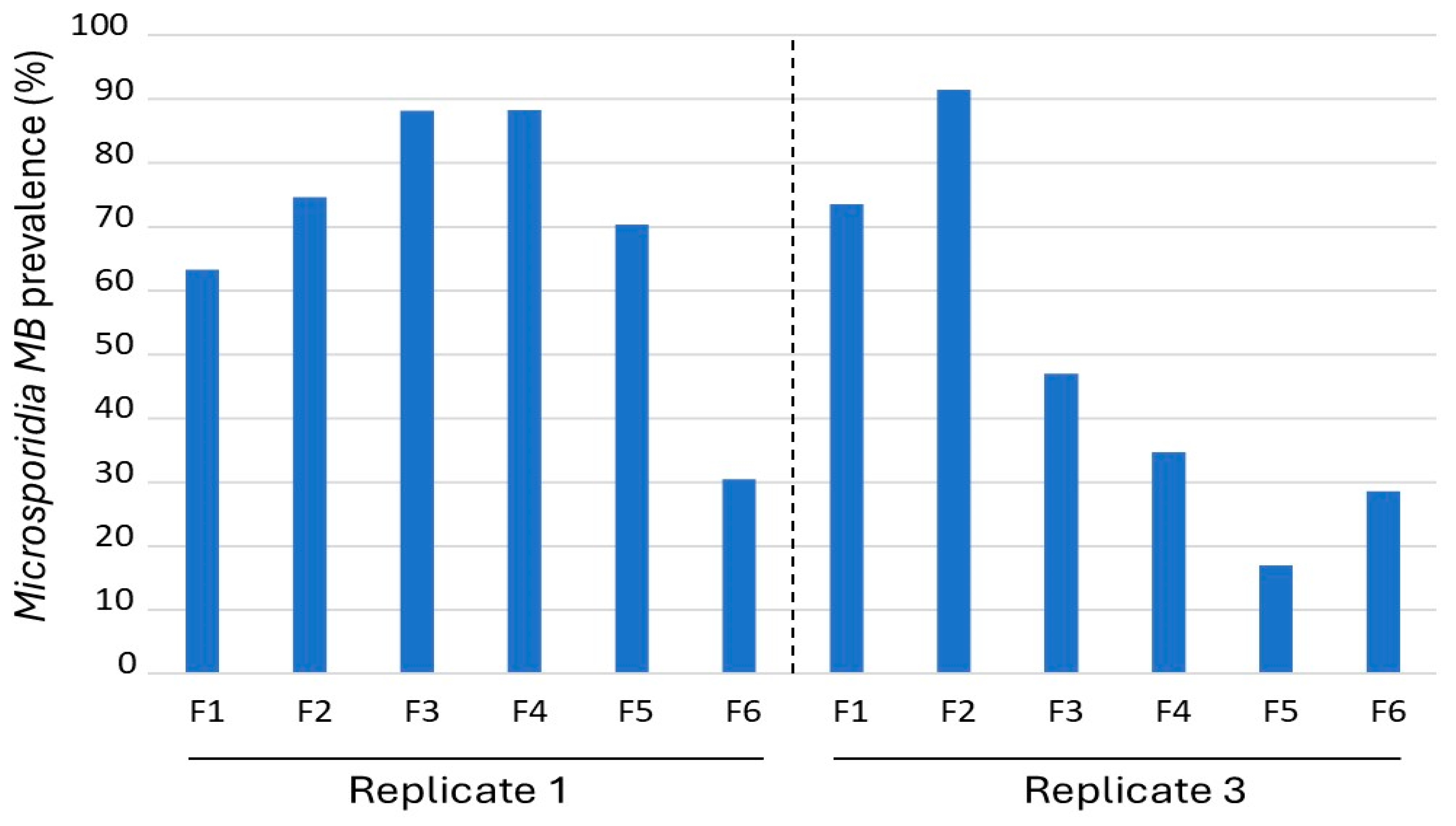
Microsporidia MB prevalence was determined to predict the prevalence of the symbiont in mosquito populations across generations (Figure 2). For replicate 1, the Microsporidia MB prevalence in the mosquito population increased from F1 (63.3%) to F4 (88.3%). The Microsporidia MB prevalence for replicate 3 also showed a similar trend with an increase from F1 (73.6%) to F2 (91.5%) (Figure 2). However, Microsporidia MB prevalence declined in both replicates, with 30% or less Microsporidia MB prevalence by the 6th generation. Sex was not associated with being infected in replicate 1 (χ2 = 0.00, df = 1, p = 0.97) or replicate 3 (χ2 = 0.84, df = 1, p = 0.77).
In replicate 1, Microsporidia MB prevalence declined after the F4 generation, whereas in replicate 3, the decline began earlier, after the second generation (Figure 2). Regression analysis revealed a significant positive correlation between Microsporidia MB prevalence and average temperature (Supplementary Materials) during the adult stage for each generation (R = 0.74, F = 11.83, p < 0.01) (Figure 3). In contrast, average humidity during the adult stage for each generation showed no significant effect on prevalence (R = 0.31, F = 1.06, p = 0.32) (Figure 3).

3.3. Microsporidia MB Intensity Across Generations and Sex

Like the prevalence, Microsporidia MB intensity increased, followed by a decrease across generations (Figure 4 and Supplementary Materials Tables S1 and S2). However, unlike Microsporidia MB prevalence, regression analysis revealed that both average temperature (R = 0.02, F = 0.85, p = 0.35) and average humidity during the adult stage for each generation showed no significant effect on Microsporidia MB intensity (R = 0.30, F = 1.90, p = 0.17) for both replicates 1 and 3.
A Wilcoxon signed-rank test showed that Microsporidia MB intensity was statistically significant between females and males in replicate 1 (Z = −10.50, p < 0.00) and replicate 3 (Z = −5.65, p < 0.00) (Figure 5). In replicate 1, the median Microsporidia MB intensity of females was 5.20 and for male was 0.34. In replicate 3, the Microsporidia MB median intensity of females was 0.81 and for males it was 0.06.

3.4. Wing Size of Mosquitoes Across Generations

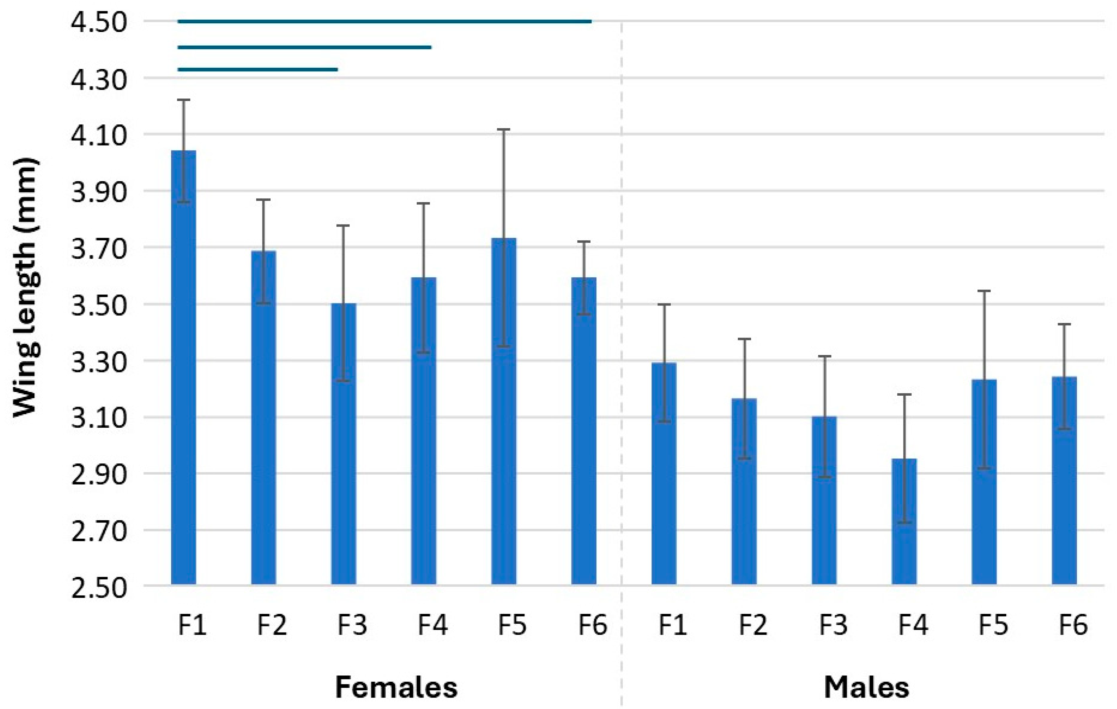
Wing length was used as a proxy for mosquito size across the generations. Wing length was only measured for replicate 1. Replicate 2 was excluded because the last generations failed to meet the minimum sample size for males (10) and females (10). In females, there was an overall significant difference in the wing length across the generations (F = 5.69, df = 5, p < 0.00). Pairwise comparison indicated a difference between the wing length of F1 generation and F3, F4, and F6, with the F1 generation having larger wings (Figure 6 and Supplementary Materials Tables S3 and S4). However, the female wing length of mosquitoes remained similar across generations after the F2 generation. In the case of males, an overall difference was detected (F = 2.50, df = 5, p > 0.04) but pairwise comparison with a Bonferroni correction did not highlight any two generations to be significantly different, indicating that the differences between groups may be too small but collectively cause a significant effect.

4. Discussion

This study monitored the spread of Microsporidia MB across generations in infected mosquito populations under semi-field conditions. We investigated how the symbiont’s prevalence and intensity are maintained across the early generations, which go through the natural bottleneck effect and revealed a link between environmental temperature and Microsporidia MB population prevalence. Additionally, we assessed the effect of these factors on mosquito body size, a proxy of both development and reproductive fitness.
Microsporidia MB prevalence and intensity increased in the earlier generations followed by a decrease in subsequent generations across the two surviving biological replicates. Each replicate started with offsprings of only mothers that were infected with Microsporidia MB, with no subsequent selection, allowing all the offsprings to contribute towards the following generation. While Microsporidia MB may be transmitted both vertically and horizontally, these transmission routes are not 100% efficient [5]. This could be attributed to the localisation of Microsporidia MB in the gonads where it is transferred from the germ line cells to the developing eggs [30,31]. In horizontal transmission the intensity of Microsporidia MB in the donor female was positively associated with the intensity in the recipient male after mating [3]. It is therefore likely that a decrease in Microsporidia MB intensity in a population will result in a decrease in population prevalence of Microsporidia MB.
An understanding of the factors that cause the reduction in Microsporidia MB prevalence and intensity is essential to optimise the symbiont infection in the mosquito population. The reported infected mosquitoes were established in semi-screened houses with ambient climatic conditions. While humidity has no impact on Microsporidia MB prevalence and intensity due to the minimal average changes in humidity over the experimental period, average temperature was positively associated with Microsporidia MB prevalence. In nature, however, higher Microsporidia MB prevalence has been associated with rainfall in An. arabiensis adults [2]. Microsporidia MB-infected larvae reared under a high average temperature of 32 °C were found to develop in a shorter time and have a higher infection rate compared to infected larvae reared under lower temperatures [25]. This study shows that the average temperature during the adult stage also contributes to higher infection rates. Subsequent generations of Wolbachia-infected mosquitoes have also been shown to be influenced by temperatures that are higher than 28 °C [32], in addition to a significant number of other symbiotic microorganisms [33]. It is noteworthy that, in addition to average temperature, other environmental factors such as water quality may also influence Microsporidia MB infection.
In both replicates, female mosquitoes had higher infection intensity compared to males. Higher Microsporidia MB intensity in females has also been reported previously when both infected males and females were reared on similar larval and adult diets [21]. In this study, the females were blood-fed which could have provided resources that Microsporidia MB exploits for replication, leading to higher intensity in females. Significantly higher relative intensities of Microsporidia MB are found in the gonads of blood-fed An. arabiensis compared to sugar-fed mosquitoes [31]. This may partially explain the higher Microsporidia MB intensity in females. Other factors to consider include the possibly lower evolutionary pressure for Microsporidia MB to infect males due to lower contribution to vertical transmission, shorter lifespans of males compared to females and therefore less time for Microsporidia MB to proliferate, variation in immune response to Microsporidia MB infection, or loss of heavy loads of Microsporidia MB following mating.
In addition to Microsporidia MB intensity, females also varied from males in how colonisation impacted their body size. While male body size remained similar across generations, the females body size was significantly smaller in the second generation, beyond which it did not change. Body size is crucial as it is a measure of both developmental and reproductive fitness [34,35,36,37]. Conveniently, body size can be maintained by adjusting the quantity and quality of diet available [21,22,38]. Noting that this result is based on a small sample from one replicate, a follow up study on adjusting the diet of female mosquitoes especially in the early generations and determining its outcome on Microsporidia MB transmission to the following will be helpful.
This study successfully determined the intergenerational spread of Microsporidia MB in Anopheles arabiensis under semi-field conditions, demonstrating its potential for malaria control while revealing key challenges in maintaining symbiont prevalence and intensity across generations. Future research should focus on optimizing environmental conditions such as temperature modulation and selective breeding of high-intensity individuals to enhance vertical and horizontal transmission rates for long-term maintenance of Microsporidia MB in Anopheles population. Additionally, studies on an improved diet for female mosquitoes and advancement in developing artificial infection methods (e.g., spore inoculation or microinjection) will support this too. These findings together with further development will be critical for scaling up production and implementing Microsporidia MB-based malaria transmission blocking strategy.

Supplementary Materials

The following supporting information can be downloaded at: https://www.mdpi.com/article/10.3390/insects16121206/s1. Table S1: The median absolute Microsporidia MB intensity across generations for replicate 1 and replicate 3. Table S2: Adjusted p-values for pairwise comparison of absolute Microsporidia MB intensity over generation in Replicate 1 and Replicate 3. Significant p-values are in bold. Table S3: The mean wing length size (S.D.) of female and male mosquitoes across generations in replicate 1. Table S4: Adjusted p-values for pairwise comparison of winglength over generation in colony 1 for female and male mosquitoes. Significant p-values are in bold. Excel S1: Data.

Author Contributions

Conceptualization, G.Y.B., L.L.K., T.B. and J.K.H.; Methodology, G.Y.B., L.L.K., T.B. and J.K.H.; Software, G.Y.B. and T.B.; Validation, G.Y.B., L.L.K., T.B. and J.K.H.; investigation, G.Y.B.; formal analysis, G.Y.B. and T.B.; data curation, G.Y.B. and T.B.; writing—original draft preparation, G.Y.B.; writing—review and editing, G.Y.B., L.L.K., T.B. and J.K.H.; supervision, L.L.K., T.B. and J.K.H.; funding acquisition, J.K.H. All authors have read and agreed to the published version of the manuscript.

Funding

The authors gratefully acknowledge the financial support for this research by the following organisations and agencies: Open Philanthropy (SYMBIOVECTOR Track A); the Bill and Melinda Gates Foundation (INV0225840); the Children’s Investment Fund Foundation (SMBV-FFT); National Research Foundation of South Africa (Ref Numbers SRUG2203311457); the Swedish International Development Cooperation Agency (Sida); the Swiss Agency for Development and Cooperation (SDC); the Australian Centre for International Agricultural Research (ACIAR); the Norwegian Agency for Development Cooperation (Norad); the German Federal Ministry for Economic Cooperation and Development (BMZ); and the Government of the Republic of Kenya. The views expressed herein do not necessarily reflect the official opinion of the donors.

Institutional Review Board Statement

Ethical approval was obtained from the Scientific and Ethics Review Unit (SERU) of the Kenya Medical Research Institute (KEMRI) for the field collection of mosquitoes from households (NON-KEMRI 4520, approved on 3 November 2022) and the arm feeding in the insectary (NON-KEMRI 593, approved on 29 March 2025).

Data Availability Statement

The original data for this study are included in the article/Supplementary Materials. Further inquiries can be directed to the corresponding authors.

Acknowledgments

We are grateful for the assistance of David Alila, Charles Amara Philip Owigo, and Kisero Auma (Shem) from ICIPE Mbita insectary team and Ian Okello and Sampson Okello. We also acknowledge the support provided by the project administrators, Faith Kyengo and Mary Mutuku. Figure 1 was created in BioRender. Bukhari, T. (2025) https://BioRender.com/aakdd1q. The authors have reviewed and edited the output and take full responsibility for the content of this publication.

Conflicts of Interest

The authors declare no conflicts of interest.

References

  1. Bukhari, T.; Pevsner, R.; Herren, J.K. Microsporidia: A promising vector control tool for residual malaria transmission. Front. Trop. Dis. 2022, 3, 957109. [Google Scholar] [CrossRef]
  2. Herren, J.K.; Mbaisi, L.; Mararo, E.; Makhulu, E.E.; Mobegi, V.A.; Butungi, H.; Mancini, M.V.; Oundo, J.W.; Teal, E.T.; Pinaud, S.; et al. A microsporidian impairs Plasmodium falciparum transmission in Anopheles arabiensis mosquitoes. Nat. Commun. 2020, 11, 2187. [Google Scholar] [CrossRef]
  3. Maina, T.; Shisia, A.; Gichuhi, J.; Bargul, J.L.; Herren, J.K.; Bukhari, T. Maximizing horizontal transmission through mating: Increased mating frequency and mating competitiveness associated with Microsporidia MB-infected Anopheles arabiensis males. Malar. J. 2025, 24, 114. [Google Scholar] [CrossRef]
  4. Nattoh, G.; Onyango, B.; Makhulu, E.E.; Omoke, D.; Ang’ang’o, L.M.; Kamau, L.; Gesuge, M.M.; Ochomo, E.; Herren, J.K. Microsporidia MB in the primary malaria vector Anopheles gambiae sensu stricto is avirulent and undergoes maternal and horizontal transmission. Parasites Vectors 2023, 16, 335. [Google Scholar] [CrossRef] [PubMed]
  5. Nattoh, G.; Maina, T.; Makhulu, E.E.; Mbaisi, L.; Mararo, E.; Otieno, F.G.; Bukhari, T.; Onchuru, T.O.; Teal, E.; Paredes, J.; et al. Horizontal Transmission of the Symbiont Microsporidia MB in Anopheles arabiensis. Front. Microbiol. 2021, 12, 647183. [Google Scholar] [CrossRef]
  6. Akorli, J.; Akorli, E.A.; Tetteh, S.N.; Amlalo, G.K.; Opoku, M.; Pwalia, R.; Adimazoya, M.; Atibilla, D.; Pi-Bansa, S.; Chabi, J.; et al. Microsporidia MB is found predominantly associated with Anopheles gambiae s.s. and Anopheles coluzzii in Ghana. Sci. Rep. 2021, 11, 18658. [Google Scholar] [CrossRef] [PubMed]
  7. Moustapha, L.M.; Sadou, I.M.; Arzika, I.I.; Maman, L.I.; Gomgnimbou, M.K.; Konkobo, M.; Diabate, A.; Bilgo, E. First identification of Microsporidia MB in Anopheles coluzzii from Zinder City, Niger. Parasites Vectors 2024, 17, 39. [Google Scholar] [CrossRef]
  8. Moustapha, L.M.; Mukhtar, M.M.; Sanda, A.N.; Adamu, S.; Aliyu, Y.Y.; Einoi, H.K.; Maigari, M.U.; Okeke, P.C.; Nwele, D.E.; Obembe, A.; et al. Spatial Distribution of Microsporidia MB Along Clinal Gradient and the Impact of Its Infection on Pyrethroid Resistance in Anopheles gambiae s.l. Mosquitoes from Nigeria and Niger Republic. Parasitologia 2025, 5, 31. [Google Scholar] [CrossRef]
  9. Tchigossou, G.; Lontsi-Demano, M.; Tossou, E.; Sovegnon, P.M.; Akoton, R.; Adanzounon, D.; Dossou, C.; Koto, M.; Ogbon, A.; Gouété, M.; et al. Seasonal variation of Microsporidia MB infection in Anopheles gambiae and Anopheles coluzzii in two different geographical localities in Benin. Malar. J. 2025, 24, 95. [Google Scholar] [CrossRef] [PubMed]
  10. El Jarkass, H.T.; Reinke, A.W. The ins and outs of host-microsporidia interactions during invasion, proliferation and exit. Cell. Microbiol. 2020, 22, e13247. [Google Scholar] [CrossRef]
  11. Texier, C.; Vidau, C.; Viguès, B.; El Alaoui, H.; Delbac, F. Microsporidia: A model for minimal parasite-host interactions. Curr. Opin. Microbiol. 2010, 13, 443–449. [Google Scholar] [CrossRef]
  12. Vávra, J.; Lukeš, J. Microsporidia and ‘The Art of Living Together’. Adv. Parasitol. 2013, 82, 253–319. [Google Scholar]
  13. Puerta-Guardo, H.; Contreras-Perera, Y.; Perez-Carrillo, S.; Che-Mendoza, A.; Ayora-Talavera, G.; Vazquez-Prokopec, G.; Martin-Park, A.; Zhang, D.; Manrique-Saide, P.; UCBE-LCB Team. Wolbachia in native populations of Aedes albopictus (Diptera: Culicidae) from Yucatan Peninsula, Mexico. J. Insect Sci. 2020, 20, 16. [Google Scholar] [CrossRef]
  14. Vinayagam, S.; Nirmolia, T.; Chetry, S.; Kumar, N.P.; Saini, P.; Bhattacharyya, D.; Bhowmick, I.P.; Sattu, K.; Patgiri, S.J. Molecular Evidence of Wolbachia Species in Wild-Caught Aedes albopictus and Aedes aegypti Mosquitoes in Four States of Northeast India. J. Trop. Med. 2023, 1, 6678627. [Google Scholar] [CrossRef] [PubMed]
  15. Mohanty, I.; Rath, A.; Pradhan, N.; Panda, B.B.; Mohapatra, P.K.; Hazra, R.K. Prevalence and transmission potential of Wolbachia in Aedes albopictus population circulating in endemic coastal districts of Odisha, India. J. Vector Borne Dis. 2021, 58, 297–305. [Google Scholar] [CrossRef]
  16. Joubert, D.A.; Walker, T.; Carrington, L.B.; De Bruyne, J.T.; Kien, D.H.; Hoang, N.L.; Chau, N.V.; Iturbe-Ormaetxe, I.; Simmons, C.P.; O’Neill, S.L. Establishment of a Wolbachia Superinfection in Aedes aegypti Mosquitoes as a Potential Approach for Future Resistance Management. PLoS Pathog. 2016, 12, e1005434. [Google Scholar] [CrossRef] [PubMed]
  17. Bayoh, M.N.; Lindsay, S.W. Effect of temperature on the development of the aquatic stages of Anopheles gambiae sensu stricto (Diptera: Culicidae). Bull. Entomol. Res. 2003, 93, 375–381. [Google Scholar] [CrossRef]
  18. Mouton, L.; Henri, H.; Bouletreau, M.; Vavre, F. Effect of temperature on Wolbachia density and impact on cytoplasmic incompatibility. Parasitology 2006, 132, 49–56. [Google Scholar] [CrossRef]
  19. Moghadam, N.N.; Thorshauge, P.M.; Kristensen, T.N.; de Jonge, N.; Bahrndorff, S.; Kjeldal, H.; Nielsen, J.L. Strong responses of Drosophila melanogaster microbiota to developmental temperature. Fly 2018, 12, 1–12. [Google Scholar] [CrossRef]
  20. Doremus, M.R.; Kelly, S.E.; Hunter, M.S. Exposure to opposing temperature extremes causes comparable effects on Cardinium density but contrasting effects on Cardinium induced cytoplasmic incompatibility. PLoS Pathog. 2019, 15, e1008022. [Google Scholar] [CrossRef]
  21. Boanyah, G.Y.; Koekemoer, L.L.; Herren, J.K.; Bukhari, T. Effect of Microsporidia MB infection on the development and fitness of Anopheles arabiensis under different diet regimes. Parasites Vectors 2024, 17, 294. [Google Scholar] [CrossRef]
  22. Carvajal-Lago, L.; Ruiz-López, M.J.; Figuerola, J.; Martínez-de la Puente, J. Implications of diet on mosquito life history traits and pathogen transmission. Environ. Res. 2021, 195, 110893. [Google Scholar] [CrossRef]
  23. Jiggins, F.M. The spread of Wolbachia through mosquito populations. PLOS Biol. 2017, 15, e2002780. [Google Scholar] [CrossRef]
  24. Wiwatanaratanabutr, I.; Kittayapong, P. Effects of crowding and temperature on Wolbachia infection density among life cycle stages of Aedes albopictus. J. Invertebr. Pathol. 2009, 102, 220–224. [Google Scholar] [CrossRef]
  25. Otieno, F.G.; Barreaux, P.; Belvinos, A.S.; Makhulu, E.E.; Onchuru, T.O.; Wairimu, A.W.; Omboye, S.M.; King’ori, C.N.; Sokame, B.M.; Nyamache, A.; et al. The dissemination potential of Microsporidia MB in Anopheles arabiensis mosquitoes is modulated by temperature. Sci. Rep. 2025, 15, 28839. [Google Scholar] [CrossRef]
  26. Degefa, T.; Yewhalaw, D.; Zhou, G.; Lee, M.C.; Atieli, H.; Githeko, A.K.; Yan, G. Indoor and outdoor malaria vector surveillance in western Kenya: Implications for better understanding of residual transmission. Malar. J. 2017, 16, 443. [Google Scholar] [CrossRef]
  27. Nepomichene, T.N.; Andrianaivolambo, L.; Boyer, S.; Bourgouin, C. Efficient method for establishing F1 progeny from wild populations of Anopheles mosquitoes. Malar. J. 2017, 16, 21. [Google Scholar] [CrossRef]
  28. Coetzee, M. Key to the females of Afrotropical Anopheles mosquitoes (Diptera: Culicidae). Malar. J. 2020, 19, 70. [Google Scholar] [CrossRef] [PubMed]
  29. Santolamazza, F.; Mancini, E.; Simard, F.; Qi, Y.; Tu, Z.; della Torre, A. Insertion polymorphisms of SINE200 retrotransposons within speciation islands of Anopheles gambiae molecular forms. Malar. J. 2008, 7, 163. [Google Scholar] [CrossRef] [PubMed]
  30. Onchuru, T.O.; Makhulu, E.E.; Ronnie, P.C.; Mandere, S.; Otieno, F.G.; Gichuhi, J.; Herren, J.K. The Plasmodium transmission-blocking symbiont, Microsporidia MB, is vertically transmitted through Anopheles arabiensis germline stem cells. PLoS Pathog. 2024, 20, e1012340. [Google Scholar] [CrossRef]
  31. Makhulu, E.E.; Onchuru, T.O.; Gichuhi, J.; Otieno, F.G.; Wairimu, A.W.; Muthoni, J.N.; Koekemoer, L.; Herren, J.K. Localization and tissue tropism of the symbiont Microsporidia MB in the germ line and somatic tissues of Anopheles arabiensis. MBio 2024, 15, e0219223. [Google Scholar] [CrossRef]
  32. Tokash-Peters, A.G.; Jabon, J.D.; Fung, M.E.; Peters, J.A.; Lopez, S.G.; Woodhams, D.C. Trans-Generational Symbiont Transmission Reduced at High Temperatures in a West Nile Virus Vector Mosquito Culex quinquefasciatus. Front. Trop. Dis. 2022, 3, 762132. [Google Scholar] [CrossRef]
  33. Corbin, C.; Heyworth, E.R.; Ferrari, J.; Hurst, G.D. Heritable symbionts in a world of varying temperature. Heredity 2017, 118, 10–20. [Google Scholar] [CrossRef] [PubMed]
  34. Ameneshewa, B.; Service, M.W. The relationship between female body size and survival rate of the malaria vector Anopheles arabiensis in Ethiopia. Med. Vet. Entomol. 1996, 10, 170–172. [Google Scholar] [CrossRef] [PubMed]
  35. Cator, L.J.; Zanti, Z. Size, sounds and sex: Interactions between body size and harmonic convergence signals determine mating success in Aedes aegypti. Parasites Vectors 2016, 9, 622. [Google Scholar] [CrossRef]
  36. Helinski, M.E.H.; Knols, B.G.J. Mating Competitiveness of Male Anopheles arabiensis Mosquitoes Irradiated with a Partially or Fully Sterilizing Dose in Small and Large Laboratory Cages. J. Med. Entomol. 2008, 45, 698–705. [Google Scholar] [CrossRef]
  37. Sawadogo, S.P.; Diabaté, A.; Toé, H.K.; Sanon, A.; Lefevre, T.; Baldet, T.; Gilles, J.; Simard, F.; Gibson, G.; Sinkins, S.; et al. Effects of age and size on Anopheles gambiae s.s. male mosquito mating success. J. Med. Entomol. 2013, 50, 285–293. [Google Scholar] [CrossRef]
  38. Mackay, A.J.; Yan, J.; Kim, C.H.; Barreaux, A.M.; Stone, C.M. Larval diet and temperature alter mosquito immunity and development: Using body size and developmental traits to track carry-over effects on longevity. Parasites Vectors 2023, 16, 434. [Google Scholar] [CrossRef]
Figure 1. The schematic diagram showing the (a) collection from houses, (b) screening of Microsporidia MB-infected G0, and (c) rearing of F1 generation from larvae to pupa stage. The pupae were transferred to cages kept in an insectary where (d) temperature and humidity were recorded daily. The female mosquitoes in the cage (blue dotted arrow) were (e) arm-fed and the eggs that were laid were reared in larval trays. The resulting pupae were transferred to the cage designated for the next generation (maroon dotted arrow). (f) Dead mosquitoes were picked daily from each cage and screened for Microsporidia MB (green dotted line) to determine Microsporidia MB prevalence and intensity per generation (g).
Insects 16 01206 g001
Figure 2. Prevalence of Microsporidia MB across generations in two replicates of An. arabiensis mosquito population.
Insects 16 01206 g002
Figure 3. Prevalence of Microsporidia MB across generations in relation to average temperature (°C) ± S.D. ((a,b) for replicate 1 and 3 respectively) and humidity (%) ± S.D. ((c,d) for replicate 1 and 3 respectively) during the adult stage of the mosquitoes. Also shown is the correlation between the Microsporidia MB prevalence across generations in both replicates, (e) average temperature, and (f) average humidity during the adult stage of An. arabiensis mosquitoes.
Insects 16 01206 g003
Figure 4. Box plots indicating the median relative Microsporidia MB intensities across generations in replicate 1 and replicate 3. Whiskers represent the IQR and dots represent the outliers. Blue lines indicate a significant difference between the generations after adjustment for multiple comparison (Bonferroni correction).
Insects 16 01206 g004
Figure 5. Box plots indicating the median relative Microsporidia MB intensities for females and males combined across the generations in replicate 1 and replicate 3. Whiskers represent the IQR and dots represent the outliers. Blue lines indicate a significant difference between females and males.
Insects 16 01206 g005
Figure 6. Female and male wing size (mm (S.D.)) across generations for replicate 1. Lines on top indicate a significant difference between the generations for females. No significant difference was found in wing length between the generations for males.
Insects 16 01206 g006
Table 1. Frequency table of adult Microsporidia MB mosquitoes (males and females) across generations in replicate 1 and 3.
Replicate 1Replicate 3
Generation (F)FemaleMaleTotalFemaleMaleTotal
1 *5010608498182
2841061908172153
3153178331162119281
46155116413172
599101200272653
62962785743 **5 **28
Each replicate was started with 200 F1 adult progeny of Microsporidia MB-infected Anopheles arabiensis. * Represents the number out of the starting 200 that was in the cage when the first oviposition was recorded. ** Sex was recorded for only 8 out of the 28 samples. These eight samples were all the Microsporidia MB infected samples in the generation.
Disclaimer/Publisher’s Note: The statements, opinions and data contained in all publications are solely those of the individual author(s) and contributor(s) and not of MDPI and/or the editor(s). MDPI and/or the editor(s) disclaim responsibility for any injury to people or property resulting from any ideas, methods, instructions or products referred to in the content.