Article

CHS-2 Is Involved in the Response of Aedes albopictus (Diptera: Culicidae) Larvae to Cadmium Stress by Mediating the Formation of the Peritrophic Membrane

1
College of Life and Environmental Sciences, Hangzhou Normal University, Hangzhou 311121, China
2
State Key Laboratory of Animal Biodiversity Conservation and Integrated Pest Management, Institute of Zoology, Chinese Academy of Sciences, Beijing 100101, China
*
Authors to whom correspondence should be addressed.
Insects 2025, 16(11), 1102; https://doi.org/10.3390/insects16111102
Submission received: 6 September 2025 / Revised: 30 September 2025 / Accepted: 27 October 2025 / Published: 29 October 2025

Simple Summary

The exposure of Aedes albopictus larvae to cadmium-contaminated water may lead to an increased resistance, thereby complicating vector control efforts. The CHS-2 gene plays a critical role in the formation of the peritrophic membrane, an essential tissue involved in the larval stress response of Ae. albopictus. However, the function of CHS-2 in mediating the response to cadmium stress remains unclear. In this study, we first exposed Ae. albopictus larvae to cadmium and examined CHS-2 expression levels alongside morphological changes in the peritrophic membrane. Subsequently, RNA interference was employed to silence the CHS-2 gene, followed by cadmium exposure to assess alterations in larval resistance in the absence of CHS-2. Our results elucidate the adaptive responses and underlying mechanisms of Ae. albopictus larvae to cadmium exposure, providing a theoretical basis for the utilization of this species in monitoring aquatic environmental contamination.

Abstract

Pollution with heavy metals, such as cadmium (Cd), can significantly affect insect growth, development, and behavior. The midgut is an essential organ for stress response. Chitin synthase-2 (CHS-2) is closely associated with forming the peritrophic membrane (PM). The fourth-instar larvae of Aedes albopictus were exposed to varying concentrations of Cd. The results showed that Cd inhibited chitin synthesis and metabolism-related genes, but thickened the midgut PM, indicating that the larvae could respond to Cd stress through the midgut PM. Silencing CHS-2 by RNA interference resulted more severe vacuolization and malformation of midgut epithelial cells without midgut PM protection. Additionally, there was an intensified redox reaction, upregulated expression of metallothionein (MT) and heat shock proteins 70 (HSP70), and increased activity of antioxidant enzymes at some scattered time points. This study confirms that CHS-2 is involved in oxidative stress induced by Cd exposure by regulating PM formation. This study also contributes to further understanding the resistance mechanism of Ae. albopictus under Cd stress, thereby establishing a theoretical foundation for the future studies of them, which is concerned with the possibility of Ae. albopictus as a water environment detection and the control of Ae. albopictus based on resistance mechanism.

Graphical Abstract

1. Introduction

Nearly all heavy metals occur naturally in an oxidized state in various ecosystems. However, once their concentration surpasses the environmental threshold, these metals threaten ecosystems, resulting in heavy metal pollution. In addition, heavy metals are stable, difficult to degrade, highly toxic and can accumulate in the food chain, making heavy metal pollution harmful and difficult to control. The pollution and degradation of soil and water in China have become increasingly prominent in recent years, exacerbated by rapid industrialization and irrational agricultural practices. Particularly, Cd, Hg, Ni, Cu, and As contamination levels are alarmingly high [1]. Most heavy metals interact with DNA molecules and nuclear proteins to cause DNA damage, leading to apoptosis or cancer [2]. Heavy metals are associated with various diseases and can impair reproductive fitness and neural development [3,4]. Exposure to heavy metals in the environment affects the growth and development of various organisms in the biosphere and indirectly impairs human life and health through accumulation in food chains [5]. Cd, a significant contributor to pollution, has garnered increasing attention in recent years. Cd pollution in China’s rivers is primarily concentrated in the Xiangjiang River Basin and regions such as Jiangsu and Zhejiang, as revealed by a survey [6].
Insects display exhibit a broad spectrum of feeding habits and remarkable adaptability. They are the most abundant and geographically dispersed organisms in the world. When insects reside in environments contaminated with heavy metals, heavy metals can permeate the insect body through respiration, ingestion, and skin penetration, reducing hemolymph energy reserves, disrupting redox balance, and impairing the integrity of cellular structures [7]. Furthermore, heavy metal exposure may enhance the susceptibility of insects to pathogenic microorganisms [8], thereby influencing their growth, development [9,10] and reproductive capabilities [11]. Heavy metal pollution, both direct and indirect, can potentially decrease aquatic insect diversity and trigger the local extinction of insect populations [12]. Insects respond to heavy metal stress through a series of reactions and mechanisms, such as the active deposition and expulsion of heavy metals, because most insects are exposed to heavy metal stress through their diet. The main components of insect digestion and absorption occurs in the midgut, where heavy metals primarily accumulate, and have the greatest impact on the insect midgut [13,14]. The uptake and accumulation of heavy metals in the insect midgut are primarily mediated by midgut epithelial cells, which contain many metal particles capable of adsorbing surplus heavy metal ions. Heavy metals deposited by these cells are subsequently excreted as intestinal metabolites during molting [15]. Simultaneously, midgut epithelial cells can secret a lamellar tubular membrane structure, the peritrophic membrane (PM), to resist the harm of pesticides, heavy metals, and pathogens [16]. The primary component of PM in the midgut is chitin, which makes the biosynthesis, assembly, and degradation of PM intimately associated with chitin synthesis and metabolism [17]. In insects, chitin is synthesized using trehalose as the initial substrate and converted through a sequential enzymatic pathway involving trehalase (TRE), hexokinase (HK), glucose-6-phosphate isomerase (G6PI), fructose-6-phosphate aminotransferase (GFAT), glucosamine-6-phosphate N-acetyltransferase (GNPNA), phosphoacetylglucosamine mutase (PAGM), UDP-N-acetylglucosamine pyrophosphorylase (UAP), and chitin synthase (CHS) [18]. CHS is divided into two types, CHS-A and CHS-B [19]. Chitin synthase-2 (CHS-2), which encodes CHS-B, is highly expressed only in insect midgut epithelial cells and is responsible for synthesizing chitin in the peritrophic membrane [20]. Previous studies demonstrated the indispensability of CHS-2 in forming PM within the midgut of Ae. albopictus. When CHS-2 is silenced, the fourth-instar of Ae. albopictus cannot generate a complete PM structure [21].
Furthermore, metallothionein (MT) and heat shock proteins (HSPs) are crucial mechanisms that enable insects to alleviate heavy metal stress. MT, a class of binding proteins, can form chelates with metal ions, thereby reducing or eliminating the toxic effects of heavy metals. As molecular chaperones, HSPs can restore the functional structure of damaged proteins, thereby exerting cytoprotective effects [22]. Recently, a growing body of research has explored the potential of MT and HSPs as biomarkers of heavy metal pollution [23,24]. Heavy metals can generate a multitude of reactive oxygen species (ROS) after entering the insect body, triggering redox reactions and subsequent activation of a cascade of antioxidant defense enzymes, including superoxide dismutase (SOD), peroxidase (POD), catalase (CAT) and glutathione peroxidase (GSH-PX), which are very sensitive to environmental pollutants. These enzymes have been used as biomarkers in ecotoxicological studies [25].
Ae. albopictus, belongs to the family Diptera and is a vital viral vector capable of transmitting various pathogens, including dengue and yellow fevers. The eggs, larvae, and pupae of Ae. albopictus grow and develop in aquatic environments, whereas adult mosquitoes reside on land. The larvae of Ae. albopictus, which reside in water and subsist on algae, protozoa, and yeast, exhibit a certain capacity to accumulate heavy metals. Excessive heavy metal infiltration may have detrimental effects on the growth, development, and reproduction of Ae. albopictus [26]. However, the mechanisms underlying the response to Ae. albopictus to heavy metal stress, particularly the response of CHS-2 to heavy metal stress and its potential functions and regulatory pathways, remains elusive. The lethal concentration 50% (LC50) of cadmium for Ae. albopictus, as determined in laboratory conditions, was 263 mg/L. In the present study, the insect vector Ae. albopictus was first exposed to various concentrations of Cd to investigate the effects of different Cd stress levels on the chitin synthesis and metabolism of Ae. albopictus. Subsequently, the CHS-2 was silenced via RNA interference (RNAi) and combined with three Cd stress concentrations to explore the response mechanism and potential adaptive functions of CHS-2 under Cd pollution stress. This clarifies the resistance adaptation and internal expression mechanisms of Ae. albopictus under Cd stress. This study supports the morphological changes in the midgut PM of Ae. albopictus larvae as potential biomarkers of environmental Cd pollution. Simultaneously, the CHS-2 gene is considered to be very important for the development of resistance to Ae. albopictus.

2. Materials and Methods

2.1. Mosquitoes

Ae. albopictus mosquitoes, sourced from Sun Yat-sen University (Guangdong Province, China), were maintained and bred through more than three generations under controlled conditions. These conditions included a temperature of 27 ± 1 °C, 70% relative humidity, and a light/dark cycle of 14:10 h, within an artificial climate chamber. The cat food was ground into powder and mixed with yeast powder at a ratio of 2:1, then fed to the first- and second-instar larvae. For the third- and fourth-instar larvae, cat chow powder alone was provided [27]. Adult mosquitoes, after eclosion, were sustained with a solution of 10% sugar water. Mice were utilized for mosquito blood-feeding to facilitate oviposition.

2.2. Cd Stress on Ae. albopictus

According to the national standard GB/T 602-2002 [28], a Cd solution (1 g/L) was prepared using CdCl2· 5 2 H2O, and successively diluted to concentrations of 0, 6.25, 12.5, 25, 50, 100, 200, 300, and 400 mg/L. Fifty fourth-instar larvae of approximately the same size were selected and placed in each cup for exposure, and the 12 h mortality rate was recorded. The concentration of Cd solution corresponding to 50% mortality was determined to be 263 mg/L using probit regression analysis. Based on the semi-lethal concentration and the results of previous laboratory experiments, Cd stress concentrations (0, 25, 50, and 100 mg/L) were selected, and larvae treated with 0 mg/L were used as negative controls. The fourth-instar larvae were exposed to the above concentrations of Cd solution, and samples were taken at 12, 24 and 36 h after exposure.

2.3. Microinjection

Using the successfully sequenced CHS-2 intermediate fragment as a template, T7 promoter sequences were added to the 5′ end of primers (Table 1) [21]. Double-stranded RNA (dsRNA) was synthesized using a T7 RiboMAXTM Express RNAi System kit (LABOOT, Hangzhou, China) and purified, and the specific experimental steps were in accordance with the instructions. The same method was used to synthesize dsRNA using the green fluorescent protein (GFP) as a negative control. Seven hundred and fifty well-developed fourth-instar larvae of the same size were randomly selected for microinjection. The dsCHS-2 (200 ng) was dissolved in 100 nL of water and injected into the soft spot between the penultimate and third-last body segments of the larvae using a microinjection device. The same method was used to inject dsGFP into another group of 750 fourth-instar larvae. The expression levels of the CHS-2 gene in Ae. albopictus larvae were reduced by 50% at both 12 h and 24 h post-injection of dsCHS-2 [21].

2.4. Gene Expression Analysis

Ten fourth-instar larvae of Ae. albopictus were collected each treatment concentration after 12 h of Cd treatment. Total RNA was extracted using the TRIzol method, and the quality and concentration were detected using agarose gel electrophoresis and microspectrophotometry (Thermo, Waltham, MA, USA). cDNA was synthesized using the Prime ScriptTM RT Reagent Kit (Haofeng, Hangzhou, China) according to the manufacturer’s instructions. Through the National Center for Biotechnology Information and relevant literature, chitin synthesis genes HK, G6PI, GFAT, GNPNA, PAGM, UAP, and CHS-2, as well as chitin metabolism enzyme genes chitinase2 (Cht2) and chitinase10 (Cht10), were selected. Primer premier 5.0 software was used to design qPCR primers (Table 2), and the expression levels were measured using qPCR. The qPCR reaction mixture (10 µL) contained 5 µL of TB Green ® Premix Ex Taq ™, 3.2 µL of ddH2O, 0.4 µL each of upstream and downstream primers and 1 µL cDNA. The qPCR procedure was as follows: 10 min at 94 °C; 30 s at 94 °C, 30 s at 59 °C, 45 s at 72 °C for a total of 30 cycles, and then 10 min at 72 °C. A default dissolution curve analysis was performed. The actin gene was used as an internal control, and the 2−ΔΔCT method was used to calculate the relative expression of genes related to chitin synthesis and metabolism after Cd treatment for 12 h.

2.5. Determination of Chitin Content in the Midgut

The midguts from 45 Ae. albopictus mosquitoes per group, exposed to different concentrations of cadmium for 12 h, were dissected and collected. The content of chitin in the midgut of larvae was determined with reference to the method of Liu et al. [29], and the principle is that chitinase hydrolyzes chitin to produce N-acetylglucosamine, and the intermediate compound produced by the cross-reaction of N-acetylglucosamine and alkali can further react with p-dimethylaminobenzaldehyde to produce chromogenic substances.

2.6. Antioxidant Enzyme Activity Assay

At 12, 24, and 36 h after dsRNA injection, 45 fourth-instar larvae exposed to four concentrations of Cd were collected, respectively. Then 0.9% normal saline was added according to the ratio of weight (g):volume (mL) = 1:9, and the supernatant was taken after ice bath breaking. SOD (Cat. No. A001-1-2) activity was measured by xanthine and xanthine oxidase reaction system using Kit (Jiancheng, Nanjing, China). POD (Cat. No. A084-3-1), CAT (Cat. No. A007-2-1) and GSH-PX (Cat. No. A005-1-2) activities were measured according to the principle of catalyzing H2O2 using Kit (Jiancheng, Nanjing, China), and the specific experimental steps were performed in accordance with the manufacturer’s instructions.

2.7. Observation of the PM of the Midgut

The fourth-instar larvae exposed to four concentrations of Cd for 12 h and the fourth-instar larvae injected with dsRNA and exposed to 50 mg/L Cd for 12, 24 and 36 h were used as materials, and the head and tail were removed according to Gregor et al. production, sectioning and staining methods [30]. After fixation, the larvae were embedded in paraffin using a paraffin embedding machine (MICROM, Walldorf, Germany) and cut into 5-µm-thick sections. After dewaxing and cleaning, the samples were stained with hematoxylin-eosin staining (HE staining). A microscope camera system (Axio Observer A1+Stemi2000, ZEISS, Oberkochen, Germany) was used for photograph and observation.

2.8. Data Analysis

IBM SPSS Statistics 23.0 was used for the statistical analysis. Data on gene expression, enzyme activity, and chitin content were analyzed using one-way ANOVA and Duncan’s test. Mortality data were analyzed using the chi-square test (p < 0.05 indicated a significant difference, annotated with “*”; p < 0.01 indicated a significant difference, annotated with “**”). Graphs were created using SigmaPlot 10.0. The normality and homogeneity of the data were assessed using three replicates for each treatment concentration and tissue sample quantity.

3. Results

3.1. Expression Changes in Genes Related to Chitin Metabolism Pathway in Ae. albopictus After Cd Stress

Acute Cd stress affects the expression of genes related to chitin synthesis and metabolism in the fourth-instar larvae of Ae. albopictus. The expression of CHS-2 gradually decreased with increasing Cd concentration. Significant inhibition of CHS-2 expression was observed at a Cd concentration of 100 mg/L (Figure 1A). Similarly, the expression of genes associated with chitin synthesis, including PAGM, UAP, GNPNA, and GFAT, was inhibited by a pronounced decrease in their expression levels at a Cd concentration of 100 mg/L (Figure 1C–F). Under low Cd stress (50 mg/L), the expression of HK and G6PI increases, whereas it significantly decreased under 100 mg/L Cd stress (Figure 1B,G). The expression of the chitin metabolism enzyme Cht2 remained unchanged at low-Cd concentrations and significantly decreased at 100 mg/L (Figure 1H). The relative expression of Cht10 also decreased with increasing Cd concentrations (Figure 1I).

3.2. Determination of Chitin Content and Observation of Midgut Structure of Aedes albopictus Larvae Under Cadmium Stress

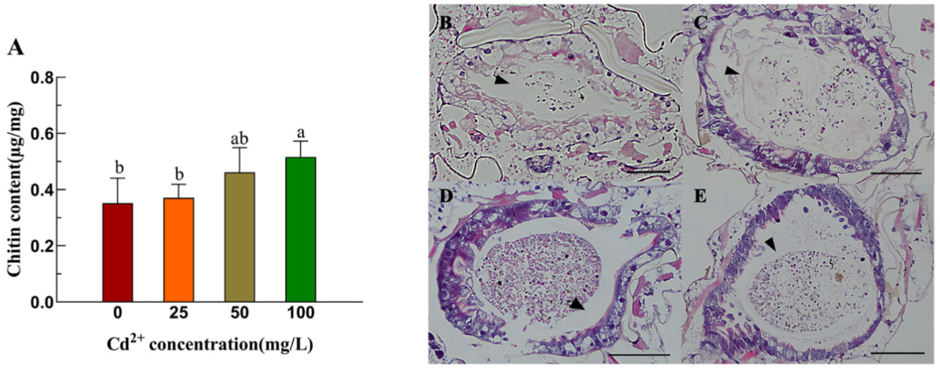
After Cd stress, the chitin content in the midgut of fourth-instar larvae of Ae. albopictus demonstrated a significant positive correlation with Cd concentration. Under 100 mg/L Cd stress, the chitin content notably increased (Figure 2A). Compared with the control group (Figure 2B), the midgut PM of the fourth-instar Ae. albopictus larvae were clearer and continuous after Cd stress, and the higher the concentration of Cd, the more obvious the PM thickening (Figure 2C–E).

3.3. Morphological Changes in the Midgut of Ae. albopictus Under Cd Stress After CHS-2 Silencing

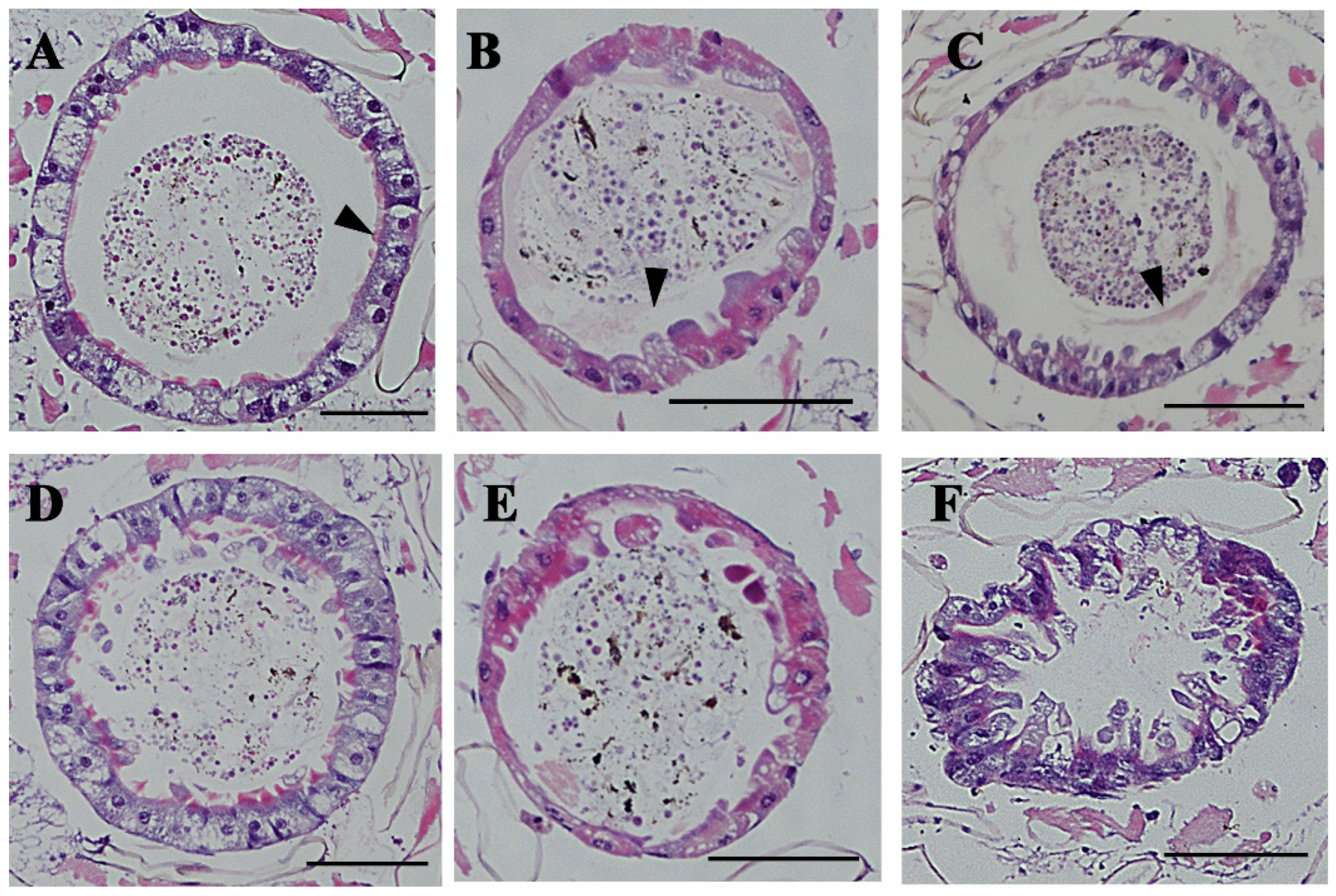
Under 50 mg/mL cadmium stress, the PM of the larvae in the dsGFP group was visible (Figure 3A,B). With increased exposure time to cadmium, the epithelial cells in the larvae midgut exhibited a slight deformity (Figure 3C). In contrast, the larval PM in the dsCHS-2 group was destroyed, and the epithelial cells were severely deformed after exposure to the Cd solution for only 12 h (Figure 3D). After 24 h, a large number of epithelial cells were convex, and some epithelial cells were vacuolized (Figure 3E). However, after exposure to a cadmium solution for 36 h, the whole midgut was severely deformed and could not maintain its normal morphology (Figure 3F).

3.4. Mortality of Ae. albopictus After CHS-2 Silencing Combined with Cd Stress

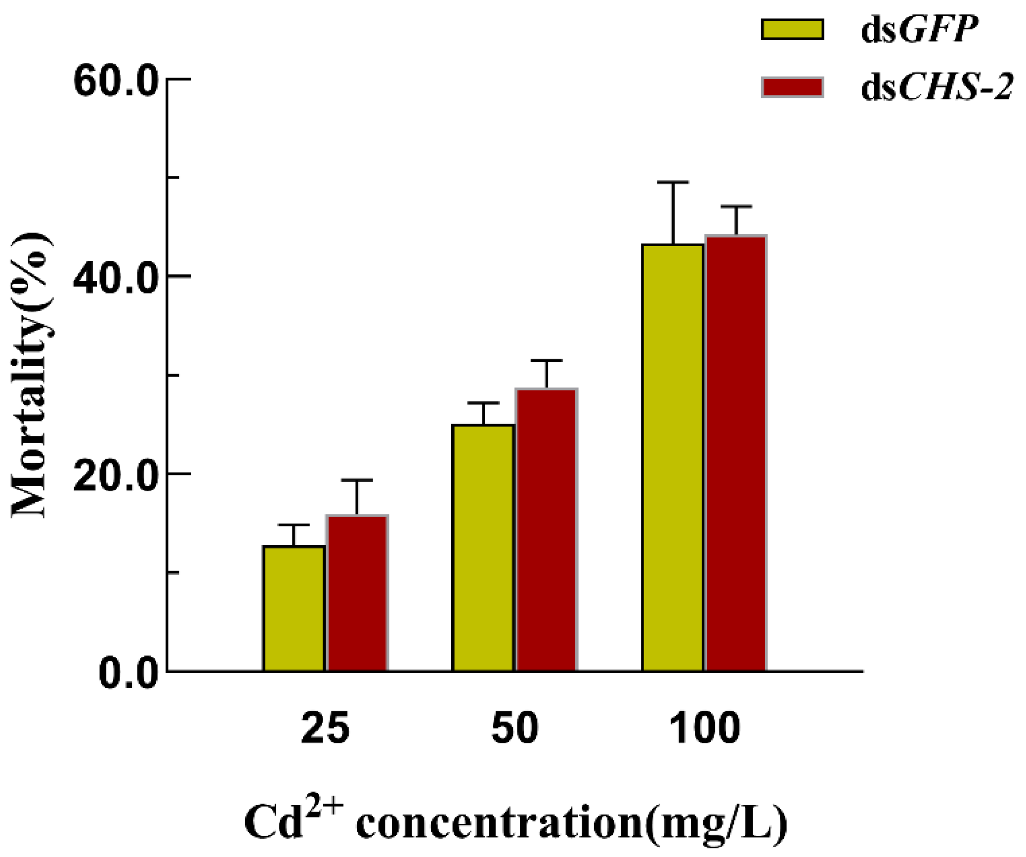
The findings of previous studies indicate that silencing the CHS-2 alone for 24 h has no discernible impact on the larval survival of Ae. albopictus [21]. However, the application of Cd for 12 h after RNA interference caused a significant increase in larval mortality, with higher Cd concentrations correlated with increased mortality. The mortality rate in the dsCHS-2 injected group following Cd stress was virtually indistinguishable from that of the control group, indicating no significant disparity (Figure 4).

3.5. Changes in HSP70 and MT Expression in Ae. albopictus After CHS-2 Silencing Combined with Acute Cd Stress

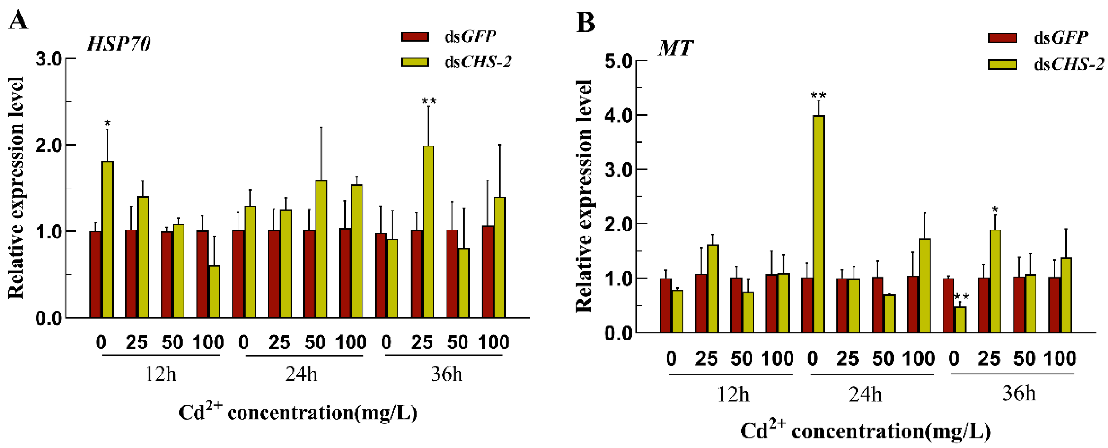
The expression of HSP70 was significantly upregulated within 12 h following silencing of CHS-2, returning to basal levels at 24 and 36 h (Figure 5A). The expression of MT initially increased and then declined after CHS-2 silencing (Figure 5B). Under low-Cd stress (25 mg/L), the expression levels of HSP70 and MT were significantly upregulated after 36 h.

3.6. Changes in the Activities of Antioxidant Enzymes in Ae. albopictus After CHS-2 Silencing Combined with Cd Stress

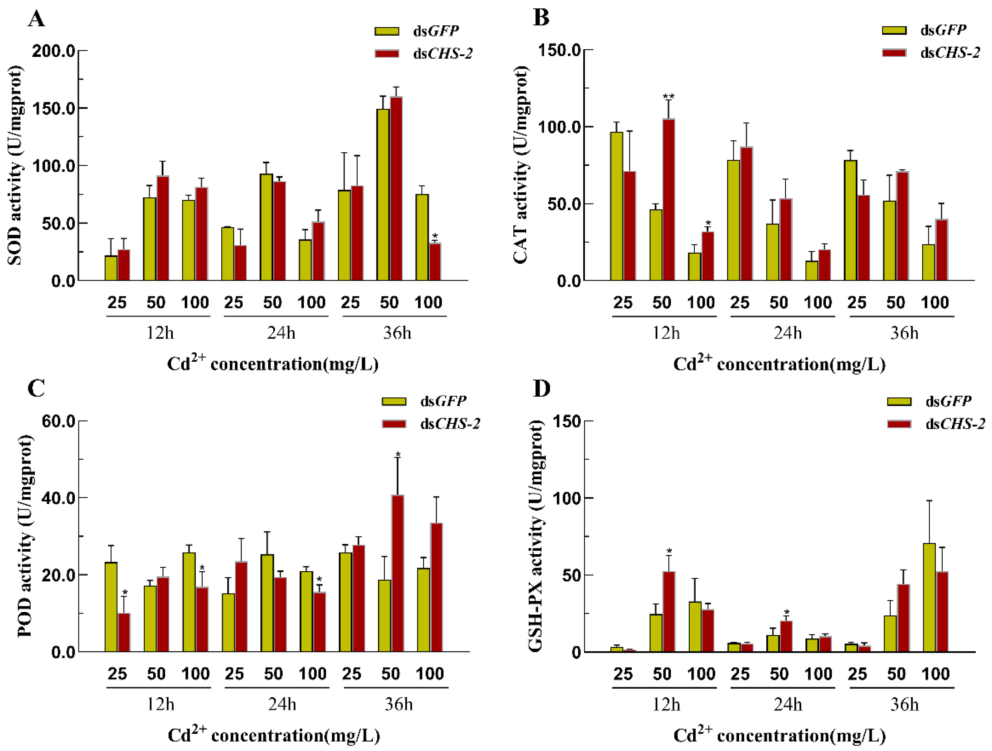
The combination of CHS-2 silencing and Cd stress partially influenced the activity of antioxidant enzymes in Ae. albopictus. Compared with the dsGFP group, significant down-regulation of SOD was observed only 36 h after 100 mg/L Cd stress (Figure 6A). CAT activity significantly increased within a brief period and subsequently returned to that of the control group (Figure 6B). POD activity was prominently down-regulated at 12 h post-25 mg/L Cd stress and 12 and 24 h after 100 mg/L Cd stress; however, it was significantly up-regulated at 36 h following 50 mg/L Cd stress (Figure 6C). GSH-PX activity was significantly up-regulated at 12 and 24 h post-50 mg/L Cd stress (Figure 6D).

4. Discussion

Cd exposure has numerous detrimental effects. For example, Cd stress has been observed to decrease the survival and reproductive rates of Anopheles gambiae [31] and impede the cell proliferation of Ae. albopictus [32], and more. Previous studies have revealed that prolonged Cd stress can result in smaller larvae and pupae, suppress pupation and emergence rates, extend the developmental cycle of Ae. albopictus, and negatively affect the reproductive success of adult Ae. albopictus [26]. The insect midgut is the main target organ for heavy metal stress, and midgut epithelial cells secrete a reticular structure, the PM, to resist heavy metal damage. Proteins, mucopolysaccharides and chitin are the main components of the PM. Therefore, CHS-2 and its PM chitin may play important roles in the response of aquatic insects to heavy metal stress, and the synthesis and metabolism of trehalose and chitin mediated by CHS-2 may play an important role in the adaptation of aquatic insects to heavy metal pollution. CHS-2 has previously been identified as predominantly expressed in the midgut of Ae. albopictus larvae and plays a pivotal role in PM formation. Upon silencing CHS-2, a significant reduction in chitin content was observed in the midgut of fourth-instar larvae of Ae. albopictus, leading to the partial or complete destruction of the PM [21]. Abedi and Brown found large amounts of dichlorodiphenyltrichloroethane (DDT) in the larval PM of Aedes aegypti mosquitoes, which are resistant to the insecticide DDT [33]. In the present study, except for HK and its downstream G6PI, the expression of genes associated with chitin synthesis and metabolic pathways in Ae. albopictus were suppressed under Cd stress (Figure 1). Lin et al. demonstrated that HK is a crucial gene regulating ROS in insects and that reducing HK expression increases ROS activity [34]. Heavy metals can generate substantial amounts of ROS in insects; thus, high HK expression at low concentrations might mitigate ROS activity (Figure 1B). As the expression of HK increased, its downstream gene G6PI also increased (Figure 1G). In contrast to the low expression of chitin synthesis genes, chitin content in the larval midgut was significantly increased (Figure 2A). This discrepancy might be attributed to the post-transcriptional regulation of CHS-2 [35]. The midgut of the fourth-instar larvae was examined for varying concentrations Cd-induced stress using HE staining and paraffin sections. These findings indicate that the midgut epithelial cells of Ae. albopictus fourth-instar larvae exhibited vacuolization under Cd stress accompanied by significant PM thickening (Figure 2C–E). This thickening was more pronounced at higher Cd concentrations, which concurred with the increased chitin content in the midgut. Wu et al. observed a similar phenomenon in the midgut of Boettcherisca peregrina larvae following Cd stress [36]. Based on the aforementioned results, we postulate that Cd stress could prompt the midgut to secrete a substantial amount of PM to facilitate the metabolism of excess Cd, subsequently leading to an increase in chitin content.
To cope with the stress caused by heavy metals, insects have evolved various adaptive mechanisms, including active heavy metal deposition and excretion, initiation of oxidative stress responses, and regulation of MT and HSP expression. First, the midgut of Ae. albopictus fourth-instar larvae exposed to 50 mg/L of Cd after silencing CHS-2 was observed. When CHS-2 were silenced, the midgut of Ae. albopictus larvae failed to develop a complete PM, thereby losing its protective effects. In comparison to simple Cd stress (Figure 2D), the vacuolization of midgut epithelial cells was exacerbated and progressively deformed as the duration of Cd stress increased (Figure 3F), which was consistent with the results of Dabor et al.’s study on the alterations in bee midgut cells induced by CdO and PbO [37]. Subsequent analysis of larval mortality revealed a positive correlation between the mortality of Ae. albopictus larvae and Cd concentrations, whereas the silencing CHS-2 did not significantly influence larval mortality under Cd stress (Figure 4). In previous experiments, silencing the CHS-2 gene alone did not affect the survival of Ae. albopictus larvae [21]. This suggests Ae. albopictus larvae cope with excessive Cd stress via alternative mechanisms following the loss of PM deposition metabolic functions. MT has been identified as the primary ligand of Cd in numerous organisms and is associated with biological tolerance to Cd [38,39]. MT is mainly distributed in the digestive tract of insects, and the Cd ingested by fleshfly larvae mostly combines with proteins to form MT in the digestive tract [40]. The induction of HSPs is a crucial mechanism insects use to counteract heavy metal stress. The expression of HSPs serves as a biological indicator of oxidative stress induced by heavy metals. The HSP70 family is particularly sensitive to Cd stress, and the expression of HSP70 in insects is significantly upregulated following Cd exposure [41,42]. In this study, the combination of CHS-2 silencing and Cd stress occasionally altered the expression of MT and HSP70. These changes appeared irregular. Given that some members of the HSP family are constitutively expressed, we speculate that the selected genes may not be directly related to cadmium stress. The precise underlying mechanisms warrant further investigation.
The ingestion of heavy metals by insects generates a multitude of reactive oxygen free radicals, which in turn stimulate the activation of a panel of antioxidant defense enzymes, including SOD, POD, CAT, and GSH-PX. SOD rapidly disintegrates ROS into H2O2 and concurrently activates CAT and POD to further decompose H2O2 into H2O and CO2 [43]. GSH-PX converts oxidized glutathione to its reduced form and aids in decomposing a portion of H2O2 [44]. It has been found that insect PM has a stronger oxidative stress response than other tissues and can secrete antioxidant enzymes, such as SOD, to counteract free radical damage [45,46]. Uncharged chitin and positively charged proteoglycans in insect PM can chelate heavy metal ions, thereby partially mitigating the damage induced by oxidative stress. Under low Cd stress, antioxidant enzyme activity is f activated in Oxya chinensis, whereas it is inhibited under high-Cd stress [47]. In the present study, the antioxidant enzymes activity in Ae. albopictus under Cd stress following CHS-2 silencing was investigated. Post-silencing of CHS-2, SOD activity remained unaltered under Cd stress, but exhibited a significant down-regulation only 36 h after 100 mg/L Cd stress (Figure 6A). The overproduction of hydrogen peroxide free radicals, triggered by strong oxidative stress reactions following the inhibition of CHS-2, was speculated to oxidize cysteine residues in SOD, leading to unchanged or even decreased SOD activity [48]. Significant increases in the activities of CAT and POD were observed under both short-term (12 h) and long-term (36 h) Cd stress conditions following the silencing of CHS-2 (Figure 6B,C), suggesting that these enzymes substituted for PM in response to oxidative stress induced by Cd at different timepoints. POD activity was significantly reduced within 24 h of CHS-2 silencing. Ma et al. found that POD affects cuticle formation in Cyprinus carpio [49], and PM is composed of cuticle proteins in addition to chitin [50]. Thus, we postulate that silencing CHS-2 leads to a failure in midgut PM formation, diminishing the requirement for cuticular proteins and, consequently, POD activity.
In conclusion, Cd stress can influence chitin synthesis and metabolism in fourth-instar larvae of Ae. albopictus, simultaneously promoting the accumulation of excess Cd in larval midgut PM. Upon silencing CHS-2, a key gene for chitin synthesis, fourth-instar larvae of Ae. albopictus failed to develop a complete PM, affecting Cd deposition and metabolism in the midgut and triggering a robust oxidative stress response. Thus, CHS-2 modulates the formation of Ae. albopictus intestinal PM, which is involved in Cd-induced oxidative stress. The PM is a semi-permeable tissue composed of chitin and proteins, offering protection against pathogens and macromolecular compounds, thus the anti-stress ability of Ae. albopictus was enhanced, who live long time in the Cd-polluted water environment, and then leading to worries of their population expansion. In the future, our research will focus on the following three points: First, we will observe the morphological changes in the PM in Ae. albopictus larvae under Cd stress in more detail, and try to use it as a potential biological indicator for environmental monitoring; Second, the stress resistance of Ae. albopictus was tested through long-term or multiple generations of Cd stress, revealing that environmental pollution may imperceptibly improve the resistance of pests, thus increasing the difficulty and cost of prevention and control, and increasing the public’s attention to environmental pollution. Third, the function and adaptation mechanisms of CHS-2 in Ae. albopictus’ response to long-term Cd stress, including its regulatory relationship with related antioxidant enzyme genes, was explored to clarify the resistance mechanism of Ae. albopictus and attempt to control Ae. albopictus by using resistance or other control methods.

Author Contributions

C.Z.: Investigation, Writing—Original Draft, Writing—Review & Editing, Visualization. Y.D.: Methodology, Investigation, Visualization. R.L.: Investigation, Writing—Original Draft. M.Z.: Validation, Writing—Original Draft. Y.C.: Visualization. B.T.: Methodology, Writing—Review & Editing. G.Q.: Writing—Review & Editing, Methodology, Project administration. S.W.: Conceptualization, Methodology, Project administration. All authors have read and agreed to the published version of the manuscript.

Funding

This work was supported by the National Natural Science Foundation of China [Grant Number 32272608 and 31672081].

Institutional Review Board Statement

All animal procedures have been approved by the Experimental Animal Ethics Committee (AEWC) of Hangzhou Normal University (Approval No. HSD20220103, January 2022).

Data Availability Statement

The original contributions presented in this study are included in the article. Further inquiries can be directed to the corresponding authors.

Conflicts of Interest

The authors declare that they have no competing interest.

References

  1. Qin, Y.H.; Tao, Y.Q. Pollution status of heavy metals and metalloids in Chinese lakes: Distribution, bioaccumulation and risk assessment. Ecotoxicol. Environ. Saf. 2022, 248, 114293. [Google Scholar] [CrossRef] [PubMed]
  2. Zhu, Y.S.; Costa, M. Metals and molecular carcinogenesis. Carcinogenesis 2020, 41, 1161–1172. [Google Scholar] [CrossRef] [PubMed]
  3. Farías, P.; Hernández-Bonilla, D.; Moreno-Macías, H.; Montes-López, S.; Schnaas, L.; Texcalac-Sangrador, J.L.; Ríos, C.; Riojas-Rodríguez, H. Prenatal Co-Exposure to Manganese, Mercury, and Lead, and Neurodevelopment in Children during the First Year of Life. Int. J. Environ. Res. Public Health 2022, 19, 13020. [Google Scholar] [CrossRef]
  4. Ma, C.C.; Iwai-Shimada, M.; Nakayama, S.F.; Isobe, T.; Kobayashi, Y.; Tatsuta, N.; Taniguchi, Y.; Sekiyama, M.; Michikawa, T.; Yamazaki, S.; et al. Association of prenatal exposure to cadmium with neurodevelopment in children at 2 years of age: The Japan Environment and Children’s Study. Environ. Int. 2021, 156, 106762. [Google Scholar] [CrossRef]
  5. Fu, Z.S.; Xi, S.H. The effects of heavy metals on human metabolism. Toxicol. Mech. Methods 2020, 30, 167–176. [Google Scholar] [CrossRef] [PubMed]
  6. Lian, G.Q.; Lee, X.Q. Concentrations, Distribution, and Pollution Assessment of Metals in River Sediments in China. Int. J. Environ. Res. Public Health 2021, 18, 6908. [Google Scholar] [CrossRef]
  7. Chen, J.; Wang, J.W.; Shu, Y.H. Review on the effects of heavy metal pollution on herbivorous insects. Ying Yong Sheng Tai Xue Bao 2020, 31, 1773–1782. [Google Scholar] [CrossRef]
  8. Jiang, D.; Tan, M.T.; Guo, Q.X.; Yan, S.C. Transfer of heavy metal along food chain: A mini-review on insect susceptibility to entomopathogenic microorganisms under heavy metal stress. Pest Manag. Sci. 2021, 77, 1115–1120. [Google Scholar] [CrossRef]
  9. Ali, S.; Ullah, M.I.; Saeed, M.F.; Khalid, S.; Saqib, M.; Arshad, M.; Afzal, M.; Damalas, C.A. Heavy metal exposure through artificial diet reduces growth and survival of Spodoptera litura (Lepidoptera: Noctuidae). Environ. Sci. Pollut. Res. 2019, 26, 14426–14434. [Google Scholar] [CrossRef]
  10. Arambourou, H.; Llorente, L.; Moreno-Ocio, I.; Herrero, O.; Barata, C.; Fuertes, I.; Delorme, N.; Méndez-Fernández, L.; Planelló, R. Exposure to heavy metal-contaminated sediments disrupts gene expression, lipid profile, and life history traits in the midge. Water Res. 2020, 168, 115165. [Google Scholar] [CrossRef]
  11. Gao, H.H.; Zhao, H.Y.; Du, C.; Deng, M.M.; Du, E.X.; Hu, Z.Q.; Hu, X.S. Life table evaluation of survival and reproduction of the aphid, Sitobion avenae, exposed to cadmium. J. Insect Sci. 2012, 12, 44. [Google Scholar] [CrossRef]
  12. Lefcort, H.; Vancura, J.; Lider, E.L. 75 years after mining ends stream insect diversity is still affected by heavy metals. Ecotoxicology 2010, 19, 1416–1425. [Google Scholar] [CrossRef] [PubMed]
  13. Hensbergen, P.J.; van Velzen, M.J.M.; Nugroho, R.A.; Donker, M.H.; van Straalen, N.M. Metallothionein-bound cadmium in the gut of the insect Orchesella cincta (Collembola) in relation to dietary cadmium exposure. Comp. Biochem. Physiol. C-Toxicol. Pharmacol. 2000, 125, 17–24. [Google Scholar] [CrossRef] [PubMed]
  14. Pedersen, S.A.; Kristiansen, E.; Andersen, R.A.; Zachariassen, K.E. Cadmium is deposited in the gut content of larvae of the beetle Tenebrio molitor and involves a Cd-binding protein of the low cysteine type. Comp. Biochem. Physiol. C-Toxicol. Pharmacol. 2008, 148, 217–222. [Google Scholar] [CrossRef] [PubMed]
  15. Su, H.H.; Hu, M.M.; Harvey-Samuel, T.; Yang, Y.Z. Accumulation and Excretion of Cadmium in Three Successive Generations of Spodoptera exigua (Lepidoptera: Noctuidae) and Impact on the Population Increase. J. Econ. Entomol. 2014, 107, 223–229. [Google Scholar] [CrossRef]
  16. Hegedus, D.; Erlandson, M.; Gillott, C.; Toprak, U. New Insights into Peritrophic Matrix Synthesis, Architecture, and Function. Annu. Rev. Entomol. 2009, 54, 285–302. [Google Scholar] [CrossRef]
  17. Liu, X.J.; Cooper, A.M.W.; Zhang, J.Z.; Zhu, K.Y. Biosynthesis, modifications and degradation of chitin in the formation and turnover of peritrophic matrix in insects. J. Insect Physiol. 2019, 114, 109–115. [Google Scholar] [CrossRef]
  18. Xu, C.D.; Liu, Y.K.; Qiu, L.Y.; Wang, S.S.; Pan, B.Y.; Li, Y.; Wang, S.G.; Tang, B. GFAT and PFK genes show contrasting regulation of chitin metabolism in Nilaparvata lugens. Sci. Rep. 2021, 11, 5246. [Google Scholar] [CrossRef]
  19. Li, L.; Wang, Y.Q.; Li, G.Y.; Song, Q.S.; Stanley, D.; Wei, S.J.; Zhu, J.Y. Genomic and transcriptomic analyses of chitin metabolism enzymes in Tenebrio molitor. Arch. Insect Biochem. Physiol. 2022, 111, e21950. [Google Scholar] [CrossRef]
  20. Merzendorfer, H.; Zimoch, L. Chitin metabolism in insects: Structure, function and regulation of chitin synthases and chitinases. J. Exp. Biol. 2003, 206, 4393–4412. [Google Scholar] [CrossRef]
  21. Zhang, C.; Ding, Y.J.; Zhou, M.; Tang, Y.; Chen, R.F.; Chen, Y.R.; Wen, Y.T.; Wang, S.G. RNAi-mediated CHS-2 silencing affects the synthesis of chitin and the formation of the peritrophic membrane in the midgut of Aedes albopictus larvae. Parasites Vectors 2023, 16, 259. [Google Scholar] [CrossRef]
  22. Joly, A.L.; Wettstein, G.; Mignot, G.; Ghiringhelli, F.; Garrido, C. Dual Role of Heat Shock Proteins as Regulators of Apoptosis and Innate Immunity. J. Innate Immun. 2010, 2, 238–247. [Google Scholar] [CrossRef] [PubMed]
  23. Morales, M.; Planelló, R.; Martínez-Paz, P.; Herrero, O.; Cortés, E.; Martínez-Guitarte, J.L.; Morcillo, G. Characterization of Hsp70 gene in Chironomus riparius: Expression in response to endocrine disrupting pollutants as a marker of ecotoxicological stress. Comp. Biochem. Physiol. C-Toxicol. Pharmacol. 2011, 153, 150–158. [Google Scholar] [CrossRef]
  24. Gu, B.N.; Liang, W.; Yang, T.Z.; Hu, Z.J.; Shen, H.D. Metallothionein, hemocyte status and superoxide dismutase/aspartate aminotransferase activity are sensitive biomarkers of cadmium stress in. Aquat. Toxicol. 2019, 215, 105284. [Google Scholar] [CrossRef]
  25. El-Gendy, A.H.; Augustyniak, M.; Toto, N.A.; Al Farraj, S.; El-Samad, L.M. Oxidative stress parameters, DNA damage and expression of HSP70 and MT in midgut of Trachyderma hispida(Forskal, 1775) (Coleoptera: Tenebrionidae) from a textile industry area. Environ. Pollut. 2020, 267, 115661. [Google Scholar] [CrossRef] [PubMed]
  26. Yu, L.Y.; Chen, X.M.; Wei, Y.; Ding, Y.J.; Wang, Q.W.; Wang, S.H.; Tang, B.; Wang, S.G. Effects of long-term cadmium exposure on trehalose metabolism, growth, and development of Aedes albopictus (Diptera: Culicidae). Ecotoxicol. Environ. Saf. 2020, 204, 111034. [Google Scholar] [CrossRef]
  27. Puggioli, A.; Balestrino, F.; Damiens, D.; Lees, R.S.; Soliban, S.M.; Madakacherry, O.; Dindo, M.L.; Bellini, R.; Gilles, J.R.L. Efficiency of Three Diets for Larval Development in Mass Rearing Aedes albopictus (Diptera: Culicidae). J. Med. Entomol. 2013, 50, 819–825. [Google Scholar] [CrossRef]
  28. GB/T 602-2002; Chemical Reagent—Preparations of Standard Solutions for Impurities. General Administration of Quality Supervision, Inspection and Quarantine of the People’s Republic of China: Beijing, China, 2002.
  29. Liu, X.Y.; Wang, S.S.; Yu, Y.Y.; Cheng, Y.S.; Hu, C.X.; Zhou, M.; Li, C.; Tang, B.; Wu, Y. Effects of Inhibiting the Expression of Chitin Synthase Gene sfCHSB on the Metabolism of Trehalose and Chitin in Spodoptera frugiperda Larvae. Agriculture 2022, 12, 2019. [Google Scholar] [CrossRef]
  30. Gregor, K.M.; Becker, S.C.; Hellhammer, F.; Schön, K.; Baumgärtner, W.; Puff, C. Histochemical staining techniques in Culex pipiens and Drosophila melanogaster (Diptera) with a comparison to mammals. Vet. Pathol. 2022, 59, 836–849. [Google Scholar] [CrossRef]
  31. Braeckman, B.; Raes, H.; VanHoye, D. Heavy-metal toxicity in an insect cell line. Effects of cadmium chloride, mercuric chloride and methylmercuric chloride on cell viability and proliferation in Aedes albopictus cells. Cell Biol. Toxicol. 1997, 13, 389–397. [Google Scholar] [CrossRef]
  32. Mireji, P.O.; Keating, J.; Hassanali, A.; Mbogo, C.M.; Muturi, M.N.; Githure, J.I.; Beier, J.C. Biological cost of tolerance to heavy metals in the mosquito Anopheles gambiae. Med. Vet. Entomol. 2010, 24, 101–107. [Google Scholar] [CrossRef] [PubMed]
  33. Abedi, Z.H.; Brown, A.W.A. Peritrophic Membrane as Vehicle for DDT and DDE Excretion in Aedes aegypti Larvae. Ann. Entomol. Soc. Am. 1961, 54, 539–542. [Google Scholar] [CrossRef]
  34. Lin, X.W.; Xu, W.H. Hexokinase is a key regulator of energy metabolism and ROS activity in insect lifespan extension. Aging 2016, 8, 245–259. [Google Scholar] [CrossRef]
  35. Zimoch, L.; Hogenkamp, D.G.; Kramer, K.J.; Muthukrishnan, S.; Merzendorfer, H. Regulation of chitin synthesis in the larval midgut of Manduca sexta. Insect Biochem. Mol. Biol. 2005, 35, 515–527. [Google Scholar] [CrossRef] [PubMed]
  36. Wu, G.X.; Gao, X.; Ye, G.Y.; Li, K.; Hu, C.; Cheng, J.A. Ultrastructural alterations in midgut and Malpighian tubules of Boettcherisca peregrina exposure to cadmium and copper. Ecotoxicol. Environ. Saf. 2009, 72, 1137–1147. [Google Scholar] [CrossRef]
  37. Dabour, K.; Al Naggar, Y.; Masry, S.; Naiem, E.; Giesy, J.P. Cellular alterations in midgut cells of honey bee workers (Apis millefera L.) exposed to sublethal concentrations of CdO or PbO nanoparticles or their binary mixture. Sci. Total Environ. 2019, 651, 1356–1367. [Google Scholar] [CrossRef]
  38. Luo, M.; Finet, C.; Cong, H.S.; Wei, H.Y.; Chung, H. The evolution of insect metallothioneins. Proc. R. Soc. B-Biol. Sci. 2020, 287, 20202189. [Google Scholar] [CrossRef]
  39. Zhang, J.; Shi, Z.H.; Gao, Z.H.; Wen, Y.T.; Wang, W.Q.; Liu, W.; Wang, X.P.; Zhu, F. Identification of three metallothioneins in the black soldier fly and their functions in Cd accumulation and detoxification. Environ. Pollut. 2021, 286, 117146. [Google Scholar] [CrossRef]
  40. Aoki, Y.; Suzuki, K.T.; Kubota, K. Accumulation of Cadmium and Induction of Its Binding-Protein in the Digestive-Tract of Fleshfly (Sarcophaga-Peregrina) Larvae. Comp. Biochem. Physiol. C-Toxicol. Pharmacol. 1984, 77, 279–282. [Google Scholar] [CrossRef]
  41. Kafel, A.; Nowak, A.; Bembenek, J.; Szczygiel, J.; Nakonieczny, M.; Swiergosz-Kowalewska, R. The localisation of HSP70 and oxidative stress indices in heads of Spodoptera exigua larvae in a cadmium-exposed population. Ecotoxicol. Environ. Saf. 2012, 78, 22–27. [Google Scholar] [CrossRef]
  42. Somasundaram, S.; Abraham, J.S.; Maurya, S.; Toteja, R.; Gupta, R.; Makhija, S. Expression and molecular characterization of stress-responsive genes (hsp70 and Mn-sod) and evaluation of antioxidant enzymes (CAT and GPx) in heavy metal exposed freshwater ciliate, Tetmemena sp. Mol. Biol. Rep. 2019, 46, 4921–4931. [Google Scholar] [CrossRef] [PubMed]
  43. Sun, H.X.; Wu, W.J.; Guo, J.X.; Xiao, R.; Jiang, F.Z.; Zheng, L.Y.; Zhang, G.R. Effects of nickel exposure on testicular function, oxidative stress, and male reproductive dysfunction in Spodoptera litura Fabricius. Chemosphere 2016, 148, 178–187. [Google Scholar] [CrossRef]
  44. Li, M.; Zheng, Y.; Liang, H.W.; Zou, L.H.; Sun, J.J.; Zhang, Y.Y.; Qin, F.; Liu, S.Z.; Wang, Z.Z. Molecular cloning and characterization of cat, gpx1 and Cu/Zn-sod genes in pengze crucian carp (Carassius auratus var. Pengze) and antioxidant enzyme modulation induced by hexavalent chromium in juveniles. Comp. Biochem. Physiol. C-Toxicol. Pharmacol. 2013, 157, 310–321. [Google Scholar] [CrossRef] [PubMed]
  45. Filipovic, A.; Mrdakovic, M.; Ilijin, L.; Vlahovic, M.; Todorovic, D.; Grcic, A.; Peric-Mataruga, V. Effect of fluoranthene on antioxidative defense in different tissues of Lymantria dispar and Euproctis chrysorrhoea larvae. Comp. Biochem. Physiol. C-Toxicol. Pharmacol. 2019, 224, 108565. [Google Scholar] [CrossRef]
  46. Peric-Mataruga, V.; Ilijin, L.; Mrdakovic, M.; Todorovic, D.; Prokic, M.; Matic, D.; Vlahovic, M. Parameters of oxidative stress, cholinesterase activity, Cd bioaccumulation in the brain and midgut of Lymantria dispar (Lepidoptera: Lymantriidae) caterpillars from unpolluted and polluted forests. Chemosphere 2019, 218, 416–424. [Google Scholar] [CrossRef] [PubMed]
  47. Zhang, Y.P.; Song, D.N.; Wu, H.H.; Yang, H.M.; Zhang, J.Z.; Li, L.J.; Ma, E.B.; Guo, Y.P. Effect of Dietary Cadmium on the Activity of Glutathione S-Transferase and Carboxylesterase in Different Developmental Stages of the Oxya chinensis (Orthoptera: Acridoidea). Environ. Entomol. 2014, 43, 171–177. [Google Scholar] [CrossRef]
  48. Stdimitrova, M.; Tishinova, V.; Velcheva, V. Combined Effect of Zinc and Lead on the Hepatic Superoxide Dismutase-Catalase System in Carp (Cyprinus-Carpio). Comp. Biochem. Physiol. C-Toxicol. Pharmacol. 1994, 108, 43–46. [Google Scholar] [CrossRef]
  49. Ma, C.P.; Guo, Z.M.; Zhang, F.L.; Su, J.Y. Molecular identification, expression and function analysis of peroxidasin in. Insect Sci. 2020, 27, 1173–1185. [Google Scholar] [CrossRef]
  50. Terra, W.R. The origin and functions of the insect peritrophic membrane and peritrophic gel. Arch. Insect Biochem. Physiol. 2001, 47, 47–61. [Google Scholar] [CrossRef]
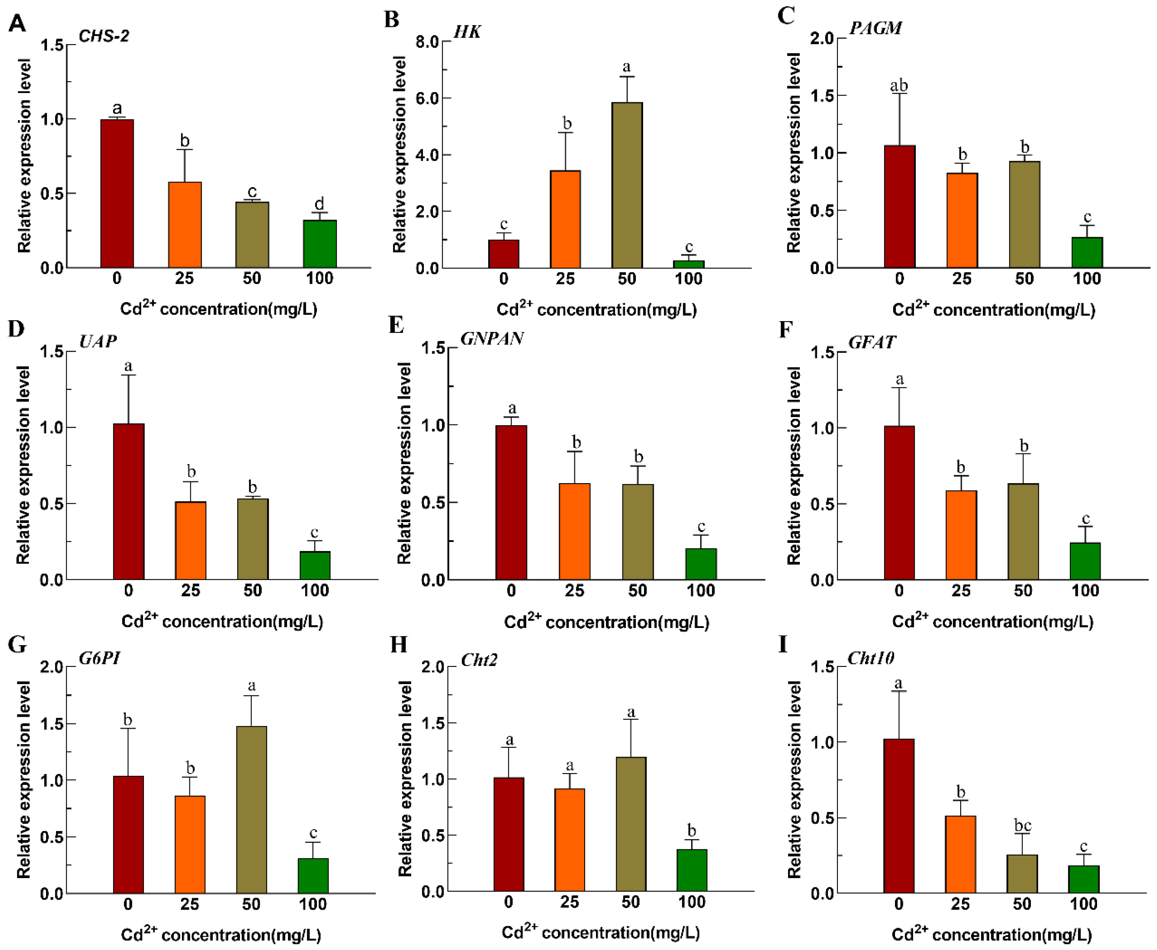
Figure 1. Relative expression of chitin metabolic pathway-related genes of Ae. albopictus after Cd stress. (A) CHS-2. (B) HK. (C) PAGM. (D) UAP. (E) GNPAN. (F) GFAT. (G) G6PI. (H) Cht2. (I) Cht10. Values represent mean ± SE. Different lowercase letters above the bar indicate that the difference is statistically significant. Relative expression levels were calculated in comparison with that of the fourth-instar larvae in 0 mg/L Cd solution, which was ascribed an arbitrary value of 1.
Insects 16 01102 g001
Figure 2. After different concentrations cadmium stresses of Ae. albopictus larva of chitin content change and intestine structure observation. (A) Chitin content in the midgut of the fourth-instar larvae of Ae. albopictus after 12 h of Cd stress. Values represent mean ± SE. Different lowercase letters above the bar indicate that the difference is statistically significant. Fourth-instar larvae 12 h exposed to Cd solutions of different concentrations were used as materials, and the head and tail were removed, embedded in paraffin, sectioned, stained with HE, and observed under a microscope. (B) 0 mg/L, (C) 25 mg/L (D) 50 mg/L (E) 100 mg/L. The scale indicates 100 μm. The triangle indicates the position of the PM.
Insects 16 01102 g002
Figure 3. Slices of the fourth-instar larvae of Ae. albopictus after CHS-2 silencing combined with 50 mg/L Cd stress. After injection of dsRNA, fourth-instar larvae were exposed to 50 mg/L Cd solution for 12, 24 and 36 h, were used as materials, and the head and tail were removed, embedded in paraffin, sectioned, stained with HE, and observed under a microscope. (A) 12 h after dsGFP injection. (B) 24 h after dsGFP injection. (C) 36 h after injection of dsGFP. (D) 12 h after injection of dsCHS-2. (E) 24 h after injection of dsCHS-2. (F) 36 h after injection of dsCHS-2. The scale is 100 μm, and the black arrow in the figure is the normal PM.
Insects 16 01102 g003
Figure 4. Mortality of Ae. albopictus after CHS-2 silencing combined with acute Cd stress. Values represent mean ± SE.
Insects 16 01102 g004
Figure 5. The relative expression of HSP70 and MT in Ae. albopictus after CHS-2 silencing combined with Cd acute stress. (A) HSP70. (B) MT. Relative expression levels were calculated in comparison with dsGFP exposed to the same concentration of Cd solution for the same time, which was ascribed an arbitrary value of 1. Values represent mean ± SE. (One-way ANOVA, Duncan test, * p < 0.05, ** p < 0.01).
Insects 16 01102 g005
Figure 6. Activity of antioxidant enzymes in Ae. albopictus after CHS-2 silencing combined with Cd stress. (A) SOD. (B) CAT. (C) POD. (D) CSH-PX. Values represent mean ± SE. (One-way ANOVA, Duncan test, * p < 0.05, ** p < 0.01).
Insects 16 01102 g006
Table 1. Primers for synthesis of dsRNA.
Primer NameForward Primer (5′-3′)Reverse Primer (5′-3′)NCBI
dsCHS-2GGATCCTAATACGACTCACTATAGGAGTCAACGCACCGTACAGGAGGATCCTAATACGACTCACTATAGGGAAGAGCAACACCAAACCGALOC109405983
dsGFPGGATCCTAATACGACTCACTATAGGAAGGGCGAGGAGCTGTTCACCGGGATCCTAATACGACTCACTATAGGCAGCAGGACCATGTGATCGCGCXM_013480425.1
The underlined is the T7 promoter.
Table 2. Primers for qRT-PCR.
Primer NameForward Primer (5′-3′)Reverse Primer (5′-3′)NCBI
actinGCTACGTCGCCCTGCACTTAGGAACGACGGCTGGAAGADQ657949.1
CHS-2GGAGACCAAAGGATGGGACGCCTGTAAGGACGATGACGAATGTXM_019679038.3
Cht2GACTCGGATGACAAGGGTTTCAGGGCTTCACATACTTCGTTXM_029879282.1
Cht10GCCACGGATACTTAGAATAGCGCTGTTTGACGGTCGTTGATTTXM_029869372.1
HKGGGAGAAGCCAGCGAAGATAGATGTCTGCTTCGGGATGTGAY705876.1
PAGMGCCATTTCTGACATGCTACTCGTTTCGGTCTTCTACTTTGAXM_019671844.2
UAPAGTGCTCTATTTACACGCTCATACTCCGACCGCTTCATTTGAPW01001510.1
GNPNACAGTCCTCCCATTTCAGCCAACGAAACATCGCCCACCXM_019707567.2
GFATTCAACGGGCAACATCCAGAAGCGTCCGATGCAAAGAXM_019671518.2
G6PIGGAGGATGACATTCGCTTCGCGGTGATACGGTTCTTGGAGXM_019673634.3
HSP70GATGTGTCCCTTTTGACCATTCGACTTCACGACGCAGTTTCTJN132154.1
MTATGCCTTGCCCATGCGACAGTCAGATCCGCAGTTGC
Disclaimer/Publisher’s Note: The statements, opinions and data contained in all publications are solely those of the individual author(s) and contributor(s) and not of MDPI and/or the editor(s). MDPI and/or the editor(s) disclaim responsibility for any injury to people or property resulting from any ideas, methods, instructions or products referred to in the content.