Article

Differences in Male-Killing Rickettsia Bacteria between Lineages of the Invasive Gall-Causing Pest Leptocybe invasa

Guangxi Colleges and Universities Key Laboratory for Cultivation and Utilization of Subtropical Forest Plantation, Guangxi Key Laboratory of Forest Ecology and Conservation, College of Forestry, Guangxi University, Nanning 530004, China
*
Author to whom correspondence should be addressed.
These authors contributed equally to this work.
Insects 2023, 14(9), 757; https://doi.org/10.3390/insects14090757
Submission received: 10 July 2023 / Revised: 22 August 2023 / Accepted: 7 September 2023 / Published: 11 September 2023
(This article belongs to the Section Insect Behavior and Pathology)

Abstract

:

Simple Summary

This study compared the differences in symbiotic bacteria between the A and B lineages of the Leptocybe invasa, so as to provide a reference for the scientific control of the pest.

Abstract

(1) Background: Leptocybe invasa (Hymenoptera: Eulophidae) is a global invasive pest that seriously damages eucalyptus plants and has caused serious harm to forestry production in many countries. Two genotypically distinct lineages of L. invasa have been detected outside of Australia, namely, lineage A and lineage B. However, the composition and abundance of endosymbiotic bacteria in L. invasa are still unclear between lineages. Therefore, the purpose of this study was to compare the bacterial communities in female adults of L. invasa of different lineages distributed in the same domain; (2) Methods: The PacBio Sequel II platform was used to compare bacterial community composition between lineages of L. invasa by sequencing the V1–V9 region of the 16S rRNA gene, and fluorescence quantitative PCR was used to compare the relative expression of Rickettsia between lineages of L. invasa; (3) Results: A total of 437 operational taxonomic units (OTUs) were obtained. These OTUs were subdivided into 20 phyla, 32 classes, 77 orders, 129 families, and 217 genera. At the genus level, the dominant bacteria in lineage A and lineage B were Rickettsia and Bacteroides, respectively. There were differences in the bacterial community of L. invasa between lineages, and the abundance and relative expression of Rickettsia in lineage A were significantly higher than those in lineage B; (4) Conclusions: There were differences in the bacterial community of L. invasa between lineages, and the abundance and relative expression of Rickettsia in lineage A were significantly higher than those in lineage B.

1. Introduction

Insects generally carry heritable endosymbiotic bacteria [1], and the main mode of transmission is maternal inheritance [2]. The interaction between endosymbiotic bacteria and insects is often complex. In the long-term process of coevolution, endosymbiotic bacteria generally live in host-specific bacteria-containing cells, and the host insects provide them with the necessary ecological conditions for growth. Endosymbiotic bacteria can impart new biological characteristics on host insects [3] and play important roles in host growth and development, reproductive regulation, environmental adaptation, genetic evolution, immune responses, and many other biological processes [4,5,6,7]. They are interdependent, interactive, and coevolutionary [8]. For example, Rickettsia can shorten the generation development period and improve the survival, egg laying, and offspring sex ratio in whitefly [9,10]. Wolbachia, an endosymbiotic bacterium, can enhance mosquitoes’ resistance to RNA viruses [11,12]. In recent years, pest control by eliminating endosymbiotic bacteria has been successfully applied. For example, the mixed use of antibiotics and insecticides can effectively reduce the number of endosymbiotic bacteria in Nilaparvata lugens Stal and improve the control effect of insecticides [13]. By eliminating the endosymbiotic bacterium Wolbachia from mosquitoes, mosquito fecundity is effectively reduced [14]. Therefore, understanding of the composition and functions of bacteria in insects is of great significance for pest control.
Leptocybe invasa Fisher & LaSalle (Hymenoptera: Eulophidae) is a globally invasive pest that seriously damages eucalyptus plants. L. invasa female adults lay their eggs in eucalyptus branches and leaves to reproduce their offspring, resulting in lumpy galls on the leaf veins, petioles, and branches that have serious effects on the normal growth of the plant [15]. The main reproductive mode of L. invasa is parthenogenesis [16], which makes the success rate of invasion and colonization very high. In addition, the amazing reproductive rate and population numbers also enable L. invasa to break out rapidly in areas invaded, causing large economic losses to forestry production in many countries. L. invasa was first discovered in the Middle East and Mediterranean in 2000 [16]. It had spread to 29 countries on five continents by 2014, and that number soared to 45 countries in 2018 [16,17,18]. In China, L. invasa was first found in Dongxing city, Guangxi Zhuang Autonomous Region, on the border with Vietnam, in 2007 [19] and has now spread to Guangxi, Hainan, Guangdong, Fujian, Sichuan, Jiangxi, Yunnan, and Taiwan Provinces [18,20]. Under the scenario of future climate warming, the potential core distribution area of L. invasa in China will be located in Yunnan, Guangxi, Guangdong, and Hainan Provinces and will spread to high latitudes (Hubei, Anhui, Zhejiang, Jiangsu, etc.) [21].
Human activities have broken the original barriers of geographical isolation of pests and led to the mixing of pests of different lineages, which has created new challenges for pest management. The invasion of pests in the form of lineage complexes may be the fundamental reason for the repeated occurrence of pests and the failure in some areas to control pests [22], such as Gonipterus scutellatus [22], Bemisia tabaci [23], and Cacopsylla chinensis [24]. Mitochondrial COI sequence data have shown that L. invasa has three lineages, A, B (B1 and B2), and C, of which lineages B2 and C only appear in Australia, while lineages A and B1 are widely distributed in areas invaded by L. invasa. L. invasa lineages A and B1 coexist in China, Laos, Vietnam, Thailand, and South Africa (lineage B in this paper refers to lineage B1) [15,25,26,27].
To date, there have been many reports on the bacteria in L. invasa. Nugnes et al. [25] found that Rickettsia in L. invasa is located in the ovary of female adults, indicating that Rickettsia is related to its parthenogenesis. Guo et al. [28] found that the content of Rickettsia in male L. invasa was much lower than that in female L. invasa, and the difference in Rickettsia content between males and females may be due to the difference in their physiological structure. Liu et al. [29] identified the types of culturable bacteria in L. invasa at different developmental stages and identified 88 kinds of bacteria belonging to 4 phyla, 27 families, and 44 genera, of which 72 kinds were isolated at the gall stage and 46 kinds at the adult stage. The most abundant bacterial species was γ-Proteobacteria. There were significant differences in total number of bacteria and community composition among developmental stages of L. invasa. Guo et al. [30] studied the bacterial diversity in 10 geographical populations of L. invasa in China and found that differences in bacterial species and abundances among geographical populations, with Rickettsia dominant in most populations. Although these studies have clarified the bacterial diversity and function of L. invasa from many angles, deficiencies remain because most researchers ignored the influence of lineage type on the symbiotic bacteria of L. invasa before carrying out the research. Even as early as 2015, Nugnes et al. proved that Rickettsia in lineages A and B of L. invasa had differentiated genetically and speculated that there may be differences in reproductive strategy between lineages A and B. Therefore, clarifying the bacterial composition and abundances in different lineages of L. invasa can supplement and verify many of the research results described above.

2. Materials and Methods

2.1. Sample Collection

The samples used in this study were collected from Luchuan City, Guangxi Zhuang Autonomous Region, China (110°15′ E, 22°17′ N) in November 2021. The host plant was DH201-2: Eucalyptus grandis × Eucalyptus tereticornis (Myrtales: Myrtaceae). Fresh eucalyptus shoots with galls were collected from woodland, and the branches were placed in a plastic bottle filled with water to maintain freshness and then transferred into a sealed net cage (40 cm × 40 cm × 80 cm) at room temperature to keep the adults from escaping. The water in the plastic bottle was renewed daily until L. invasa adults emerged. Sexes were identified by morphological observation. Female L. invasa adults were selected as test insects within 12 h of emergence.

2.2. Sample Processing and Extraction of L. invasa Total Genomic DNA

A total of 200 adult female L. invasa were collected within 12 h of emergence and starved for 12 h. The surface of each L. invasa female was disinfected with 75% alcohol 3 times in an ultraclean workbench for 30 s each time and then rinsed with sterile water 5 times. Finally, the head of each L. invasa female was cut off in sterile water. The separated head, chest, and abdomen of each female were placed in a sterilized 1.5 mL centrifuge tube, marked, and stored in a refrigerator at −20 °C until use. An animal tissue DNA extraction kit (column type, Beijing Qingke Biotechnology Co., Ltd. Beijing, China) was used to extract the total genomic DNA of female L. invasa heads. According to the manufacturer’s instructions, the quality and concentration of DNA were determined at ratios of 260 nm/280 nm and 260 nm/230 nm using a Nanodrop 2000 Ultra-micro UV spectrophotometer. Qualified DNA was stored in a −20 °C refrigerator until use.

2.3. Identification of L. invasa Lineages

Extracted L. invasa DNA templates were amplified by PCR with mtDNA COI universal primers LCO1490 (5′-GGTCAACAAATCATAAAGATATTGG-3′) and HCO2198 (5′-TAAACTT CAGGGTGACCAAAAAATCA-3′) [31]. Each PCR system contained 25.0 μL, including 12.5 μL of 2 × Hotstart PCR Master Mix (without dye) (Shanghai Shenggong Bioengineering Technology Service Co., Ltd. Shanghai, China), 0.5 μL (10 μM) of each of the forward and reverse primers, 2.0 μL of DNA template, and 9.5 μL of ddH2O. The PCR procedure was predenaturation at 95 °C for 3 min; 30 cycles of denaturation at 95 °C for 20 s, annealing at 49 °C for 20 s, and extension at 72 °C for 10 s; and a final extension at 72 °C for 3 min. PCR products were mixed with 1 μL of 6× loading buffer and detected by 1% agarose gel electrophoresis. Each gel was then cut, purified, and sent to Shanghai Shenggong Bioengineering Technology Service Co., Ltd., Shanghai, China for sequencing. The COI sequence for the L. invasa were downloaded from the GenBank molecular database (ON166667-ON166668, MH093001-MH093481, JQ289999-JQ290005, KP233972-KP233993, KP233954, and MZ378835-MZ379154). The COI sequence GenBank accession numbers obtained in this study were ON166667 (Lineage A) and ON166668 (Lineage B) (because the COI gene sequence is consistent in the L. invasa lineage, we only uploaded one sequence of each lineage). Using MEGAX v7.0 software, all COI sequences were compared, and a sample neighbor-joining (NJ) phylogenetic tree was established with the Kimura 2-parameter model. L. invasa lineages were identified according to the results of the phylogenetic tree. When the number of female L. invasa adults of lineages A and B to be identified reached 50, their chests and abdomens were mixed according to lineage type in an ultraclean workbench.

2.4. Extraction of Total Microbial DNA from L. invasa and Amplification of the 16S rRNA Gene Sequence

L. invasa lineage A and B samples were ground with liquid nitrogen, and the powder was placed in sterile centrifuge tubes. The PowerSoil® DNA Isolation kit was used to extract the total microbial DNA of each lineage sample. The quality and concentration of DNA were detected by a Nanodrop 2000 Ultra-micro UV spectrophotometer. Primers 27F: 5’-AGRGTTTGATYNTGGCTCAG-3’ and 1492R: 5’-TASGGHTACCTTGTTASGACTT-3’ were used to amplify the full length sequence of 16S rRNA gene (V1-V9) [32]. The sequencing library was obtained according to the preparation process for the 16S Amplification SMRTbell® library, and the library was sequenced on the PacBio Sequel II platform [33]. Database construction and high-throughput sequencing were completed by Beijing Biomarker Technologies Co., Ltd., Beijing, China (GenBank registered number: PRJNA845099).

2.5. Data Optimization and Statistical Analysis

According to the barcode sequence of each sample, Limma 1.9.0 software was used to separate sample data from the off-board data. After correction by CCS v3.4.1 software, the sequences that were too long or too short and those with too few subreads were filtered out to obtain raw reads. Finally, primer excision, simple sequence repeat (SSR) removal, and chimera removal were carried out to obtain clean FASTQ files. UPARSE (usearch 10) software was used to cluster the filtered data for all samples into operational taxonomic units (OTUs) according to a 97% similarity level. Diversity analysis was then carried out, and the alpha diversity index was calculated by QIIME 1.9.1 software to evaluate the species richness and diversity in each single sample. Differences in community structure and species abundances between samples were analyzed by counting the species with relative abundance greater than 1%.

2.6. Phylogenetic Analysis of Rickettsia Endosymbionts from L. invasa Based on 16S rRNA Gene Sequences

The GenBank accession numbers of Rickettsia 16S rRNA full length gene sequences from L. invasa lineages A and B obtained in this study were OR466117 (lineage A) and OR466118 (lineage B), respectively (1421 bp, because the Rickettsia 16S rRNA gene sequence is consistent within L. invasa lineages, we uploaded only one sequence from each lineage). Then, we downloaded 16S rRNA v3–v4 region gene sequences of L. invasa from various regions around the world, as well as 16S rRNA gene sequences of different lineages of Rickettsia, from the GenBank database. We used MEGAXv7.0 software to align and trim all 16S rRNA gene sequences, resulting in 21 sequences of 744 bp length for the 16S rRNA gene. Subsequently, we employed MEGAXv7.0 software to construct a neighbor-joining (NJ) phylogenetic tree using the Kimura 2-parameter model.

2.7. Fluorescence Quantitative PCR Verification

This experiment utilizes L. invasa β-Actin as the reference gene and the gltA gene of the symbiotic bacterium Rickettsia as the target gene. Primers and probes have been designed accordingly, and the SYBR GreenI method is employed to assess the infection density of Rickettsia within L. invasa. The primers used for amplifying the gltA gene of Rickettsia in this experiment are gltA-F: 5′-AG AATTACCAAGCATCGAGCAG-3′and gltA-R: 5′-GCCGCAAGCATAATAGCCATA-3′. The primers for the β-Actin gene are β-Actin-F: 5′-GCCGTGTTCCCGTCCATCGT-3′ and β-Actin-R: 5′-GCGGTTGCCATTT CCTGCTC-3′ [34].
Using the ChamQ Universal SYBR qPCR Master Mix fluorescence quantification kit, the quantitative analysis of Rickettsia was performed on 20 samples from each lineage (A and B) of L. invasa, following the instructions provided in the user manual. Quantitative real-time reverse transcription polymerase chain reaction (qRT-PCR) reactions were conducted using the LightCycler® 480 real-time fluorescence quantification PCR system. The reaction mixture consisted of 10.0 µL of 2×ChamQ Universal SYBR qPCR Master Mix, 0.4 µL of forward primer, 0.4 µL of reverse primer, and 1 µL of template DNA, and was adjusted to a total volume of 20 µL with ddH2O. The qRT-PCR reaction program comprised an initial denaturation step at 95 °C for 30 s, followed by 40 cycles of denaturation at 95 °C for 10 s, annealing at 60 °C for 30 s, and extension. The melt curve analysis was performed with the following conditions: denaturation at 95 °C for 15 s, annealing at 60 °C for 60 s, and denaturation at 95 °C for 30 s. Following the completion of the reaction, the primer specificity of the qRT-PCR was assessed through melt curve analysis. The reliability of the chosen reference gene and primer design was determined based on the standard curve. The cycle threshold (Ct) values for the gltA gene of Rickettsia and the β-Actin gene of L. invasa were recorded. The expression level of the symbiotic bacteria Rickettsia within L. invasa was calculated using the 2-ΔΔCt method.

3. Results

3.1. Identification of L. invasa Lineages

The NJ phylogenetic tree based on mitochondrial COI sequence data showed that the adult female L. invasa sampled in this study belonged to lineage A and lineage B, and the mitochondrial COI sequence is consistent within each L. invasa lineage (Figure 1).

3.2. Bacterial Diversity and Abundance in L. invasa Lineages A and B

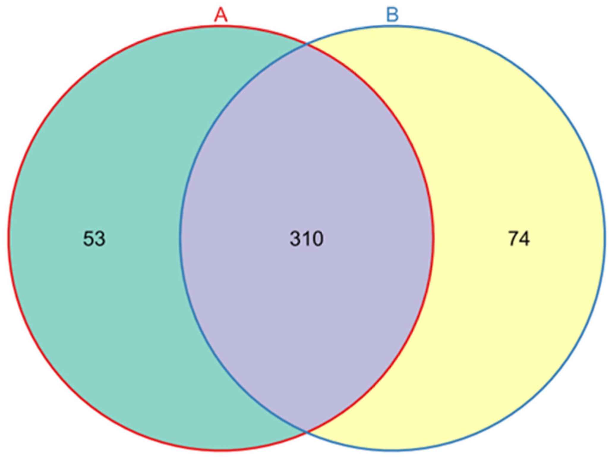
A total of 15,038 high-quality 16S rRNA gene sequences were generated from L. invasa lineage A and B samples, and an average of 7519 high-quality 16S rRNA gene sequences were generated from each sample. After grouping OTUs according to sequence similarity, a total of 437 OTUs were obtained. A total of 363 and 384 OTUs were obtained for the lineage A and B samples, respectively, including 53 OTUs unique to lineage A and 74 OTUs unique to lineage B (Figure 2). There were 20 phyla, 32 classes, 77 orders, 129 families, 217 genera, and 261 species noted (Table 1).
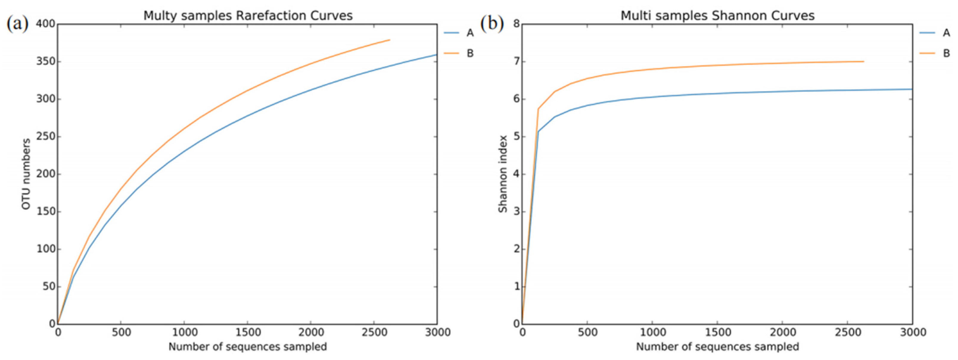
The dilution curve and Shannon index curve were used to evaluate whether the numbers of samples sequenced were sufficient. The results are shown in Figure 3. The dilution curves indicate that the samples of L. invasa lineages A and B have not entered the plateau stage, which suggests it is possible to find new bacterial species with increasing sequencing effort, but the Shannon index curves indicate that the samples of L. invasa lineages A and B have entered the plateau stage, suggesting that the diversity of bacteria in the samples can be fully displayed at this sequencing depth. Therefore, the degree of bacterial sequencing in this study meets the requirements of subsequent bioinformatics analysis.
Alpha diversity was estimated by five indices: Chao1, Shannon index, Simpson index, ACE, and coverage (Table 2). The Chao1 (447.7917 vs. 462.2692) and ACE (460.7090 vs. 474.4288) values of lineage A were lower than those of lineage B. There was also good consistency between the Simpson index and Shannon index, indicating that the diversity of the bacterial community of lineage B was higher than that of lineage A. The coverage of L. invasa lineages A and B was higher than 95%, indicating that the probability of detecting bacteria was much higher than that of not detecting bacteria.

3.3. Bacterial Community Structure in L. invasa Lineages A and B

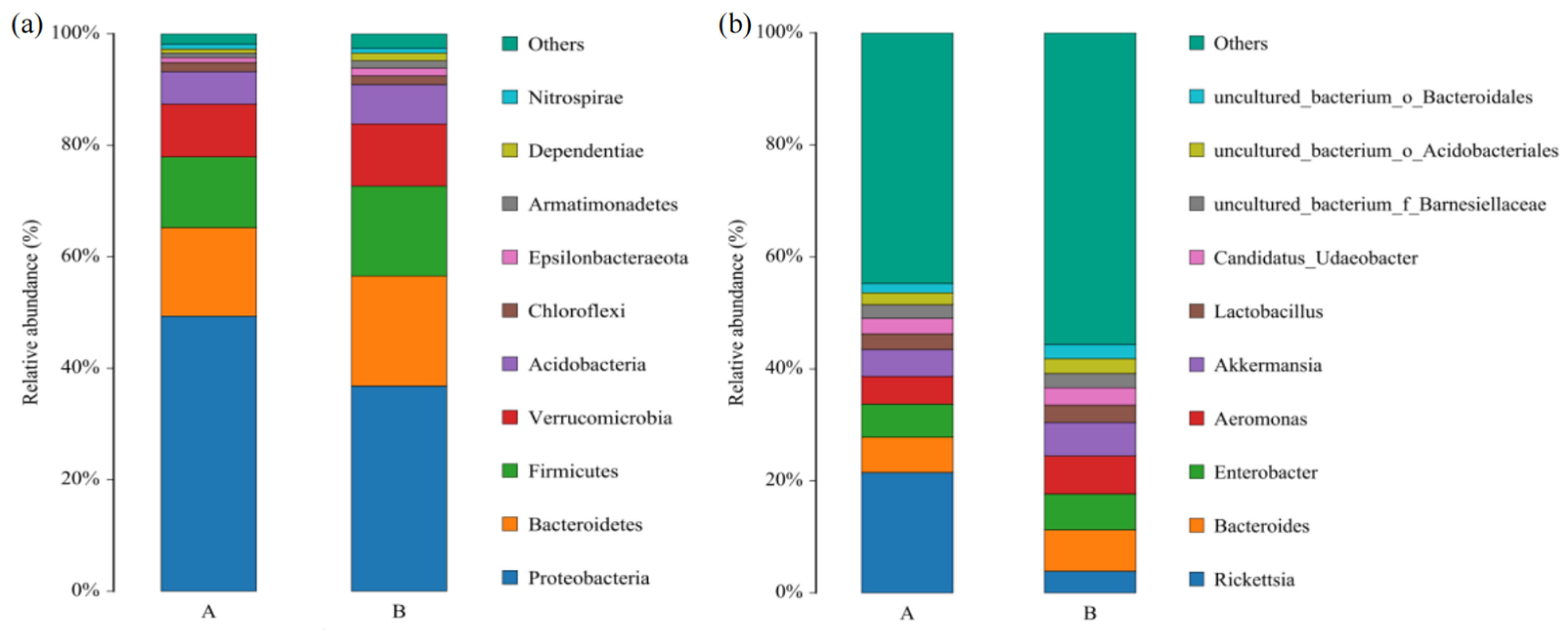
At the phylum level, the bacteria in L. invasa lineages A and B mainly belonged to Proteobacteria, Bacteroidetes, and Firmicutes, and their relative abundances were 49.34%, 36.81%, and 15.87%, respectively. In addition, bacteria with abundance greater than 1% also included Verrucomicrobia, Acidobacteria, and Chloroflexi (Figure 4a).
At the genus level, the dominant bacteria in L. invasa lineage A mainly included Rickettsia, Bacteroides, Enterobacter, Aeromonas, and Akkermansia, with abundances of 21.50%, 6.27%, 5.92%, 4.98%, and 4.78%, respectively. The dominant bacteria in lineage B mainly included Bacteroides, Aeromonas, Enterobacter, Akkermansia, and Rickettsia, with abundances of 7.37%, 6.82%, 6.42%, 5.95%, and 3.87%, respectively. The bacterial species in L. invasa lineages A and B were essentially identical, but there was a significant difference in the relative abundance of Rickettsia (Figure 4b).

3.4. Phylogenetic Analysis of L. invasa Rickettsia Based on 16S rRNA Gene Sequence

The phylogenetic tree based on Rickettsia 16S rRNA gene sequences showed Rickettsia inhabiting L. invasa lineage A and lineage B to be differentiated (Figure 5). By searching and comparing the original data for all 16S rRNA gene sequences of each lineage, no mixing of Rickettsia from L. invasa lineages A and B was found.

3.5. Fluorescent Quantitative PCR Validation

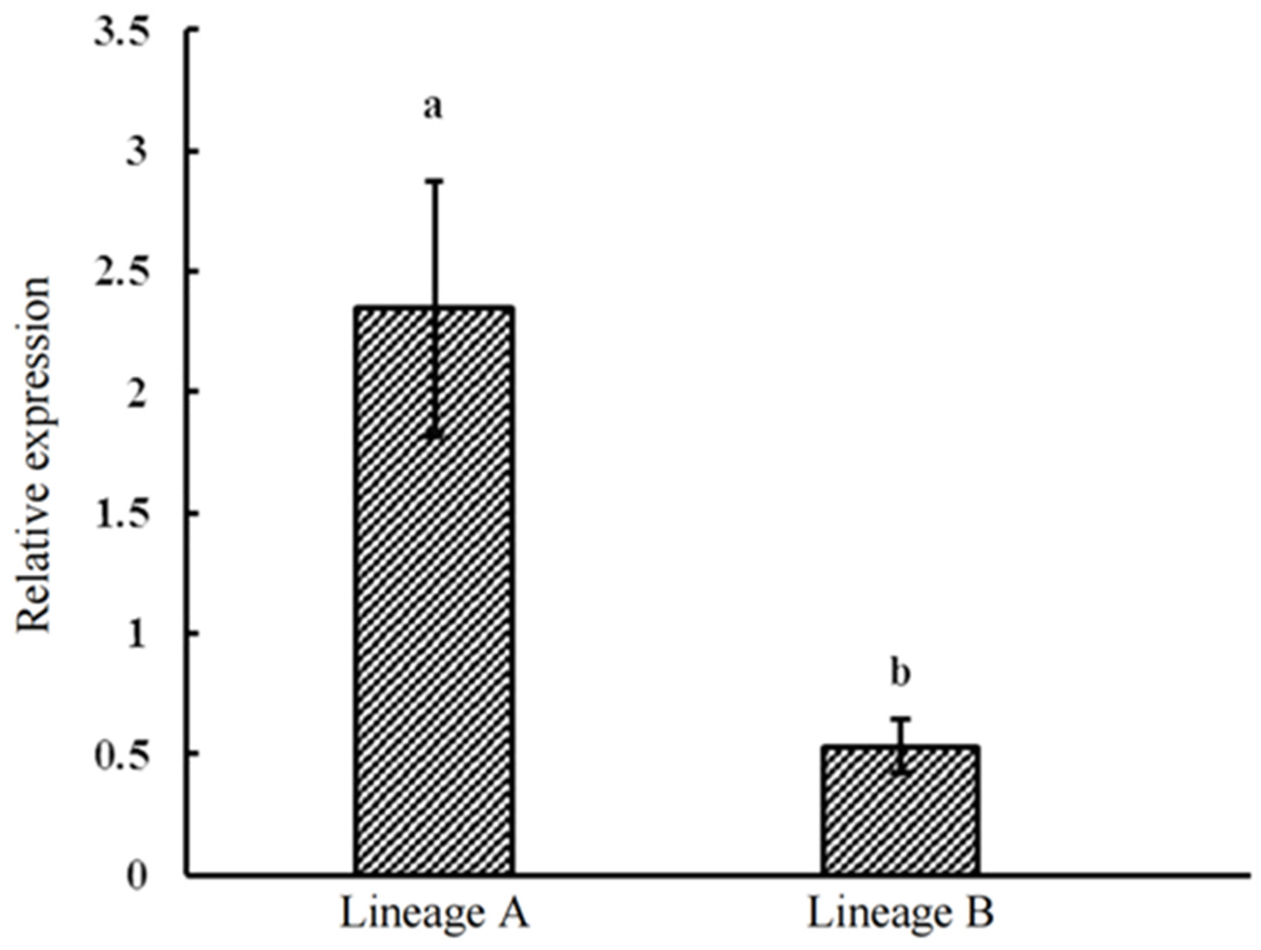
The obtained melting curves for the gltA gene of Rickettsia and the β-Actin gene of L. invasa through qRT-PCR exhibit singular peaks, characterized by narrow and sharp peak lines. This indicates a high level of primer specificity for the two amplified genes in qRT-PCR, resulting in a single amplification product. Ct values were obtained after qRT PCR amplification of continuously diluted DNA of different concentrations and draw standard curves (Supporting Information). Through the investigation of the β-Actin gene of L. invasa lineages A (20 individuals) and B (20 individuals), as well as the Rickettsia gltA gene, the results reveal the presence of Rickettsia within the bodies of both A and B lineages of L. invasa female adults. Based on the results of a univariate analysis, it was found that the relative expression level of Rickettsia within the bodies of the L. invasa varies between different lineages. Specifically, the relative expression level (a,b) of Rickettsia in lineage A (2.35 ± 0.53) is significantly higher than in lineage B (0.53 ± 0.11) (Figure 6), which is consistent with the high-throughput sequencing results.

4. Discussion

The genetic characteristics of insects affect the species of endosymbiotic bacteria they host [35]. For example, Acyrthosiphon pisum populations of the same biotype carry similar endosymbiotic bacterial communities, while the composition of endosymbiotic bacterial communities varies among biotypes [36]. Bemisia tabaci lineage B in Israel mainly hosts Rickettsia and Hamiltonella, while Bemisia tabaci lineage Q mainly hosts Rickettsia and Arsenophonus, and a few individuals are infected with Wolbachia [37]. Chu et al. [38] examined 24 Bemisia tabaci populations and found that Wolbachia was not detected in Bemisia tabaci populations of lineages B and Q, but Wolbachia was detected in the Zhejiang population in China and Kenya populations other than lineages B and Q, and Wolbachia in the Bemisia tabaci populations in Zhejiang and Kenya belonged to different genotypes. This study found that both L. invasa lineages A and B contained Rickettsia, but Rickettsia was differentiated between lineages, which was consistent with the results of Nugnes et al. [25].
Endosymbiotic bacteria in insects often spread in a strict vertical matrilineal manner, that is, from female carrier insects to offspring through eggs [39]. Nugnes et al. [25] found that Rickettsia in L. invasa is located in the ovary of female adults and believed that Rickettsia is the main cause of parthenogenesis. Results reported by Guo et al. [28] also supported this conjecture; the abundance of Rickettsia in male L. invasa was much lower than that in female L. invasa, and this difference in Rickettsia abundance between males and females may stem from differences in their physiological structure. Shan et al. [40] also revealed the pathway and mechanism by which Rickettsia reaches and enters the offspring during the oogenesis and embryonic development of the host insect. It is worth mentioning that not all transmission of symbiotic bacteria among insects is vertical, and a low incidence of horizontal transmission also occurs [41]. The proposed horizontal transmission routes mainly include plant-mediated horizontal transmission, parasitic wasp-mediated horizontal transmission, and sexual mating-mediated horizontal transmission [42]. This study sequenced and analyzed the microorganisms in L. invasa lineages A and B distributed in the same domain. It was found that different types of Rickettsia did not mix between lineages, indicating that Rickettsia in L. invasa showed strict maternal inheritance.
The genetic characteristics of host insects affect the abundance of endosymbiotic bacteria. For example, the population abundance of Buchnera in Acyrthosiphon pisum is affected by lineage [43]. The abundance of Wolbachia in Leptopilina heterotoma has also been found to be significantly affected by its genotype, with the role of host insect genotype related to ambient temperature [44]. The abundances of the two endosymbiotic bacteria Moranella and Tremblaya in Planococcus citri are affected by the genotype of the host insect, and the abundances of the endosymbiotic bacterial populations are determined by maternal inheritance of the host pest [45]. This study found that in the Luchuan population in Guangxi, the abundance and relative expression of endosymbiotic bacterium Rickettsia in L. invasa lineage A were significantly higher than those in lineage B. We believe that these differences derive from the differences in genetic characteristics between lineages.
In addition, different geographical populations or climatic conditions also lead to differences in the species of endosymbiotic bacteria in host insect populations. Guo et al. [30] studied the bacterial diversity of L. invasa in 10 geographical populations in China and found differences in the bacterial diversity and abundance of L. invasa among geographical populations. Rickettsia was dominant in most populations, but the abundances of Rickettsia in Deyang, Sichuan, Qinzhou, Guangxi, and Sanming, Fujian, were significantly lower than those of other populations. The L. invasa samples used in this study were collected from Luchuan City, Guangxi Province, and the abundance of Rickettsia was detected to be low, which may be related to the sampled population in the field.
Dittrich-Schröder et al. [15] studied the population genetics of L. invasa around the world and found introgressive hybridization between L. invasa lineages A and B in Vietnam. Peng et al. [27] conducted population genetic research on 14 geographical populations of L. invasa in China and found that 7 geographical populations included both lineages A and B. Of these, there was obvious introgressive hybridization between lineages in Wuzhou, Guangxi, and Panzhihua, Sichuan. The genetic diversity of L. invasa lineage A is significantly higher than that of lineage B. These researchers believe that this is caused by the introgression of a large number of genes into lineage A from L. invasa lineage B and speculate that L. invasa lineage B may be more inclined to sexual reproduction. With the reports of introgressive hybridization between L. invasa lineages, the differences in reproductive strategies between L. invasa lineages have attracted much attention. Previous studies have also shown that male L. invasa adults have never been reported in areas where L. invasa lineage A has invaded, such as Italy, Tunisia, or Argentina, and there are few males in Turkey (male to female ratio 0–0.5%), while male L. invasa adults appear more frequently in areas where L. invasa lineage B has invaded, such as Thailand, India, and China (7.2%), so it is speculated that lineage B tends to produce males [25,28]. In Hymenoptera, where parthenogenesis is the main mode of reproduction, a threshold density of Rickettsia bacteria in eggs is necessary to trigger the development of female embryos, and the phenomenon of male parthenogenesis may occur when Rickettsia is removed with antibiotics. For example, Pnigalio soemius, which employs parthenogenesis as the main reproductive mode, produces male offspring 24 h after consuming 20 mg/mL rifampicin [46]. Therefore, we believe that differences in Rickettsia abundance between lineages of L. invasa lead to differences in reproductive strategy between lineages. The low abundance of Rickettsia in lineage B may lead to higher numbers of males and a greater tendency towards sexual reproduction, while the high abundance of Rickettsia in lineage A may make this lineage more inclined towards parthenogenesis, so it is more dominant in invasive colonization.
There are obvious differences in genetics and endosymbiotic bacteria between lineages of L. invasa. We suggest that it is necessary to rethink the research conclusions regarding L. invasa and the effectiveness of control measures. It is urgent for researchers to distinguish lineages and clarify the research target species before selecting control measures or conducting physiological and biochemical studies to eliminate the impact of differences in lineages on research results.

5. Conclusions

Rickettsia exhibited genetic differentiation between L. invasa lineages. The abundance and relative expression of Rickettsia in lineage A were significantly higher than those in lineage B.

Supplementary Materials

The following supporting information can be downloaded at: https://www.mdpi.com/article/10.3390/insects14090757/s1, Figure S1: The Standard curve of gltA gene.

Author Contributions

Conceptualization, H.W. and X.P.; Data curation, H.W.; Formal analysis, Z.Y.; Funding acquisition, Z.Y.; Methodology, Z.Y.; Software, H.W.; Validation, H.W.; Visualization, H.W. and Z.Y.; Writing—original draft, X.P.; Writing—review and editing, X.P. All authors have read and agreed to the published version of the manuscript.

Funding

This research was funded by the National Natural Science Foundation of China (No. 31971664, No. 32360390).

Data Availability Statement

The data that support the findings of this study are openly available in the NCBI data repository by searching for accession numbers ON166667-ON166668, OR466117- OR466118 and PRJNA845099.

Conflicts of Interest

The authors declare no conflict of interest.

References

  1. Moran, N.A.; McCutcheon, J.P.; Nakabachi, A. Genomics and evolution of heritable bacterial symbionts. Annu. Rev. Genet. 2008, 42, 165–190. [Google Scholar] [CrossRef] [PubMed]
  2. Oliver, K.M.; Degnan, P.H.; Burke, G.R.; Moran, N.A. Facultative symbionts in aphids and the horizontal transfer of ecologically important traits. Annu. Rev. Entomol. 2010, 55, 247–266. [Google Scholar] [CrossRef] [PubMed]
  3. Latorre, A.; Manzano-Marín, A. Dissecting genome reduction and trait loss in insect endosymbionts. Ann. N. Y. Acad. Sci. 2017, 1389, 52–75. [Google Scholar] [CrossRef] [PubMed]
  4. Cardoza, Y.J.; Klepzig, K.D.; Raffa, K.F. Bacteria in oral secretions of an endophytic insect inhibit antagonistic fungi. Ecol. Entomol. 2006, 31, 636–645. [Google Scholar] [CrossRef]
  5. Douglas, A.E. Multiorganismal insects: Diversity and function of resident microorganisms. In: Berenbaum MR, edito. Annu. Rev. Entomol. 2015, 60, 17–34. [Google Scholar] [CrossRef]
  6. Feldhaar, H. Bacterial symbionts as mediators of ecologically important traits of insect hosts. Ecol. Entomol. 2011, 36, 533–543. [Google Scholar] [CrossRef]
  7. Oliver, K.M.; Degnan, P.H.; Hunter, M.S.; Moran, N.A. Bacteriophages encode factors required for protection in a symbiotic mutualism. Science 2009, 325, 992–994. [Google Scholar] [CrossRef]
  8. Baumann, P.; Baumann, L.; Lai, C.Y.; Rouhbakhsh, D.; Moran, N.A.; Clark, M.A. Genetics, physiology, and evolutionary relationships of the genus Buchnera: Intracellular symbionts of aphids. Annu. Rev. Microbiol. 1995, 49, 55–94. [Google Scholar] [CrossRef]
  9. Chiel, E.; Inbar, M.; Mozes-Daube, N.; White, J.A.; Hunter, M.S.; Zchori-Fein, E. Assessments of fitness effects by the facultative symbiont Rickettsia in the Sweetpotato Whitefly (Hemiptera: Aleyrodidae). Ann. Entomol. Soc. Am. 2009, 102, 413–418. [Google Scholar] [CrossRef]
  10. Himler, A.G.; Adachi-Hagimori, T.; Bergen, J.E.; Kozuch, A.; Kelly, S.E.; Tabashnik, B.E.; Chiel, E.; Duckworth, V.E.; Dennehy, T.J.; Zchori-Fein, E.; et al. Rapid spread of a bacterial symbiont in an invasive Whitefly is driven by fitness Benefits and Female Bias. Science 2011, 332, 254–256. [Google Scholar] [CrossRef]
  11. Bourtzis, K.; Dobson, S.L.; Xi, Z.Y.; Rasgon, J.L.; Calvitti, M.; Moreira, L.A.; Bossin, H.C.; Moretti, R.; Baton, L.A.; Hughes, G.L.; et al. Harnessing mosquito-Wolbachia symbiosis for vector and disease control. Acta Trop. 2014, 132, S150–S163. [Google Scholar] [CrossRef] [PubMed]
  12. Rainey, S.M.; Shah, P.; Kohl, A.; Dietrich, I. Understanding the Wolbachia-mediated inhibition of arboviruses in mosquitoes: Progress and challenges. J. Gen. Virol. 2014, 95, 517–530. [Google Scholar] [CrossRef] [PubMed]
  13. Shentu, X.P.; Li, D.T.; Xu, J.F.; She, L.; Yu, X.P. Effects of fungicides on the yeast-like symbiotes and their host, Nilaparvata lugens Stal (Hemiptera: Delphacidae). Pestic. Biochem. Physiol. 2016, 128, 16–21. [Google Scholar] [CrossRef]
  14. Zheng, X.Y.; Zhang, D.J.; Li, Y.J.; Yang, C.; Wu, Y.; Liang, X.; Liang, Y.K.; Pan, X.L.; Hu, L.C.; Sun, Q.; et al. Incompatible and sterile insect techniques combined eliminate mosquitoes. Nature 2019, 572, 56–61. [Google Scholar] [CrossRef]
  15. Dittrich-Schröder, G.; Hoareau, T.B.; Hurley, B.P.; Wingfield, M.J.; Lawson, S.; Nahrung, H.F.; Slippers, B. Population genetic analyses of complex global insect invasions in managed landscapes: A Leptocybe invasa (Hymenoptera) case study. Biol. Invasions 2018, 20, 2395–2420. [Google Scholar] [CrossRef]
  16. Mendel, Z.; Protasov, A.; Fisher, N.; La Salle, J. Taxonomy and biology of Leptocybe invasa gen. & sp. n. (Hymenoptera: Eulophidae), an invasive gall inducer on Eucalyptus. Aust. J. Entomol. 2004, 43, 101–113. [Google Scholar]
  17. Le, N.H.; Nahrung, H.F.; Griffiths, M.; Lawson, S.A. Invasive Leptocybe spp. and their natural enemies: Global movement of an insect fauna on eucalypts. Biol. Control. 2018, 125, 7–14. [Google Scholar] [CrossRef]
  18. Zheng, X.L.; Li, J.; Yang, Z.D.; Xian, Z.H.; Wei, J.G.; Lei, C.L.; Wang, X.P.; Lu, W. A review of invasive biology, prevalence and management of Leptocybe invasa Fisher & La Salle (Hymenoptera: Eulophidae: Tetrastichinae). Afr. Entomol. 2014, 22, 68–79. [Google Scholar]
  19. Wu, Y.J.; Jiang, X.J.; Li, D.W.; Luo, J.T.; Zhou, G.F.; Chang, M.S.; Yang, Z.Q. Leptocybe invasa, a New invasive forest pest making galls on twigs and leaves of eucalyptus trees in China (Hymenoptera: Eulophidae). Sci. Silvae Sin. 2009, 45, 161–163+182–183. [Google Scholar]
  20. Zheng, X.L.; Huang, Z.Y.; Li, J.; Yang, Z.D.; Yang, X.H.; Lu, W. Reproductive biology of Leptocybe invasa Fisher & La Salle (Hymenoptera: Eulophidae). Neotrop. Entomol. 2018, 47, 19–25. [Google Scholar]
  21. Zhang, H.; Song, J.Y.; Zhao, H.X.; Li, M.; Han, W.H. Predicting the distribution of the invasive species Leptocybe invasa: Combining MaxEnt and geodetector models. Insects 2021, 12, 92. [Google Scholar] [CrossRef] [PubMed]
  22. Schröder, M.L.; Slippers, B.; Wingfield, M.J.; Hurley, B.P. Invasion history and management of Eucalyptus snout beetles in the Gonipterus scutellatus species complex. J. Pest. Sci. 2020, 93, 11. [Google Scholar] [CrossRef]
  23. Chu, D.; Guo, D.; Pan, H.P.; Zhang, Y.J.; Wan, F.H. Cryptic invasion of alien species: Types and effects. Acta Entomol. Sin. 2012, 55, 218–225. [Google Scholar]
  24. Tsai, C.L.; Lee, H.C.; Cho, G.; Liao, Y.C.; Yang, M.M.; Yeh, W.B. Invasive and quarantine risks of Cacopsylla chinensis (Hemiptera: Psyllidae) in East Asia: Hybridization or gene flow between differentiated lineages. J. Econ. Entomol. 2020, 113, 2890–2899. [Google Scholar] [CrossRef]
  25. Nugnes, F.; Gebiola, M.; Monti, M.M.; Gualtieri, L.; Giorgini, M.; Wang, J.G.; Bernardo, U. Genetic diversity of the invasive gall wasp Leptocybe invasa (Hymenoptera: Eulophidae) and of its Rickettsia endosymbiont, and associated Sex-Ratio differences. PLoS ONE 2015, 10, e124660. [Google Scholar] [CrossRef] [PubMed]
  26. Peng, X.; Wang, H.T.; Guo, C.H.; Yang, Z.D.; Zhou, J.; Wang, X.; Ding, Z.R. EST-SSR Development and cryptic species Identification for the invasive gall-pest Leptocybe invasa (Hymenoptera: Eulophidae). Sci. Silvae Sin. 2021, 57, 140–151. [Google Scholar]
  27. Peng, X.; Wang, H.T.; Guo, C.H.; Hu, P.; Xu, L.; Zhou, J.; Ding, Z.R.; Yang, Z.D. Genetic diversity analysis of the invasive gall pest Leptocybe invasa (Hymenoptera: Eulophidae) from China. PLoS ONE 2021, 16, e258610. [Google Scholar] [CrossRef]
  28. Guo, C.H.; Peng, X.; Zheng, X.L.; Wang, X.Y.; Wang, R.R.; Huang, Z.Y.; Yang, Z.D. Comparison of bacterial diversity and abundance between sexes of Leptocybe invasa Fisher & La Salle (Hymenoptera: Eulophidae) from China. PeerJ 2020, 8, e8411. [Google Scholar]
  29. Liu, Y.P.; Xu, L.T.; Zhang, Z.Q.; Huang, Z.Y.; Fang, D.X.; Zheng, X.L.; Yang, Z.D.; Lu, M. Isolation, identification, and analysis of potential functions of culturable bacteria associated with an invasive gall wasp, Leptocybe invasa. Microb. Ecol. 2021, 83, 151–166. [Google Scholar] [CrossRef]
  30. Guo, C.H.; Peng, X.; Wang, H.T.; Zheng, X.L.; Hu, P.; Zhou, J.; Ding, Z.R.; Wang, X.; Yang, Z.D. Bacterial diversity of Leptocybe invasa Fisher & La Salle (Hymenoptera: Eulophidae) from different geographical conditions in China. Arch. Insect Biochem. Physiol. 2021, 108, e21847. [Google Scholar]
  31. Folmer, O.; Black, M.; Hoeh, W.; Lutz, R.; Vrijenhoek, R. DNA primers for amplification of mitochondrial cytochrome c oxidase subunit I from diverse metazoan invertebrates. Mol. Mar. Biol. Biotechnol. 1994, 3, 294–299. [Google Scholar] [PubMed]
  32. Singer, E.; Bushnell, B.; Coleman-Derr, D.; Bowman, B.; Bowers, R.M.; Levy, A.; Gies, E.A.; Cheng, J.F.; Copeland, A.; Klenk, H.P.; et al. High-resolution phylogenetic microbial community profiling. ISME J. 2016, 10, 2020–2032. [Google Scholar] [CrossRef] [PubMed]
  33. Earl, J.P.; Adappa, N.D.; Krol, J.; Bhat, A.S.; Balashov, S.; Ehrlich, R.L.; Palmer, J.N.; Workman, A.D.; Blasetti, M.; Sen, B.; et al. Species-level bacterial community profiling of the healthy sinonasal microbiome using Pacific Biosciences sequencing of full-length 16S rRNA genes. Microbiome 2018, 6, 190. [Google Scholar] [CrossRef] [PubMed]
  34. Lu, T.C. Study on the Biology, Risk Analysis and Control Strategy of Leptocybe invasa Fisher & LaSalle. Ph.D. Thesis, Fujian Agriculture and Forestry University, Fuzhou, China, 2017. [Google Scholar]
  35. Liu, X.D.; Zhang, Y.C. Infection pattern and dynamics of endosymbionts in aphids and their effects on population differentiation of hosts. J. Nanjing Agric. Univ. 2018, 41, 209–217. [Google Scholar]
  36. Gauthier, J.P.; Outreman, Y.; Mieuzet, L.; Simon, J.C.; Duperron, S. Bacterial communities associated with Host-Adapted populations of Pea Aphids revealed by deep sequencing of 16S Ribosomal DNA. PLoS ONE 2015, 10, e120664. [Google Scholar] [CrossRef] [PubMed]
  37. Skaljac, M.; Zanic, K.; Ban, S.G.; Kontsedalov, S.; Ghanim, M. Co-infection and localization of secondary symbionts in two whitefly species. BMC Microbiol. 2010, 10, 142. [Google Scholar] [CrossRef] [PubMed]
  38. Chu, D.; Cong, B.; Zhang, Y.J.; Xu, B.Y.; Wu, Q.J.; Zhu, G.R. Detection and phylogenetic analysis of Wolbachia in different Bemisia tabaci biotypes. Acta Entomol. Sin. 2005, 48, 518–525. [Google Scholar]
  39. An, P.; Xu, Y.P.; Wu, J.M.; Zheng, R.E.; Yu, X.P. Vertical transmission of insect symbionts. Acta Entomol. Sin. 2019, 62, 877–884. [Google Scholar]
  40. Shan, H.W.; Liu, Y.Q.; Luan, J.B.; Liu, S.S. New insights into the transovarial transmission of the symbiont Rickettsia in whiteflies. Sci. China Life Sci. 2021, 64, 1174–1186. [Google Scholar] [CrossRef]
  41. Ahmed, M.Z.; Li, S.J.; Xue, X.; Yin, X.J.; Ren, S.X.; Jiggins, F.M.; Greeff, J.M.; Qiu, B.L.; Hurst, G. The intracellular bacterium Wolbachia uses parasitoid wasps as phoretic vectors for efficient horizontal transmission. PLoS Pathog. 2015, 11, e1004672. [Google Scholar] [CrossRef]
  42. Liu, Y.; Fan, Z.Y.; Li, Y.; Peng, J.; Chen, X.Y.; Qiu, B.L. Plant-mediated horizontal transmission of insect endosymbionts and their biological effects on recipients. Chin. J. Appl. Entomol. 2020, 57, 272–283. [Google Scholar]
  43. Martinez, A.J.; Weldon, S.R.; Oliver, K.M. Effects of parasitism on aphid nutritional and protective symbioses. Mol. Ecol. 2014, 23, 1594–1607. [Google Scholar] [CrossRef] [PubMed]
  44. Mouton, L.; Henri, H.; Charif, D.; Boulétreau, M.; Vavre, F. Interaction between host genotype and environmental conditions affects bacterial density in Wolbachia symbiosis. Biol. Lett. 2007, 3, 210–213. [Google Scholar] [CrossRef] [PubMed]
  45. Parkinson, J.F.; Gobin, B.; Hughes, W.O.H. Heritability of symbiont density reveals distinct regulatory mechanisms in a tripartite symbiosis. Ecol. Evol. 2016, 6, 2053–2060. [Google Scholar] [CrossRef] [PubMed]
  46. Giorgini, M.; Bernardo, U.; Monti, M.M.; Nappo, A.G.; Gebiola, M. Rickettsia Symbionts Cause Parthenogenetic Reproduction in the Parasitoid Wasp Pnigalio soemius (Hymenoptera: Eulophidae). Appl. Environ. Microbiol. 2010, 76, 2589–2599. [Google Scholar] [CrossRef]
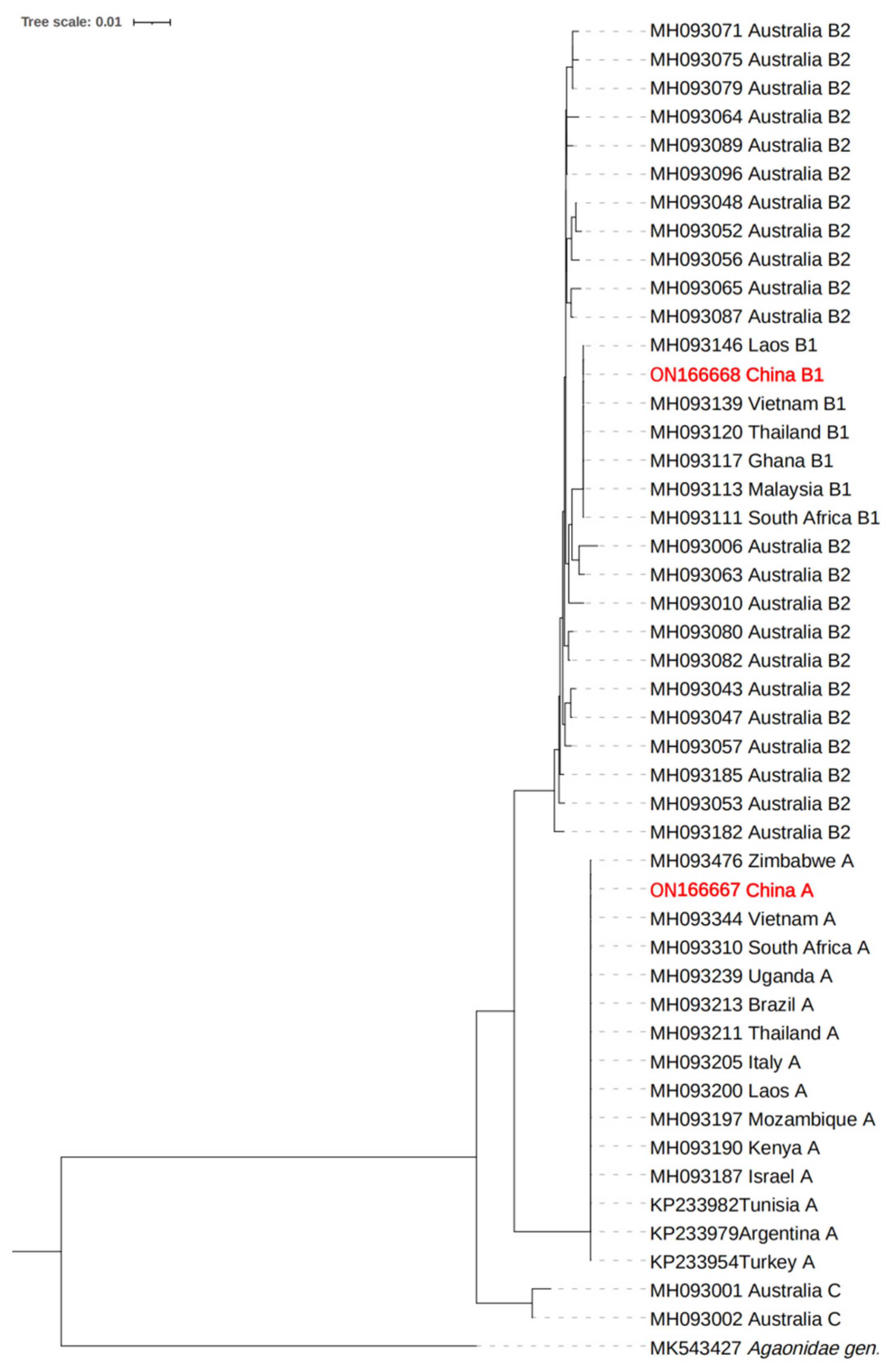
Figure 1. The NJ phylogenetic tree of COI sequence of L. invasa.
Insects 14 00757 g001
Figure 2. Venn diagram of OTUs of endosymbiotic bacteria found in lineage A and lineage B of L. invasa.
Insects 14 00757 g002
Figure 3. Dilution curves (a) and Shannon index curves (b) of the number of bacterial OTUs observed in L. invasa.
Insects 14 00757 g003
Figure 4. Relative abundances of the top 10 bacterial phyla (a) and genera (b) in lineage A and lineage B of L. invasa.
Insects 14 00757 g004
Figure 5. Phylogenetic analysis of L. invasa Rickettsia based on 16S rRNA gene sequences.
Insects 14 00757 g005
Figure 6. Relative expression of Rickettsia from different lineages of L. invasa.
Insects 14 00757 g006
Table 1. Statistics of classification levels for endosymbiotic bacteria found in L. invasa lineages A and B.
Lineage TypePhylumClassOrderFamilyGenusSpecies
Lineage A182971112185226
Lineage B203176125204240
Lineage A-specific01141321
Lineage B-specific236173235
Lineage in common182870108172205
Total203277129217261
Table 2. Statistics of alpha diversity indices of the bacteria in female adults of L. invasa lineages A and B.
Lineage TypeACEChao 1ShannonSimpsonCoverage
Lineage A460.7090447.79176.2690.94190.9641
Lineage B474.4288462.26927.0130.98030.9595
Disclaimer/Publisher’s Note: The statements, opinions and data contained in all publications are solely those of the individual author(s) and contributor(s) and not of MDPI and/or the editor(s). MDPI and/or the editor(s) disclaim responsibility for any injury to people or property resulting from any ideas, methods, instructions or products referred to in the content.