Article

Screening of Entomopathogenic Fungal Culture Extracts with Honeybee Nosemosis Inhibitory Activity

1
Department of Agricultural Biology, College of Agriculture, Life & Environment Science, Chungbuk National University, Cheongju 28644, Republic of Korea
2
Process Development Team, R&D Division, Plolagen Co., Ltd., Seoul 03722, Republic of Korea
*
Author to whom correspondence should be addressed.
Insects 2023, 14(6), 538; https://doi.org/10.3390/insects14060538
Submission received: 24 April 2023 / Revised: 6 June 2023 / Accepted: 8 June 2023 / Published: 9 June 2023

Abstract

:

Simple Summary

Nosemosis in honeybees caused by the Nosema spp. infection is not only related to colony collapse but also increases the susceptibility of honeybees to various other pathogens; thus, it is necessary to develop effective control methods for nosemosis. Research on various control methods has been conducted, but clearly defined methods are still rare. In our study, the possibility of controlling honeybee nosemosis using entomopathogenic fungal culture extracts with various biological activities was evaluated. Many of the tested entomopathogenic fungal culture extracts showed high germination inhibitory activity on Nosema ceranae spores. Among them, fungal culture extracts with high spore germination inhibitory activity and fungicidal activity was used to evaluate honeybee nosemosis inhibitory activity. As a result, it was confirmed for the first time that the fungal culture extract could not only suppress honeybee nosemosis but also increase the lifespan of honeybees. Entomopathogenic fungal culture extracts are expected to have applications in controlling honeybee nosemosis.

Abstract

This study aimed to select the most effective culture extracts for controlling honeybee nosemosis using 342 entomopathogenic fungi of 24 species from 18 genera. The germination inhibitory activity of the fungal culture extract on Nosema ceranae spores was evaluated using an in vitro germination assay method. Among 89 fungal culture extracts showing germination inhibitory activity of approximately 80% or more, 44 fungal culture extracts that maintained their inhibitory activity even at a concentration of 1% were selected. Finally, the honeybee nosemosis inhibitory activity was evaluated using the cultured extracts of five fungal isolates having a Nosema inhibitory activity of approximately 60% or more, even when the extract was removed after treatment. As a result, the proliferation of Nosema spores was reduced by all fungal culture extract treatments. However, only the treatment of the culture extracts from Paecilomyces marquandii 364 and Pochonia bulbillosa 60 showed a reduction in honeybee mortality due to nosemosis. In particular, the extracts of these two fungal isolates also increased the survival of honeybees.

1. Introduction

The honeybee (Apis mellifera L. (Hymenoptera: Apidae)) performs an important role in the pollination of flowering plants, which are essential for the production of fruits, nuts, and seeds upon which animals, including humans, rely for food [1,2,3,4]. In addition to their role in agriculture, honeybees are also important for maintaining biodiversity in natural ecosystems [5]. They help pollinate wildflowers, which provide habitat and food for a variety of other species. Honeybees are also vital to the production of honey, beeswax, and other bee products that humans have used for thousands of years [6,7]. However, unfortunately, honeybee colony collapse has become frequent, with global bee populations rapidly declining in recent years due to habitat loss, pesticide use, climate change, and disease [8,9]. This is a major concern for the health of both food systems and natural ecosystems. Efforts are therefore underway worldwide to protect and conserve honeybee populations, including promoting habitat restoration, reducing pesticide use, and supporting research on bee health and behavior [9].
Bee colony collapse is a complex phenomenon that can be caused by multiple environmental and human-related factors [8,9,10]. Habitat loss and fragmentation, climate change, pesticides, mites, and other parasites, and diseases and pathogens have been highlighted as major causes of honeybee colony collapse [11,12,13,14]. Notably, bee colony collapse is often the result of a combination of these factors rather than a single cause. To address honeybee colony collapse, it is important to take a multipronged approach that addresses both environmental and human-related factors. Of these factors, honeybee disease, known as nosemosis, is known to have the greatest impact on honeybees [11,14,15,16]. Nosema infection can have severe negative effects on honeybee colonies, including reduced foraging activity, decreased colony growth and productivity, increased mortality, increased susceptibility to other stressors, reduced winter survival, decreased queen bee productivity, and reduced immune function [16,17,18,19,20,21,22,23]. Nosemosis is caused by two species of microsporidia, Nosema apis and N. ceranae. Nosema ceranae is more common and more lethal than N. apis [16,24,25,26]. Between the prevention and control of nosemosis, preventive methods are mainly relied on. Representative preventive methods include good beekeeping practices, such as keeping the hive clean and providing adequate nutrition and moisture to the bees, hygienic behaviors, such as removing diseased and dead brood from the hive, and avoiding stressors, such as exposure to pesticides and poor nutrition [9,27,28,29]. In contrast, for direct control, an antibiotic called fumagillin is used, but its use is controversial due to its potential negative effects on the environment and other organisms [30,31,32,33]. Research efforts to replace it have reported that probiotics, plant essential oils, propolis, plant extracts, royal jelly, etc., can reduce the production of Nosema spores in honeybees and improve the survival rate of Nosema-infected bees [22,23,34,35,36,37].
Entomopathogenic fungi, which are fungi that infect and kill insects, have been widely studied and used as materials for microbial insecticides for pest control [38,39,40,41]. Metabolites of these entomopathogenic fungi have various physiological activities, such as insecticidal activity, antibacterial activity, antioxidant activity, immunomodulatory activity, and cytotoxic activity, and potential applications are being sought in various fields, such as agriculture, medicine, and biotechnology [42,43,44,45].
In our study, entomopathogenic fungal culture extracts with proliferation inhibitory activity against N. ceranae were screened from various entomopathogenic fungi for the prevention and control of nosemosis, and the effect of improving the survival rate of honeybees upon Nosema infection was evaluated. The purpose of this study was to provide basic data on the possibility of using culture extracts of entomopathogenic fungi for the prevention or control of nosemosis in honeybees.

2. Materials and Methods

2.1. Honeybees and N. ceranae

A colony of the honeybee A. mellifera was reared with 50% (w/v) sucrose solution, and bee bread as the main food, and adults that emerged within 24 h were used in the experiment. Honeybees highly infected with N. ceranae were provided by the Sericulture and Apiculture Division of the Rural Development Administration, Republic of Korea.

2.2. Entomopathogenic Fungal Culture Extract

In this study, 342 isolates of entomopathogenic fungi from 18 different genera were used (Table S1) [46]. In the same fungal species, fungal isolates indicate particular fungi from a particular environment or region. Fungal isolates were initially suspended in 1 mL of Sabouraud dextrose broth medium containing yeast extract (SDYB: 10 g of Bacto peptone, 40 g of dextrose, 10 g of yeast extract in 1000 mL of distilled water, and pH 6.0). Cultures were inoculated with agar blocks (6 mm in diameter) of fungi cultured in potato dextrose agar (PDA) medium for 2 weeks and grown in the dark at 25 °C with shaking at 150 rpm. After 10 days, the cultures were centrifuged at 10,000× g for 10 min. After removing the pellet, the supernatant was filtered using a LaboPass™ Mini Plasmid DNA Purification Kit column (Cosmo Genetech Co. Ltd., Seoul, Republic of Korea) to remove spores and mycelia. The ethyl acetate fractionation method was used to separate hydrophobic substances from fungal culture filtrates. After adding the same volume of ethyl acetate as the culture medium, vortexing for 20 min, and centrifugation at 4000× g rpm for 5 min, the supernatant was collected. Afterward, ethyl acetate was evaporated using gaseous nitrogen, and the remaining extract pellet was dissolved in 2% acetone in the same volume as the culture filtrate and used in the next experiment. The prepared culture extract was stored at −76 °C until use. To prepare a culture extract after quantitative inoculation of entomopathogenic fungi, fungal conidia that were harvested after being cultured in PDA medium for 2 weeks were used to prepare a conidial suspension using a 0.02% Tween-80 solution. The conidia were then counted using a hemocytometer. The conidial suspension was inoculated in 30 mL PDB medium in 50 µL at a concentration of 9 × 10⁵ conidia/mL and cultured for 10 days. After culturing, spores and mycelia were removed from the culture medium, and culture extracts were prepared using the ethyl acetate fraction method as described above.

2.3. Purification of Nosema Spores

To produce infective spores, honeybees were placed in plastic cages and inoculated with 1 × 10⁶ spores of N. ceranae in sucrose solution (50% w/v in water). To obtain purified Nosema spores, after 10 days, the midgut tissues from heavily infected honeybees were individually separated using forceps and washed with phosphate-buffered saline [47]. The isolated midgut was ground in 200 μL of sterile distilled water in a Bullet Blender® Homogenizer (Scientific Instrument Services Inc., Palmer, MA, USA) set to speed 8 with 2 mm diameter tungsten carbide beads (Sigma–Aldrich, St. Louis, MO, USA) for 2 min. The homogenate volume was increased to 1 mL to increase the filtration efficiency, and the mixture was filtered through Qualitative No. 2 filter paper (Advantec MFS Inc., Dublin, OH, USA) with an 8–11 μm pore size to remove tissue debris [48]. The filtered suspension was overlaid very gently on discontinuous 25%, 50%, 75%, and 90% Percoll® (Sigma–Aldrich, St. Louis, MO, USA) gradient and centrifuged twice at 15,000× g for 30 min at 20 °C [49]. A small but dense band just above the bottom of the tube was collected and resuspended in sterile distilled water. After final centrifugation at 15,000× g for 10 min at 20 °C, the spore pellet was resuspended in sterile distilled water. The spore concentration was measured by counting with a hemocytometer [50]. The viability of Nosema spores was determined by the in vitro germination method as described below, and those with a viability of 95% or more were used in the experiment.

2.4. In Vitro Germination Assay

Aliquots of purified Nosema spores (10 μL; 1 × 10³ spores) were spotted onto glass slide reaction cells (12 wells; Paul Marienfeld GmbH & Co. KG, Lauda-Königshofen, Germany) and air-dried for 2 h at room temperature. Germination was triggered by adding 1.5 μL of 0.1 M sucrose in distilled water to the air-dried spores [51]. After maintaining the covered glass slide at room temperature for 6 h, the germinated spores were observed under a light microscope (magnification, 400×) (Nikon Instech Co., Ltd., Tokyo, Japan). The germination rate was calculated as the percentage of total observed spores that had germinated.

2.5. Safety Test of Fungal Culture Extract

Twenty honeybees that emerged within 24 h were transferred to one cage and allowed to adapt for 3 days before being used in the experiment. To evaluate the safety of the fungal culture extract, 50% (w/v) sucrose solution mixed with culture extract was fed once to the honeybees, and as a control for comparison, 50% (w/v) sucrose solution containing the same concentration of acetone used to dissolve the culture extract was fed once. The survival rate of honeybees was observed and recorded every day for 14 days after treatment, and the experiment was repeated three times for each treatment group.

2.6. Inhibitory Activity of Fungal Culture Extracts on Nosema Infection in Honeybees

To evaluate the effect of fungal culture extract on Nosema infection in honeybees, 25 honeybees that emerged within 24 h were used for each experimental group. Honeybees were orally infected with purified Nosema spores by making a 50% sucrose suspension at 1 × 10⁶ spores/mL and administering 1 mL to each experimental group. Treatment of fungal culture extract was carried out before and after Nosema inoculation. For the control group, honeybees were infected with Nosema, and no culture extract treatment was used. To evaluate the inhibitory effect of Nosema infection, the survival rate of honeybees was observed and recorded every day for 14 days. The production of Nosema spores was examined by isolating and counting spores from the midgut of honeybees on the 14th day after infection to evaluate the effect of inhibition on Nosema growth. The experiment was repeated three times for each treatment group.

2.7. Statistical Analysis

Spore germination and honeybee survival rate results were analyzed with SPSS statistical software v12.0 (SPSS, Inc., Chicago, IL, USA). Data were subjected to a one-way analysis of variance (ANOVA), and comparisons among groups were performed with the SNK test. Data are expressed as the means ± standard errors (SEs), and statistical significance was set at the conventional α < 0.05 level.

3. Results

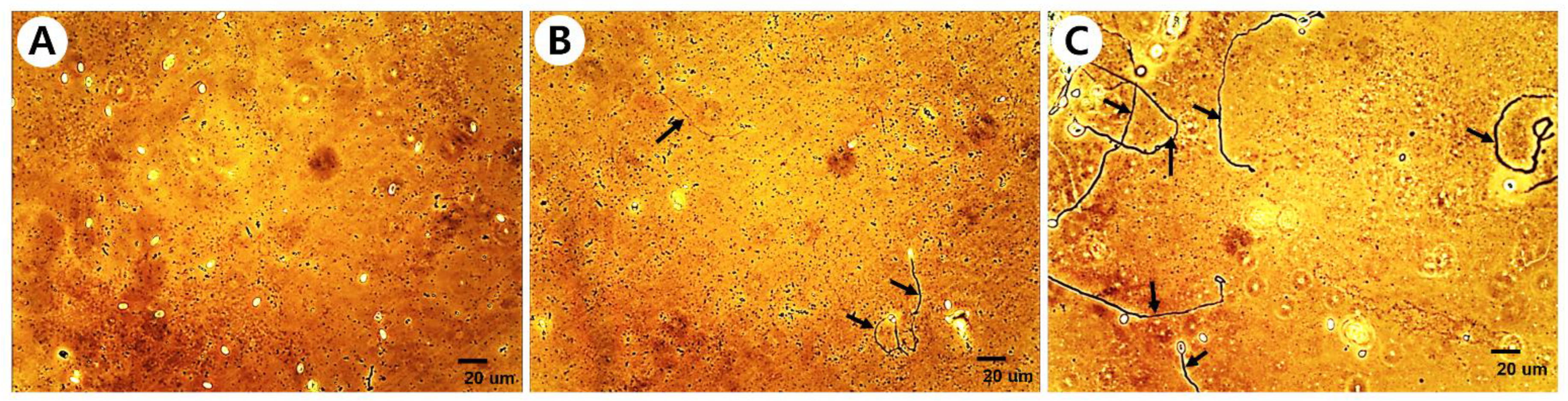
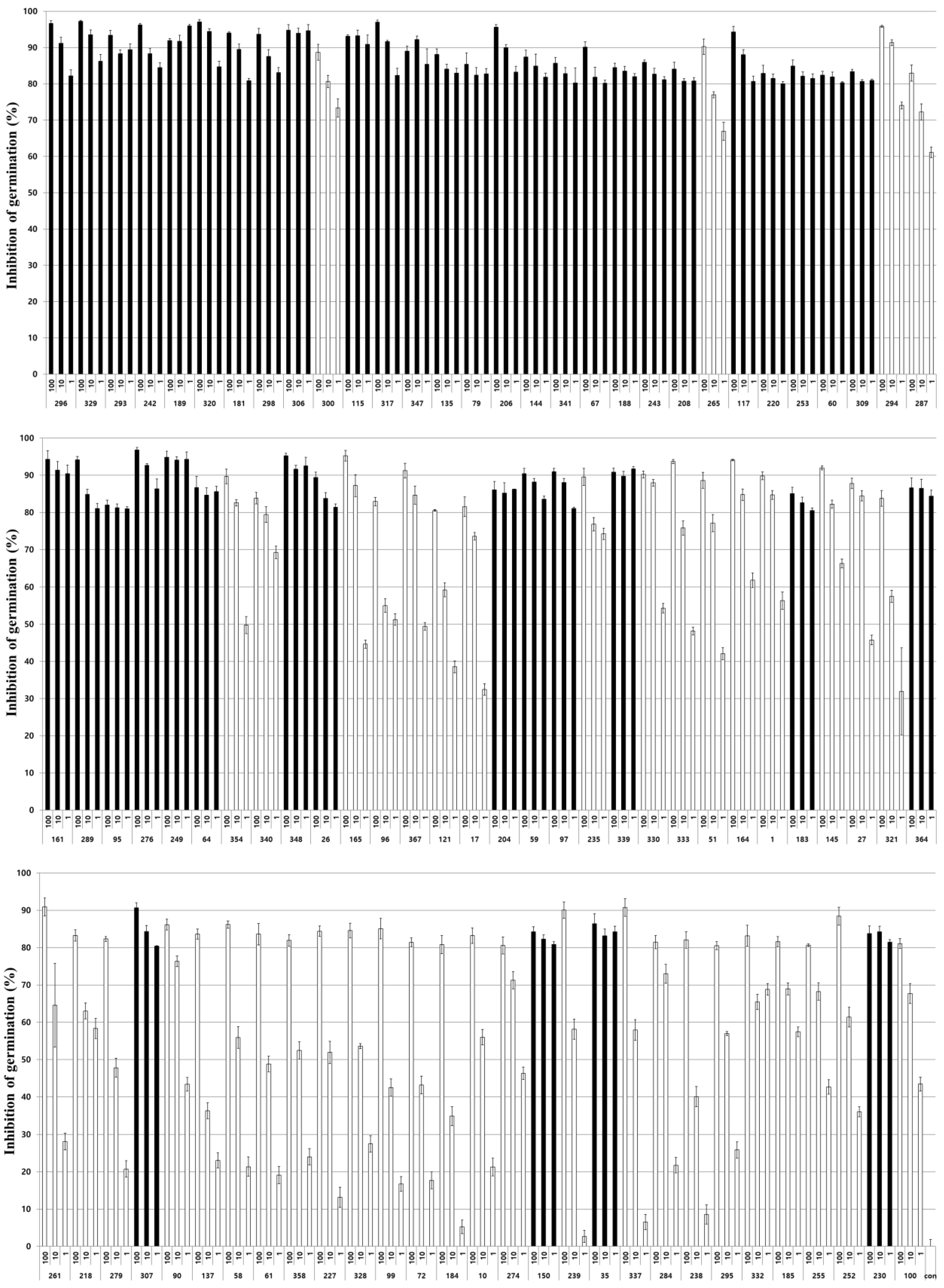
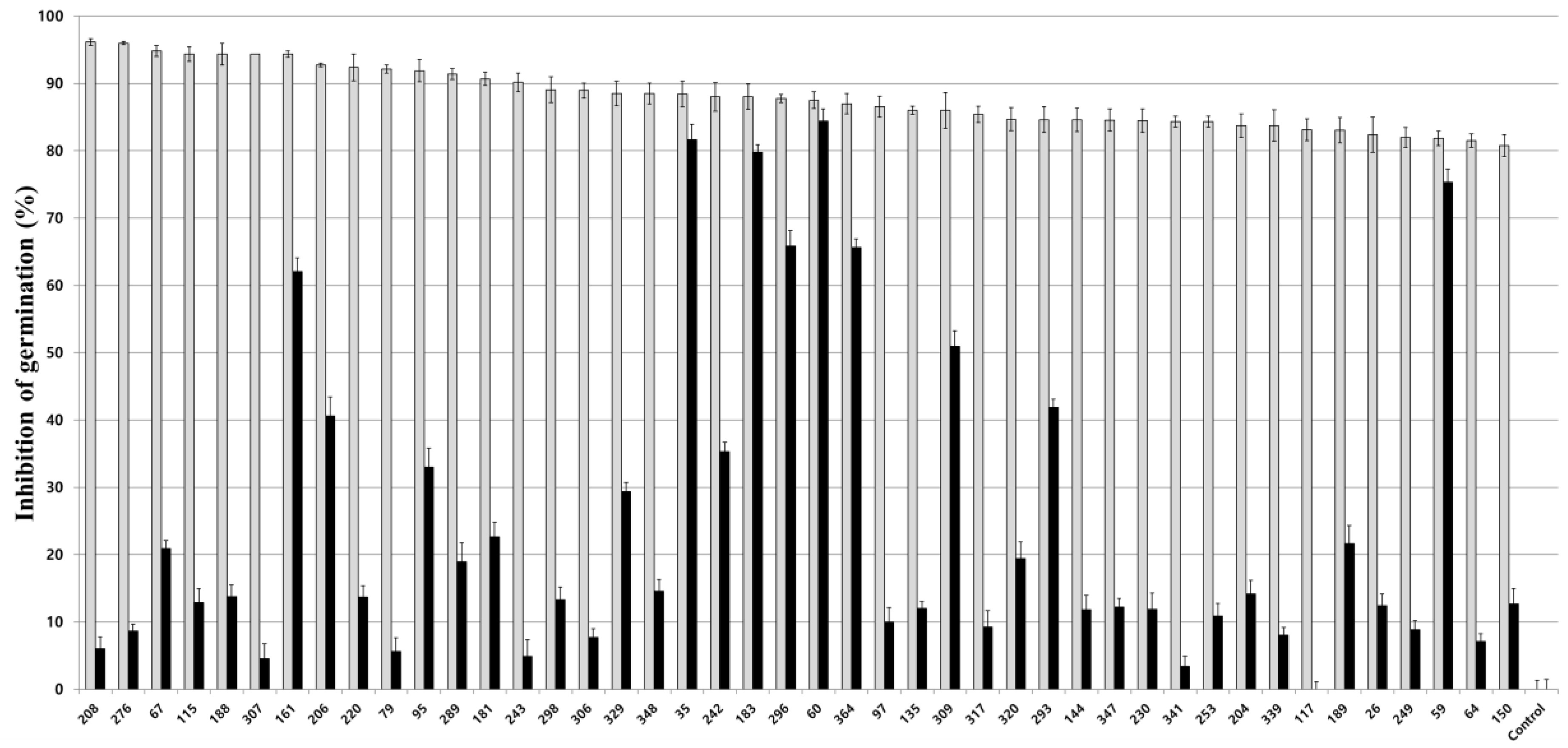
3.1. Inhibitory Activity of Entomopathogenic Fungal Culture Extracts on Nosema Germination

To evaluate the inhibitory activity of the entomopathogenic fungal culture extract on Nosema spore germination, the influence of acetone, a solvent used in preparing the culture extract, on Nosema spore germination was first evaluated. As a result of treating Nosema spores with various concentrations of acetone from 0.25% to 20%, no significant influence of acetone on the germination of Nosema spores was observed at all concentrations (Figure S1). Therefore, it was confirmed that the 0.2% concentration of acetone used in our experiment had no influence on the germination of Nosema spores. All fungal isolates used in the experiment are shown in Table S1. As a result of evaluating the germination inhibitory activity of the culture extracts of 342 fungal isolates on Nosema spores, the germination of spores was inhibited in a variety of ways from 0% to a maximum of approximately 96.5% (Table S2). Most of the spores did not germinate when the germination inhibitory activity was greater than approximately 80%, some spores germinated when the germination inhibitory activity was greater than 60%, and many spores germinated when the activity was less than 60% (Figure 1). Among the 342 fungal isolates, culture extracts from 89 isolates showed spore germination inhibitory activity of more than 80%, and 20 isolates showed activity of more than 90% (Table S2). By classification of entomopathogenic fungi, approximately 80% or more of the germination inhibitory activity against Nosema was observed in 15 species of 10 genera among 24 species from 18 genera (Table 1). A total of 50 of the 126 isolates of Beauveria spp., 18 of the 81 isolates of Metarhizium spp., 11 of the 40 isolates of Cordyceps spp., 3 of the 17 isolates of Pochonia spp., and 2 of the 12 isolates of Paecilomyces spp. showed germination inhibitory activity on Nosema spores. As various activities were confirmed in various fungi, culture extracts of 89 isolates showing spore germination inhibitory activity of 80% or more were prepared again by quantitative inoculation of fungi, and the germination inhibitory activity on Nosema spores was re-evaluated.

3.2. Inhibitory Activity of Fungal Culture Extracts on Nosema Spore Germination

For 89 selected entomopathogenic fungi, culture extracts were prepared by inoculation with the same conidia concentration, then the extracts were diluted in distilled water at concentrations of 100, 10, and 1% to evaluate the germination inhibitory activity on Nosema spores (Figure 2). As a result, 44 of the 89 fungal culture extracts showed a high spore germination inhibitory activity of 80% or more, even at a diluted concentration of 1% and the extract stock solution. In particular, the decrease in activity according to the concentration of the extract of these isolates did not exceed 20% of the activity difference between the original stock solution and the diluted concentration of 1%. The activity of the remaining 45 isolates decreased significantly depending on the concentration of the extract, showing activity from approximately 50% to 5% at 1% extract concentration. A total of 25 isolates of Beauveria spp., 6 isolates of Met. anisopliae, 6 isolates of Cordyceps spp., 2 isolates of Paecilomyces spp., 2 isolates of Pochonia spp., and 3 other fungal isolates showed high activity similar to the stock solution even in the 100-fold diluted extract (Table 2). The following experiment was conducted using these 44 fungal culture extracts showing high spore germination inhibitory activity even at 100-fold dilution concentrations.

3.3. Mechanism of Inhibitory Activity of Fungal Culture Extracts on Nosema Spore Germination

To investigate the mechanism of inhibitory activity of 44 fungal culture extracts on Nosema spore germination, each fungal extract was treated on Nosema spores for 2 h, and after removal of the treated extracts, spore germination was observed. As a result, the fungal isolates showing a germination inhibition rate of approximately 60% or more even after removal of the culture extract were M. anisopliae 296, Pae. marquandii 364, Poc. bulbillosa 60, Bea. brongniartii 183, and Bea. bassiana 35, 161, and 59 (Figure 3). Among the remaining other fungal isolates, five isolates showed a germination inhibition rate of approximately 30% or more even after removing the extract, and the extracts of the other isolates showed no anti-germination activity or very low inhibition activity of approximately 5% or 20%. If the fungal culture extract showed spore germination inhibitory activity even after removal, it was determined that Nosema spores were inactivated by the culture extract, and the Nosema spore germination inhibitory activity of the culture extract was judged to be fungicidal activity. The inhibitory effect of fungal extracts on honeybee nosemosis was continuously evaluated using the culture extracts of 6 fungal isolates with 60% or more fungicidal activity, except for Bea. bassiana 161, which showed the lowest fungicidal activity.

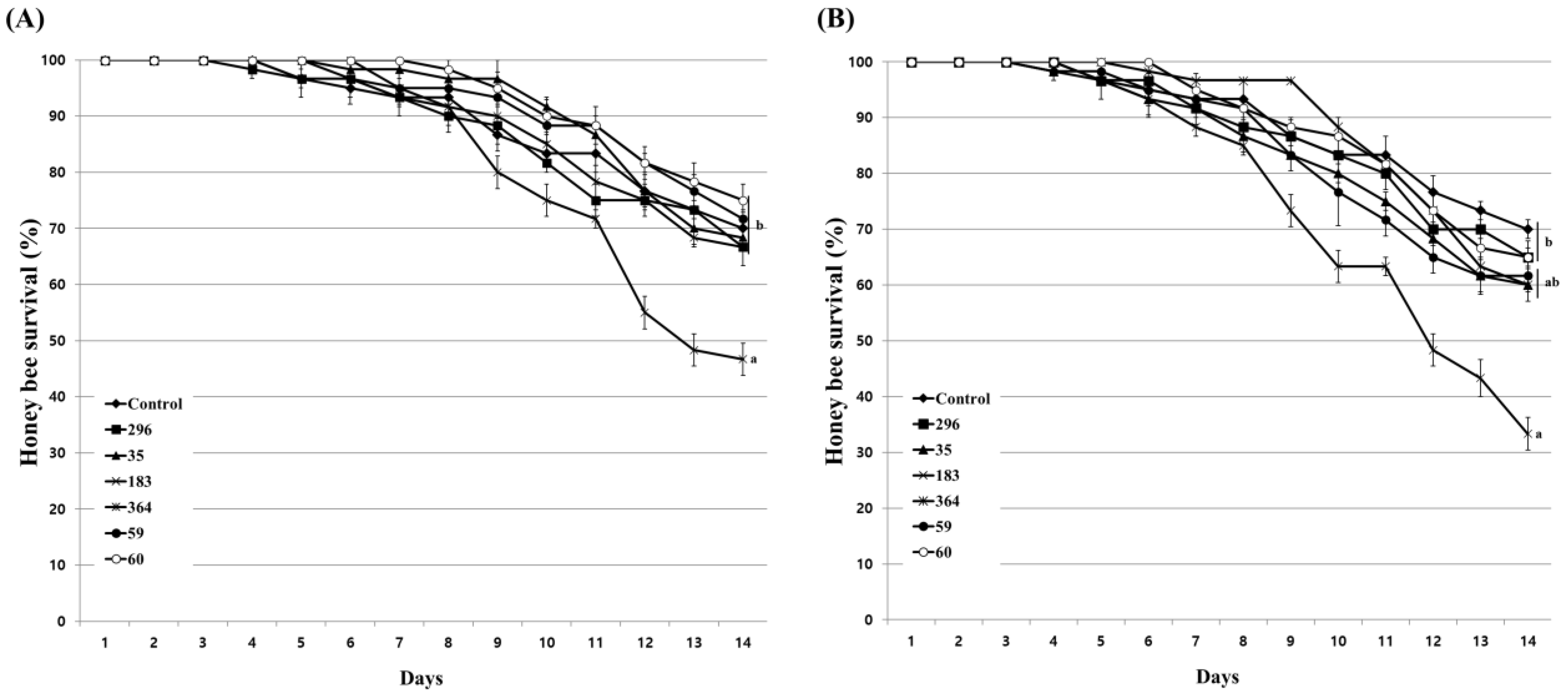
3.4. Influence of Fungal Culture Extracts on Honeybees

Prior to the evaluation of the inhibitory activity of the culture extract on Nosema infection in honeybees, the influence of the culture extracts on the lifespan of honeybees were evaluated. Feeding with acetone, used as a solvent for the preparation of culture extract, showed a similar survival rate as that of the untreated honeybee group at concentrations of up to 2%. However, at a concentration of 4%, the survival rate of honeybees decreased after 4 days of treatment, and the final survival rate showed a difference of approximately 5% from the untreated group (Figure S2). When evaluating the nosemosis inhibitory activity of the fungal culture extract on honeybees, the concentration of acetone actually used does not exceed 0.2%. Therefore, these results indicate that acetone has little effect on the lifespan of honeybees. The influence of fungal culture extracts on honeybees was evaluated at concentrations of 1% and 10%. As a result of treatment with 1% diluted culture extract, 5 fungal isolates except Bea. brongniartii 183 had no significant influence on the survival rate of honeybees (Figure 4A). Even at 10% dilution, only Bea. brongniartii 183 partially reduced the survival rate of honeybees, so it was excluded from further experiments (Figure 4B).

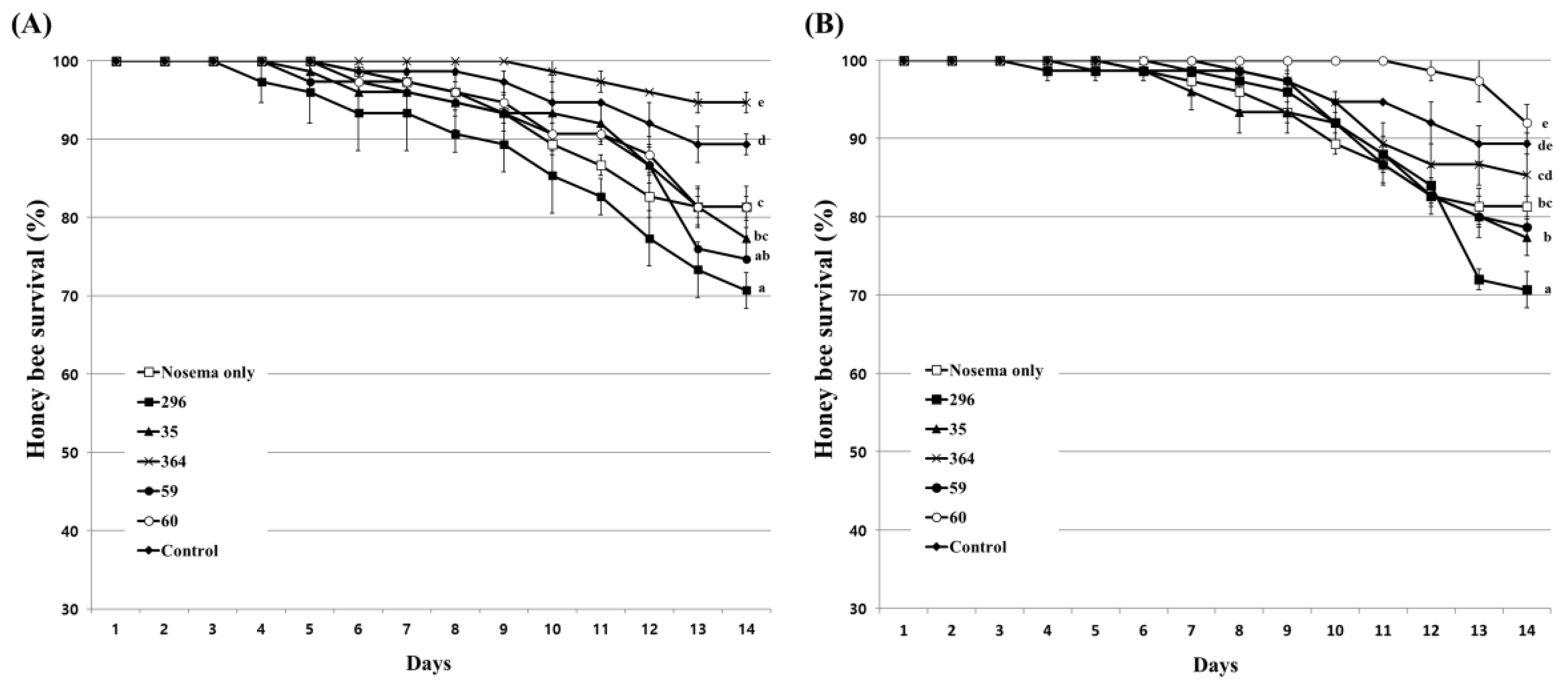
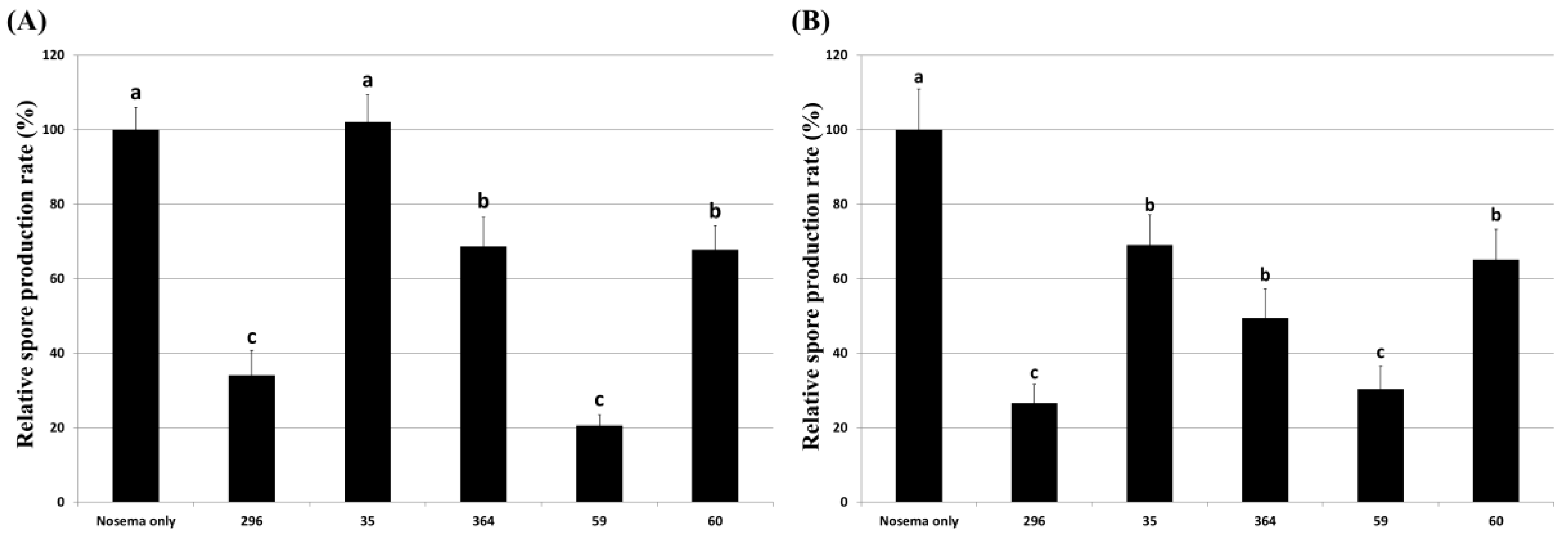
3.5. Inhibitory Effect of Culture Extract on Honeybee Nosemosis

To evaluate the effect of inhibiting honeybee nosemosis by fungal culture extracts, honeybees were treated with each culture extract at a concentration of 5%, and the survival rate of honeybees and Nosema spore production were evaluated. The nosemosis inhibitory effect of the culture extract was evaluated in two ways: honeybees treated with the extract before Nosema infection and after Nosema infection. After treating each culture extract before infection with Nosema, Pae. marquandii 364 culture extract only improved the survival rate of honeybees by approximately 13% compared to Nosema-infected honeybees (Figure 5A). Other fungal culture extracts showed similar or lower survival rates of honeybees compared to Nosema-infected honeybees. Treatment of culture extracts of M. anisopliae 296, Pae. marquandii 364, Bea. bassiana 59, and Poc. bulbillosa 60 reduced spore production by approximately 65%, 32%, 80%, and 33%, respectively, compared to that of honeybees infected with Nosema alone (Figure 6A).
When honeybees were infected with Nosema, then treated with culture extracts, the extracts of Poc. bulbillosa 60 and Pae. marquandii 364 showed approximately 11% and 8% increases in honeybee survival rates, respectively, while the survival rates of the others decreased slightly comparing to Nosema-infected honeybees (Figure 5B). Nosema spore production was reduced compared to Nosema-infected honeybees in all fungal culture extract treatments (Figure 6B). These results suggested that the culture extracts of Pae. marquandii 364 and Poc. bulbillosa 60 were effective in inhibiting honeybee nosemosis.

4. Discussion

This study was conducted to screen and select culture extracts of entomopathogenic fungi that are effective for the control of honeybee nosemosis. Among the 342 entomopathogenic fungal isolates of 24 species from 18 genera tested, the inhibitory effect of fungal culture extracts on Nosema spore germination was shown in more than 95% of the isolates (Table 1 and Table S2). These results suggested that the germination inhibitory substances on Nosema spores were contained in the fungal culture extract and were consistent with previous studies showing that entomopathogenic fungal metabolites had various biological activities. Reportedly, entomopathogenic fungal metabolites have antimicrobial activity against various bacteria and fungi, so sufficient antifungal activity could be expected for unicellular fungal parasites, such as Nosema, in our study [42,43,44,45]. To our knowledge, this is the first report showing that culture extracts of various diverse types of fungi have anti-Nosema activity.
To determine the most effective culture extract among various fungal culture extracts with anti-Nosema activity, six fungal culture extracts showing high anti-germination effects at a 100-fold diluted concentration and even when removed after treatment with the extract were selected and used for further testing anti-Nosema activity (Figure 2 and Figure 3). In our study, the germination inhibitory activity of the culture extract on Nosema spores was evaluated by dividing the fungistatic activity, which is inhibited only when the extract is present, and the fungicidal activity, which inactivates the spores by the extract [52]. It was judged that the fungicidal active culture extract was more effective in controlling nosemosis, and such fungal isolates were selected. In addition, the nosemosis inhibitory activity of the culture extract was shown as a preventive effect in one fungal isolate (Figure 5A) and a control effect in two fungal isolates (Figure 5B). However, the survival rate of honeybees in the experimental groups treated with the culture extract was lower than that of the control group not treated with the culture extract. These results are presumed to be due to the toxicity of the culture extract against honeybees. Although the honeybee survival rate was not significantly reduced compared to that in control under treatment with 10% of each culture extract, it is presumed that the low toxicity of the culture extract increased the susceptibility of honeybees to Nosema, thereby lowering the survival rate. However, it was confirmed that the production of Nosema spores was reduced by treatment with the culture extract (Figure 6). This result suggested that all the culture extracts could inhibit the proliferation of Nosema. As a particularly noteworthy result, treatment with the culture extracts of Poc. bulbillosa 60 and Pae. marquandii 364 showed a higher honeybee survival rate than uninfected honeybees, confirming that this culture extracts not only inhibit nosemosis but also increase the lifespan of honeybees. However, it has been shown that these effects may vary depending on the concentration of these culture extracts. When only the culture extract was treated at a 10% concentration, the survival rate of honeybees was not higher than that of the control group, but at a 1% concentration, the culture extracts of Poc. bulbillosa 60 and Bea. bassiana 59 partially increased the survival rate of honeybees (Figure 4). To date, there has been no report on substances showing positive activity on the lifespan of honeybees among entomopathogenic fungal culture extracts. Further research should identify the metabolites that showed positive effects in this work. Additionally, it should be determined whether the same metabolites have anti-Nosema activity and a positive effect on the lifespan of honeybees.
Various entomopathogenic fungal metabolites have been reported, and the most representative substance is beauvericin from Bea. bassiana and destruxins from Metarhizium spp., which are the most actively studied and utilized [43,53,54,55,56]. These substances have both antifungal activity and insecticidal activity. Other substances known to have antifungal activity include muscodorin, oosporein, patulin, enniatins, pradimicin, flavoglaucin, and terpenoids [43,45,55,57,58]. However, among these substances, inhibitory activity against Nosema has not been reported. Furthermore, since various fungal metabolites show various antifungal activities, the possibility that anti-Nosema active substances exist among fungal metabolites is considered sufficient. In previous reports on Poc. bulbillosa and Pae. marquandii, which showed the highest Nosema inhibitory activity in our study, we could not find any Nosema inhibitory activity. However, the antimicrobial activity and insecticidal activity of the metabolites in Poc. chlamydosporia, Pae. variotii, and Pae. lilacinus have been reported [59,60,61]. Through further research on the metabolites of Poc. bulbillosa 60 and Pae. marquandii 364, it may be possible to develop an effective control agent for honeybee nosemosis.

5. Conclusions

Culture extracts of 342 entomopathogenic fungal isolates of 24 species from 18 genera were used to evaluate the germination inhibitory activity on Nosema spores to search for fungal culture extracts with effective inhibitory activity against Nosema. As a result, inhibitory activity was observed in all fungal culture extracts except for 2 genera and 1 species of fungus, and high inhibitory activity of approximately 80% or more was shown in fungal isolates of 15 species and 10 genera. Among them, the culture extracts of Poc. bulbillosa 60 and Pae. marquandii 364, which have fungicidal activity at low concentrations, not only effectively inhibited honeybee nosemosis but also prolonged the lifespan of honeybees.

Supplementary Materials

The following supporting information can be downloaded at: https://www.mdpi.com/article/10.3390/insects14060538/s1, Table S1. Entomopathogenic fungal isolates were used in the experiment. Table S2. Germination inhibition activity of entomopathogenic fungal culture extracts against Nosema ceranae spores. Figure S1. Viability of Nosema ceranae spores by acetone treatment at different concentrations. After treating the spores with acetone at each concentration, the germination rate was determined by an in vitro germination assay. Figure S2. Honeybee survival by acetone treatment at different concentrations. A mixture of acetone and 50% sucrose solution was fed to honeybees. Afterward, the survival rate of honeybees was determined for 14 days. The control group was fed only a 50% sucrose solution. Data show the mean ± SE.

Author Contributions

Conceptualization: S.-D.W. and D.-J.K.; design of experiment and methodology: S.-D.W. and D.-J.K.; laboratory analysis: D.-J.K., R.-M.W. and K.-S.K.; data curation: D.-J.K., R.-M.W. and K.-S.K.; writing, original draft, review, and editing: S.-D.W. and D.-J.K. All authors have read and agreed to the published version of the manuscript.

Funding

This work was supported by the Korea Institute of Planning and Evaluation for Technology in Food, Agriculture and Forestry (IPET) through the Agricultural Machinery/Equipment Localization Technology Development Program, funded by the Ministry of Agriculture, Food and Rural Affairs (MAFRA) (No. 321054-05).

Data Availability Statement

The data are available at reasonable request from the corresponding author.

Conflicts of Interest

The authors declare no conflict of interest. Author Dong-Jun Kim was employed by the company Plolagen Co., Ltd. The remaining authors declare that the research was conducted in the absence of any commercial or financial relationships that could be construed as a potential conflict of interest.

References

  1. Gallai, N.; Salles, J.M.; Settele, J.; Vaissière, B.E. Economic valuation of the vulnerability of world agriculture confronted with pollinator decline. Ecol. Econ. 2009, 68, 810–821. [Google Scholar] [CrossRef]
  2. Breeze, T.D.; Bailey, A.P.; Balcombe, K.G.; Potts, S.G. Pollination services in the UK: How important are honeybees? Agric. Ecosyst. Environ. 2011, 142, 137–143. [Google Scholar] [CrossRef] [Green Version]
  3. Ollerton, J.; Winfree, R.; Tarrant, S. How many flowering plants are pollinated by animals? Oikos 2011, 120, 321–326. [Google Scholar] [CrossRef]
  4. Garibaldi, L.A.; Steffan-Dewenter, I.; Winfree, R.; Aizen, M.A.; Bommarco, R.; Cunningham, S.A.; Kremen, C.; Carvalheiro, L.G.; Harder, L.D.; Afik, O.; et al. Wild pollinators enhance fruit set of crops regardless of honey bee abundance. Science 2013, 339, 1608–1611. [Google Scholar] [CrossRef] [PubMed]
  5. Papa, G.; Maier, R.; Durazzo, A.; Lucarini, M.; Karabagias, I.K.; Plutino, M.; Bianchetto, E.; Aromolo, R.; Pignatti, G.; Ambrogio, A.; et al. The honey bee Apis mellifera: An insect at the interface between human and ecosystem health. Biology 2022, 11, 233. [Google Scholar] [CrossRef]
  6. Samarghandian, S.; Farkhondeh, T.; Samini, F. Honey and health: A review of recent clinical research. Pharmacogn. Res. 2017, 9, 121–127. [Google Scholar]
  7. Luo, X.; Dong, Y.; Gu, C.; Zhang, X.; Ma, H. Processing technologies for bee products: An overview of recent developments and perspectives. Front. Nutr. 2021, 8, 727181. [Google Scholar] [CrossRef] [PubMed]
  8. Steinhauer, N.; Kulhanek, K.; Antúnez, K.; Human, H.; Chantawannakul, P.; Chauzat, M.P.; Vanengelsdorp, D. Drivers of colony losses. Curr. Opin. Insect Sci. 2018, 26, 142–148. [Google Scholar] [CrossRef]
  9. Hristov, P.; Shumkova, R.; Palova, N.; Neov, B. Factors associated with honey bee colony losses: A mini-review. Vet. Sci. 2020, 7, 166. [Google Scholar] [CrossRef]
  10. Stanimirovic, Z.; Glavinic, U.; Ristanic, M.; Aleksic, N.; Jovanovic, N.; Vejnovic, B.; Stevanovic, J. Looking for the causes of and solutions to the issue of honey bee colony losses. Acta Vet-Beograd 2019, 69, 1–31. [Google Scholar] [CrossRef] [Green Version]
  11. Higes, M.; Martín-Hernández, R.; Garrido-Bailón, E.; González-Porto, A.V.; García-Palencia, P.; Meana, A.; Del Nozal, M.J.; Mayo, R.; Bernal, J.L. Honeybee colony collapse due to Nosema ceranae in professional apiaries. Environ. Microbiol. Rep. 2009, 1, 110–113. [Google Scholar] [CrossRef] [PubMed]
  12. Vanengelsdorp, D.; Evans, J.D.; Saegerman, C.; Mullin, C.; Haubruge, E.; Nguyen, B.K.; Frazier, M.; Frazier, J.; Cox-Foster, D.; Chen, Y.; et al. Colony collapse disorder: A descriptive study. PLoS ONE 2009, 4, e6481. [Google Scholar] [CrossRef] [PubMed] [Green Version]
  13. Le Conte, Y.; Ellis, M.; Ritter, W. Varroa mites and honey bee health: Can Varroa explain part of the colony losses? Apidologie 2010, 41, 353–363. [Google Scholar] [CrossRef] [Green Version]
  14. Cornman, R.S.; Tarpy, D.R.; Chen, Y.; Jeffreys, L.; Lopez, D.; Pettis, J.S.; van Engelsdorp, D.; Evans, J.D. Pathogen webs in collapsing honey bee colonies. PLoS ONE 2012, 7, e43562. [Google Scholar] [CrossRef] [Green Version]
  15. Adl, S.M.; Simpson, A.G.; Lane, C.E.; Lukeš, J.; Bass, D.; Bowser, S.S.; Brown, M.W.; Burki, F.; Dunthorn, M.; Hampl, V.; et al. The revised classification of Eukaryotes. J. Eukaryotic Microbiol. 2012, 59, 429–514. [Google Scholar] [CrossRef] [Green Version]
  16. Galajda, R.; Valenčáková, A.; Sučik, M.; Kandráčová, P. Nosema disease of European honey bees. J. Fungi 2021, 7, 714. [Google Scholar] [CrossRef]
  17. Fries, I. Nosema in European honey bees (Apis mellifera): An emerging threat to bee health. Insect Sci. 2010, 17, 418–424. [Google Scholar]
  18. Mayack, C.; Naug, D. Energetic stress in the honeybee Apis mellifera from Nosema ceranae infection. J. Invertebr. Pathol. 2010, 105, 20–27. [Google Scholar] [CrossRef]
  19. Goblirsch, M.J.; Spivak, M.; Kurtti, T.J. Nosema spp. infections in honey bees (Apis mellifera): Literature review and a survey of prevalence in North America. Insect Sci. 2013, 20, 1–22. [Google Scholar]
  20. Huang, W.F.; Solter, L.F.; Yau, P.M. Nosema ceranae causes asynchronous emergence of honey bee workers and reduces colony productivity. Apidologie 2018, 49, 11–22. [Google Scholar]
  21. Higes, M.; Martín-Hernández, R.; Meana, A. Nosema ceranae in European honey bees (Apis mellifera): An emerging infectious disease threatening apiaries and beekeeping. Insects 2020, 11, 886. [Google Scholar]
  22. Glavinic, U.; Stevanovic, J.; Ristanic, M.; Rajkovic, M.; Davitkov, D.; Lakic, N.; Stanimirovic, Z. Potential of fumagillin and Agaricus blazei mushroom extract to reduce Nosema ceranae in honey bees. Insects 2021, 12, 282. [Google Scholar] [CrossRef] [PubMed]
  23. Glavinic, U.; Rajkovic, M.; Vunduk, J.; Vejnovic, B.; Stevanovic, J.; Milenkovic, I.; Stanimirovic, Z. Effects of Agaricus bisporus mushroom extract on honey bees infected with Nosema ceranae. Insects 2021, 12, 915. [Google Scholar] [CrossRef] [PubMed]
  24. Klee, J.; Besana, A.M.; Genersch, E.; Gisder, S.; Nanetti, A.; Tam, D.Q.; Chin, T.X.; Puerta, F.; Ruz, J.M.; Kryger, P.; et al. Wide spread dispersal of the microsporidian Nosema ceranae, an emergent pathogen of the western honey bee, Apis mellifera. J. Invertebr. Pathol. 2007, 96, 1–10. [Google Scholar] [CrossRef] [PubMed]
  25. Chen, Y.P.; Evans, J.D.; Simith, I.B.; Pettis, J.S. Nosema ceranae is a long-present and wide-spread microsporidian infection of the European honey bee (Apis mellifera) in the United States. J. Invertebr. Pathol. 2008, 97, 186–188. [Google Scholar] [CrossRef]
  26. Emsen, B.; Guzman-Novoa, E.; Hamiduzzaman, M.M.; Eccles, L.; Lacey, B.; Ruiz-Perez, R.A.; Nasr, M. Higher prevalence and levels of Nosema ceranae than Nosema apis infections in Canadian honey bee colonies. Parasitol. Res. 2016, 115, 175–181. [Google Scholar] [CrossRef]
  27. Goulson, D.; Nicholls, E.; Botías, C.; Rotheray, E.L. Bee declines driven by combined stress from parasites, pesticides, and lack of flowers. Science 2015, 347, 1255957. [Google Scholar] [CrossRef]
  28. Vanengelsdorp, D.; Evans, J.D.; Donovall, L.; Mullin, C.; Frazier, M.; Frazier, J.; Tarpy, D.R.; Hayes, J.; Pettis, J.S. Entombed pollen: A new condition in honey bee colonies associated with increased risk of colony mortality. J. Invertebr. Pathol. 2009, 101, 147–149. [Google Scholar] [CrossRef]
  29. Requier, F.; Garnery, L.; Kohl, P.L.; Njovu, H.K.; Pirk, C.W.W.; Crewe, R.M.; Steffan-Dewenter, I. The conservation of native honey bees is crucial. Trends Ecol. Evol. 2019, 34, 789–798. [Google Scholar] [CrossRef]
  30. Stanimirovic, Z.; Stevanovic, J.; Bajic, V.; Radovic, I. Evaluation of genotoxic effects of fumagillin (dicyclohexylamine) by citogenetic tests in vivo. Mutat. Res. 2006, 628, 1–10. [Google Scholar]
  31. Pajuelo, A.G.; Torres, C.; Bermejo, F.J.O. Colony losses: A double blind trial on the influence of supplementary protein nutrition and preventative treatment with fumagillin against Nosema ceranae. J. Apic. Res. 2008, 47, 84–86. [Google Scholar] [CrossRef]
  32. Williams, G.R.; Shutler, D.; Little, C.M.; Burgher-MacLellan, K.L.; Rogers, R.E.L. The microsporidian Nosema ceranae, the antibiotic Fumagilin-B®, and western honey bee (Apis mellifera) colony strength. Apidologie 2011, 42, 15–22. [Google Scholar] [CrossRef] [Green Version]
  33. Williams, G.R.; Sampson, M.A.; Shutler, D.; Rogers, R.E. Does fumagillin control the recently detected invasive parasite Nosema ceranae in western honey bees (Apis mellifera)? J. Apic. Res. 2018, 57, 714–721. [Google Scholar] [CrossRef]
  34. Sabaté, D.C.; Cruz, M.S.; Benítez-Ahrendts, M.R.; Audisio, M.C. Beneficial effects of Bacillus subtilis subsp. subtilis Mori2, a honey-associated strain, on honeybee colony performance. Probiot. Antimicrob. Proteins 2012, 4, 39–46. [Google Scholar] [CrossRef]
  35. Glavinic, U.; Blagojevic, J.; Ristanic, M.; Stevanovic, J.; Lakic, N.; Mirilovic, M.; Stanimirovic, Z. Use of thymol in Nosema ceranae control and health improvement of infected honey bees. Insects 2022, 13, 574. [Google Scholar] [CrossRef]
  36. Klassen, S.S.; VanBlyderveen, W.; Eccles, L.; Kelly, P.G.; Borges, D.; Goodwin, P.H.; Petukhova, T.; Wang, Q.; Guzman-Novoa, E. Nosema ceranae infections in honey bees (Apis mellifera) treated with pre/probiotics and impacts on colonies in the field. Vet. Sci. 2021, 8, 107. [Google Scholar] [CrossRef]
  37. Kunat-Budzyńska, M.; Budzyński, M.; Schulz, M.; Strachecka, A.; Gancarz, M.; Rusinek, R.; Ptaszyńska, A.A. Natural substances, probiotics, and synthetic agents in the treatment and prevention of honeybee nosemosis. Pathogens 2022, 11, 1269. [Google Scholar] [CrossRef]
  38. Lacey, L.A.; Frutos, R.; Kaya, H.K.; Vail, P. Insect pathogens as biological control agents: Do they have a future? Biol. Control 2001, 21, 230–248. [Google Scholar] [CrossRef] [Green Version]
  39. Zimmermann, G. Review on safety of the entomopathogenic fungus Metarhizium anisopliae. Biocontrol Sci. Technol. 2007, 17, 879–920. [Google Scholar] [CrossRef]
  40. Vega, F.E.; Goettel, M.S.; Blackwell, M.; Chandler, D.; Jackson, M.A.; Keller, S.; Koike, M.; Maniania, N.K.; Monzon, A.; Ownley, B.H.; et al. Fungal entomopathogens: New insights on their ecology. Fungal Ecol. 2009, 2, 149–159. [Google Scholar] [CrossRef] [Green Version]
  41. Butt, T.M.; Coates, C.J.; Dubovskiy, I.M.; Ratcliffe, N.A. Entomopathogenic fungi: New insights into host-pathogen interactions. Adv. Genet. 2016, 94, 307–364. [Google Scholar] [PubMed]
  42. Isaka, M.; Kittakoop, P.; Kirtikara, K.; Hywel-Jones, N.L.; Thebtaranonth, Y. Bioactive substances from insect pathogenic fungi. Acc. Chem. Res. 2005, 38, 813–823. [Google Scholar] [CrossRef] [PubMed]
  43. Wang, Q.; Xu, L. Beauvericin, a bioactive compound produced by fungi: A short review. Molecules 2012, 17, 2367–2377. [Google Scholar] [CrossRef] [Green Version]
  44. Shin, T.Y.; Bae, S.M.; Kim, D.J.; Yun, H.G.; Woo, S.D. Evaluation of virulence, tolerance to environmental factors and antimicrobial activities of entomopathogenic fungi against two-spotted spider mite, Tetranychus urticae. Mycoscience 2017, 58, 204–212. [Google Scholar] [CrossRef]
  45. Zhang, L.; Fasoyin, O.E.; Molnár, I.; Xu, Y. Secondary metabolites from hypocrealean entomopathogenic fungi: Novel bioactive compounds. Nat. Prod. Rep. 2020, 37, 1181–1206. [Google Scholar] [CrossRef]
  46. Shin, T.Y.; Lee, W.W.; Ko, S.H.; Choi, J.B.; Bae, S.M.; Choi, J.Y.; Lee, K.S.; Je, Y.H.; Jin, B.R.; Woo, S.D. Distribution and characterisation of entomopathogenic fungi from Korean soils. Biocontrol Sci. Technol. 2013, 23, 288–304. [Google Scholar] [CrossRef]
  47. Gisder, S.; Hedtke, K.; Möckel, N.; Frielitz, M.C.; Linde, A.; Genersch, E. Five-year cohort study of Nosema spp. in Germany: Does climate shape virulence and assertiveness of Nosema ceranae? Appl. Environ. Microbiol. 2010, 76, 3032–3038. [Google Scholar] [CrossRef] [Green Version]
  48. Kim, D.J.; Yun, H.G.; Kim, I.H.; Gwak, W.S.; Woo, S.D. Efficient method for the rapid purification of Nosema ceranae spores. Mycobiology 2017, 45, 204–208. [Google Scholar] [CrossRef] [Green Version]
  49. Chen, Y.P.; Evans, J.D.; Murphy, C.; Gutell, R.; Zuker, M.; Gundensen-Rindal, D.; Pettis, J.S. Morphological, molecular, and phylogenetic characterization of Nosema ceranae, a microsporidian parasite isolated from the European honey bee, Apis mellifera. J. Eukaryotic Microbiol. 2009, 56, 142–147. [Google Scholar] [CrossRef] [PubMed] [Green Version]
  50. Shimanuki, H.; Knox, D.A. Diagnosis of Honey Bee Diseases; Agriculture Handbook No. AH-690; U.S. Department of Agriculture: Washington, DC, USA, 2000; p. 61.
  51. Olsen, P.E.; Rice, W.A.; Liu, T.P. In vitro germination of Nosema apis spores under conditions favorable for the generation and maintenance of sporoplasms. J. Invertebr. Pathol. 1986, 47, 65–73. [Google Scholar] [CrossRef]
  52. Graybill, J.R.; Burgess, D.S.; Hardin, T.C. Key issues concerning fungistatic versus fungicidal drugs. Eur. J. Clin. Microbiol. Infect. Dis. 1997, 16, 42–50. [Google Scholar] [CrossRef]
  53. Klarić, M.S.; Pepeljnjak, S. Bovericin: Kemizam, bioloski aspekti i rasirenost [Beauvericin: Chemical and biological aspects and occurrence]. Arh. Hig. Rada Toksikol. 2005, 56, 343–350. [Google Scholar] [PubMed]
  54. Liu, B.L.; Tzeng, Y.M. Development and applications of destruxins: A review. Biotechnol. Adv. 2012, 30, 1242–1254. [Google Scholar] [CrossRef]
  55. Wang, X.; Gong, X.; Li, P.; Lai, D.; Zhou, L. Structural diversity and biological activities of cyclic depsipeptides from fungi. Molecules 2018, 23, 169. [Google Scholar] [CrossRef] [Green Version]
  56. Wang, H.; Peng, H.; Li, W.; Cheng, P.; Gong, M. The toxins of Beauveria bassiana and the strategies to improve their virulence to insects. Front. Microbiol. 2021, 12, 705343. [Google Scholar] [CrossRef] [PubMed]
  57. Houard, J.; Aumelas, A.; Noël, T.; Pages, S.; Givaudan, A.; Fitton-Ouhabi, V.; Villain-Guillot, P.; Gualtieri, M. Cabanillasin, a new antifungal metabolite, produced by entomopathogenic Xenorhabdus cabanillasii JM26. J. Antibiot. 2013, 66, 617–620. [Google Scholar] [CrossRef] [PubMed]
  58. Ashraf, S.A.; Elkhalifa, A.E.O.; Siddiqui, A.J.; Patel, M.; Awadelkareem, A.M.; Snoussi, M.; Ashraf, M.S.; Adnan, M.; Hadi, S. Cordycepin for health and wellbeing: A potent bioactive metabolite of an entomopathogenic Cordyceps medicinal fungus and Its nutraceutical and therapeutic potential. Molecules 2020, 25, 2735. [Google Scholar] [CrossRef]
  59. Lacatena, F.; Marra, R.; Mazzei, P.; Piccolo, A.; Digilio, M.C.; Giorgini, M.; Woo, S.L.; Cavallo, P.; Lorito, M.; Vinale, F. Chlamyphilone, a novel Pochonia chlamydosporia metabolite with insecticidal activity. Molecules 2019, 24, 750. [Google Scholar] [CrossRef] [Green Version]
  60. Qin, F.; Li, Y.; Lin, R.; Zhang, X.; Mao, Z.; Ling, J.; Yang, Y.; Zhuang, X.; Du, S.; Cheng, X.; et al. Antibacterial radicicol analogues from Pochonia chlamydosporia and their biosynthetic gene cluster. J. Agric. Food Chem. 2019, 67, 7266–7273. [Google Scholar] [CrossRef]
  61. Dai, Z.B.; Wang, X.; Li, G.H. Secondary metabolites and their bioactivities produced by Paecilomyces. Molecules 2020, 25, 5077. [Google Scholar] [CrossRef]
Figure 1. Spore germination inhibitory activity of fungal culture extract on Nosema ceranae. (A) more than 80% inhibition; (B) more than 60% inhibition; (C) less than 60% inhibition. The scale bar is 20 μm. Arrows indicate mycelia that have grown after germination.
Insects 14 00538 g001
Figure 2. Inhibition rate of Nosema ceranae spore germination in response to 89 entomopathogenic fungal culture extracts. Each extract was used to treat Nosema ceranae spores at concentrations of 100%, 10%, and 1%, then the germination rate was determined. Fungal isolates exhibiting an inhibition rate of 80% or more are indicated by black bars. Data show the mean ± SE.
Insects 14 00538 g002
Figure 3. Evaluation of the fungistatic and fungicidal activities of entomopathogenic fungal culture extracts on Nosema ceranae spores. After treatment with the fungal culture extract on Nosema ceranae spores, germination was observed without removing the extract (grey bar) or after removing the extract (black bar). Data show the mean ± SE.
Insects 14 00538 g003
Figure 4. Honeybee survival under 1% (A) and 10% (B) fungal culture extract treatments. A mixture of fungal culture extract and 50% sucrose solution was fed to honeybees. Afterward, the survival rate of honeybees was determined for 14 days. The control group was fed only a 50% sucrose solution. Data show the mean ± SE. Values with different letters are significantly different (p < 0.05, SNK test in one-way ANOVA) at 14 days.
Insects 14 00538 g004
Figure 5. Survival rate of honeybees against Nosema ceranae infection. Honeybees were treated with 5% fungal culture extract before (A) and after (B) Nosema infection. The control group and Nosema-only group were fed a 50% sucrose solution and a mixture containing Nosema spores, respectively. Data show the mean ± SE. Values with different letters are significantly different (p < 0.05, SNK test in one-way ANOVA) at 14 days.
Insects 14 00538 g005
Figure 6. Nosema spore production in Nosema-infected honeybees treated with 5% fungal culture extract before (A) or after (B) Nosema ceranae infection. The relative ratio of the spore production of each treatment group to the spore production of honeybees infected with Nosema only was shown. Data show the mean ± SE. Values with different letters are significantly different (p < 0.05, SNK test in one-way ANOVA).
Insects 14 00538 g006
Table 1. Entomopathogenic fungi showing more than 80% Nosema ceranae germination inhibition activity.
FungusNo. of Tested IsolatesNo. of Isolates Showing the Inhibition of Spore Germination
All fungal isolates34289 (26%) *
Acremonium strictum10 (0%)
Aspergillus lentulus.50 (0%)
Aspergillus versicolor31 (33.3%)
Beauveria bassiana11048 (43.6%)
Beauveria brongniartii81 (12.5%)
Beauveria pseudobassiana81 (12.5%)
Bionectria ochroleuca70 (0%)
Clonostachys rosea10 (0%)
Cordyceps farinosa125 (41.7%)
Cordyceps fumosorosea61 (16.7%)
Cordyceps javanica225 (22.7%)
Fusarium oxysporum20 (0%)
Lecanicillium spp.81 (12.5%)
Metarhizium anisopliae6415 (23.4%)
Metarhizium lepidiotae10 (0%)
Metarhizium pemphigus163 (18.8%)
Mucoromycotina spp.10 (0%)
Myrothecium spp.50 (0%)
Paecilomyces lilacinus71 (14.3%)
Paecilomyces marquandii51 (20%)
Paraconiothyrium sporulosum21 (50%)
Phialocephala spp.10 (0%)
Pochonia bulbillosa163 (18.8%)
Pochonia rubescens10 (0%)
Simplicillium aogashimaense11 (100%)
Simplicillium sp.20 (0%)
Tolypocladium album231 (4.3%)
Tolypocladium cylindrosporum30 (0%)
Verticillium insectorum10 (0%)
* Relative % to the number of isolates (species or genus).
Table 2. Entomopathogenic fungi showing the inhibition of Nosema ceranae spore germination by more than 80% in 1% concentration culture extract.
FungusNo. of Isolate (%)
Aspergillus versicolor1 (2.3) *
Beauveria bassiana24 (54.5)
Beauveria brongniartii1 (2.3)
Cordyceps farinosa4 (9.1)
Cordyceps fumosorosea1 (2.3)
Cordyceps javanica1 (2.3)
Lecanicillium spp.1 (2.3)
Metarhizium anisopliae6 (13.6)
Paecilomyces lilacinus1 (2.3)
Paecilomyces marquandii1 (2.3)
Pochonia bulbillosa2 (4.5)
Tolypocladium album1 (2.3)
* Relative % to number of isolates (species or genus).
Disclaimer/Publisher’s Note: The statements, opinions and data contained in all publications are solely those of the individual author(s) and contributor(s) and not of MDPI and/or the editor(s). MDPI and/or the editor(s) disclaim responsibility for any injury to people or property resulting from any ideas, methods, instructions or products referred to in the content.

