Article

RNAi-Mediated Silencing of Putative Halloween Gene Phantom Affects the Performance of Rice Striped Stem Borer, Chilo suppressalis

1
Department of Entomology, Faculty of Agriculture, Gomal University, Dera Ismail Khan 29220, Pakistan
2
College of Plant Protection, Nanjing Agricultural University, Nanjing 210095, China
3
Guangdong Key Laboratory of Animal Conservation and Resource Utilization, Guangdong Public Laboratory of Wild Animal Conservation and Utilization, Institute of Zoology, Guangdong Academy of Sciences, Guangzhou 510260, China
4
Institute of Molecular Biology and Biotechnology, The University of Lahore, 1-Km Defense Road, Lahore 54000, Pakistan
5
Honeybee Research Institute, National Agricultural Research Centre, Park Road, Islamabad 45500, Pakistan
6
Department of Entomology and Wildlife Ecology, University of Delaware, Newark, DE 19716, USA
7
College of Agriculture, Bahadur Sub Campus Layyah, Bahauddin Zakariya University, Multan 31200, Pakistan
*
Authors to whom correspondence should be addressed.
These authors contributed equally to this work.
Insects 2022, 13(8), 731; https://doi.org/10.3390/insects13080731
Submission received: 30 June 2022 / Revised: 11 August 2022 / Accepted: 12 August 2022 / Published: 15 August 2022
(This article belongs to the Special Issue Managing Invasive Insects: Good Intentions, Hard Realities)

Abstract

:

Simple Summary

Ecdysteroids have a significant role in insect development and reproduction. Ecdysteroidogensis is regulated by a group of genes known as Halloween genes. We provide experimental evidence that Chilo suppressalis-phantom (Cs-Phm) in rice striped stem borer is a Halloween gene phantom and has important role in specific steps in ecdysteroidogensis. Three lines of experiments were done on typical protein domains of Cs-Phm, RNAi-mediated knockdown of Cs-Phm, and the rescue of larval development in dsRNA-treated groups. The results strongly suggest that Cs-Phm plays a critical role in ecdysteroidogensis in rice striped stem borer.

Abstract

The physiological and biochemical characterization of the “Halloween” genes has fundamental importance in the biosynthesis pathway of ecdysteroids. These genes were found to catalyze the final phases of ecdysteroid biosynthesis from dietary cholesterol to the molting hormone 20-hydroxyecdysone. We report the characterization of the Cs-Phm in a major insect pest in agriculture, the rice striped stem borer, Chilo suppressalis (C. suppressalis). A full-length transcript of Cs-Phm was amplified with an open reading frame (ORF) of 478 amino acids through 5′ and 3′ RACE. Cs-Phm shows five insect-conserved P450 motifs: Helix-C, Helix-I, Helix-K, PERF, and heme-binding motifs. Phylogenetic analysis clearly shows high similarity to Lepidoptera and evolutionary conservation in insects. The relative spatial and temporal transcript profile shows that Cs-Phm is highly expressed in the prothoracic glands and appears throughout the larval development, but with low expression at the start of the larval instar. It seems to peak in 3–4 days and decreases again before the larvae molt. Double-stranded RNA (dsRNA) injection of Cs-Phm at the larval stage efficiently knocked down the target gene and decreased its expression level. The dsRNA-treated group showed significantly decreased ecdysteroid titers, which leads to delayed larval development and higher larval mortality. Negative effects of larval development were rescued by treating 20E in the dsRNA-treated group. Thus, in conclusion, our results suggest that Cs-Phm is functionally conserved in C. suppressalis and encodes functional CYP that contributes to the biogenesis of 20E.

1. Introduction

Insect development and growth are coordinated by steroid hormones for developmental transitions [1,2]. Metamorphosis and molting are precisely timed developmental transitions that are strictly regulated by ecdysone in insects [3,4,5]. During larval pupal development, the neuropeptide prothoracicotropic hormone (PTTH) stimulates the synthesis of ecdysone in the prothoracic glands through the activation of mitogen-activated protein kinase (MAPK) and/or phosphoinositide 3-kinase (PI3K) [4,6,7]. Finally, ecdysone is converted to the active hormone 20-hydroxyecdysone (20E) in several peripheral tissues (principally in body fat or the midgut) [8,9,10,11].
Steroid hormones′ universal precursor is cholesterol [6]. However, like vertebrates, insects cannot produce it [12]. The biosynthesis of ecdysone includes oxidation and hydroxylation steps during postembryonic development [4]. The initial biosynthetic step of 20-hydroxyecdysone (20E) starts in the endoplasmic reticulum by the conversion of cholesterol to 7-dehydrocholesterol (7dc) by the Rieske-domain protein, Neverland [13]. Subsequently, the oxidation of 7dC to diketol occurs through a series of assumed and unverified “black box” reactions [14]. Then, hydroxylation steps proceed along this pathway. For the hydroxylation reactions, P450 protein is the only catalyzing enzyme [15].
Recent studies have shown that Halloween genes encode cytochrome P450 mono-oxygenases involved in the biosynthesis of 20E, such as spook (CYP307a1), phantom (CYP306a1), disembodied (CYP302a1), shadow (CYP315a1), and shade (CYP314a1) [8,16,17,18,19,20,21,22]. Aside from spook (CYP307a1), no paralogs have been identified for the other Halloween genes. In contrast, two additional paralogs exist for spook, spookier (CYP307a2), and spookiest (CYP307B1) [23]. Mutant embryos of the Halloween genes show a similar phenotype characterized by low ecdysteroid titers, dorsal closure, failure of head involution, hindgut abnormality, failure to induce ecdysteroid receptor genes, and lethality [18,19,24].
In this study, we cloned the full length of a Halloween gene, Cs-Phm, which is speculated to encode an enzyme involved in the biosynthesis of ecdysteroids. The relative spatio-temporal expression patterns were analyzed using quantitative real time PCR (qRT-PCR). We also investigated whether dsRNA-mediated knockdown of Cs-Phm induces delayed larval development and mortality. To validate our results, topical application of 20E was used to rescue the negative effects of dsRNA-injected insects. The results of this study provide basic information on Cs-Phm and its functional importance in ecdysteroidogenesis in the rice striped stem borer, C. suppressalis.

2. Materials and Methods

2.1. Insects

Individuals of C. suppressalis were collected from rice fields in Nanjing (32.00° N, 118.50° E), Jiangsu province, China, and cultured routinely in an insectary. The collected strain has been reared in an insectary on rice seedlings at 25 ± 3 °C and a light:dark ratio of 16:8 with approximately 80% relative humidity.

2.2. Total RNA Isolation and cDNA Synthesis

Total RNA was extracted from 4th-instar larvae (~3 pooled larvae) using TRIzol reagent (Invitrogen, Grand Island, NY, USA) and an RNeasy micro kit (Qiagen Inc., Valencia, CA, USA) according to the manufacturer′s instructions. Genomic DNA was removed from RNA samples with RNase-free DNase I using the protocol of the DNA-free kit (Roche Diagnostics, Mannheim, Germany). The RNA purity and concentration were measured with a NanoDropTM ND1000 spectrophotometer (NanoDrop Technologies, Rockland, DE, USA). cDNA was synthesized using a PrimeScriptTM Ist Strand cDNA Synthesis Kit (TaKaRa Bio., Dalian, China) from 1 μg of total RNA by following the manufacturer′s instructions. It was then used as a template for PCR.

2.3. Amplification of cDNA Fragment

The annotation-based Cs-Phm transcript was searched for in the C. suppressalis draft genome [25]. Based on the transcript, primers were designed using the software Primer Premier version 5.0 [26] and are listed in Table 1. PCR was performed according to the following program: 94 °C for 3 min followed by 30 cycles at 94 °C for 30 s, 55 °C for 40 s, and 2 min at 72 °C, with a final elongation at 72 °C for 10 min. Each 50 μL PCR reaction mixture contained 2 μL of cDNA template, 0.5 μL of LA Taq polymerase, 5 μL of MgCl2 (25 mM), 5 μL of 10 × LA Taq buffer, 8 μL of dNTP mixture (2.5 mM/each), 1 μL of sense primers, 1 μL of antisense primers (10 μM) (TaKaRa Bio, Dalian, China), and 27.5 μL of double-distilled H2O. The PCR product was cleaned using a Wizard DNA Gel Extraction Kit by following the manufacturer′s protocol (Promega, Madison, WI, USA) and then sequenced.

2.4. Rapid Amplification of cDNA Ends (3′ RACE and 5′ RACE)

To amplify the full-length cDNA of Cs-Phm, we carried out rapid amplification of cDNA ends (RACE): 5′ and 3′ RACE-ready cDNA were prepared using a SMARTer RACE cDNA amplification kit (Takara Bio, Dalian, China) from the total RNA that was extracted from whole bodies of 4th-instar larvae (~3 pooled larvae). Gene-specific primers (GSP) and nested gene-specific primers were designed by partial sequences using Primer Premier 5.0 to amplify the 5′ and 3′ ends. The first PCR was performed with GSP and universal primer mix according to the following program: incubation for 3 min at 94 °C, 5 cycles at 94 °C for 30 s, 72 °C for 3 min; 5 cycles for 30 s at 94 °C, 70 °C for 30 s, 3 min at 72 °C; and 25 cycles at 94 °C for 30 s, 68 °C for 30 s, and 72 °C for 3 min, with final elongation at 72 °C for 10 min. In the nested gene-specific primer PCR, the first round of PCR product was diluted by 100-fold and then used as a template with nested gene-specific primer and nested universal primer mix. PCR was performed according to the following program: incubation for 3 min at 94 °C, followed by 20 cycles at 94 °C for 30 s, 68 °C for 30 s, and 72 °C for 3 min. The final extension was 72 °C for 10 min.
The amplified PCR products were separated by 1.2% agarose gel, cleaned using a Wizard DNA Gel Extraction Kit, and cloned into easy vector pGEM-T (Promega, Madison, WI, USA). Several of the longest clones were sequenced from both directions with the help of an automated sequencer (Model-3730, Applied Biosystems, Foster City, CA, USA). After amplifying the full-length cDNA of Cs-Phm, we designed forward and reverse primers using Primer Premier 5.0 software [26] to confirm the complete open reading frame (Table 1). The DNAStar program (http://www.dnastar.com accessed on 12 April 2019) was used to predict the open reading frame. The resulting Cs-Phm sequence was submitted to GenBank® (KF701127).

2.5. Phylogenetic Analysis

The following phantom amino acid sequences were downloaded from the NCBI GenBank® database (http://www.ncbi.nlm.nih.gov accessed on 12 April 2019): Spodoptera littoralis (ACM45975); Ha—Helicoverpa armigera (AID54855); Mb—Mamestra brassicae (BAN66311); Ms—Manduca sexta (ABC96068); Bm—Bombyx mori (BAM73853); Dp—Danaus plexippus (EHJ76071); Zn—Zootermopsis nevadensis (KDR21920); Ae—Aedes aegypti (EAT43717); Ad—Anopheles darling (ENT60005); Dm—Drosophila melanogaster (NP573319); and Tc—Tribolium castaneum (XP968477). The neighbor-joining technique was used to build a phylogenetic tree with 1000 bootstrap replicates using MEGA version 5.0 with the complete deletion of gaps and a Poisson model [27].
Table 1. Primers used for validation of Cs-Phantom, 5′ and 3′ RACE, and quantitative real-time PCR.
PrimerSequence (5′ to 3′)Amplicon Size (bp)
Primers used in RT-PCR
Phm-RT-FAGCAACCTCATTTGACCCTA483
Phm-RT-RGGCACCCTTCTTCTTGGA
Primers used in 5′-RACE
Phm-5GSPCACCCTTCTTCTTGGATCTGACGAAACC
Phm-5NGSPCAGCACATATGATACCATTGCCACGC
Primers used in 3′-RACE
Phm-3GSPACTGGAAAAACGCATCGCTGCTGGC
Phm-3NGSPGGTTTCGTCAGATCCAAGAAGAAGGGTG
Primers used in PCR for End to End
Phm-FATGGGGAGTCGCTGGTCA1717
Phm-RCCCGCACCGACGTGGTAT
Primers used for synthesizing the dsRNAs
Phm-dsRNA-F
Phm-dsRNA-R
GFP-dsRNA-F
GFP-dsRNA-R
Primers used in qRT-PCR


taatacgactcactataggTGACACCAAAAGGAGCGGAA
taatacgactcactataggGCCCACTGGAGAGGTATCAC
taatacgactcactataggTCACGGATACAACCTCTTT
taatacgactcactataggAGTTCAGCGTGTCCG


544

414
Phm-qFGCCAGGTGATAGGTTGTGTT204
Phm-qRATGAGGTTGCTTGGGATCTATG

2.6. Quantitative Reverse Transcription Analysis

Whole-body total RNA was isolated from the third- to sixth-instar larvae and pupae to determine the Cs-phm expression pattern at different developmental stages. Epidermis tissues, prothoracic glands, midgut, Malpighian tubules, and body fat RNA was isolated from fourth-instar larvae to measure Cs-phm′s spatial expression using TRIzol reagent according to the manufacturer′s directions (Invitrogen, Grand Island, NY, USA). Pooled samples of three animals were used for developmental expression, and pooled samples of 10–20 animals were used to collect samples for tissue expression.
Genomic DNA was removed from RNA samples with RNase-free DNase I using the protocol of the DNA-free kit (Roche Diagnostics, Mannheim, Germany). qRT-PCR analysis was performed using three biological replicates for each sample. We used an online program, PrimerQuest (http://www.idtdna.com/scitools/applications/primerquest/ accessed on 12 April 2019), to design qRT-PCR primers (Table 1). In our previous study, we found that Actin-A1 was the most stable housekeeping gene in C. suppressalis, so it was also used in this study [28].
Real-Time PCR ABI 7500 System (Takara Bio, Dalian, China) was performed with 96-well plates using SYBR PremixEx TaqTM (Takara Bio, China) in technical triplicate reactions by following the manufacturer’s protocol. The reaction mixture had a final volume of 20 μL and contained 2 μL of cDNA template, 1 μL of forward primer (10 μmol/L), 1 μL of reverse primer (10 μmol/L), 10 μL of SYBR Premix Ex Taq (Takara Bio), and 0.4 μL of Rox Reference Dye (50×). A non-template negative control and a reverse-transcription negative control (without reverse transcriptase) were included for each primer set to detect contamination of primer–dimer and genomic DNA in the reaction.
The qRT-PCR program included pre-denaturing at 95 °C for 30 s and then 40 cycles at 95 °C for 5 s and 60 °C for 34 s. After amplification, the samples were heated at 95 °C for 15 s to determine the melting curves, followed by cooling down for 1 min at 60 °C, and finally heating for 15 s at 95 °C. All data were analyzed using the 2−ΔΔCt method [29]. For ΔΔCT calculation to be valid, the amplification efficiencies of target and reference gene must be approximately equal. A cDNA preparation was diluted by 100-fold to investigate how ΔCT (CT,Cs-Phm-CT,Actin-A1) varies with the template dilution. A plot of the log CDNA dilution versus ΔCT was made. The absolute value of slope was 0.0472, which is close to 0. This calculation clearly showed that efficiencies of target and reference gene were similar; therefore, ΔΔCT calculation for the relative quantification of Cs-phm may be used [30].

2.7. dsRNA Preparation and Microinjection

A T7 RibomaxTM Express RNAi System (Promega) was used to prepare double-strand RNA (dsRNA), and pEASY-T3 vector (TransGen Biotech, Beijing, China) was used to clone the cDNA of green fluorescent protein (GFP) and Cs-Phm. T7 promoter sites were added by PCR using diluted plasmid templates and modified GSPs (Table 1). Wizard HSV Gel (Promega) was used to purify PCR products for templates, and dsRNA was synthesized by the T7 RibomaxTM Express RNAi System using the manufacturer′s protocol. Two complementary RNA transcripts were prepared and hybridized to synthesize dsRNA. The products were treated with RNase and DNase I with incubation at 37 °C for at least 30 min to remove any single-stranded RNA and DNA. Isopropanol was used to precipitate the synthesized dsRNA, which was then resuspended in nuclease-free water.
The dsRNA quantity was analyzed at 260 nm using a NanoDropTM ND1000 spectrophotometer (Nano-Drop Technologies, Rockland, DE, USA). The dsRNA product aliquots were measured with a 1.2% w/v agarose gel run in TAE buffer (40 mmol/L Tris acetate, 2 mmol/L Na2EDTA·2H2O). Resulting bands were visualized by ethidium bromide staining. The required dsRNA concentration of 2400 ng/μL was diluted by adding nuclease-free water.
On day 2, fourth-instar larvae were injected with 2 μL of dsRNA between the third and fourth segments using an InjectMan NI-2 microinjection system (Eppendorf, Germany) in two groups. dsGFP injection was used as a negative control in one group, and dsCs-Phm injection was used for treatment in the other group. Needles were prepared using glass capillaries (outer diameter 1.00 mm, and inner diameter 0.50 mm) and a micropipette puller (Model P-87, Sutter Instruments Company, Novato, CA, USA). To avoid dsRNA leakage, needles were kept in the insect body for at least 30 s at the injection point.

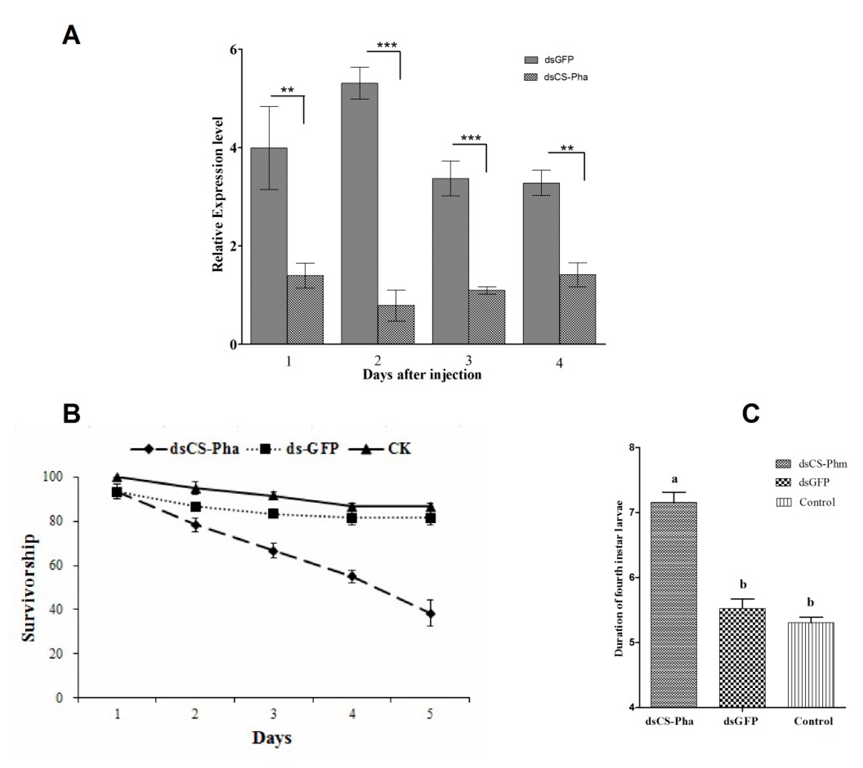
2.8. Cs-Phm Relative Expression Level in dsRNA-Treated Insects

Phenotypic observations were recorded in the first group, and mRNA abundance after dsRNA injection was estimated in the second group (40 individuals for each treatment). We collected samples at 1, 2, 3, and 4 days after dsRNA injection to estimate mRNA abundance. The mRNA abundance of Cs-Phm in the dsRNA-treated group was normalized with the data at the same time point with the negative control. Independent triplicate biological replications were used in all experiments.
Isolation of total RNA and cDNA preparation were the same as explained above. We used the Actin-A1 housekeeping gene (ChiloDB Accession Number = CSUOGS101387-TA) as a reference gene in all experiments [25,28]. An ABI 7500 Real-Time PCR System (Applied Biosystems) was used according to the instructions of the manufacturers to measure the mRNA abundance. The molting and mortality percentage were calculated at 3 days post-injection. The data were analyzed by analysis of variance (ANOVA) using SPSS for Windows (SPSS version 24, Chicago, IL, USA) and are presented as the means ± SE. All graphs were prepared by using Graph Pad Prism 5.

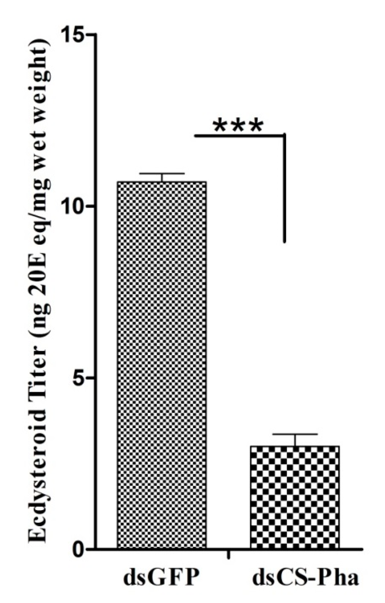
2.9. 20-Hydroxyecdysone Titer Measurement

Samples were collected at 72 h after dsRNA injection. Each sample contained 3 larvae and was repeated in biological triplicate. The whole larval bodies were homogenized with 70% methanol and incubated at 60 °C for 10 min, followed by centrifugation at 10,000× g for 10 min. 70% methanol was used to extract the resulting pellets, and supernatants were dried under low pressure. Hexane and 70% methanol were mixed with dried residues to remove apolar lipids [31]. A standard procedure to remove the hexane phase was adopted. Methanol was desiccated, and the sample redissolved in 300 μL of 70% methanol.
High-performance liquid chromatography (HPLC) with an XDB-C18 column (4.60 × 250 mm, 5 μm, Agilent Technologies, Santa Clara, CA, USA) was used to measure ecdysteroids, and 20-E was separated using a binary gradient elution. Mobile phase A was water containing 0.1% formic acid, and mobile phase B was acetonitrile containing 0.1% formic acid. The analysis was done using a described method [32]. For this, 20E (Sigma, St. Louis, MO, USA) was used as a standard for 20-hydroxyecdysone measurement.

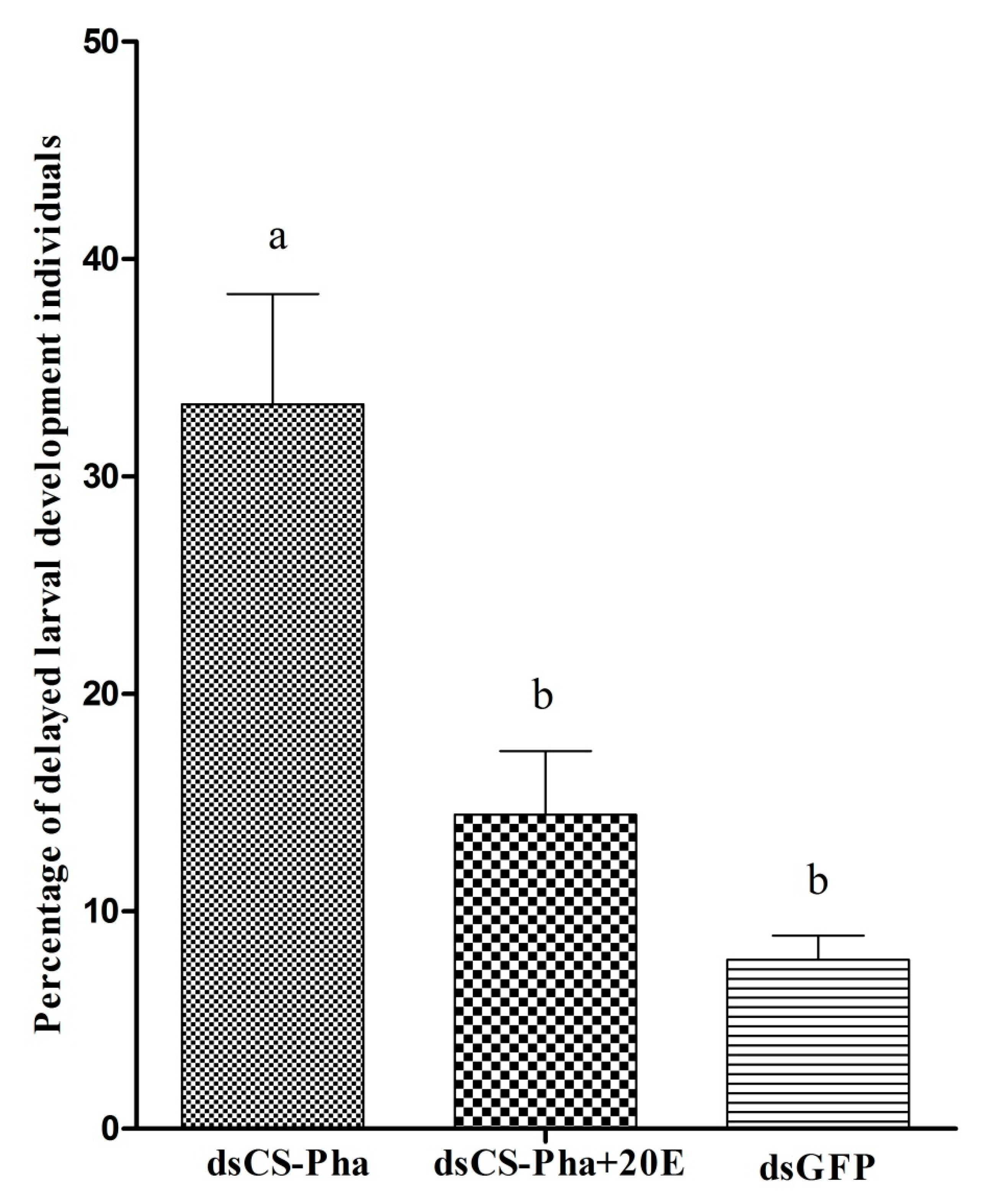
2.10. Rescue Analysis

We used 20E (Sigma, St. Louis, MO, USA) to rescue the dsCs-Phm phenotype. Topical application of a 0.25 μL aliquot of acetone with or without 20E (1.00 mg/mL) on the thoracic surface of the larvae was used on dsCs-Phm-injected larvae at 24 h post-injection. Each group had 40 individuals with three biological replicates.

2.11. Statistical Analysis

The data are presented as the means ± SE, analyzed by ANOVA, and compared by A Tucky–Kramer test using SPSS for Windows (SPSS Inc., Chicago, IL, USA).

3. Results

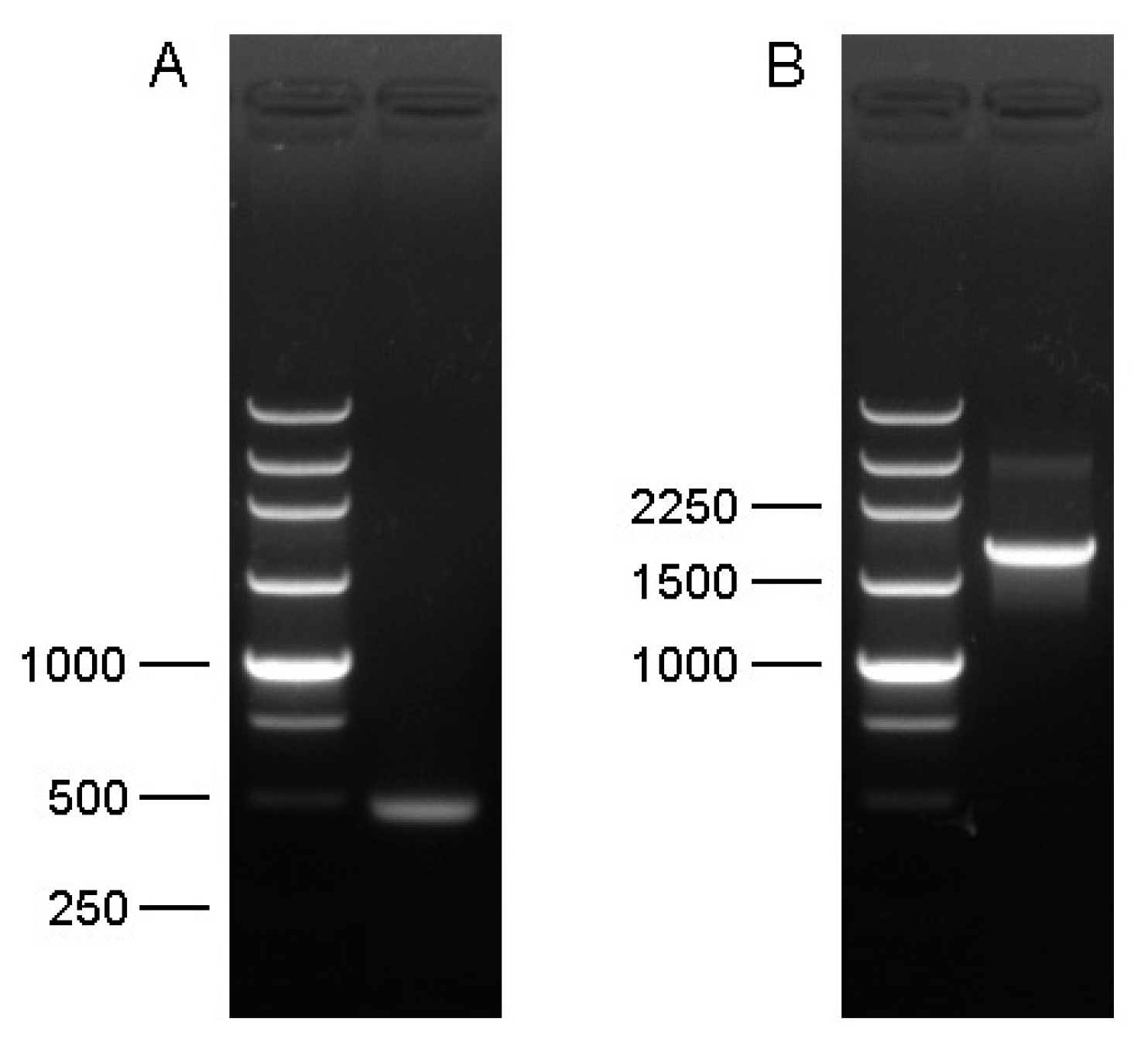
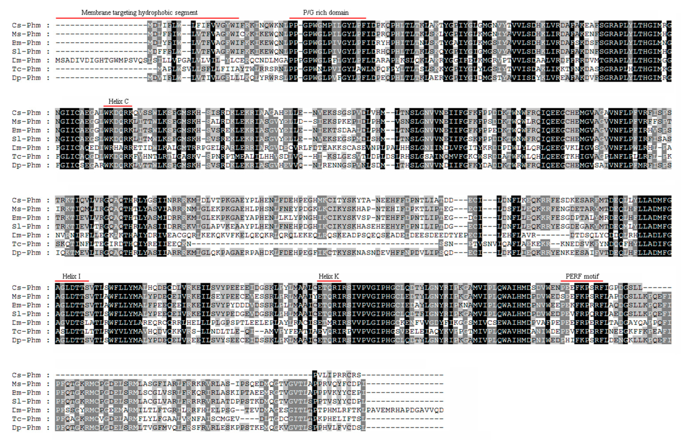
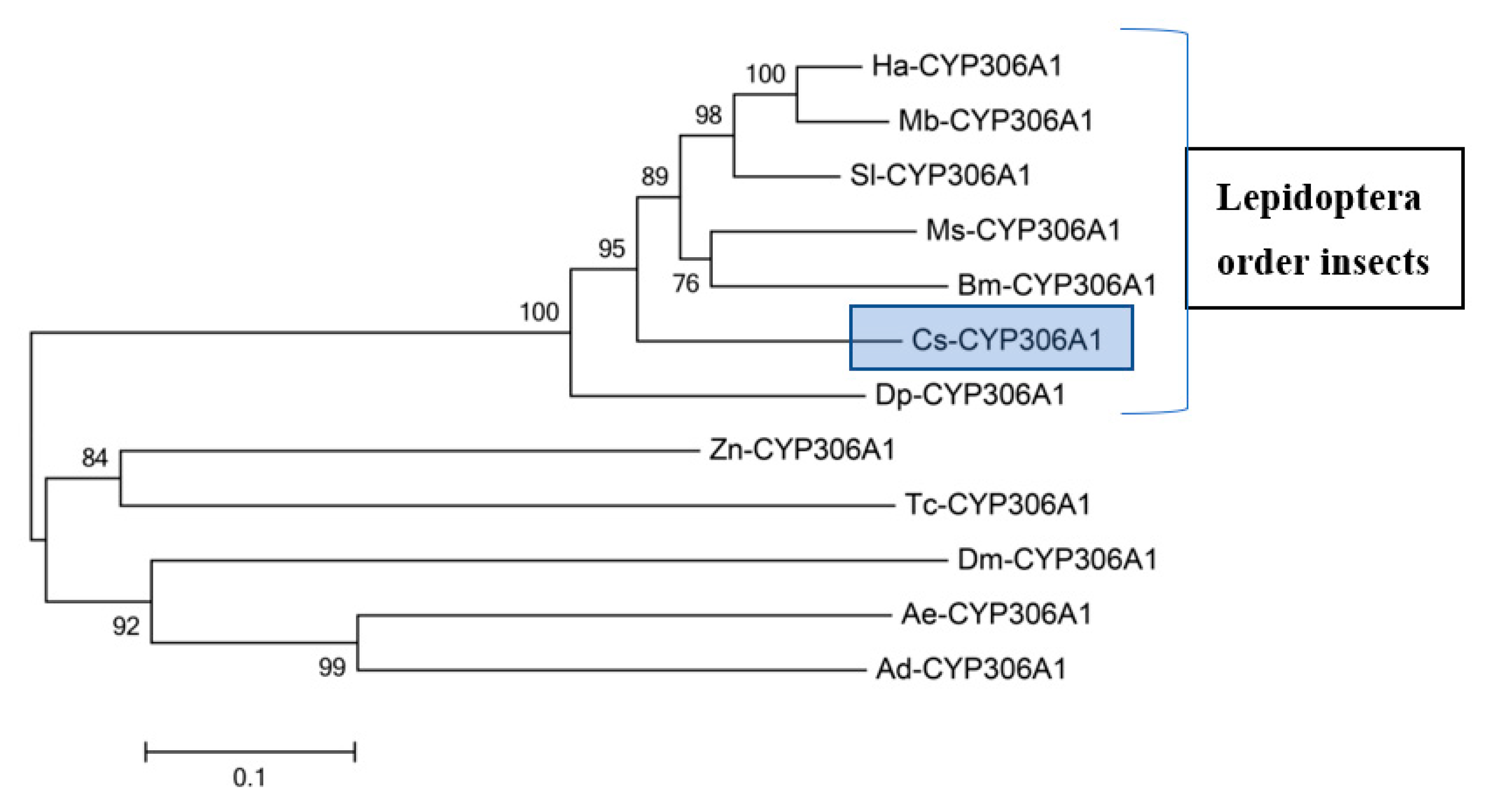
3.1. Molecular Cloning and Sequence Analysis of Cs-Phm

A full-length Cs-Phm sequence of a 2045 bp ORF encoding a protein of 478 amino acids was obtained through 5′ and 3′ RACE (Figure 1). To assign the specific name, we submitted it to the Nomenclature Committee for Standardized Cytochrome P450 [33]. Moreover, the resulting full-length sequence was submitted to the National Centre for Biotechnology (GenBank, NCBI) (accession number KF701127). Protein alignment of Cs-Phm with other insects′ known phantom proteins showed similar cytochrome P450 conserved motifs (Figure 2).
Insects Cytochrome P450 has five conserved P450 motifs: WxxxR (Helix-C), GxE.DTT/S (Helix-I) ExxR (Helix-K), PxxFxPxRF (PERF), and PFxxGxRxCxG/A (Heme-binding), where x means any amino acid [34]. We observed that the first motif WxxxR is located in helix-C, which is believed to form a charge pair with the propionate of the heme by arginine. The second conserved motif, AGxxT, is located in helix-I, which is used to transfer a proton groove on the distal side of heme. The third conserved P450 motif is located in helix-K (ExxR), which stabilizes the enzyme through a set of salt bridge interactions.
The PERF (PxxFxPxRF) motif is the fourth domain and is present in the sequence in an aromatic region. Moreover, PFxxGxRxCxG/A (heme-binding domain) serves as a ligand to heme iron by conserved cysteine [34]. The phylogenetic tree was constructed with the neighbor joining method in MEGA version 5.0 and CLUSTALX, which clearly shows high similarity (≥73%) and identity (≥63%) of Cs-Phm with insects belonging to the Lepidoptera order compared to others (Figure 3).

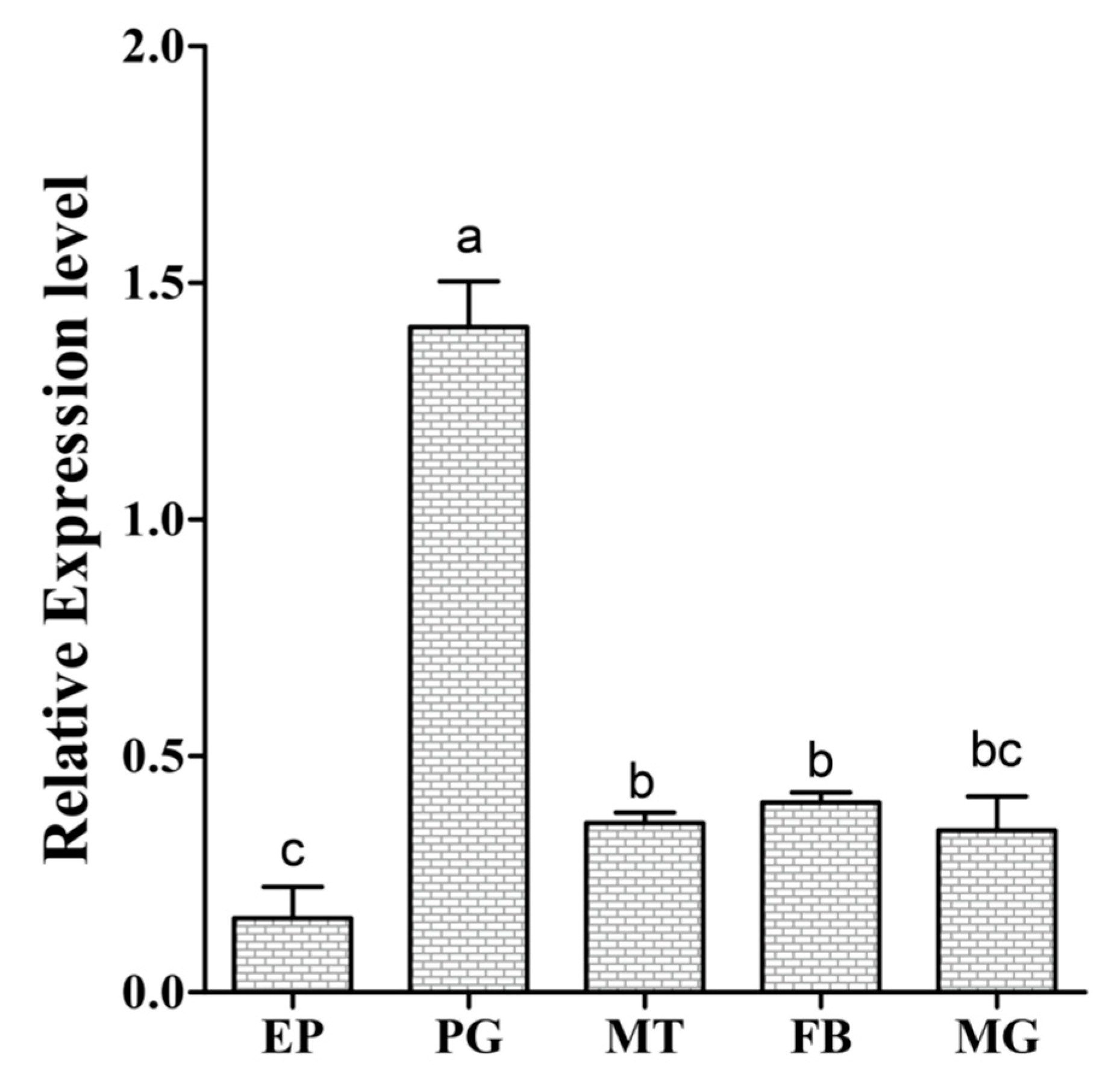
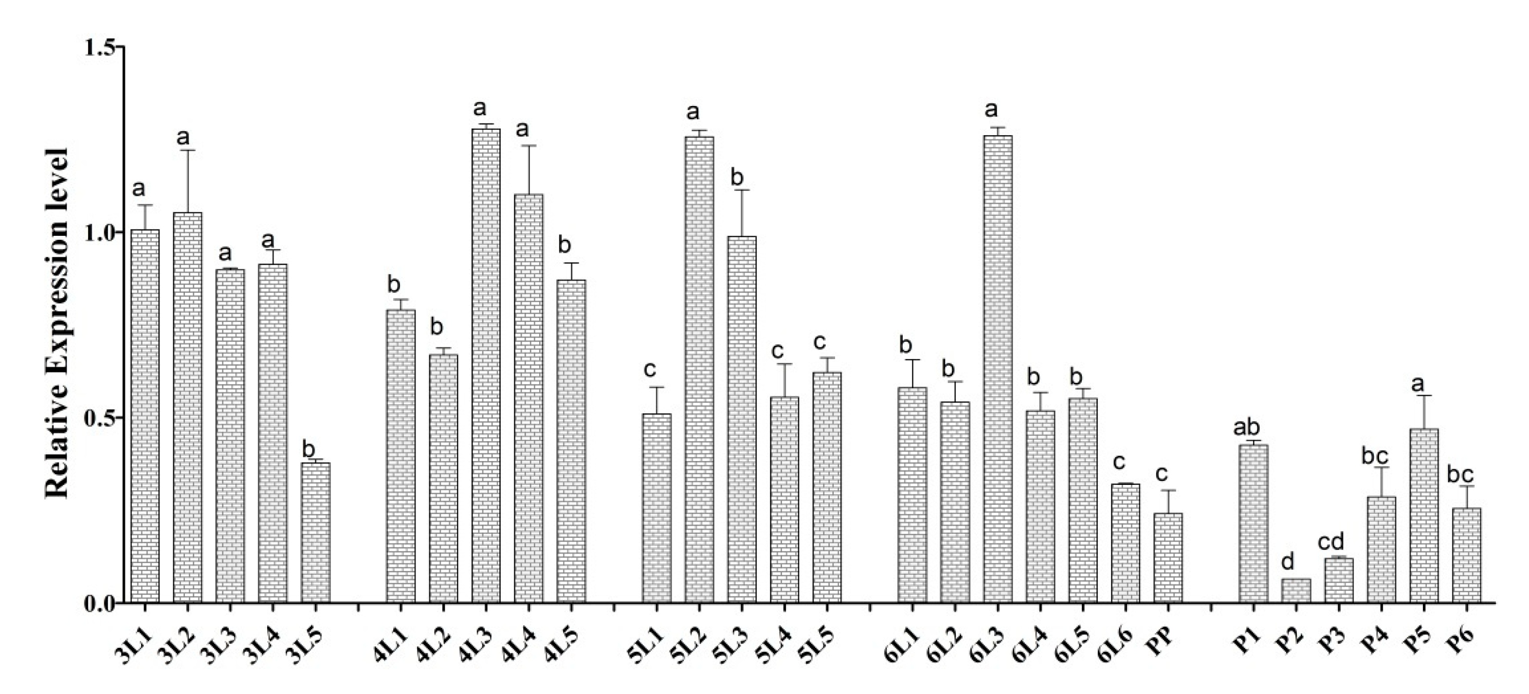
3.2. Spatial Transcript Profiles

qRT-PCR was used to understand the functional importance of Cs-Phm in the selected tissues. The relative spatial transcript profile from the epidermis tissues, prothoracic glands, midgut, Malpighian tubules, and body fat showed that Cs-phm is mainly expressed in the prothoracic glands (Figure 4). A low transcript level of Cs-phm also appeared in the midgut, Malpighian tubules, and body fat.

3.3. Temporal Transcript Profiles

The Cs-phm relative transcript was measured throughout 3th- to 6th-instar larval development and pupal development to determine the expression pattern at different developmental stages (3rd-, 4th-, 5th-, and 6th-instar larvae and pupae with average times of 5, 5, 5, 7, and 6 days at our insectary temperature, respectively). Cs-Phm was present throughout the larval development, but at the beginning, it had low expression during the larval instar and seemed to peak on days 3–4. It decreased again before the larvae molt. However, in the 3rd-instar larvae, the Cs-phm level was relatively high throughout the instar. A similar relative expression trend was observed throughout the third to sixth larval instars. In contrast to larvae, high expression was observed during the first day of pupal development, followed by a significant decrease and then a peak at day 4 before pupal transition (Figure 5).

3.4. Effects of dsRNA Injection

The efficacy of dsCs-Phm injection was robust (Figure 6). The relative expression level of Cs-Phm in dsCs-Phm-injected insects after 1, 2, 3, and 4 days was reduced by 64.91%, 85.11%, 67.56%, and 56.81%, respectively, compared to insects injected with dsGFP (Figure 6A). Mortality was calculated in fourth-instar larvae injected with dsCs-Phm and compared to the control group. We observed 61.67% mortality in the larvae injected with dsCs-Phm, while only 13.33% and 18.33% mortality was observed in the control and the group injected with dsGFP, respectively (Figure 6B). Larval development was also observed in the treated and control group. Larvae that were injected with dsCs-Phm showed significantly delayed larval development compared to the control group. The average fourth-instar larval stage was longer in the dsCs-Phm RNAi group than the controls, spanning ~ 7d rather than ~5 d (Figure 6C). The data clearly showed that larval development was significantly delayed in larvae injected with dsCs-Phm.

3.5. Ecdysteroid Titer Measurement

Larvae injected with dsCs-Phm showed significantly decreased ecdysteroid levels compared to the dsGFP-injected larvae in the control group (Figure 7). The highly decreased ecdysteroid titer clearly indicates the involvement of Cs-Phm in ecdysteroid biosynthesis.

3.6. Rescue Experiment

Topical application of 20E (Sigma, St. Louis, MO, USA) at 24 h post-injection on the thoracic surface of larvae was used to address whether delayed larval molting in dsCs-Phm larvae was due to decreased ecdysteroid titer. It was observed that larvae injected with dsCs-Phm had 33.32% delayed larval molting compared to the control group with dsGFP injection, which had only 7.78% delayed larval molting. The percentage of delayed larval molting was completely rescued with topical application of 20E (Sigma, St. Louis, MO, USA) at 24 h post-injection at 14.44%, which is similar to the control group with dsGFP injection (Figure 8).

4. Discussion

As metamorphosis and molting are fundamental phenomena during arthropod development, the Halloween genes in insects are well conserved [8,9,35,36,37,38,39], as in other arthropods [40,41]. In this report, we demonstrated the characterization and the relative transcript levels of Halloween gene Cs-Phm in the rice striped stem borer, C. suppressalis. It has been identified in several insects species that Halloween genes are involved in ecdysteroidogenesis [8,19,24,42]. Helix-C, Helix-I, Helix-K, PERF motif, and heme-binding domain are well-conserved typical cytochrome P450 motifs [43]. Previous studies have reported similar P450 motifs of Halloween genes in Lepidoptera, Diptera, Hemiptera, Hymenoptera, Orthoptera, and Coleoptera [20,35,36,37,39,42,44,45].
Cs-Phm has Helix-C, Helix-I, Helix-K, PERF, and heme-binding motifs. Consistent with the structural features, the similar P450 motifs in Cs-Phm are believed to have similar functions. Similarly sequences with an N-terminal part have microsomal enzyme character [34]. Moreover, phantom microsomal localization is also detected in phantom transfected Drosophila S2 cells [19]. Cs-Phm shows similar microsomal localization in sequence. This correlation strongly suggests that with the structural features and motifs, Cs-Phm is functionally conserved in the rice striped stem borer. Moreover, the spatial distribution of Cs-Phm supports our hypothesis that strong expression is limited in the prothoracic gland.
As in previous qRT-PCR and in situ hybridization studies, it has been described in different insects such as Drosophila melanogaster, Manduca sexta, Schistocerca gregaria, Bombyx mori, and Spodoptera littoralis that phantom Halloween genes in immature insect stages are highly expressed in the prothoracic gland cells [19,35,36]. The significantly high relative expression of Cs-Phm in the prothoracic gland strongly suggests that it is involved in ecdysteroids biosynthesis because it is a major site of ecdysone production.
Consistent with our results, Lepidoptera and Diptera insect species show similar relative expression [9,17,18,42,46]. Trace amounts of Cs-Phm transcripts were also found in other tissues besides PG, suggesting a possible additional role of this gene might in other physiological functions in insects [47,48,49]. Furthermore, the relative expression pattern of phantom coincides with circulating ecdysteroid quantity in the hemolymph [19]. Therefore, it can be speculated that the temporal expression pattern of phantom is related to the 20E titer in the hemolymph. The observed temporal transcript pattern of Cs-Phm in the rice striped stem borer reflects the repetitive surges of ecdysteroid biosynthesis that act to coordinate molting and metamorphosis. Consequently, it seemed reasonable to expect the functional importance of Cs-Phm for the biosynthesis of ecdysteroids.
To validate our hypothesis that Cs-Phm is involved in the biosynthesis of ecdysteroids, we downregulated it using RNAi to evaluate the possible effects on larval development. The target gene was effectively knocked down by injection of dsCs-Phm. RNAi knockdown significantly downregulated the relative transcript level in the dsCs-Phm-injected group which led to a significantly reduced ecdysteroid titer. Similar findings have also been reported of reduced ecdysteroid titers with the loss of Halloween enzymes [17,18,24,35,36,37,49,50]. We also noted that negative effects in immature stages, such as mortality and delayed larval development, were observed in dsCs-Phm-injected insects because of the reduced ecdysteroid titer. Similar findings have been documented to those of lepidopteran species Bombyx mori, C. suppressalis, and Plutella xylostella that downregulated Halloween genes animals resulted in longer developmental duration, lower pupation, phenotypic defects as well as mortality [21,51,52]. Moreover, these findings are similar to those of Wan et al., for hemipteran insects Laodelphax striatellus, where mortality and delayed developmental effects were found in downregulated Halloween gene animals [53].
Consistent with our speculation, similar findings were observed in Toxoneuron nigriceps with the parasitization of Heliothis virescens, where downregulation of Halloween gene induces downregulation of ecdysone production [54]. We also confirmed that the negative effects of dsCs-Phm-injected insects can be rescued by topical application of 20E. The observed insects were almost completely rescued by application of 20E, and negative effects such as delayed development were relieved.

5. Conclusions

The Halloween gene phantom is evolutionarily conserved in C. suppressalis. The spatio-temporal expression patterns and RNAi knockdown of Cs-Phm showed a significant correlation with the ecdysteroid titer. This suggests the importance of Cs-Phm in ecdysone biosynthesis by prothoracic glands. We expect that in future studies, knowledge of Cs-Phm will lead to practical application in managing this insect pest.

Author Contributions

Conceptualization, M.F.S. and A.I.; methodology, M.F.S., A.I. and J.I.; data curation, M.F.S., A.I., A.A.; formal analysis, M.F.S. and A.I.; funding acquisition, J.L. and A.I.; investigation, M.F.S. and A.I.; software, J.I., Z.A.Q. and A.A.; validation, J.I., Z.A.Q. and A.U.; project administration, J.L.; resources, A.U., A.A.K. and Z.A.Q.; visualizations; A.U. and A.A.K.; supervision, J.L. and A.I.; writing—original draft, M.F.S. and A.I.; writing—review and editing, M.F.S., A.I., A.A., Z.A.Q., A.A., A.A.K., J.I. and J.L. All authors have read and agreed to the published version of the manuscript.

Funding

This research study was funded by GDAS Special Project of Science and Technology Development (No. 2020GDASYL-20200301003), and GDAS Action Capital Project to build a comprehensive industrial technology innovation center (No. 2022GDASZH-2022010106).

Institutional Review Board Statement

Not applicable.

Informed Consent Statement

Not applicable.

Data Availability Statement

The data presented in the study are available in the article.

Conflicts of Interest

The authors declare no conflict of interest. The funders had no role in the design of the study; in the collection, analyses, or interpretation of data; in the writing of the manuscript, or in the decision to publish the results.

References

  1. Danielsen, E.T.; Moeller, M.E.; Rewitz, K.F. Chapter Two—Nutrient Signaling and Developmental Timing of Maturation. In Current Topics in Developmental Biology; Ann, E.R., Michael, B.O.C., Eds.; Academic Press: Cambridge, MA, USA, 2013; Volume 105, pp. 37–67. [Google Scholar]
  2. Miller, W.L.; Auchus, R.J. The molecular biology, biochemistry, and physiology of human steroidogenesis and its disorders. Endocr. Rev. 2010, 32, 81–151. [Google Scholar] [CrossRef]
  3. Thummel, C.S. Molecular Mechanisms of Developmental Timing in C. elegans and Drosophila. Dev. Cell 2001, 1, 453–465. [Google Scholar] [CrossRef]
  4. Gilbert, L.I.; Rybczynski, R.; Warren, J.T. Control and biochemical nature of the ecdysteroidogenic pathway. Annu. Rev. Entomol. 2002, 47, 883–916. [Google Scholar] [CrossRef] [PubMed]
  5. Thummel, C.S.; Chory, J. Steroid signaling in plants and insects—Common themes, different pathways. Genes Dev. 2002, 16, 3113–3129. [Google Scholar] [CrossRef] [PubMed]
  6. Schumann, I.; Kenny, N.; Hui, J.; Hering, L.; Mayer, G. Halloween genes in panarthropods and the evolution of the early moulting pathway in Ecdysozoa. R. Soc. Open Sci. 2018, 5, 180888. [Google Scholar] [CrossRef]
  7. Moulos, P.; Samiotaki, M.; Panayotou, G.; Dedos, S.G. Combinatory annotation of cell membrane receptors and signalling pathways of Bombyx mori prothoracic glands. Sci. Data 2016, 3, 1–12. [Google Scholar] [CrossRef]
  8. Petryk, A.; Warren, J.T.; Marqués, G.; Jarcho, M.P.; Gilbert, L.I.; Kahler, J.; Parvy, J.-P.; Li, Y.; Dauphin-Villemant, C.; O’Connor, M.B. Shade is the Drosophila P450 enzyme that mediates the hydroxylation of ecdysone to the steroid insect molting hormone 20-hydroxyecdysone. Proc. Natl. Acad. Sci. USA 2003, 100, 13773–13778. [Google Scholar] [CrossRef]
  9. Rewitz, K.; Rybczynski, R.; Warren, J.T.; Gilbert, L.I. The Halloween genes code for cytochrome P450 enzymes mediating synthesis of the insect moulting hormone. Biochem. Soc. Trans. 2006, 34, 1256–1260. [Google Scholar] [CrossRef]
  10. Lehmann, M. Endocrine and physiological regulation of neutral fat storage in Drosophila. Mol. Cell. Endocrinol. 2018, 461, 165–177. [Google Scholar] [CrossRef]
  11. Ruang-Rit, K.; Park, Y. Endocrine system in supernumerary molting of the flour beetle, Tribolium freemani, under crowded conditions. Insect Biochem. Mol. Biol. 2018, 101, 76–84. [Google Scholar] [CrossRef]
  12. Clark, A.; Bloch, K. The absence of sterol synthesis in insects. J. Biol. Chem. 1959, 234, 2578–2582. [Google Scholar] [CrossRef]
  13. Sumiya, E.; Ogino, Y.; Toyota, K.; Miyakawa, H.; Miyagawa, S.; Iguchi, T. Neverland regulates embryonic moltings through the regulation of ecdysteroid synthesis in the water flea Daphnia magna, and may thus act as a target for chemical disruption of molting. J. Appl. Toxicol. 2016, 36, 1476–1485. [Google Scholar] [CrossRef]
  14. Warren, J.T.; O’Connor, M.B.; Gilbert, L.I. Studies on the black box: Incorporation of 3-oxo-7-dehydrocholesterol into ecdysteroids by Drosophila melanogaster and Manduca sexta. Insect Biochem. Mol. Biol. 2009, 39, 677–687. [Google Scholar] [CrossRef] [PubMed]
  15. Gilbert, L.I.; Warren, J.T. A Molecular Genetic Approach to the Biosynthesis of the Insect Steroid Molting Hormone. In Vitamins & Hormones; Gerald, L., Ed.; Academic Press: Cambridge, MA, USA, 2005; Volume 73, pp. 31–57. [Google Scholar]
  16. Chávez, V.M.; Marqués, G.; Delbecque, J.P.; Kobayashi, K.; Hollingsworth, M.; Burr, J.; Natzle, J.E.; O’Connor, M.B. The Drosophila disembodied gene controls late embryonic morphogenesis and codes for a cytochrome P450 enzyme that regulates embryonic ecdysone levels. Development 2000, 127, 4115–4126. [Google Scholar] [CrossRef] [PubMed]
  17. Warren, J.T.; Petryk, A.; Marqués, G.; Jarcho, M.; Parvy, J.-P.; Dauphin-Villemant, C.; O’Connor, M.B.; Gilbert, L.I. Molecular and biochemical characterization of two P450 enzymes in the ecdysteroidogenic pathway of Drosophila melanogaster. Proc. Natl. Acad. Sci. USA 2002, 99, 11043–11048. [Google Scholar] [CrossRef] [PubMed]
  18. Niwa, R.; Matsuda, T.; Yoshiyama, T.; Namiki, T.; Mita, K.; Fujimoto, Y.; Kataoka, H. CYP306A1, a cytochrome P450 enzyme, is essential for ecdysteroid biosynthesis in the prothoracic glands of Bombyx and Drosophila. J. Biol. Chem. 2004, 279, 35942–35949. [Google Scholar] [CrossRef]
  19. Warren, J.T.; Petryk, A.; Marqués, G.; Parvy, J.-P.; Shinoda, T.; Itoyama, K.; Kobayashi, J.; Jarcho, M.; Li, Y.; O’Connor, M.B. Phantom encodes the 25-hydroxylase of Drosophila melanogaster and Bombyx mori: A P450 enzyme critical in ecdysone biosynthesis. Insect Biochem. Mol. Biol. 2004, 34, 991–1010. [Google Scholar] [CrossRef]
  20. Namiki, T.; Niwa, R.; Sakudoh, T.; Shirai, K.-i.; Takeuchi, H.; Kataoka, H. Cytochrome P450 CYP307A1/Spook: A regulator for ecdysone synthesis in insects. Biochem. Biophys. Res. Commun. 2005, 337, 367–374. [Google Scholar] [CrossRef]
  21. Shahzad, M.F.; Li, Y.; Ge, C.; Sun, Y.; Yang, Q.; Li, F. Knockdown of Cs-Spook induces delayed larval molting in Rice Striped Stem Borer Chilo suppressalis. Arch. Insect Biochem. Physiol. 2014, 88, 179–191. [Google Scholar] [CrossRef]
  22. Wan, P.J.; Jia, S.; Li, N.; Fan, J.M.; Li, G.Q. A Halloween gene shadow is a potential target for RNA-interference-based pest management in the small brown planthopper Laodelphax striatellus. Pest Manag. Sci. 2015, 71, 199–206. [Google Scholar] [CrossRef]
  23. Rewitz, K.F.; O’Connor, M.B.; Gilbert, L.I. Molecular evolution of the insect Halloween family of cytochrome P450s: Phylogeny, gene organization and functional conservation. Insect Biochem. Mol. Biol. 2007, 37, 741–753. [Google Scholar] [CrossRef] [PubMed]
  24. Zhang, X.Y.; He, Q.H.; Zhang, T.T.; Wu, H.H.; Zhang, J.Z.; Ma, E.B. Characteristics of Halloween genes and RNA interference-mediated functional analysis of LmCYP307a2 in Locusta migratoria. Insect Sci. 2022, 29, 51–64. [Google Scholar] [CrossRef]
  25. Yin, C.; Liu, Y.; LIu, J.; Xiao, H.; Huang, S.; Lin, Y.; Han, Z.; Li, F. ChiloDB: A genomic and transcriptome database for an important rice insect pest Chilo suppressalis. Database 2014, 2014, bau065. [Google Scholar] [CrossRef]
  26. Ren, L.; Zhu, B.; Zhang, Y.; Wang, H.; Li, C.; Su, Y.; Ba, C. The research of applying primer premier 5.0 to design PCR primer. J. Jinzhou Med. Coll. 2004, 25, 43–46. [Google Scholar]
  27. Tamura, K.; Peterson, D.; Peterson, N.; Stecher, G.; Nei, M.; Kumar, S. MEGA5: Molecular evolutionary genetics analysis using maximum likelihood, evolutionary distance, and maximum parsimony methods. Mol. Biol. Evol. 2011, 28, 2731–2739. [Google Scholar] [CrossRef] [PubMed]
  28. Teng, X.; Zhang, Z.; He, G.; Yang, L.; Li, F. Validation of reference genes for quantitative expression analysis by real-time RT-PCR in four lepidopteran insects. J. Insect Sci. 2012, 12, 60. [Google Scholar] [CrossRef]
  29. Pfaffl, M.W. A new mathematical model for relative quantification in real-time RT–PCR. Nucleic Acids Res. 2001, 29, e45. [Google Scholar] [CrossRef]
  30. Livak, K.J.; Schmittgen, T.D. Analysis of relative gene expression data using real-time quantitative PCR and the 2−ΔΔCT method. Methods 2001, 25, 402–408. [Google Scholar] [CrossRef]
  31. Dinan, L.N.; Donnahey, P.L.; Rees, H.H.; Goodwin, T.W. High-performance liquid chromatography of ecdysteroids and their 3-epi, 3-dehydro and 26-hydroxy derivatives. J. Chromatogr. A 1981, 205, 139–145. [Google Scholar] [CrossRef]
  32. Zhou, J.; Qi, Y.; Hou, Y.; Zhao, J.; Li, Y.; Xue, X.; Wu, L.; Zhang, J.; Chen, F. Quantitative determination of juvenile hormone III and 20-hydroxyecdysone in queen larvae and drone pupae of Apis mellifera by ultrasonic-assisted extraction and liquid chromatography with electrospray ionization tandem mass spectrometry. J. Chromatogr. B 2011, 879, 2533–2541. [Google Scholar] [CrossRef]
  33. Nelson, D.R. Cytochrome P450 and the individuality of species. Arch. Biochem. Biophys. 1999, 369, 1–10. [Google Scholar] [CrossRef] [PubMed]
  34. Werck-Reichhart, D.; Feyereisen, R. Cytochromes P450: A success story. Genome Biol. 2000, 1, 3003.3001–3003.3009. [Google Scholar] [CrossRef] [PubMed]
  35. Iga, M.; Smagghe, G. Identification and expression profile of Halloween genes involved in ecdysteroid biosynthesis in Spodoptera littoralis. Peptides 2010, 31, 456–467. [Google Scholar] [CrossRef] [PubMed]
  36. Marchal, E.; Badisco, L.; Verlinden, H.; Vandersmissen, T.; Van Soest, S.; Van Wielendaele, P.; Vanden Broeck, J. Role of the Halloween genes, Spook and Phantom in ecdysteroidogenesis in the desert locust, Schistocerca gregaria. J. Insect Physiol. 2011, 57, 1240–1248. [Google Scholar] [CrossRef] [PubMed]
  37. Christiaens, O.; Iga, M.; Velarde, R.; Rougé, P.; Smagghe, G. Halloween genes and nuclear receptors in ecdysteroid biosynthesis and signalling in the pea aphid. Insect Mol. Biol. 2010, 19, 187–200. [Google Scholar] [CrossRef]
  38. Rewitz, K.F.; Rybczynski, R.; Warren, J.T.; Gilbert, L.I. Developmental expression of Manduca shade, the P450 mediating the final step in molting hormone synthesis. Mol. Cell. Endocrinol. 2006, 247, 166–174. [Google Scholar] [CrossRef]
  39. Yamazaki, Y.; Kiuchi, M.; Takeuchi, H.; Kubo, T. Ecdysteroid biosynthesis in workers of the European honeybee Apis mellifera L. Insect Biochem. Mol. Biol. 2011, 41, 283–293. [Google Scholar] [CrossRef]
  40. Rewitz, K.F.; Gilbert, L.I. Daphnia Halloween genes that encode cytochrome P450s mediating the synthesis of the arthropod molting hormone: Evolutionary implications. BMC Evol. Biol. 2008, 8, 60. [Google Scholar] [CrossRef]
  41. Iga, M.; Kataoka, H. Recent studies on insect hormone metabolic pathways mediated by cytochrome P450 enzymes. Biol. Pharm. Bull. 2012, 35, 838–843. [Google Scholar] [CrossRef]
  42. Ono, H.; Rewitz, K.F.; Shinoda, T.; Itoyama, K.; Petryk, A.; Rybczynski, R.; Jarcho, M.; Warren, J.T.; Marqués, G.; Shimell, M.J. Spook and Spookier code for stage-specific components of the ecdysone biosynthetic pathway in Diptera. Dev. Biol. 2006, 298, 555–570. [Google Scholar] [CrossRef]
  43. Feyereisen, R. Insect cytochrome P450. Compr. Mol. Insect Sci. 2005, 4, 1–77. [Google Scholar]
  44. Luan, J.-B.; Ghanim, M.; Liu, S.-S.; Czosnek, H. Silencing the ecdysone synthesis and signaling pathway genes disrupts nymphal development in the whitefly. Insect Biochem. Mol. Biol. 2013, 43, 740–746. [Google Scholar] [CrossRef]
  45. Hentze, J.L.; Moeller, M.E.; Jørgensen, A.F.; Bengtsson, M.S.; Bordoy, A.M.; Warren, J.T.; Gilbert, L.I.; Andersen, O.; Rewitz, K.F. Accessory gland as a site for prothoracicotropic hormone controlled ecdysone synthesis in adult male insects. PLoS ONE 2013, 8, e55131. [Google Scholar] [CrossRef] [PubMed]
  46. Niwa, R.; Sakudoh, T.; Namiki, T.; Saida, K.; Fujimoto, Y.; Kataoka, H. The ecdysteroidogenic P450 Cyp302a1/disembodied from the silkworm, Bombyx mori, is transcriptionally regulated by prothoracicotropic hormone. Insect Mol. Biol. 2005, 14, 563–571. [Google Scholar] [CrossRef] [PubMed]
  47. Scott, J.G. Cytochromes P450 and insecticide resistance. Insect Biochem. Mol. Biol. 1999, 29, 757–777. [Google Scholar] [CrossRef]
  48. Bernhardt, R. Cytochromes P450 as versatile biocatalysts. J. Biotechnol. 2006, 124, 128–145. [Google Scholar] [CrossRef]
  49. Zhou, X.; Ye, Y.-Z.; Ogihara, M.H.; Takeshima, M.; Fujinaga, D.; Liu, C.-W.; Zhu, Z.; Kataoka, H.; Bao, Y.-Y. Functional analysis of ecdysteroid biosynthetic enzymes of the rice planthopper, Nilaparvata lugens. Insect Biochem. Mol. Biol. 2020, 123, 103428. [Google Scholar] [CrossRef]
  50. Yoshiyama-Yanagawa, T.; Enya, S.; Shimada-Niwa, Y.; Yaguchi, S.; Haramoto, Y.; Matsuya, T.; Shiomi, K.; Sasakura, Y.; Takahashi, S.; Asashima, M. The conserved Rieske oxygenase DAF-36/Neverland is a novel cholesterol-metabolizing enzyme. J. Biol. Chem. 2011, 286, 25756–25762. [Google Scholar] [CrossRef]
  51. Peng, L.; Wang, L.; Zou, M.-M.; Vasseur, L.; Chu, L.-N.; Qin, Y.-D.; Zhai, Y.-L.; You, M.-S. Identification of halloween genes and RNA interference-mediated functional characterization of a Halloween gene Shadow in Plutella xylostella. Front. Physiol. 2019, 10, 1120. [Google Scholar] [CrossRef]
  52. Niwa, R.; Namiki, T.; Ito, K.; Shimada-Niwa, Y.; Kiuchi, M.; Kawaoka, S.; Kayukawa, T.; Banno, Y.; Fujimoto, Y.; Shigenobu, S. Non-molting glossy/shroud encodes a short-chain dehydro genase/reductase that functions in the ‘Black Box’of the ecdysteroid biosynthesis pathway. Development 2010, 137, 1991–1999. [Google Scholar] [CrossRef]
  53. Wan, P.-J.; Jia, S.; Li, N.; Fan, J.-M.; Li, G.-Q. RNA interference depletion of the Halloween gene disembodied implies its potential application for management of planthopper Sogatella furcifera and Laodelphax striatellus. PLoS ONE 2014, 9, e86675. [Google Scholar] [CrossRef] [PubMed]
  54. Salvia, R.; Nardiello, M.; Scieuzo, C.; Scala, A.; Bufo, S.A.; Rao, A.; Vogel, H.; Falabella, P. Novel factors of viral origin inhibit TOR pathway gene expression. Front. Physiol. 2018, 9, 1678. [Google Scholar] [CrossRef] [PubMed]
Figure 1. Elelctrophoretic analysis of (A) partial fragment and (B) end-to-end RT-PCR for full-length transcript of Cs-Phm.
Insects 13 00731 g001
Figure 2. Alignment of CYP306A1 sequences originating from C. suppressalis (KF701127), Ms—Manduca sexta (ABC96068), Bm—Bombyx mori (BAM73853), Sl—Spodoptera littoralis (ACM45975), Dm—Drosophila melanogaster (NP573319), Tc—Tribolium castaneum (XP968477) and Dp—Danaus plexippus (EHJ76071). Amino acids shaded in black, dark grey, and light grey demonstrated 100%, >80%, and >60% conservation, respectively.
Insects 13 00731 g002
Figure 3. Phylogenetic tree of a Halloween gene Cs-Phm. The tree was generated using MEGA version 5.0 with a bootstrap value of 1000 trials for each branch position based on the whole amino acid sequences by neighbor-joining method excluding the gap position. Phantom-like proteins originate from Sl—Spodoptera littoralis (ACM45975), Ha—Helicoverpa armigera (AID54855), Mb—Mamestra brassicae (BAN66311), Ms—Manduca sexta (ABC96068), Bm—Bombyx mori (BAM73853), Dp—Danaus plexippus (EHJ76071), Zn—Zootermopsis nevadensis (KDR21920), Ae—Aedes aegypti (EAT43717), Ad—Anopheles darling (ENT60005), Dm—Drosophila melanogaster (NP573319) and Tc—Tribolium castaneum (XP968477). The percentiles of bootstrap values (1000 replicates) are indicated. The amino acid divergence is shown in the scale bar.
Insects 13 00731 g003
Figure 4. The relative Cs-Phm transcript level measured in various larval tissues using qRT-PCR. The larval tissues (EP—epidermis; PG—prothoracic glands; MT—Malpighian tubules; FB—body fat; and MG—midgut) were dissected from 4th day of 5th-instar larvae. The columns indicate average values, and vertical bars represent SE. Different letters indicate significant differences at p value < 0.05.
Insects 13 00731 g004
Figure 5. The relative Cs-phm transcript level measured throughout 3rd- to 6th-instar larvae and pupae after 24 h intervals using qRT-PCR. The columns indicate average values, and vertical bars represent SE. Different letters indicate significant differences at p value < 0.05.
Insects 13 00731 g005
Figure 6. Effects of dsCs-Phm injection on the (A) relative expression of Cs-phm (B) larval survival (C) and larval development of rice striped stem borer, C. suppressalis. dsCs-Phm was injected on day 2 in 4th-instar larvae. The relative transcript level of Cs-Phm was measured after 1, 2, 3, and 4 days after dsCs-Phm injection. Each sample collected for Cs-Phm relative expression measurement contained two larvae. The larval survival and larval development in 4th-instar larvae was calculated from three batches with 40 individuals in each batch. The columns indicate average values, and vertical bars represent SE. ** and *** indicate statistical differences between treatments at p < 0.01 and p < 0.001, respectively. Different letters in vertical bars represent significant differences at p value < 0.05.
Insects 13 00731 g006
Figure 7. Ecdysteroid titers of dsGFP and dsCs-Phm-injected larvae measured with HPLC. The columns indicate average values, and vertical bars represent SE. *** indicates statistical differences between treatments at d p < 0.001.
Insects 13 00731 g007
Figure 8. Delayed larval individuals subjected to dsCs-Phm, dsCs-Phm +20E, and dsGFP. The columns indicate average values, and vertical bars represent SE. Letters above the bars indicate statistical differences between treatments at p < 0.05.
Insects 13 00731 g008
Publisher’s Note: MDPI stays neutral with regard to jurisdictional claims in published maps and institutional affiliations.

