Article

Phenotypic Plasticity of the Mimetic Swallowtail Butterfly Papilio polytes: Color Pattern Modifications and Their Implications in Mimicry Evolution

The BCPH Unit of Molecular Physiology, Department of Chemistry, Biology and Marine Science, Faculty of Science, University of the Ryukyus, Okinawa 903-0213, Japan
*
Author to whom correspondence should be addressed.
Insects 2022, 13(7), 649; https://doi.org/10.3390/insects13070649
Submission received: 16 June 2022 / Revised: 5 July 2022 / Accepted: 16 July 2022 / Published: 19 July 2022
(This article belongs to the Special Issue Phenotypic Plasticity of Insects)

Abstract

:

Simple Summary

Diverse butterfly wing color patterns are evolutionary products in response to environmental changes in the past. Environmental stress, such as temperature shock, is known to induce color pattern modifications in various butterfly species, and this phenotypic plasticity plays an important role in color pattern evolution. However, the potential contributions of phenotypic plasticity to mimicry evolution have not been evaluated. Here, we focused on the swallowtail butterfly Papilio polytes, which has nonmimetic and mimetic forms in females, to examine its plastic phenotypes. Cold shock and heat shock treatments in the nonmimetic form induced color pattern modifications that were partly similar to those of the mimetic form, and nonmimetic females were more sensitive than males and mimetic females. These results suggest that phenotypic plasticity in nonmimetic females might have provided a basis of natural selection for mimetic color patterns during evolution.

Abstract

Butterfly wing color patterns are sensitive to environmental stress, such as temperature shock, and this phenotypic plasticity plays an important role in color pattern evolution. However, the potential contributions of phenotypic plasticity to mimicry evolution have not been evaluated. Here, we focused on the swallowtail butterfly Papilio polytes, which has nonmimetic and mimetic forms in females, to examine its plastic phenotypes. In the nonmimetic form, medial white spots and submarginal reddish spots in the ventral hindwings were enlarged by cold shock but were mostly reduced in size by heat shock. These temperature-shock-induced color pattern modifications were partly similar to mimetic color patterns, and nonmimetic females were more sensitive than males and mimetic females. Unexpectedly, injection of tungstate, a known modification inducer in nymphalid and lycaenid butterflies, did not induce any modification, but fluorescent brightener 28, another inducer discovered recently, induced unique modifications. These results suggest that phenotypic plasticity in nonmimetic females might have provided a basis of natural selection for mimetic color patterns during evolution.

1. Introduction

The phenotypes of organisms are determined by genotypes and environmental factors. Most, if not all, organisms show phenotypic plasticity in response to environmental stress during development. The contributions of phenotypic plasticity to natural selection, genetic assimilation, and speciation have been an active research area in ecology, evolutionary biology, and developmental biology [1,2,3,4,5,6,7,8]. There are at least two different types of phenotypic plasticity, “developmental conversion” and “phenotypic modulation” (sensu Smith-Gill (1983) [8]), although their distinctions may not always be clear [7,8].
Representative cases of developmental conversion are found in many insects, such as social castes in bees, ants, and termites, solitarious and gregarious forms of locusts, large and small horns with various shapes in beetles, winged and wingless forms in aphids, and seasonal color pattern polyphenism in butterflies [6,7,9]. In some butterflies, such as Araschnia levana in Europe and Asia, Precis octavia in Africa, and Junonia almana in Southeast Asia, wing color patterns differ drastically in different seasons [9]. The seasonal polyphenism in butterflies is controlled by ecdysteroid hormones [9,10,11,12]. Developmental conversion is understood as the expression of genetically determined alternative pathways in response to environmental cues such as temperature and light conditions. This type of plasticity is supposed to be adaptive as an evolutionary consequence. Similarly, there are numerous cases of genetically determined polymorphism of color patterns in butterflies, among which sexual dimorphism is well-known [13,14]. Furthermore, females often have two or multiple forms within a species. Such phenotypic variation within a species is supposed to be adaptive and is often controlled by regulatory genes that are not programmed to respond to environmental cues. A good example is a mimetic swallowtail, the common Mormon butterfly Papilio polytes, distributed widely in Southeast Asia [15,16,17].
Butterflies also show phenotypic modulation, which is the topic of the present study. This phenotypic plasticity is not adaptive at least at first and is likely an expression of a system failure during development in response to severe environmental conditions. In other words, this phenotypic plasticity was not built in as alternative genetic pathways. Nonetheless, this system failure is mild and completely viable. In butterflies, phenotypic modulation is known to cause two types of color pattern modifications: modifications induced by temperature shock and those induced via the general stress response [18,19,20]. Studies of color pattern modifications induced by temperature shock from evolutionary and developmental viewpoints were pioneered by Shapiro (1980, 1981) [21,22] and Nijhout (1984) [9,23]. Subsequently, Otaki (1998) [24] discovered that similar, if not identical, color pattern modifications can be induced by injections of oxyanions (such as sodium tungstate) into pupae. Other modification inducers, such as acid carboxypeptidase [25] and heparin and other sulfated polysaccharides [26], were also discovered later. These modification inducers or temperature shock treatments have been used for many lepidopteran species to understand developmental physiological aspects of color pattern formation [27,28,29,30,31,32,33]. These temperature-shock-type (TS-type) modifications appear to be induced via a systemic factor (i.e., a cold shock hormone) [24,34] as well as a local factor (i.e., an extracellular matrix) [24,35,36].
Importantly, color patterns of TS-type modifications of a given species are often similar to natural color patterns of different but related species in Nymphalidae, as discussed in Nymphalis [21,22] and Junonia [9,37]. The evolution of Vanessa (sensu stricto) nicely parallels the degrees of color pattern “modifications”, suggesting that environmental temperature shock might have played an important role in color pattern evolution and speciation in this genus [38,39,40]. Color pattern modifications induced via the general stress response also appear to have contributed to the evolution of Vanessa (Cynthia) kershawi [41]. Lycaenid butterflies are also modified [42], and color pattern modifications in the pale grass blue butterfly in the field may be considered a real-time evolution of color patterns [43] and may be one of the best cases of genetic assimilation in the field [5]. A similar case is also found in the pierid butterfly Colias erate [27]. These nymphalid, lycaenid, and pierid cases of phenotypic plasticity have provided evidence for the contribution of phenotypic plasticity to the color pattern evolution of butterflies. However, to our knowledge, no papilionid cases of phenotypic plasticity that might have contributed to color pattern evolution have been reported, and potential contributions of phenotypic plasticity to the evolution of dimorphic or multimorphic color patterns within a species, such as mimetic and nonmimetic forms of P. polytes, have not yet been evaluated.
The female-limited mimicry not only in P. polytes but also in many butterflies has been explained by the high cost of expressing mimetic color patterns that can compensate for predation, which is not affordable for males [44,45,46,47,48,49]. While this line of explanation is reasonable, it does not take phenotypic plasticity into account. Similarly, although genes involved in the mimetic phenotype in P. polytes have been identified as doublesex (dsx) [50,51,52,53,54] and other genes, such as cortex [55], the question of how mimicry evolved in the first place requires multiple approaches, including studies on phenotypic plasticity. Here, we focused on the mimetic swallowtail butterfly, the common Mormon, P. polytes, in Okinawa, Japan [15,16,17], to examine its phenotypic plasticity. In Okinawa, this butterfly mimics the unpalatable swallowtail butterfly Pachliopta aristolochiae, distributed sympatrically (Figure 1). While males of P. polytes have a single nonmimetic form, females have a mimetic form (forma polytes) and a nonmimetic form (forma cyrus) (Figure 1). In the present study, we obtained color pattern modifications in this butterfly induced by cold shock or heat shock. Additionally, we injected a known modification inducer, sodium tungstate [24], and a recently discovered inducer, fluorescent brightener 28 (FB28) [56]. The induced modifications were compared to the mimetic color patterns, and we discussed the possible origin of the mimetic color patterns in this butterfly from the viewpoint of phenotypic plasticity.

2. Materials and Methods

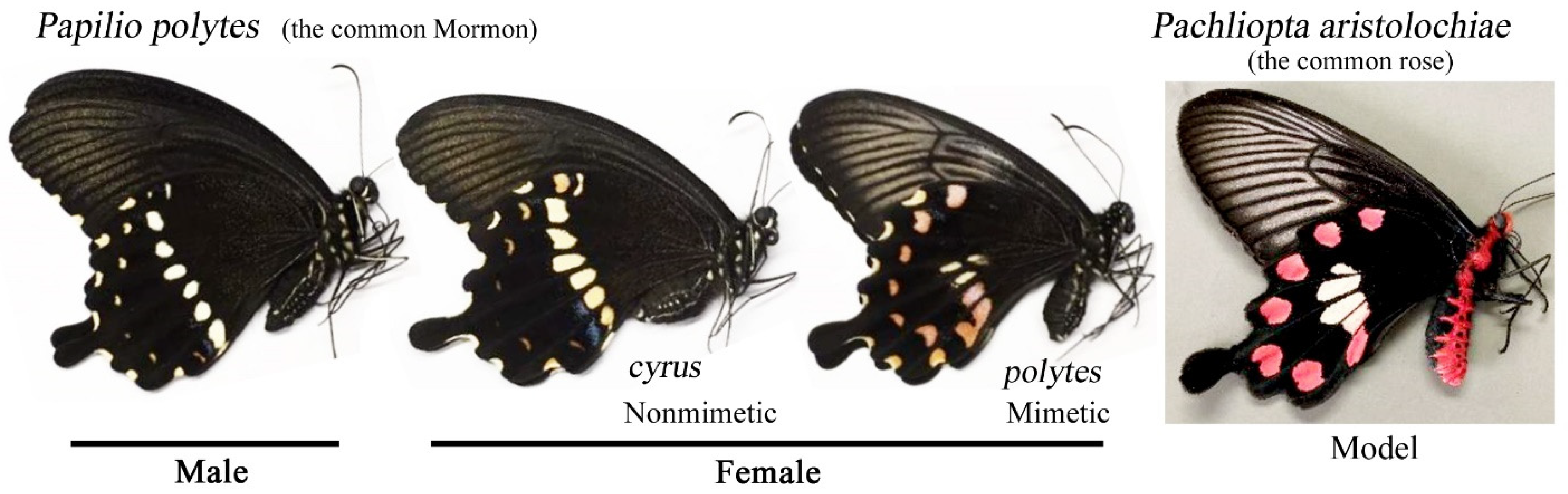
2.1. Nomenclature of Spots and Key Differences between Nonmimetic and Mimetic Forms

We focused on the ventral hindwings of P. polytes in this study because it was in the ventral hindwings that color pattern modifications were mainly observed. An exception is the ventral forewings, which showed an increase in sparse yellow scales. The nomenclature of the spots in the ventral hindwings in the present study is shown in Figure 2a. Seven submarginal reddish spots (SRS1–SRS7) and seven medial white spots (MWS1–MWS7) together with one or two medial blue spots (MBS1–MBS2) are the main color patterns of males and nonmimetic females. Marginal white spots (occasionally slightly reddish) were not examined. The SRSs are often simply called the “red spots” in other studies, but their color is closer to reddish brown and is clearly different from that of the “red spots” in the model species P. aristolochiae. Indeed, pigments in these two species are chemically different [57]. Therefore, these submarginal spots in P. polytes are called the “reddish spots” in this paper. The MBSs show structural color and may be sparse patterns [9]. In mimetic females, seven SRSs are maintained but are larger in size and darker in color than those in nonmimetic females. In contrast, the MWSs 1, 5, 6, and 7 are absent in mimetic females. MBS1 is also absent. Additionally, two medial reddish spots (MRS1 and MRS2) and the central white spot (CWS) are present only in mimetic females.
Key differences between the nonmimetic and mimetic forms are summarized in Figure 2b. There are three points. First, the SRSs, especially the most anterior one, are different in size and darkness: they are smaller in size and lighter in color in the nonmimetic form than in the mimetic form. Second, the MWSs are different: anterior ones exist in the nonmimetic form but do not exist in the mimetic form. Third, the MRSs are different: they do not exist in the nonmimetic form and exist in the mimetic form. The white spots near the MRSs are compromised in the mimetic form.
In this and many other papilionid butterflies, the background is dark-colored, and spots are light-colored. This coloration seems to be inconsistent with the binary color rule [30,31,58]. The binary color rule posits that color pattern elements are expressed in dark colors, and the background is expressed in light colors. Mechanistically, the induction model for color pattern determination states that dark (activator) signals always accompany light (inhibitor) signals and that light signals may behave independently [58,59,60]. This issue can be resolved by introducing a reversible positive–negative relationship, but the detailed explanations await future studies. Therefore, at present, we did not call these spots in P. polytes elements.

2.2. Butterflies and the Host Plant Leaves

Nonmimetic and mimetic females of P. polytes were caught in Shiohira (Itoman City), Maesato (Itoman City), Yamashiro (Itoman City), and Senbaru (Nishihara Town), Okinawa Prefecture, for egg collection in the laboratory from May 2020 to October 2021. These localities are all located in the southern region of Okinawa-jima Island. No clear difference in wing color patterns was observed among these females except for the nonmimetic and mimetic forms. Only individuals with low levels of wing damage were used to collect approximately 100 eggs per female individual. Egg collection was started on the same day as collection in the field.
One of the host plants of this butterfly in Okinawa, Toddalia asiatica, was exclusively used for egg collection and rearing. This plant is a small bush tree often found in the seacoast. Leaves were collected from various sites in Itoman City and from the Nishihara Campus of the University of the Ryukyus. When this plant was found in a city park or in a private land, permissions for leaf collection were obtained from Itoman City or from the personal owner of the land.

2.3. Sibling Groups

In this study, we set up sibling groups (SGs) based on mother butterflies, SG1 to SG8, in accordance with the experimental order of time (Appendix A, Table A1). Two groups, SG2 and SG6, were obtained from mimetic mothers, and other groups were obtained from nonmimetic mothers. For pilot experiments, a few nonmimetic mothers other than SG1–SG8 were used for egg collection. Mimetic mothers produced both mimetic and nonmimetic offspring individuals. Nonmimetic mothers produced either only nonmimetic offspring individuals or both mimetic and nonmimetic offspring individuals.

2.4. Egg Collection and Larval Rearing

For egg collection, a female was confined in a glass tank (300 mm × 300 mm × 300 mm) together with a bunch of the host plant collected from the University campus under the conditions of L16:D8 at 26 ± 2 °C in the laboratory. Twofold-diluted POCARI SWEAT (Otsuka Pharmaceutical, Tokyo, Japan) was administered to adults every day. A plant bunch was replaced with a new bunch before withering. The plant with deposited eggs was transferred to a plastic container (168 mm × 168 mm × 57 mm). Depending on the larval body size, we adjusted the number of larvae per container: 20 larvae or more for the first and second instar larvae, five to ten individuals for the third and fourth instar larvae, and just a single individual for the last instar larvae. Larvae were reared under the conditions of L16:D8 at 26 ± 2 °C. The host plant leaves were washed before use, and the leaves given to larvae were replaced with fresh leaves every day. When pupated, the pupae were randomly allocated to either the experimental (treatment) or control (no treatment) group. Each individual was determined to be a male, a nonmimetic female, or a mimetic female after eclosion.

2.5. Cold Shock and Heat Shock Treatments

For cold shock, within 12 h post-pupation, the pupae were transferred to an incubator set at −4.0 °C or −6.0 °C for 24 h or 48 h in a series of pilot experiments. Similarly, for heat shock, within 12 h post-pupation, the pupae were transferred to an incubator set at 38 °C, 39 °C, 40 °C, 43 °C, or 45 °C for 48 h in a series of pilot experiments. After the treatment, the pupae were transferred back to the original laboratory conditions. Soon after eclosion, adult individuals were frozen to minimize wing damage. The cold shock and heat shock conditions for subsequent experiments were determined in reference to the normality rate (the percentage of individuals that eclosed normally regardless of wing color pattern modifications) obtained in the pilot experiments.

2.6. Injections of Tungstate and FB28

An aqueous solution (1.0 M) of sodium tungstate (Sigma-Aldrich, St. Louis, MO, USA) or an aqueous solution (30%) of FB28 (fluorescent brightener 28 disodium salt) (Sigma-Aldrich, St. Louis, MO, USA) was injected into the abdomen of pupae within 12 h post-pupation. The injection volume was made variable for sodium tungstate (see Section 3) but was 2.0 μL for FB28. The pupae were removed from the wall of the container before injection, and injection was performed using an Ito (Terumo) microsyringe (MS-25) (Fuji, Shizuoka, Japan). After injection, they were placed back to the original position on the wall using adhesive tape.

2.7. Evaluation of Modifications

The following points were examined to judge whether a given individual was modified: (1) white spot size (reduction or enlargement), (2) reddish spot size (reduction or enlargement), (3) reddish spot elongation, (4) reddish spot darkness, (5) novel spot emergence, (6) increase in the number of blue scales as a sparse pattern, and (7) increase in the number of yellow scales as a sparse pattern.
To evaluate the reddish spot darkness semiquantitatively, modification scores (Score 1 to Score 4) were assigned to the most anterior spot (SRS7) of treated and nontreated individuals based on the darkness of the reddish spots by visual inspection (Figure 2c). The least reddish (beige) one was assigned Score 1, the pale reddish one was assigned Score 2, the more reddish one was assigned Score 3, and the most reddish one was assigned Score 4.
Individuals that eclosed with wrinkled wings or that could not eclose (confinement within the pupal case) were not considered cases of normal eclosion. Individuals that died of parasitic insects were excluded from the number of treated individuals. Individuals with one or more modifications that were clearly different from nontreated individuals were counted to determine the number of modified individuals. The modification rate was obtained according to the following calculation: the number of modified individuals divided by the number of adult individuals that eclosed successfully. Spot size reduction was defined as a size smaller than “mean – 2 SD (standard deviation)” of the size of the no-treatment group, and spot size enlargement was defined as a size larger than “mean + 2 SD” of the size of the no-treatment group.

2.8. Image Acquisition and Processing

Wings were detached by scissors from the thorax and scanned with an EPSON scanner GT-F500 (Tokyo, Japan). The right wings were used unless they were damaged. To evaluate an increase or decrease in the size of the white spots (MWS1–MWS7) and the reddish spots (SRS1–SRS7), ImageJ (bundled with 64-bit Java 1.8.01_112; https://imagej.nih.gov, accessed on 10 December 2021) was used. A wing image area was specified manually using a polygon selections tool. Alternatively, an image was converted to a 16-bit black and white image, the minimum threshold was set at 0, and the maximum threshold was set at 225 for males and nonmimetic females and at 215 for mimetic females. The size was set at 0.30–infinity (mm2) in “Analyze particles” to measure the whole wing area. To measure the white and reddish spot areas, the minimum threshold was set at 50. Using the area values obtained above, we obtained the white spot size and the reddish spot size with respect to the whole wing area. These values were expressed as percentages. Scale-level images were obtained using a Keyence Digital Microscope VHX-7000 (Osaka, Japan).

2.9. Statistical Analyses

We performed Student’s t-tests using Microsoft Excel (Office 365) for the reddish spot size (area value) and the white spot size (area value) with respect to the whole wing area between the treatment and no-treatment groups. Due to multiple comparisons, p-values were adjusted by the Bonferroni method. For score distributions, Fisher’s exact test was performed using Python 3.9.4. Distributions of the numbers of modified and nonmodified individuals were compared between the treatment and no-treatment groups. In rare cases, no SRS7 was found, in which case such individuals were excluded from the analysis. Mimetic females were not subjected to this analysis because of the high variability of the reddish spots even among a given sibling group.

3. Results

3.1. Pilot Experiments for Cold Shock and Heat Shock Conditions

To determine operational temperatures for experimental treatments that effectively induce color pattern modifications, we first examined a response profile to cold shock temperatures as a pilot experiment (Figure 3a). We examined the normality rate (the number of individuals that eclosed normally regardless of color pattern modifications). The normality rate of the −6 °C treatment was much smaller than that of the −4 °C treatment, indicating that the −6 °C treatment was too severe for this butterfly to survive. In contrast, the cold shock duration was set at either 24 h or 48 h, and these two durations did not differ much, but the latter was slightly more severe. Therefore, we decided to use the treatment conditions of −4 °C for 48 h for subsequent cold shock experiments.
We then examined a response profile to heat shock temperatures as another pilot experiment (Figure 3b). Temperatures from 26 °C to 39 °C for 48 h showed high normality rates, but the treatment temperature at 40 °C showed a dramatic decrease in normality rate. Therefore, we used the treatment conditions of 39 °C for 48 h for subsequent heat shock experiments.

3.2. Modifications Induced by Cold Shock

3.2.1. Modification Patterns

We performed cold shock treatment at −4 °C for 48 h (Appendix A, Table A2) as determined in the pilot experiments. In this case, normality rates for no treatment and cold shock treatment in all samples were 91.6% (n = 154) and 39.7% (n = 247), respectively, suggesting that treated pupae experienced a risk of survival. In males, we observed the following color pattern modifications (Figure 4a, Table 1): enlargement of the white spots (n = 5), enlargement of the reddish spots (n = 25), darkening of the reddish spots (n = 29), and increase in the blue scales (n = 2). The modification rate was 87.0% (n = 46) in the treatment group and 3.3% (n = 61) in the no treatment control group, indicating that the treatment was successfully performed.
In nonmimetic females, we observed the following color pattern modifications (Figure 4b, Table 1): enlargement of the white spots (n = 5), enlargement of the reddish spots (n = 8), darkening of the reddish spots (n = 28), and increase in the blue scales (n = 4). In addition, we observed the emergence of medial reddish spots (n = 4) (Figure 4b). This modification was not observed in males. Medial reddish spots are not present in nonmimetic females but are present in mimetic females. Furthermore, the submarginal reddish spots were elongated toward the proximal side (Figure 4b). The modification rate was 96.9% (n = 32) in the treatment group and 17.9% (n = 39) in the no-treatment control group.
In mimetic females, we observed the following color pattern modifications (Figure 4c, Table 1): enlargement of the white spots (n = 2), enlargement of the submarginal reddish spots (n = 3), and enlargement of the medial reddish spots (n = 1). This latter individual also had novel medial reddish spots at the anterior compartments (Figure 4c). The modification rate was 37.5% (n = 16) in the treatment group and 0% (n = 21) in the no-treatment control group.

3.2.2. Quantitative Evaluation

We compared modification rates among males, nonmimetic females, and mimetic females (Figure 5a). Nonmimetic females showed the largest modification rate in response to cold shock treatment, although nonmimetic females also had a relatively high “modification” rate with no treatment. The modification rate of males was slightly lower than that of nonmimetic females. Mimetic females showed the lowest modification rate.
The white spot (MWS1–MWS7) size (the white spot area value divided by the whole wing area value) was compared between the treatment and no-treatment groups (Figure 5b). In males, the white spot size was significantly larger in the treatment group than in the no-treatment group (p = 0.010). In nonmimetic females, there was no significant difference between the treatment group and no-treatment group (p = 0.32), although some individuals showed enlargement. In mimetic females, there was no significant difference (p = 0.16).
Similarly, the submarginal reddish spot (SRS1–SRS7) size (the reddish spot area value divided by the whole wing area value) was compared (Figure 5c). The reddish spots were enlarged significantly in males (p = 1.0 × 10⁻⁶), mimetic females (p = 0.0029), and mimetic females (p = 0.020), although these p-values increased in this order.
The SRS scores were evaluated here (Figure 5d). In males with treatment (n = 46), we observed Score 1 (n = 8), Score 2 (n = 11), Score 3 (n = 17), and Score 4 (n = 10). In contrast, without treatment (n = 57), Score 1 was the majority (n = 55), Score 2 was the minority (n = 2), and Score 3 and Score 4 were not observed. Here, Score 1 was considered not modified (no change), and other scores were considered modified. The distributions of these two groups were significantly different (p = 6.9 × 10⁻¹⁸, Fisher’s exact test), suggesting that the darkening of the reddish spots was induced by the cold shock treatment.
In nonmimetic females with treatment (n = 32), we observed Score 1 (n = 1), Score 2 (n = 3), Score 3 (n = 14), and Score 4 (n = 14). In contrast, without treatment (n = 38), we observed Score 1 (n = 17), Score 2 (n = 13), Score 3 (n = 5), and Score 4 (n = 3). Here, Score 1 and Score 2 were considered not modified (no change), and Score 3 and Score 4 were considered modified. The distributions of these two groups were significantly different (p = 1.8 × 10⁻⁸, Fisher’s exact test), suggesting again that the darkening of the reddish spots was induced by the cold shock treatment.

3.3. Modifications Induced by Heat Shock

3.3.1. Modification Patterns

We performed heat shock treatment at 39 °C for 48 h (Appendix A, Table A3) as determined in the pilot experiments. In this case, normality rates for no treatment and heat shock treatment were 75.5% (n = 110) and 43.1% (n = 195), respectively, suggesting that treated pupae experienced a risk of survival. In males, we observed the following color pattern modifications (Figure 6a, Table 1): reduction of white spots (n = 7), reduction of reddish spots (n = 3), and darkening of reddish spots (n = 15). In addition, an increase in yellow scales at the basal area (n = 5) was observed. Furthermore, although minor, an increase in medial blue scales (n = 2) and emergence of novel medial spots (n = 2), enlargement of white spots (n = 1), and enlargement of reddish spots (n = 2) were also observed. Both enlargement and reduction were observed after the same heat shock treatment, and this aspect of the heat shock treatment differed from that of the cold shock treatment. The modification rate was 70.6% (n = 41) in the treatment group and 0% (n = 34) in the no-treatment group.
Similarly, in the nonmimetic females (Figure 6b, Table 1), we observed a reduction in white spots (n = 9), darkening of reddish spots (n = 15), and an increase in medial blue scales (n = 5). In addition, although minor, we observed enlargement of the white spot (n = 1), reduction of reddish spots (n = 2), enlargement of reddish spots (n = 1), and an increase in yellow scales in the basal area (n = 1). The modification rate was 92.6% (n = 27) in the treatment group and 40.1% (n = 27) in the no-treatment group.
In the mimetic females (Figure 6c, Table 1), we observed reduction of white spots (n = 1), enlargement of the white spot (n = 2), reduction of reddish spots (n = 6), enlargement of reddish spot (n = 4), an increase in medial blue scales (n = 3), and an increase in yellow scales in the basal area (n = 2). The darkening of reddish spots was not differentiated due to high natural variations even in the no-treatment group. The modification rate was 55.0% (n = 20) in the treatment group and 0% (n = 16) in the no-treatment group.

3.3.2. Quantitative Evaluation

We compared modification rates among males, nonmimetic females, and mimetic females (Figure 7a). Nonmimetic females showed the largest modification rate in response to the heat shock treatment, although nonmimetic females also had a relatively high “modification” rate with no treatment. The modification rate of males was lower than that of nonmimetic females. Mimetic females showed the lowest modification rate. These results were consistent with those of the cold shock treatment (Figure 5a).
Here, we examined white spot (MWS1–MWS7) size (white spot area value divided by the whole wing area value) and reddish spot (SRS1–SRS7) size (reddish spot area value divided by the whole wing area value). White spot size was significantly smaller in the treatment group than in the no-treatment group in males (p = 0.017, t-test) and in nonmimetic females (p = 0.020, t-test), but this difference was not observed in mimetic females (Figure 7b). On the other hand, the reduction of reddish spot size was not significant in males (p = 0.81, t-test), in nonmimetic females (p = 0.69, t-test), and in mimetic females (p = 0.93, t-test) (Figure 7c).
The SRS scores were evaluated here (Figure 7d). In males, in the no-treatment group (n = 36), all individuals scored 1 (n = 36). In the treatment group (n = 24), we observed scores of 1 (n = 9), 2 (n = 6), 3 (n = 4), and 4 (n = 5). A score of 1 was considered not modified (no change), and scores of 2, 3, and 4 were considered modified. The distributions of these two groups were significantly different (p = 2.5 × 10⁻⁸, Fisher’s exact test), suggesting that the darkening of the reddish spots was induced by the heat shock treatment. In nonmimetic females, we observed scores of 1 (n = 7), 2 (n = 10), 3 (n = 8), and 4 (n = 2) in the no-treatment group (n = 27), and we observed scores of 1 (n = 1), 2 (n = 6), 3 (n = 2), and 4 (n = 13) in the treatment group. Scores of 1 and 2 were considered not modified (no change), and scores of 3 and 4 were considered modified. They were significantly different (p = 0.045, Fisher’s exact test), suggesting that the darkening of reddish spots was induced in the nonmimetic females.

3.3.3. Sibling-Dependent Response

For the heat shock treatment above, we used two sibling groups (SG1 and SG5) for males and nonmimetic females. Here, we examined modification responses within a single sibling group, SG1 or SG8, and compared their results (Appendix A, Table A4). White spot size (area value) did not show a significant difference in SG1 and SG5 males or in SG1 nonmimetic females but showed a significant difference in SG5 nonmimetic females (Figure 8a). Reddish spot size (area value) did not show any significant difference in SG1 and SG5 (Figure 8b). These results suggest that a sibling dependence (genetic dependence) of modification induction was present at least in white spot size.

3.4. Modification Inducers: Tungstate and FB28

We injected a sodium tungstate solution (1.0 M) with various injection volumes (1.0–5.0 and 25.0 μL). An increase in injection volume simply decreased the normality rate without modifications (Figure 9a). No modification was observed (Figure 9b, Table 1). We also injected FB28, a novel modification inducer found in a series of injection experiments, into the blue pansy butterfly Junonia orithya, where FB28 induced TS-type modifications similar to those from cold shock and tungstate [56]. In the present study, FB28 induced distinct inward elongation of reddish spots (Figure 9c, Table 1), which was similar to one of the modifications induced by the cold shock treatment (Figure 4b) or heat shock treatment (Figure 6c). In total, normality rates for no treatment and FB28 treatment were 93.6% (n = 47) and 84.6% (n = 13), respectively.

3.5. Summary of Modifications

The results of the present study are summarized in Table 1. Both white and reddish spot areas tended to be enlarged by cold shock and reduced by heat shock, but both treatments made reddish spots darker. Reddish spot darkening and enlargement induced by cold shock in males and nonmimetic females are reminiscent of the natural color patterns of mimetic females, suggesting a contribution of phenotypic plasticity to mimetic color pattern evolution. Interestingly, the inward elongation of the submarginal reddish spots was induced not only by temperature shock treatments but also by FB28 treatment.

4. Discussion

4.1. Modification Induction in Papilionid Butterflies and P. polytes

In this study, we successfully obtained physiologically induced color pattern modifications in the common Mormon butterfly P. polytes. Some Papilio butterflies have already been subjected to chemical injections [25,27,29,30]. In one study, Papilio xuthus was subjected to injections of acid carboxypeptidase [25]. Injections of sodium tungstate were made in two papilionid butterflies, P. xuthus and Graphium sarpedon, but without modifications [27], which is consistent with the present study and suggests mechanistic differences in developmental color pattern formation from nymphalid and lycaenid butterflies. In contrast, the high sensitivity of P. polytes to FB28 was unexpected. Cold-shock hormones (CSHs) [34] or their receptors may be different among families of butterflies.
Despite these pharmacological experiments, to our knowledge, the present study is the first systematic quantitative report on the successful induction of color pattern modifications by temperature shock treatments in papilionid butterflies. The use of temperature shock may not be as robust as chemical inducers but is more relevant in development and evolution in the real world; temperature differences are likely an important inducer of phenotypic plasticity in the field. A recent qualitative report on cold shock results using Pterourus (Papilio) glaucus [61] is interesting, but quantitative analyses are necessary to accurately evaluate effects of cold shock treatment.
In the heat shock treatment, modifications varied more than those in the cold shock treatment, as expected from previous studies in J. orithya [37] and Vanessa indica [39]. We observed modification differences between two sibling groups, SG1 and SG8, indicating lineage dependence (hence genetic dependence) of modifications. This result suggests the importance of genetic and/or maternal effects for the evolution of mimetic patterns. Indeed, the lineage-dependence of lepidopteran wing color patterns is known [14,62]. Nevertheless, overall, the heat-shock response may be considered in an opposite direction from the cold-shock response based on the reduction of the central white spots and the reddish submarginal spots in all sexes and forms. Importantly, these modifications in males and nonmimetic females were similar to the natural color patterns of mimetic females.
In P. polytes, males, nonmimetic females, and mimetic females all showed modifications but at different rates. Nonmimetic females showed the highest modification rate, and mimetic females showed the lowest modification rate in both the cold shock and heat shock treatments, indicating a higher potential for plasticity in nonmimetic females, which is reasonable if one considers that the mimetic form is a result of genetic assimilation of modification-sensitive genotypes. Considering that males and nonmimetic females show similar color patterns but very different modification rates, this sensitivity difference indicates sex-biased mechanisms of phenotypic plasticity. Moreover, the lowest sensitivity of the mimetic form strongly suggests that the mimetic form may be an end-product of plasticity-based evolution. Further studies would be needed to demonstrate this process.
In the fruit fly Drosophila melanogaster, it appears that heat-shock proteins serve as a reservoir of mutations and that heat shock treatment beyond the function of heat-shock proteins causes phenotypic expression of various aberrations [63]. Because injection of geldanamycin, a heat-shock-protein inhibitor, does not cause highly variable phenotypes in J. orithya, heat-shock proteins may not play a major role in color pattern modifications [18], but further investigation in this line may be necessary.

4.2. Real-Time Evolution via Phenotypic Plasticity

Most butterfly color patterns are considered evolutionary consequences of “modifications” of the nymphalid groundplan [9,57,64,65,66,67]. In this sense, experimentally induced modifications are often indicative of evolutionary history and mechanisms. The present study showed that the color pattern evolution mechanisms postulated in nonmimetic nymphalid, lycaenid, and pierid butterflies appeared to be applicable to mimicry evolution. Natural selection by birds [68] and sympatric abundance of model species [69,70,71,72] are important in mimicry evolution in P. polytes, but a pallet of phenotypes from which some can be selected for fitness may be provided by phenotypic plasticity in response to environmental stress not only in nonmimicry color pattern evolution but also in mimicry evolution.
For modified color patterns to be assimilated in a population, modifications should be functionally relevant. A gene responsible for a given plastic phenotype may be epistatically linked with different genes that have a survival advantage, as suggested in the case of Vanessa butterflies [38,39,40,41] and Zizeeria maha [43]. In the case of P. polytes, modifications can be readily functional if they are similar to the color patterns of unpalatable species. It is reasonable to conclude that the present study added a potential example of real-time evolution (genetic assimilation) of color patterns based on phenotypic plasticity.
However, in the case of P. polytes, not all differences between nonmimetic and mimetic color patterns were induced by physiological treatments. The central white spot (CWS) was not induced in this study. Likewise, enlargement of medial white spots in middle compartments together with reduction in other compartments were not achieved. These results demonstrate that the mimetic color pattern evolution of P. polytes may be more complex than the color pattern evolution of nonmimetic species such as Z. maha and the genus Vanessa.
The Ryukyu Archipelago (Okinawa) is the northern range margin of P. polytes, a local population that may be relatively easily affected by evolutionary pressure. Highly variable field-caught specimens of P. polytes collected in Okinawa during the period of 2019–2021 (Figure 10) suggest that P. polytes color patterns are potentially responding to environmental stress in the field even now. In other words, the color pattern evolution of P. polytes may be ongoing in Okinawa, and the process of genetic assimilation has not yet been completed. Sympatric dimorphism of P. polytes in Okinawa may be an indication of real-time color-pattern evolution and incomplete genetic assimilation, an equilibrium of which could shift in response to the abundance of the model individuals and predation pressure from birds.
The above interpretation is reasonable based on the following studies. Historically, on Okinawa-jima Island, the model species P. aristolochiae was found in 1993 [69]. Until that time, the medial white spots in the mimetic females of P. polytes were relatively small, but after the arrival of the model species, the medial white spots increased in size [70], suggesting that predation pressure by birds drives natural selection for the mimetic color patterns. The mimicry rates of P. polytes on islands are determined by the density of individuals of the model species [69,70,71,72], which has been supported by genetic analyses [73] and population dynamics’ models [74].
It has been reported that reddish spots change in size during the summer from May to August in Okinawa [75]. This change has been attributed to ultraviolet (UV) radiation [75] but may also be induced by natural heat stress. It has been reported that when field-caught females were irradiated with UV, their offspring showed color pattern modifications, although regrettably, such wing images have not been presented [75]. At present, it is reasonable to conclude that temperature shock is an important environmental stressor that could drive evolution in butterflies, including P. polytes, based on the present study, but a potential contribution of UV stress should also be investigated in the future.
In the same study [75], UV-induced melaninization was interpreted as a trade-off between two adaptive states: a mimetic phenotype for protection against predators and a melaninization phenotype for wing protection against UV irradiation. This trade-off hypothesis is interesting but needs further thought because melaninization (i.e., reduction of the white or reddish spots in size or general darkening) can be induced nonspecifically by cold shock, heat shock, modification inducers, and general-stress inducers in a variety of butterflies and moths, which are nonadaptive phenotypic modulation. Melaninization is, therefore, unlikely to be adaptive because it is not specific to UV irradiation or this species. However, there is a possibility that this nonadaptive plasticity may be evolving to be adaptive in Okinawa.
Experimental treatments of butterflies for revealing their phenotypic plasticity do not have to be similar to the environmental stress that their ancestors might have possibly experienced. In this sense, the results of pharmacological treatments are relevant as long as information on phenotypic plasticity can be obtained, assuming that the present species retain phenotypic plasticity of their ancestors at least to some extent. Nonetheless, it is tempting to ask what kind of environmental stress the ancestors might have received. Butterfly species that naturally show a resemblance of TS-type modifications of other related species are often distributed in high-altitude or high-latitude areas where the temperature difference within a day may be high [27,38,39,40,41,42,43]. These cases suggest that natural cold shock might have played a role in color pattern evolution.
Populations in the Ryukyu Archipelago (Okinawa) are considered the northern range margin populations of P. polytes, but there is no high-altitude area in these subtropical islands. Indeed, air temperatures in Okinawa cannot reach the cold shock and heat shock conditions used in this study (−4 and 39 °C, respectively). However, we believe that there are reasonable occasions in the field where pupae are subjected to serious temperature stress or “natural temperature shock”, even in temperate and subtropical regions. Temperature stress may be more serious when pupae receive direct sunshine that raises pupal body temperatures quickly even in winter. In an opposite manner, pupae may be soaked in water after a local flood, which chills pupae effectively in winter. In these cases, pupae should be accidentally moved to a life-threatening microenvironment by natural or artificial disasters. We speculate that such temperature shocks to individual pupae above or below air temperatures are not very rare in the field, even in subtropical regions such as Okinawa. To generalize, a natural temperature shock to pupae may be a driving factor for butterfly and other insect diversity via phenotypic plasticity. This hypothesis should be tested through various field and laboratory experiments in the future.

4.3. Evolution of Mimetic and Nonmimetic Female-Limited Dimorphism

In many butterflies, only females show mimicry, as seen in P. polytes. The evolution of female-limited mimetic color patterns in papilionid butterflies has been enigmatic since Darwin and Wallace. Genes responsible for this color pattern diversity in P. polytes have been identified [50,51,52,53,54,55], but identifying genes for color patterns may not be sufficient to explain the sex dependence.
It is important to consider that female-limited dimorphism or polymorphism is not rare in nonmimetic butterflies. Examples from Japanese butterflies are as follows [17]. The pierid butterfly C. erate shows white and yellow forms in females, but there is only a yellow form in males. The lycaenid butterfly, Chrysozephyrus brillantinus, has four forms (A, B, AB, and O types) in females, but there is only a single form in males that is different from any other female form. In the nymphalid butterfly J. orithya, females show various color patterns, including orange and blue forms, but males show only a single blue form. Dimorphic mimetic color patterns in P. polytes may be considered a case of female-limited color pattern diversity that is widely seen in butterflies.
Explanations for female-limited mimicry are multiple. Females are often under higher predation pressure than males due to their higher nutritious quality of eggs in the abdomen [44,45]. The mimetic form has some disadvantages; it is less active [47,48] and has shorter life expectancy [49]. The mimetic forms are not always effective; their effectiveness depends on the number of sympatric individuals of the model species [69,70,71,72,73,74].
The present study adds a new important perspective that is not mutually exclusive to the explanations above. Phenotypic plasticity that provides a pallet of phenotypes for natural selection is larger in females than in males. This possibility is supported by the fact that the lycaenid butterfly Z. maha shows female-biased modifications [43], although this species is not related to mimicry at all. Similarly, the wing color of females is more variable than that of males in Pieris napi, a nonmimetic species [76].

5. Conclusions

The common Mormon butterfly P. polytes exhibited unique wing color pattern modifications as phenotypic plasticity (phenotypic modulation) in response to cold shock and heat shock treatments and to FB28 injections but not to tungstate injections, suggesting similarities to and differences from lycaenid and nymphalid butterflies. Modifications induced in males and nonmimetic females at least partly resembled the natural color patterns of the mimetic females, suggesting that the mimetic color patterns in P. polytes might have evolved from plastic nonmimetic phenotypes in response to environmental stress through a genetic assimilation process. Nonmimetic females had a higher modification rate than males and mimetic females did, similar to the case of the nonmimetic lycaenid species Zizeeria maha. Female-limited color patterns that are widely seen in butterflies (including nonmimetic ones) may be related to sex-biased modification rates in butterflies. The present study sheds light on the development and evolution of female-limited mimetic wing color patterns not only in P. polytes but also in other butterflies.

Author Contributions

Conceptualization, methodology, software, validation, visualization, data curation, writing—original draft preparation, J.M.O. and T.S.; formal analysis, investigation, resources, T.S.; supervision, project administration, funding acquisition, writing—review and editing, J.M.O. All authors have read and agreed to the published version of the manuscript.

Funding

This research was funded by the basic fund from the University of the Ryukyus. The APC was funded by the same fund.

Institutional Review Board Statement

Not applicable.

Informed Consent Statement

Not applicable.

Data Availability Statement

The data presented in this study are available in this article and in Appendix A. Additional data that support the findings of this study are available from the authors upon request.

Acknowledgments

The authors are grateful to Itoman City and the personal owner of the host plant trees for their kind permission to collect the host plant leaves. The authors are also grateful to W. Taira and other members of the BCPH Unit of Molecular Physiology for helpful discussion.

Conflicts of Interest

The authors declare no conflict of interests. The funders had no role in the design of the study; in the collection, analyses, or interpretation of data; in the writing of the manuscript; or in the decision to publish the results.

Appendix A

Here, we present tables regarding sibling groups used in this study (Table A1) and the number of individuals that had modifications after a treatment (Table A2, Table A3 and Table A4).
Table A1. Sibling groups (SGs) used in this study.
Sibling GroupMotherTreatment
SG1NonmimeticHeat shock and cold shock
SG2MimeticCold shock
SG3NonmimeticCold shock
SG4NonmimeticCold shock
SG5NonmimeticHeat shock
SG6MimeticTungstate injection (1 M, 5 or 25 μL)
SG7NonmimeticTungstate injection (1 M, 1–4 μL)
SG8NonmimeticFB28 injection (30%, 2 μL)
Note: Groups used for pilot studies and other trials without success are not included in this table. The heat shock and cold shock conditions were performed at 39 °C for 48 h and at −4 °C for 48 h, respectively.
Table A2. Number of modified individuals after the cold shock treatment.
ModificationsMaleNonmimetic FemaleMimetic Female
NTCSNTCSNTCS
Total number of individuals614639322116
White spot reduction000000
White spot enlargement050502
Reddish spot reduction000000
Reddish spot enlargement0250803
Reddish spot darkening22972800
Reddish spot elongation000100
Novel spot emergence000401
Increase in blue scales020400
Increase in yellow scales (forewing)000000
Increase in yellow scales (hindwing)000000
Number of modified individuals24073106
Modification rate (%)3.3%87.0%17.9%96.9%0%37.5%
Note: NT and CS indicate no treatment and cold shock treatment, respectively.
Table A3. Number of modified individuals after the heat shock treatment.
ModificationsMaleNonmimetic FemaleMimetic Female
NTHSNTHSNTHS
Total number of individuals413427271620
White spot reduction070601
White spot enlargement010102
Reddish spot reduction030106
Reddish spot enlargement020104
Reddish spot darkening015101500
Reddish spot elongation000000
Novel spot emergence0211100
Increase in blue scales020503
Increase in yellow scales (forewing)030400
Increase in yellow scales (hindwing)050102
Number of modified individuals0241125011
Modification rate (%)0%70.6%40.1%92.6%0%55.0%
Note: NT and HS indicate no treatment and heat shock treatment, respectively.
Table A4. Number of modified individuals in sibling groups after the heat shock treatment.
ModificationsMaleNonmimetic Female
SG1SG5SG1SG5
NTHSNTHSNTHSNTHS
Total number of individuals1010312412121515
White spot reduction04030303
White spot enlargement01000100
Reddish spot reduction03000001
Reddish spot enlargement02000001
Reddish spot darkening030123077
Reddish spot elongation00000800
Novel spot emergence00020318
Increase in blue scales01010302
Increase in yellow scales (forewing)00030103
Increase in yellow scales (hindwing)01040001
Number of modified individuals08016311814
Modification rate (%)0%80.0%0%66.7%25.0%91.7%53.3%93.3%
Note: NT and HS indicate no treatment and heat shock treatment, respectively.

References

  1. Waddington, C.H. Canalization of development and the inheritance of acquired characters. Nature 1942, 150, 563–565. [Google Scholar] [CrossRef]
  2. West-Eberhard, M.J. Developmental Plasticity and Evolution; Oxford University Press: New York, NY, USA, 2003. [Google Scholar]
  3. Nijhout, H.F.; Davidowitz, G. Developmental perspectives on phenotypic variation, canalization, and fluctuating asymmetry. In Developmental Instability: Causes and Consequences; Polak, M., Ed.; Oxford University Press: New York, NY, USA, 2003; pp. 3–13. [Google Scholar]
  4. Pigliucci, M.; Murren, C.J.; Schlichting, C.D. Phenotypic plasticity and evolution by genetic assimilation. J. Exp. Biol. 2006, 209, 2362–2367. [Google Scholar] [CrossRef] [PubMed] [Green Version]
  5. Gilbert, S.F.; Epel, D. Ecological Developmental Biology: Integrating Epigenetics, Medicine, and Evolution; Sinauer: Sunderland, MA, USA, 2009. [Google Scholar]
  6. Pfennig, D.W.; Wund, M.A.; Snell-Rood, E.C.; Cruickshank, T.; Schlichting, C.D.; Moczek, A.P. Phenotypic plasticity’s impacts on diversification and speciation. Trends Ecol. Evol. 2010, 25, 459–467. [Google Scholar] [CrossRef] [PubMed]
  7. Whitman, D.W.; Agrawal, A.A. What is phenotypic plasticity and why it is important. In Phenotypic Plasticity of Insects: Mechanisms and Consequences; Whitman, D., Ananthakrishnan, T.N., Eds.; CRC Press: Enfield, NH, USA, 2009; pp. 1–63. [Google Scholar]
  8. Smith-Gill, S.J. Developmental plasticity: Developmental conversion versus phenotypic modulation. Am. Zool. 1983, 23, 47–55. [Google Scholar] [CrossRef] [Green Version]
  9. Nijhout, H.F. The Development and Evolution of Butterfly Wing Patterns; Smithsonian Institution Press: Washington, DC, USA, 1991; pp. 119–131. [Google Scholar]
  10. Van der Burg, K.R.L.; Reed, R.D. Seasonal plasticity: How do butterfly wing pattern traits evolve environmental responsiveness? Curr. Opin. Genet. Dev. 2021, 69, 82–87. [Google Scholar] [CrossRef]
  11. Daniels, E.V.; Murad, R.; Mortazavi, A.; Reed, R.D. Extensive transcriptional response associated with seasonal plasticity of butterfly wing patterns. Mol. Ecol. 2014, 23, 6123–6134. [Google Scholar] [CrossRef] [Green Version]
  12. Van der Burg, K.R.L.; Lewis, J.J.; Brack, B.J.; Fandino, R.A.; Mazo-Vargas, A.; Reed, R.D. Genomic architecture of a genetically assimilated seasonal color pattern. Science 2020, 370, 721–725. [Google Scholar] [CrossRef]
  13. Allen, C.E.; Zwaan, B.J.; Brakefield, P.M. Evolution of sexual dimorphism in the Lepidoptera. Annu. Rev. Entomol. 2011, 56, 445–464. [Google Scholar] [CrossRef]
  14. Oliver, J.C.; Monteiro, A. On the origins of sexual dimorphism in butterflies. Proc. Biol. Sci. 2011, 278, 1981–1988. [Google Scholar] [CrossRef] [Green Version]
  15. Clarke, C.A.; Sheppard, P.M. The genetics of the mimetic butterfly Papilio polytes L. Trans. Linn. Soc. Lond. 1972, 23, 495–566. [Google Scholar]
  16. Igarashi, S. Papilionidae and Their Early Stages; Kodansha: Tokyo, Japan, 1979. (In Japanese) [Google Scholar]
  17. Shirôzu, T. The Standard of Butterflies in Japan; Gakken: Tokyo, Japan, 2006. (In Japanese) [Google Scholar]
  18. Otaki, J.M.; Ogasawara, T.; Yamamoto, H. Tungstate-induced color-pattern modifications of butterfly wings are independent of stress response and ecdysteriod effect. Zool. Sci. 2005, 22, 635–644. [Google Scholar] [CrossRef] [PubMed]
  19. Otaki, J.M. Reversed type of color-pattern modifications of butterfly wings: A physiological mechanism of wing-wide color-pattern determination. J. Insect Physiol. 2007, 53, 526–537. [Google Scholar] [CrossRef] [PubMed]
  20. Hiyama, A.; Taira, W.; Otaki, J.M. Color-pattern evolution in response to environmental stress in butterflies. Front. Genet. 2012, 3, 15. [Google Scholar] [CrossRef] [Green Version]
  21. Shapiro, A.M. Canalization of the phenotype of Nymphalis antiopa (Lepidoptera: Nymphalidae) from subarctic and montane climates. J. Res. Lepid. 1980, 19, 82–87. [Google Scholar] [CrossRef]
  22. Shapiro, A.M. Phenotypic plasticity in temperate and subarctic Nymphalis antiopa (Nymphalidae): Evidence for adaptive canalization. J. Lepid. Soc. 1981, 35, 124–131. [Google Scholar]
  23. Nijhout, H.F. Colour pattern modification by coldshock in Lepidoptera. J. Embryol. Exp. Morphol. 1984, 81, 287–305. [Google Scholar] [CrossRef]
  24. Otaki, J.M. Color-pattern modifications of butterfly wings induced by transfusion and oxyanions. J. Insect Physiol. 1998, 44, 1181–1190. [Google Scholar] [CrossRef]
  25. Umebachi, Y.; Osanai, M. Perturbation of the wing color pattern of a swallowtail butterfly, Papilio xuthus, induced by acid carboxypeptidase. Zool. Sci. 2003, 20, 325–331. [Google Scholar] [CrossRef] [Green Version]
  26. Serfas, M.S.; Carroll, S.B. Pharmacologic approaches to butterfly wing patterning: Sulfated polysaccharides mimic or antagonize cold shock and alter the interpretation of gradients of positional information. Dev. Biol. 2005, 287, 416–424. [Google Scholar] [CrossRef] [Green Version]
  27. Otaki, J.M.; Yamamoto, H. Species-specific color-pattern modifications of butterfly wings. Dev. Growth Differ. 2004, 46, 1–14. [Google Scholar] [CrossRef]
  28. Otaki, J.M. Physiologically induced color-pattern changes in butterfly wings: Mechanistic and evolutionary implications. J. Insect Physiol. 2008, 54, 1099–1112. [Google Scholar] [CrossRef] [PubMed]
  29. Sourakov, A. Giving eyespots a shiner: Pharmacologic manipulation of the Io moth wing pattern. F1000Research 2017, 6, 1319. [Google Scholar] [CrossRef] [Green Version]
  30. Sourakov, A. Emperors, admirals and giants, zebras, tigers and woolly bears: Casting a broader net in exploring heparin effects on Lepidoptara wing patterns. F1000Research 2018, 7, 1842. [Google Scholar] [CrossRef] [PubMed] [Green Version]
  31. Connahs, H.; Rhen, T.; Simmons, R.B. Physiological perturbation reveals modularity of eyespot development in the painted lady butterfly, Vanessa cardui. PLoS ONE 2016, 11, e0161745. [Google Scholar] [CrossRef] [Green Version]
  32. Piszter, G.; Kertész, K.; Horváth, Z.E.; Bálint, Z.; Biró, L.P. Reproducible phenotype alteration due to prolonged cooling of the pupae of Polyommatus icarus butterflies. PLoS ONE 2019, 14, e0225388. [Google Scholar] [CrossRef] [PubMed]
  33. Kertész, K.; Piszter, G.; Horváth, Z.E.; Bálint, Z.; Biró, L.P. Changes in structural and pigmentary colours in response to cold stress in Polyommatus icarus butterflies. Sci. Rep. 2017, 7, 1118. [Google Scholar] [CrossRef] [Green Version]
  34. Mahdi, S.H.; Gima, S.; Tomita, Y.; Yamasaki, H.; Otaki, J.M. Physiological characterization of the cold-shock-induced humoral factor for wing color-pattern changes in butterflies. J. Insect Physiol. 2010, 56, 1022–1031. [Google Scholar] [CrossRef]
  35. Dhungel, B.; Otaki, J.M. Local pharmacological effects of tungstate on the color-pattern determination of butterfly wings: A possible relationship between the eyespot and parafocal element. Zool. Sci. 2009, 26, 758–764. [Google Scholar] [CrossRef] [Green Version]
  36. Otaki, J.M. Butterfly eyespot color pattern formation requires physical contact of the pupal wing epithelium with extracellular materials for morphogenic signal propagation. BMC Dev. Biol. 2020, 20, 6. [Google Scholar] [CrossRef] [Green Version]
  37. Mahdi, S.H.A.; Yamasaki, H.; Otaki, J.M. Heat-shock-induced color-pattern changes of the blue pansy butterfly Junonia orithya: Physiological and evolutionary implications. J. Therm. Biol. 2011, 36, 312–321. [Google Scholar] [CrossRef]
  38. Otaki, J.M.; Yamamoto, H. Color-pattern modifications and speciation in butterflies of the genus Vanessa and its related genera Cynthia and Bassaris. Zool. Sci. 2004, 21, 967–976. [Google Scholar] [CrossRef] [PubMed] [Green Version]
  39. Otaki, J.M. Phenotypic plasticity of wing color patterns revealed by temperature and chemical applications in a nymphalid butterfly Vanessa indica. J. Therm. Biol. 2008, 33, 128–139. [Google Scholar] [CrossRef]
  40. Otaki, J.M. Physiological side-effect model for diversification of non-functional or neutral traits: A possible evolutionary history of Vanessa butterflies (Lepidoptera, Nymphalidae). Trans. Lepid. Soc. Jpn. 2008, 59, 87–102. [Google Scholar] [CrossRef]
  41. Otaki, J.M. Stress-induced color-pattern modifications and evolution of the painted lady butterflies Vanessa cardui and Vanessa kershawi. Zool. Sci. 2007, 24, 811–819. [Google Scholar] [CrossRef]
  42. Otaki, J.M. Color-pattern modifications and speciaion in lycaenid butterflies. Trans. Lepid. Soc. Jpn. 2003, 54, 197–205. [Google Scholar] [CrossRef]
  43. Otaki, J.M.; Hiyama, A.; Iwata, M.; Kudo, T. Phenotypic plasticity in the range-margin population of the lycaenid butterfly Zizeeria maha. BMC Evol. Biol. 2010, 10, 252. [Google Scholar] [CrossRef] [Green Version]
  44. Ohsaki, N. Preferential predation of female butterflies and the evolution of Batesian mimicry. Nature 1995, 378, 173–175. [Google Scholar] [CrossRef]
  45. Ohsaki, N. A common mechanism explaining the evolution of female-limited and both-sex Batesian mimicry in butterflies. J. Anim. Ecol. 2005, 74, 728–734. [Google Scholar] [CrossRef]
  46. Kunte, K. Mimetic butterflies support Wallace’s model of sexual dimorphism. Proc. Biol. Sci. 2008, 275, 1617–1624. [Google Scholar] [CrossRef] [Green Version]
  47. Westerman, E.L.; Letchinger, R.; Tenger-Trolander, A.; Massardo, D.; Palmer, D.; Kronforst, M.R. Does male preference play a role in maintaining female limited polymorphism in a Batesian mimetic butterfly? Behav. Processes 2018, 150, 47–58. [Google Scholar] [CrossRef]
  48. Westerman, E.L.; Antonson, N.; Kreutzmann, S.; Peterson, A.; Pineda, S.; Kronforst, M.R.; Olson-Manning, C.F. Behaviour before beauty: Signal weighting during mate selection in the butterfly Papilio polytes. Ethology 2019, 125, 565–574. [Google Scholar] [CrossRef] [PubMed]
  49. Katoh, M.; Tatsuta, H.; Tsuji, K. Mimicry genes reduce pre-adult survival rate in Papilio polytes: A possible new mechanism for maintaining female-limited polymorphism in Batesian mimicry. J. Evol. Biol. 2020, 33, 1487–1494. [Google Scholar] [CrossRef] [PubMed]
  50. Kunte, K.; Zhang, W.; Tenger-Trolander, A.; Palmer, D.H.; Martin, A.; Reed, R.D.; Mullen, S.P.; Kronforst, M.R. doublesex is a mimicry supergene. Nature 2014, 507, 229–232. [Google Scholar] [CrossRef] [PubMed]
  51. Nishikawa, H.; Iijima, T.; Kajitani, R.; Yamaguchi, J.; Ando, T.; Suzuki, Y.; Sugano, S.; Fujiyama, A.; Kosugi, S.; Hirakawa, H.; et al. A genetic mechanism for female-limited Batesian mimicry in Papilio butterfly. Nat. Genet. 2015, 47, 405–409. [Google Scholar] [CrossRef] [PubMed]
  52. Iijima, T.; Yoda, S.; Fujiwara, H. The mimetic wing pattern of Papilio polytes butterflies is regulated by a doublesex-orchestrated gene network. Commun. Biol. 2019, 2, 257. [Google Scholar] [CrossRef]
  53. Komata, S.; Kitamura, T.; Fujiwara, H. Batesian mimicry has evolved with deleterious effects of the pleiotropic gene doublesex. Sci. Rep. 2020, 10, 21333. [Google Scholar] [CrossRef]
  54. Deshmukh, R.; Lakhe, D.; Kunte, K. Tissue-specific developmental regulation and isoform usage underlie the role of doublesex in sex differentiation and mimicry in Papilio swallowtails. R. Soc. Open Sci. 2020, 7, 200792. [Google Scholar] [CrossRef]
  55. Nadeau, N.; Pardo-Diaz, C.; Whibley, A.; Supple, M.A.; Saenko, S.V.; Wallbank, R.W.R.; Wu, G.C.; Maroja, L.; Ferguson, L.; Hanly, J.J.; et al. The gene cortex controls mimicry and crypsis in butterflies and moths. Nature 2016, 534, 106–110. [Google Scholar] [CrossRef] [Green Version]
  56. Otaki, J.M. FB28 Injection into Junonia Orithya; University of the Ryukyus: Okinawa, Japan, 2021; unpublished data. [Google Scholar]
  57. Nishikawa, H.; Iga, M.; Yamaguchi, J.; Saito, K.; Kataoka, H.; Suzuki, Y.; Sugano, S.; Fujiwara, H. Molecular basis of wing coloration in a Batesian mimic butterfly, Papilio polytes. Sci. Rep. 2013, 3, 3184. [Google Scholar] [CrossRef] [Green Version]
  58. Otaki, J.M. Color-pattern analysis of eyespots in butterfly wings: A critical examination of morphogen gradient models. Zool. Sci. 2011, 28, 403–413. [Google Scholar] [CrossRef]
  59. Otaki, J.M. Generation of butterfly wing eyespot patterns: A model for morphological determination of eyespot and parafocal element. Zool. Sci. 2011, 28, 817–827. [Google Scholar] [CrossRef] [PubMed]
  60. Otaki, J.M. Structural analysis of eyespots: Dynamics of morphogenic signals that govern elemental positions in butterfly wings. BMC Syst. Biol. 2012, 6, 17. [Google Scholar] [CrossRef] [PubMed] [Green Version]
  61. Perlman, D.L.; Perlman, M.P. An investigation into the effects of coldshock on the eastern tiger swallowtail butterfly Pterourus (Papilio) glaucus (C. Linnaeus 1758) (Lepidoptera: Papilionidae). Holarctic Lepid. 2019, 13, 1–43. [Google Scholar]
  62. Woestmann, L.; Saastamoinen, M. The importance of trans-generational effects in Lepidoptera. Curr. Zool. 2016, 62, 489–499. [Google Scholar] [CrossRef] [Green Version]
  63. Rutherford, S.L.; Lindquist, S. Hsp90 as a capacitor for morphological evolution. Nature 1998, 396, 336–342. [Google Scholar] [CrossRef]
  64. Nijhout, H.F. Elements of butterfly wing patterns. J. Exp. Zool. 2001, 291, 213–225. [Google Scholar] [CrossRef]
  65. Otaki, J.M. Color pattern analysis of nymphalid butterfly wings: Revision of the nymphalid groundplan. Zool. Sci. 2012, 29, 568–576. [Google Scholar] [CrossRef]
  66. Otaki, J.M. Morphological and spatial diversity of the discal spot on the hindwings of nymphalid butterflies: Revision of the nymphalid groundplan. Insects 2020, 11, 654. [Google Scholar] [CrossRef]
  67. Otaki, J.M. The fractal geometry of the nymphalid groundplan: Self-similar configuration of color pattern symmetry systems in butterfly wings. Insects 2021, 12, 39. [Google Scholar] [CrossRef]
  68. Uesugi, K. The adaptive significance of Batesian mimicry in the swallowtail butterfly, Papilio polytes (Insecta, Papiliondae): Associative learning in a predator. Ethology 1996, 102, 762–775. [Google Scholar] [CrossRef]
  69. Uesugi, K. Temporal change in records of the mimetic butterfly Papilio polytes with establishment of its model Pachliopa aristolochiae in the Ryukyu Islands. Jpn. J. Entomol. 1991, 59, 183–198. [Google Scholar]
  70. Katoh, M.; Tatsuta, H.; Tsuji, K. Rapid evolution of a Batesian mimicry trait in a butterfly responding to arrival of a new model. Sci. Rep. 2017, 7, 6369. [Google Scholar] [CrossRef] [PubMed] [Green Version]
  71. Palmer, D.H.; Tan, Y.Q.; Finkbeiner, S.D.; Briscoe, A.D.; Monteiro, A.; Kronforst, M.R. Experimental field tests of Batesian mimicry in the swallowtail butterfly Papilio polytes. Ecol. Evol. 2018, 8, 7657–7666. [Google Scholar] [CrossRef] [PubMed] [Green Version]
  72. Tsurui-Sato, K.; Sato, Y.; Kato, E.; Katoh, M.; Kimura, R.; Tatsuta, H.; Tsuji, K. Evidence for frequency-dependent selection maintaining polymorphism in the Batesian mimic Papilio polytes in multiple islands in the Ryukyus, Japan. Ecol. Evol. 2019, 9, 5991–6002. [Google Scholar] [CrossRef] [PubMed] [Green Version]
  73. Sato, Y.; Tsurui-Sato, K.; Katoh, M.; Kimura, R.; Tatsuta, H.; Tsuji, K. Population genetic structure and evolution of Batesian mimicry in Papilio polytes from the Ryukyu Islands, Japan, analyzed by genotyping-by-sequencing. Ecol. Evol. 2020, 11, 872–886. [Google Scholar] [CrossRef]
  74. Sekimura, T.; Fujihashi, Y.; Takeuchi, Y. A model for population dynamics of the mimetic butterfly Papilio polytes in the Sakishima Islands, Japan. J. Theor. Biol. 2014, 361, 133–140. [Google Scholar] [CrossRef]
  75. Katoh, M.; Tatsuta, H.; Tsuji, K. Ultraviolet exposure has an epigenetic effect on a Batesian mimetic trait in the butterfly Papilio polytes. Sci. Rep. 2018, 8, 13416. [Google Scholar] [CrossRef]
  76. Tuomaala, M.; Kaitala, A.; Rutowski, R.L. Females show greater changes in wing colour with latitude than males in the green-veined white butterfly, Pieris napi (Lepidoptera: Pieridae). Biol. J. Linn. Soc. 2012, 107, 899–909. [Google Scholar] [CrossRef]
Figure 1. Nonmimetic and mimetic color patterns of the common Mormon butterfly Papilio polytes and its model, the common rose butterfly Pachilopta aristolochiae. The female color patterns of P. polytes are dimorphic in Okinawa, Japan: the nonmimetic cyrus form and the mimetic polytes form.
Insects 13 00649 g001
Figure 2. Color patterns among males, nonmimetic females, and mimetic females in P. polytes used in this study. (a) Nomenclature of color pattern spots. Asterisks indicate spots unique to mimetic females. The red circle indicates SRS7, which was examined for the darkness of reddish color. (b) Three main differences in color pattern spots between nonmimetic and mimetic females. In addition to SRSs, MWSs, and MRSs, CWS is unique to the mimetic form (not indicated in this panel). (c) Reddish spot scores for modifications. The most anterior submarginal reddish spot (SRS7) was used to assign scores visually in accordance with these images.
Insects 13 00649 g002
Figure 3. Normality rates after temperature shock treatments to determine optimal experimental temperature conditions. (a) Cold shock for 24 and 48 h at various temperatures. (b) Heat shock for 48 h at various temperatures.
Insects 13 00649 g003
Figure 4. Modifications induced by cold shock in P. polytes. Red arrows indicate modified patterns. In many individuals, modifications are found wing-wide, but only representative patterns are indicated by arrows. (a) Male. (b) Nonmimetic Female. Insets are enlargements of SRS7 (circled). The novel reddish spot is superimposed by the blue scales. (c) Mimetic female. An inset of the nontreated individual is an enlargement of SRS7. The rightmost panel is an enlargement of the medial portion of the treated individual.
Insects 13 00649 g004
Figure 5. Quantitative evaluations of cold-shock-induced modifications in P. polytes. Asterisks indicate levels of statistical significance; * p < 0.05; ** p < 0.01; *** p < 0.001. (a) Modification rates among males, nonmimetic females, and mimetic females. (b) White spot area. (c) Reddish spot area. (d) Distributions of reddish spot scores.
Insects 13 00649 g005
Figure 6. Modifications induced by heat shock in P. polytes. Red arrows indicate modified patterns. In many individuals, modifications were found wing-wide, but only representative modified patterns are indicated by arrows. For the color patterns of the no-treatment groups, see Figure 4. (a) Male. (b) Nonmimetic female. An inset is an enlargement of SRS7 (circled). (c) Mimetic female.
Insects 13 00649 g006
Figure 7. Quantitative evaluations of the heat-shock-induced modifications in P. polytes. Asterisks indicate statistical significance; * p < 0.05. (a) Modification rates among males, nonmimetic females, and mimetic females. (b) White spot area. (c) Reddish spot area. (d) Distributions of reddish spot scores.
Insects 13 00649 g007
Figure 8. Quantitative evaluations of heat-shock-induced modifications in two sibling groups (SG1 and SG5) in P. polytes. Asterisks indicate statistical significance; ** p < 0.01. (a) White spot area. (b) Reddish spot area.
Insects 13 00649 g008
Figure 9. Pharmacological treatments of P. polytes. For the color patterns of the no-treatment groups, see Figure 6. (a) Normality rate in response to injection volume of a sodium tungstate solution (1.0 M) in P. polytes. (b) Injection of sodium tungstate (1.0 M, 2.0 μL). No modification was observed. (c) Injection of FB28 (30%, 2 μL). Distinct inward elongation of SRSs was observed.
Insects 13 00649 g009
Figure 10. Various color patterns of field-caught female individuals of P. polytes on Okinwa-jima Island in 2019–2021. Red arrows indicate naturally modified patterns. (a) Nonmimetic female. Novel spots, which may correspond to MRS1 and MRS2 in the mimetic female, are present in these individuals. (b) Mimetic female. Enlargement and elongation of the reddish spots and novel spot emergence are observed.
Insects 13 00649 g010
Table 1. Summary of wing color pattern modifications induced by experimental treatments.
Treatment ModeWhite Spot AreaReddish Spot AreaReddish Spot DarknessNovel Spot
Cold shockEnlarged in malesEnlarged, ElongatedDarkenedEmerged only in females
Heat shockReducedReduced (+Enlarged), ElongatedDarkenedEmerged in both sexes
Sodium tungstateNo changeNo changeNo changeNo change
FB28Not applicableElongatedNot applicableNo change
Note: In the FB28 treatment, both white and reddish spots were obscured by the elongated reddish spots, and the quantification of area values was not performed (not applicable).
Publisher’s Note: MDPI stays neutral with regard to jurisdictional claims in published maps and institutional affiliations.

