Article

Identification of 35 C-Type Lectins in the Oriental Armyworm, Mythimna separata (Walker)

1
School of Plant Protection, Anhui Agricultural University, Hefei 230036, China
2
Anhui Province Key Laboratory of Integrated Pest Management on Crops, Anhui Agricultural University, Hefei 230036, China
*
Author to whom correspondence should be addressed.
Insects 2021, 12(6), 559; https://doi.org/10.3390/insects12060559
Submission received: 28 May 2021 / Revised: 12 June 2021 / Accepted: 14 June 2021 / Published: 16 June 2021
(This article belongs to the Section Insect Physiology, Reproduction and Development)

Abstract

:

Simple Summary

The oriental armyworm Mythimna separata is a lepidopteral agricultural pest that causes serious damage to many crops, such as maize, wheat, and sorghum. To control this pest, it is advisable to take comprehensive measures, including the use of chemical pesticides, microbial pesticides, and cultural practices. However, microbial pesticides (entomopathogens) can be eliminated by the insect immune system. C-type lectins (CTLs) are a family of pattern-recognition receptors that recognize carbohydrates and mediate immune responses. C-type lectins in the oriental armyworm have not yet been identified and characterized. In this study, a transcriptome of M. separata larvae was constructed and a total of 35 CTLs containing single or dual carbohydrate-recognition domains (CRDs) were identified from unigenes. Phylogenetic analyses, sequence alignments and structural predictions were performed. Gene expression profiles in different developmental stages, naïve larval tissues, and bacteria/fungi-challenged larvae were analyzed. Overall, our findings indicate that most dual-CRD CTLs are expressed in mid-late-stage larvae, pupae, and adults. Bacterial and fungal challenges can stimulate the expression of many CTLs in larval hemocytes, fat body, and midgut. Our data suggest the importance of CTLs in immune responses of M. separata.

Abstract

Insect C-type lectins (CTLs) play vital roles in modulating humoral and cellular immune responses. The oriental armyworm, Mythimna separata (Walker) (Lepidoptera: Noctuidae) is a migratory pest that causes significant economic loss in agriculture. CTLs have not yet been systematically identified in M. separata. In this study, we first constructed a transcriptome of M. separata larvae, generating a total of 45,888 unigenes with an average length of 910 bp. Unigenes were functionally annotated in six databases: NR, GO, KEGG, Pfam, eggNOG, and Swiss-Prot. Unigenes were enriched in functional pathways, such as those of signal transduction, endocrine system, cellular community, and immune system. Thirty-five unigenes encoding C-type lectins were identified, including CTL-S1~CTL-S6 (single CRD) and IML-1~IML-29 (dual CRD). Phylogenetic analyses showed dramatic lineage-specific expansions of IMLs. Sequence alignment and structural modeling identified potential ligand-interacting residues. Real-time qPCR revealed that CTL-Ss mainly express in eggs and early stage larvae, while IMLs mainly express in mid-late-stage larvae, pupae, and adults. In naïve larvae, hemocytes, fat body, and epidermis are the major tissues that express CTLs. In larvae challenged by Escherichia coli, Staphylococcus aureus, or Beauveria bassiana, the expression of different CTLs was stimulated in hemocytes, fat body and midgut. The present study will help further explore functions of M. separata CTLs.

1. Introduction

Insects depend on the innate immune system to recognize and eliminate pathogens [1]. Germline-encoded pattern recognition receptors (PRRs) can recognize pathogen-associated molecular patterns (PAMPs) to trigger immune responses. Common PAMPs include bacterial lipopolysaccharide, lipoteichoic acid, peptidoglycans, fungal glucans, viral capsid, nucleic acids, and parasitic molecular patterns [2]. Numerous insect PRRs have been identified, such as C-type lectins (CTLs), peptidoglycan-recognition proteins (PGRPs), and β-1,3-glucan recognition proteins (βGRPs) [3,4,5,6].
CTLs are ubiquitous in plants, invertebrates, fungi, bacteria, and vertebrates. CTLs contain at least one carbohydrate-recognition domain (CRD) or C-type lectin-like domain (CTLD). Insect CTLs can be classified based on the domain architecture: CTL-S has a single CRD; immulectin (IML) has dual CRD; CTL-X has CRD and other motifs [7,8]. CTL-S and CTL-X are identified in many insect orders, while IMLs almost only exist in Lepidoptera [5]. Most CRDs consist of 110~130 amino acids and one to four Ca2+ binding sites. Ca2+ helps to maintain a stable protease-resistant structure. Ca2+ at site 2 mediates the recognition of carbohydrates through coordination bonds formed with hydroxyl groups on the sugar ring and amino acid side chains. CRDs with Glu–Pro–Asn (EPN) and Gln–Pro–Asp (QPD) motifs generally recognize mannose-type and galactose-type ligands, respectively. The differential arrangement of 3-OH and 4-OH on the pyranose ring is a major determinant in ligand preference [9]. In rat mannose-binding protein MBP-A, Glu193, Asn205, Asp206, Glu185, Asn187, Ca2+, and mannose form a ternary complex through a network of coordination and hydrogen bonds [10].
CTLs are involved in regulating humoral responses (phenoloxidase activation and antimicrobial peptide production) and cellular responses (encapsulation, nodulation, and phagocytosis). Manduca sexta immulectin-1 and immulectin-2 stimulate prophenol oxidase [11,12]. M. sexta immulectin-4 enhanced hemocyte encapsulation and melanization [13]. Two Drosophila CTLs can enhance encapsulation and melanization [14]. Knockdown of a CTL in Tribolium castaneum caused a significant decrease in antimicrobial peptides and transcription factors under lipopolysaccharide and peptidoglycan stimulation [15]. Helicoverpa armigera C-type lectin 7 can enhance hemocytes-mediated encapsulation and melanization [16]. H. armigera CTL14 depletion decreased the resistance to fungal challenge [17]. Bombyx mori lipopolysaccharide-binding protein participates in hemocyte nodule formation [18]. B. mori multibinding protein can trigger nodule reaction [19]. Some pathogens can subvert or use host CTLs to assist infection. The parasitoid, Pteromalus puparum, can suppress immune responses of the host, Pieris rapae, by silencing host CTL expression [20]. An Aedes aegypti C-type lectin (mosGCTL-1) facilitates West Nile Virus infection [21].
Caterpillars of the oriental armyworm feed on young seedlings or leaves of maize, wheat, sorghum, and millets. A large armyworm population may cause defoliation or damage corn cobs [22]. Using entomopathogens for pest control can efficiently prevent pesticide resistance [23]. Understanding the molecular interactions between entomopathogens and insects may help to develop new strategies for pest management [24]. Although a few immune factors in M. separata have been identified, less is known about its CTL family [25,26,27]. This study aims to construct a transcriptome of M. separata larvae and identify CTLs. In addition, the bioinformatic analyses and expression pattern assays will help to elucidate their roles in the development and immune system.

2. Materials and Methods

2.1. Insects and Microorganisms

Mythimna separata were collected in an experimental field at Anhui Agricultural University, Hefei, China. Larvae and adults were reared at 25 °C (photoperiod 12L:12D). Microorganisms used in the study (Escherichia coli DH5α, Staphylococcus aureus, and Beauveria bassiana ARSEF 2860) were kindly given by Dr. Erjun Ling from the Institute of Plant Physiology and Ecology, Shanghai, China.

2.2. RNA Sample Preparation, Library Construction, and Sequencing

Fourth instar larvae were frozen in liquid nitrogen and stored at −80 °C. Total RNA was isolated using the Trizol Reagent (Invitrogen Life Technologies, Carlsbad, CA, USA). RNA concentration was determined using a NanoDrop spectrophotometer (Thermo Scientific, Waltham, MA, USA). RNA quality and integrity were determined by RNA agarose gel electrophoresis and Agilent Bioanalyzer 2100 system. Three micrograms of RNA were used as input material for the RNA sample preparations. Sequencing libraries were generated using the TruSeq RNA Sample Preparation Kit (Illumina, San Diego, CA, USA). To select cDNA fragments of the preferred 200 bp in length, the library fragments were purified using the AMPure XP system (Beckman Coulter, Beverly, CA, USA). DNA fragments with ligated adaptor molecules on both ends were selectively enriched using Illumina PCR Primer Cocktail in a 15 cycle PCR reaction. Products were purified and quantified using the Agilent high sensitivity DNA assay on a Bioanalyzer 2100 system. The sequencing library was sequenced on a NovaSeq 6000 platform (Illumina) by Personal Biotechnology Co., Ltd (Nanjing, Jiangsu, China).

2.3. De Novo Transcriptome Analysis Flow

Raw data were filtered to remove low-quality reads using Cutadapt v2.7 to generate clean data (>10 bp overlap: AGATCGGAAG; 20% base error rate was allowed) [28]. Trinity v2.5.1 with the default setting was used to montage clean reads to generate transcript sequence files [29]. The longest transcript of each gene (Unigene) was extracted as the representative sequence of the gene. Databases used in gene annotation include NR (NCBI non-redundant protein sequences), GO (Gene Ontology), KEGG (Kyoto Encyclopedia of Genes and Genome), eggNOG (evolutionary genealogy of genes: Non-supervised Orthologous Groups), Swiss-Prot, and Pfam.

2.4. Identification of CTLs from the Transcriptome and Bioinformatic Analyses

To identify unigenes that encode CTLs, other annotated CTLs (Manduca sexta, Bombyx mori, Spodoptera litura, Helicoverpa armigera, Apis mellifera) were used as queries for tblastn searches [30]. Putative CTL sequences were confirmed by searching the NCBI non-redundant protein database using blastx (cut-off E-value: 1e-100). Candidate sequences were amplified from larval cDNA by PCR and sequenced (Table S1). CRD regions were predicted by ScanProsite (https://prosite.expasy.org/scanprosite/) (accessed on 10 May 2021). Sequences were aligned with MUSCLE and decorated in Jalview [31]. Phylogenetic trees were constructed in MEGA X with the neighbor-joining method and visualized with Figtree v1.4.4 [32]. Ca2+ and sugar-binding sites were predicted by I-TASSER [33]. Molecular graphics were generated using Chimera v1.14 [34]. Sequence logos were generated with WebLogo [35]. Heatmaps were generated using TBtools [36].

2.5. Analyses of the Expression Profiles by Real-Time qPCR (RT-qPCR)

To explore the expression profile in different developmental stages, samples from six stages (eggs, early stage larvae, mid-stage larvae, late-stage larvae, pupa, and adults) were ground in liquid nitrogen and stored at −80 °C. To explore the expression profile in naïve larval tissues, fifth instar larvae were anesthetized on ice and dissected to collect hemocytes, fat body, midgut, epidermis, and Malpighian tube. Collected tissues were immediately homogenized in SparkZol reagent (Sparkjade Biotechnology Co., Ltd., Jinan, Shandong, China). For the gene induction analysis, E. coli, S. aureus, and B. bassiana conidia were inactivated with 3% formaldehyde. Moreover, 104 bacteria were resuspended in PBS (10 mM phosphate buffer, 37 mM NaCl, 2.7 mM KCl, pH 7.4) and injected into fifth instar larvae with a microsyringe. PBS was injected as the negative control. B. bassiana conidia were resuspended in PBS with 0.05% tween-80, and 4 × 10⁴ conidia were injected into larvae. PBS with 0.05% tween-80 was injected as the negative control. Hemocytes, fat body, and midgut were collected 6 and 24 h post-injection. Three larvae were used in each group, and all experiments were performed in three replicates. cDNA was synthesized from 1 μg total RNA using SPARKscript II RT Plus Kit (with gDNA Eraser). RT-qPCR (95 °C 30 s, 40 cycles of 95 °C 5 s, 60 °C 30 s) was performed using MonAmp™ ChemoHS qPCR Mix (Monad Biotech Co., Ltd., Suzhou, Jiangsu, China) with the CFX96 real-time PCR detection system (Bio-Rad, Hercules, CA, USA). The relative gene expression level was calculated by the 2−ΔΔCT method. Primer information was provided in Table S1.

3. Results

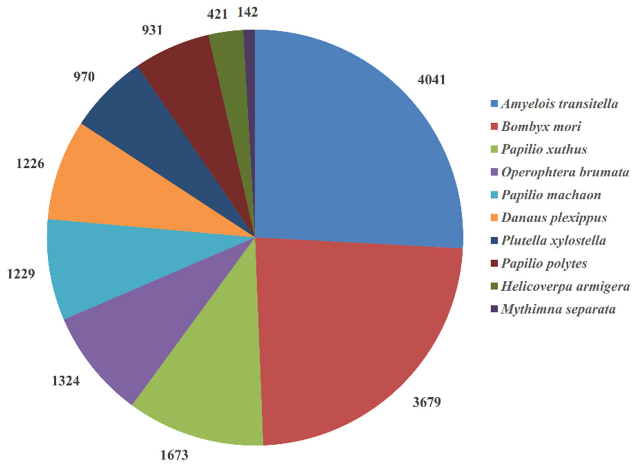
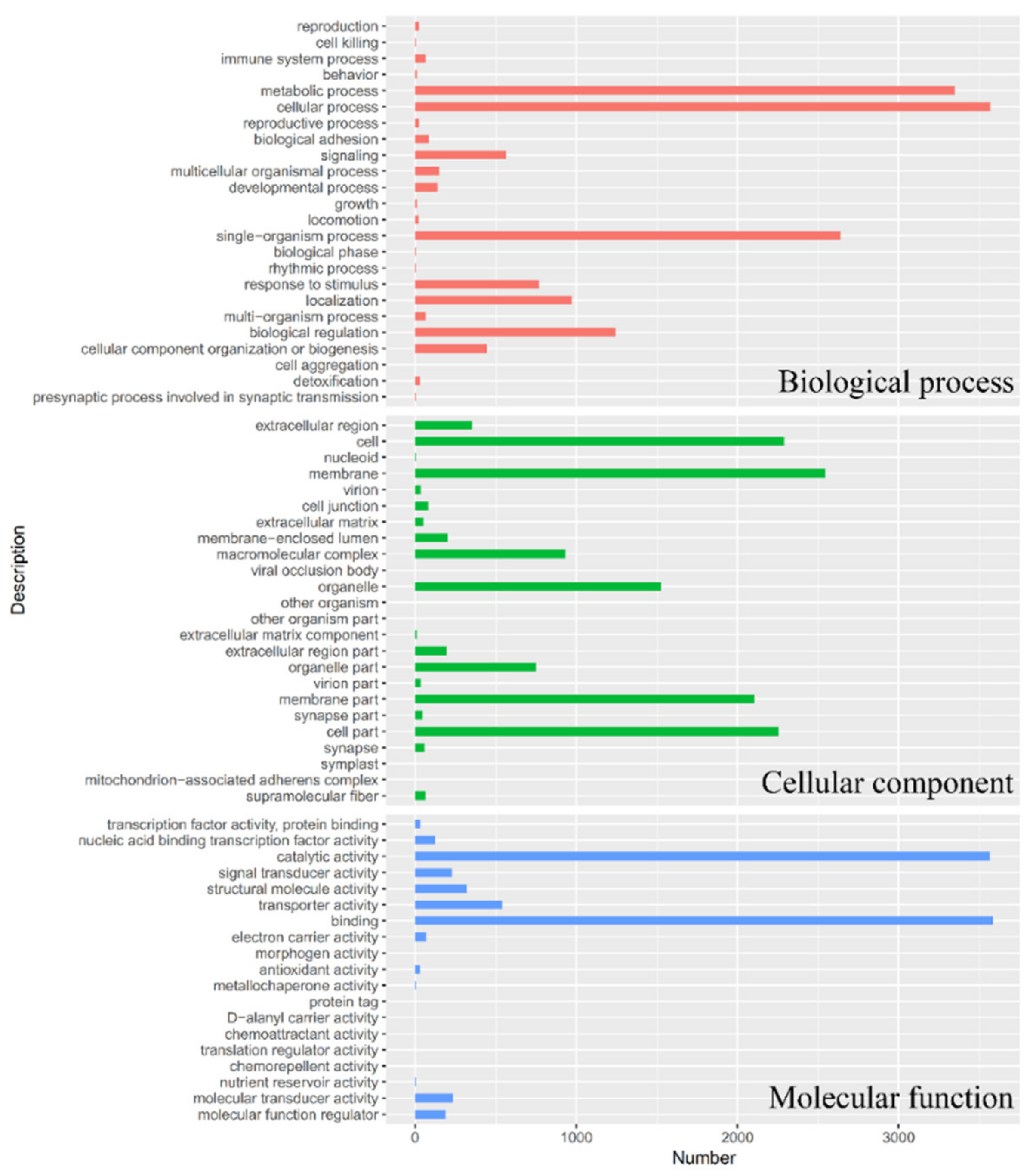
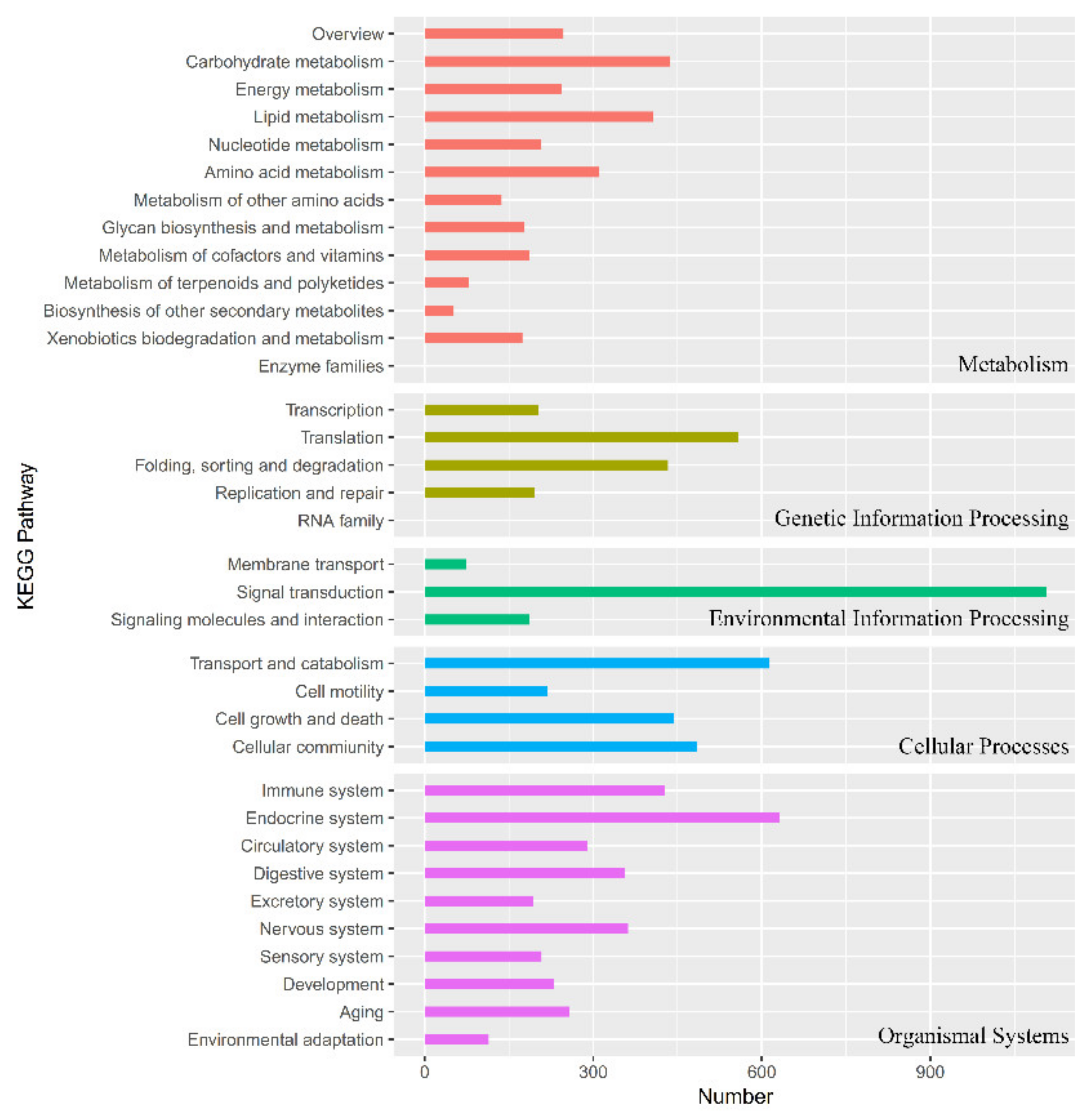
3.1. Transcriptome Sequencing, Unigene Assembly, and Functional Annotation

A cDNA library was constructed for M. separata larvae and sequenced using the Illumina platform. This run produced 47,594,876 raw reads and 44,966,148 clean reads (Clean reads: 94.47%, Q20: 97.45%, Q30: 93.07%). The clean reads were assembled into 81,837 transcripts with a mean length of 1135 bp. Transcripts were further assembled into 45,888 unigenes with a mean length of 910 bp (N50 = 1687 bp). The transcriptome dataset was deposited in Sequence Read Archive (PRJNA702891).
Unigenes were annotated in six databases: NR, GO, KEGG, Pfam, eggNOG, and Swiss-Prot (Table 1). Approximately 18,157 unigenes were annotated to the NR database. The top 10 species with the most matches in the NR database were shown in Figure 1. In total, 4041 unigenes were matched to Amyelois transitella, followed by Bombyx mori (3679), Papilio xuthus (1673), Operophtera brumata (1324), Papilio machaon (1229), Danaus plexippus (1226), Plutella xylostella (970), Papilio polytes (931), Helicoverpa armigera (421), and Mythimna separata (142).
Functions of unigenes were classified by GO assignments with BLAST2GO (Figure 2). A total of 36,622 unigenes were categorized into 55 categories, which belonged to three main categories: biological process (23), cellular component (19), and molecular function (13). The top 10 categories were binding (3584), cellular process (3569), catalytic activity (3564), metabolic process (3346), single-organism process (2640), membrane (2547), cell (2291), cell part (2257), membrane part (2102), and organelle (1524). For KEGG pathway annotation (Figure 3), the top 10 pathways were signal transduction (1106), endocrine system (632), transport and catabolism (614), translation (559), cellular community (484), cell growth, and death (444), carbohydrate metabolism (436), folding, sorting and degradation (433), immune system (428), and lipid metabolism (406).

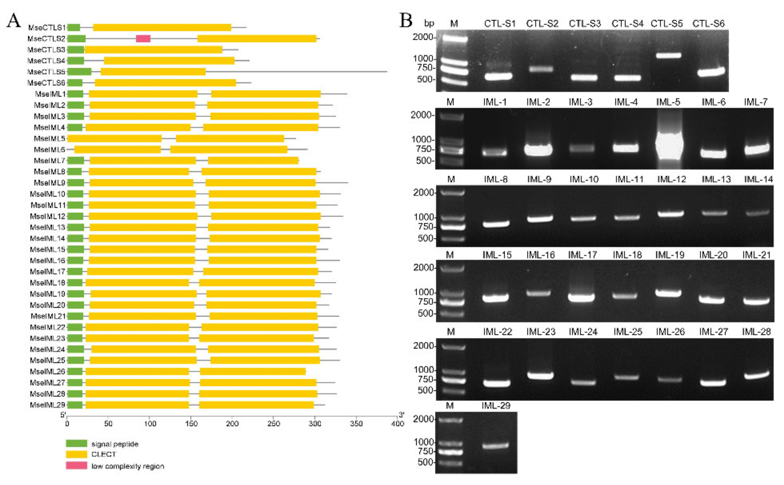
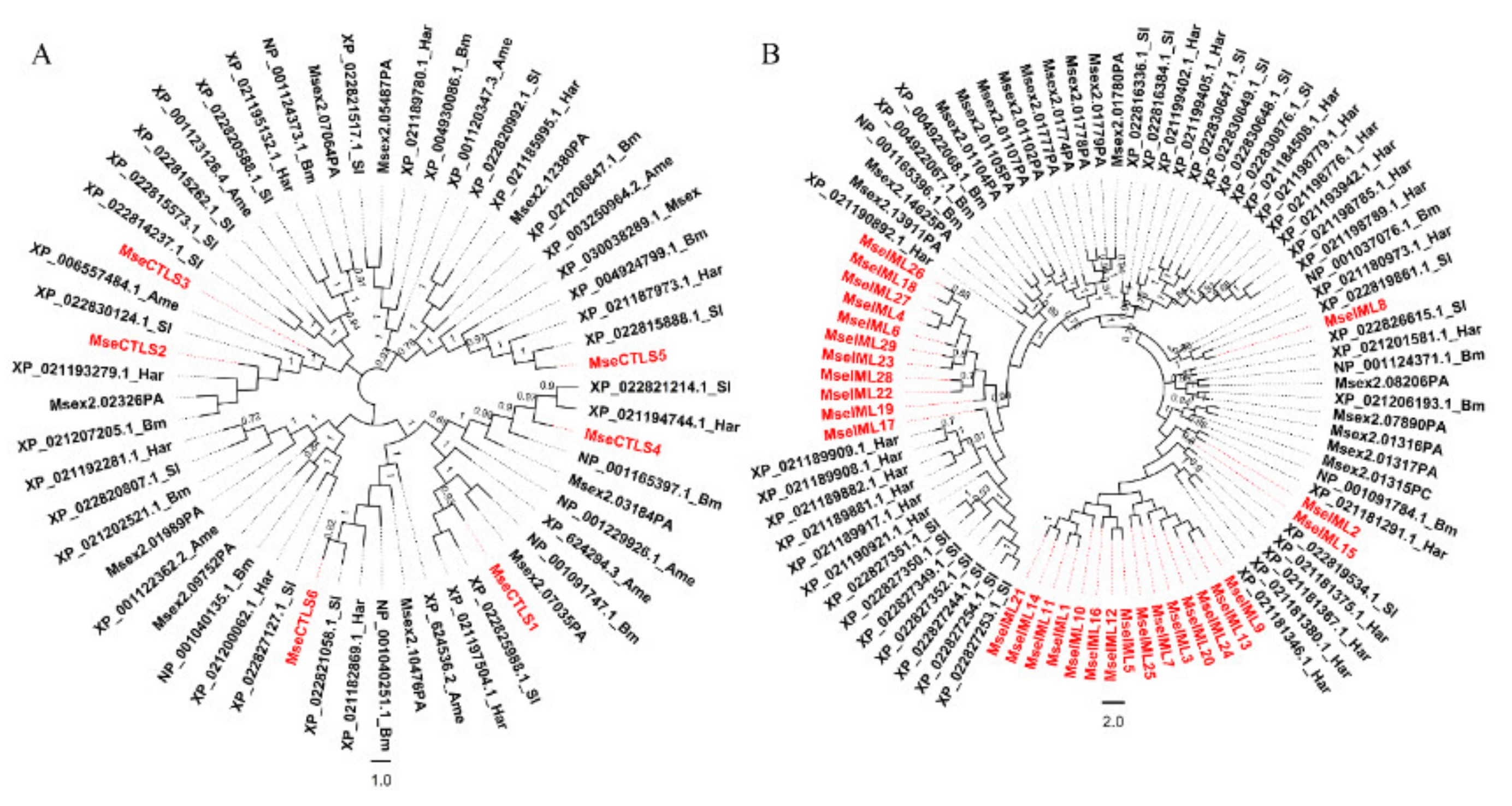
3.2. Identification of CTLs

Thirty-five unigenes that encode single-CRD or dual-CRD proteins were identified and named CTL-S1–CTL-S6 and IML-1–IML-29 (Figure 4A and Table 2). The coding sequences were amplified from cDNA and correctly sequenced (Figure 4B). To determine the phylogenetic relationships, 57 CTL-S and 90 IML were aligned to construct NJ trees, respectively [37]. All M. separata CTL-S clustered with their respective lepidopteran orthologs, except that CTL-S2 and CTL-S3 clustered together (Figure 5A). In contrast, IMLs showed lineage-specific expansions: IML-26/18/27/4/6/29/23/28/22/19 and IML-21/14/11/1/10/16/12/5/25/7/3/20/24/13/9 formed two clusters, indicating that they originated from independent expansions. IML-2 is a 1:1 ortholog of H. armigera (XP_021181291.1). IML-8 is a 1:1 ortholog of S. litura (XP_022819861.1). IML-17 is clustered with six H. armigera and seven S. litura orthologs (Figure 5B). These phylogenetic relationships are consistent with previous reports of B. mori and M. sexta CTLs [7,8].

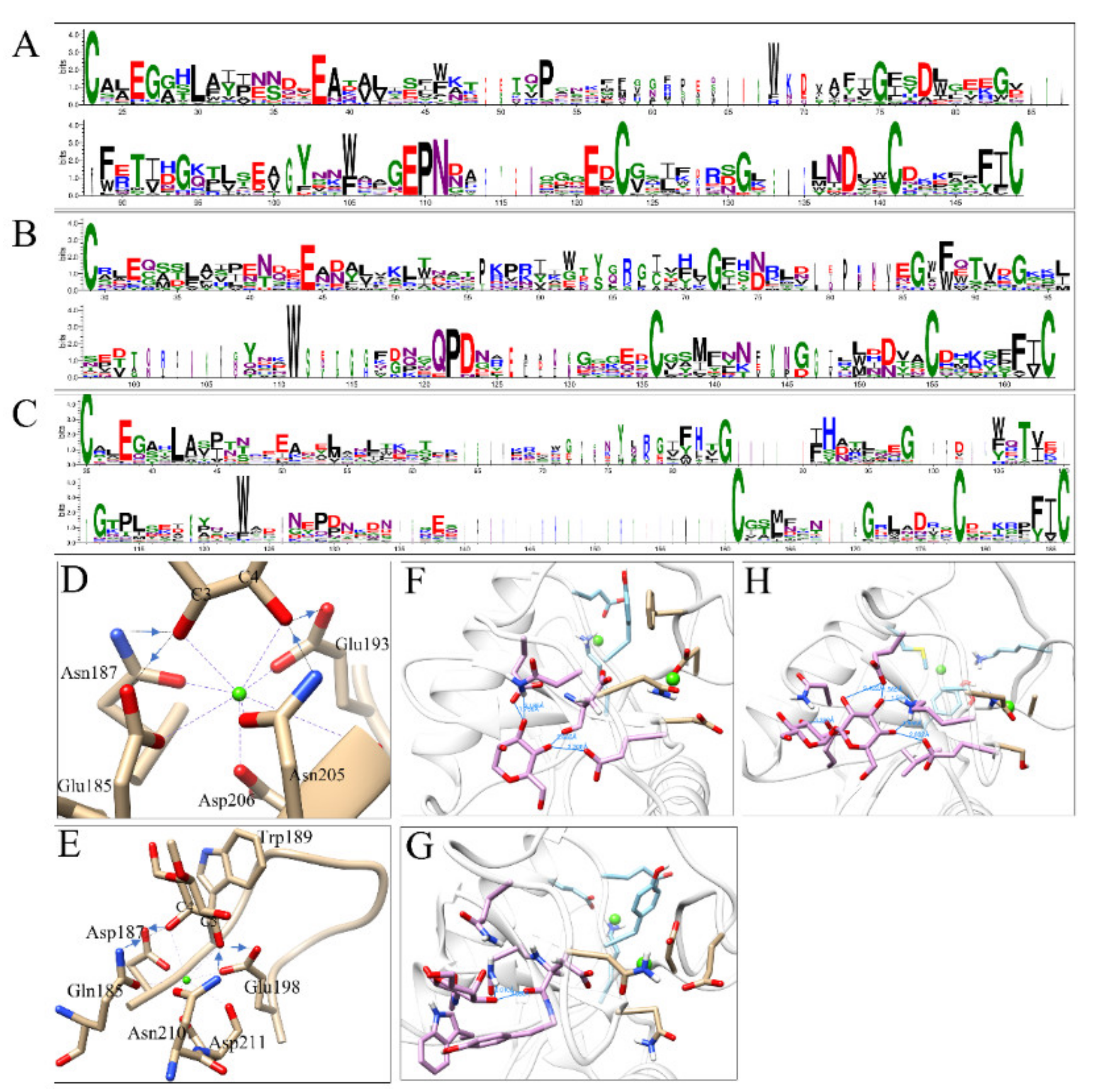
3.3. Sequence and Structure Analyses of CRDs

M. separata CRDs were analyzed by I-TASSER for ligand type and interacting residues. Of 64 CRDs, 15 contained ‘EPN’ motifs, 12 contained ‘QPD’ motifs, and 37 had noncanonical motifs (Table 3). Sequence logos showed the conserved residues in each type of CRD. Four cysteines were completely conserved in all CRDs. Other conserved residues include: E26/G27/L30/E38/P53/W68/G75/D78/G83/F89/T91/G94/Y102/W105/E109/P110/N111/E121/G131/D138/F147/I148 in ‘EPN’-type CRDs (Figure 6A and Figure S1A); E44/G72/G86/F88/W88/T90/G93/W111/Q121/P122/D123/D152/N152/F161/I162 in ‘QPD’-type CRDs (Figure 6B and Figure S1B); E38/L42/G84/H92/G98/T107/W123/F184/I185 in ‘noncanonical’-type CRDs (Figure 6C and Figure S1C). In MBP-A, Glu185, Asn187, Glu193, Asn205, and Asp206 can form coordination and hydrogen bonds with Ca2+ and 3-OH/4-OH, respectively (Figure 6D). The sequence alignment shows that in ‘EPN’-type CRDs, Glu193 is completely conserved, Asn205 is conserved in 10 out of 15 CRDs, and Asp206 is conserved in 14 out of 15 CRDs. In the MBP-A mutant (QPDWG), Gln185–Asp187–Glu198–Asn210–Asp211 forms a similar coordination and hydrogen bond network. However, the reversed hydrogen donor/acceptor and the adjacent Trp189 switched the preferred ligand to galactose (Figure 6E). The sequence comparison showed that Trp189 and Asn210 were not conserved in insect CRDs. Glu198 was conserved in 5 out of 12 CRDs. Asp211 was conserved in 8 out of 12 CRDs. In the predicted structures of IML-10-CRD1 (‘EPN’, Figure 6F), IML-14-CRD1 (‘QPD’, Figure 6G), and IML-17-CRD1 (‘EPD’, Figure 6H), sugar ligands were accommodated in proper orientations and positions through hydrogen bonds formed between hydroxyl groups and amino acids.

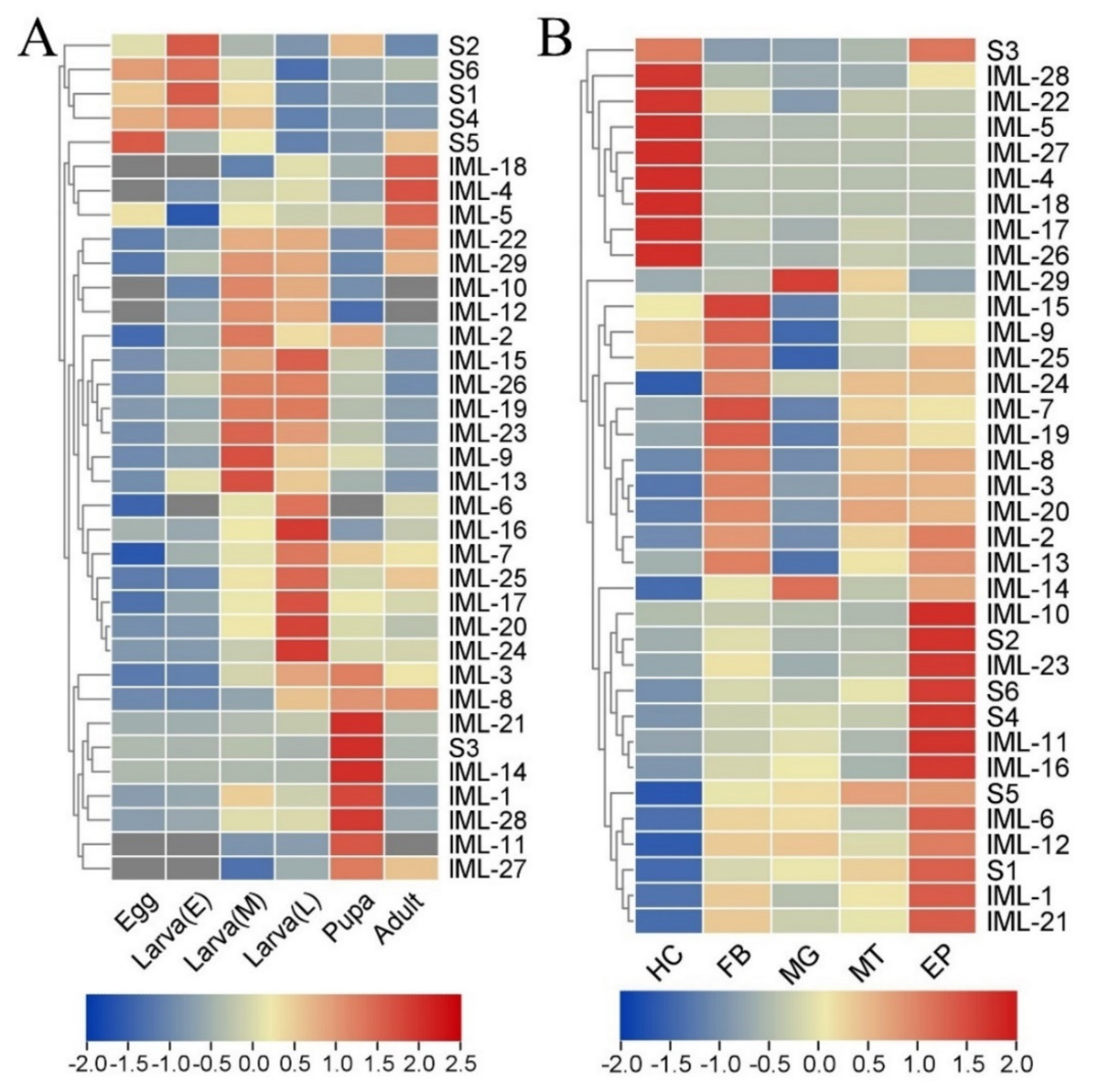
3.4. Spatial and Temporal Expression Profiles

To explore the possible functions of CTLs, the expression profile in different developmental stages was analyzed by RT-qPCR. The hierarchical clustering analysis shows distinct expression patterns: CTL-S1, CTL-S2, CTL-S4, CTL-S5, and CTL-S6 mainly express in eggs and early stage larvae; IML-4, IML-5, and IML-18 express in adults; IML-3, IML-8, IML-21, CTL-S3, IML-14, IML-1, IML-28, IML-11, and IML-27 mainly express in pupa; IML-6, IML-16, IML-7, IML-25, IML-17, IML-20, and IML-24 express in late-stage larvae; IML-22, IML-29, IML-10, IML-12, IML-2, IML-15, IML-26, IML-19, IML-23, IML-9, and IML-13 express in early and mid-stage larvae (Figure 7A). Larval tissues can produce lots of immune factors to resist the invasion of pathogens. Thus, the expression profile in larval hemocytes, fat body, midgut, Malpighian tube, and epidermis was analyzed. The clustering analysis shows that hemocytes, fat body, and epidermis are the major tissues expressing CTL genes. Notably, IML-14 and IML-29 are mostly expressed in the midgut (Figure 7B).

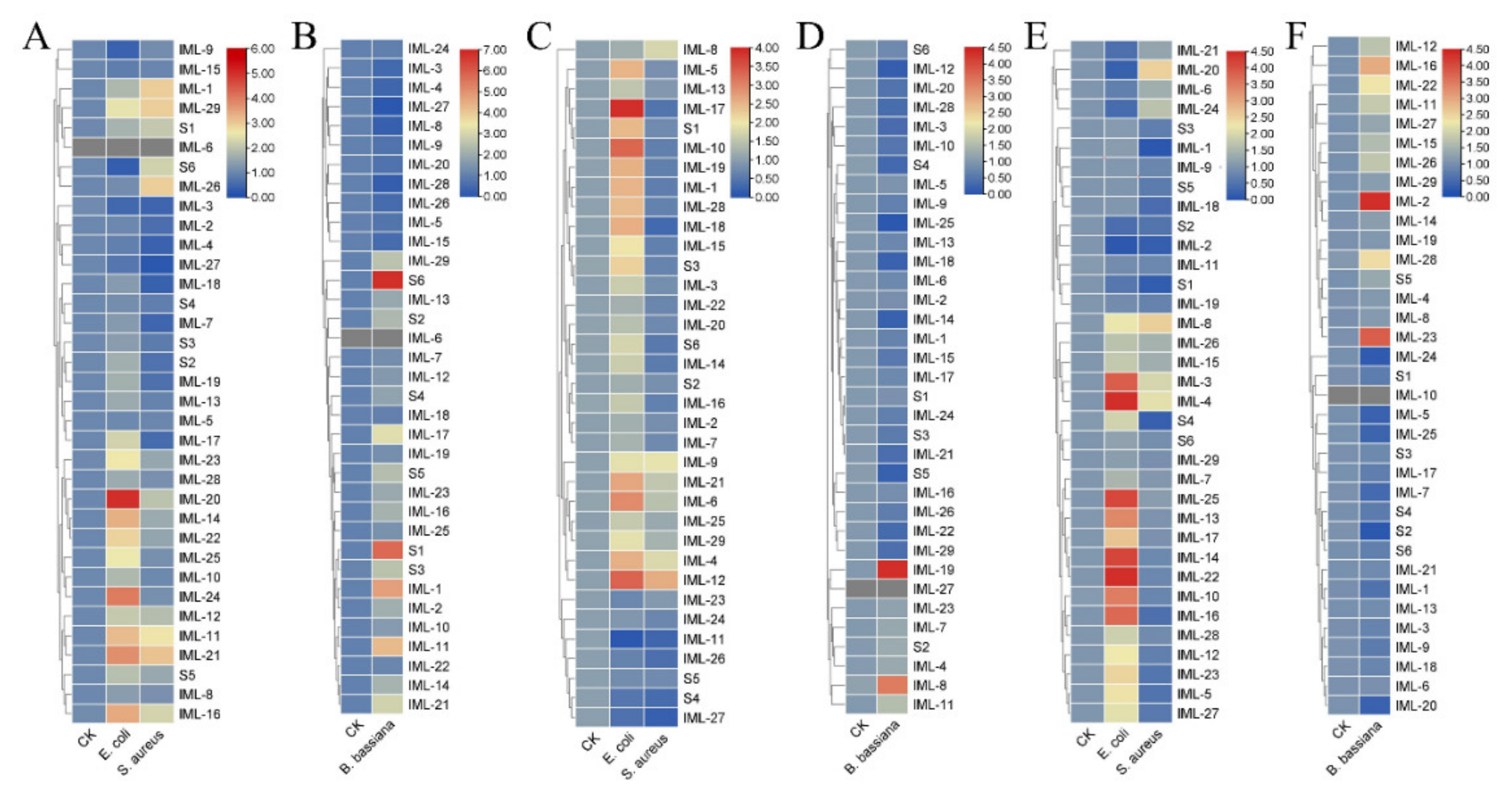
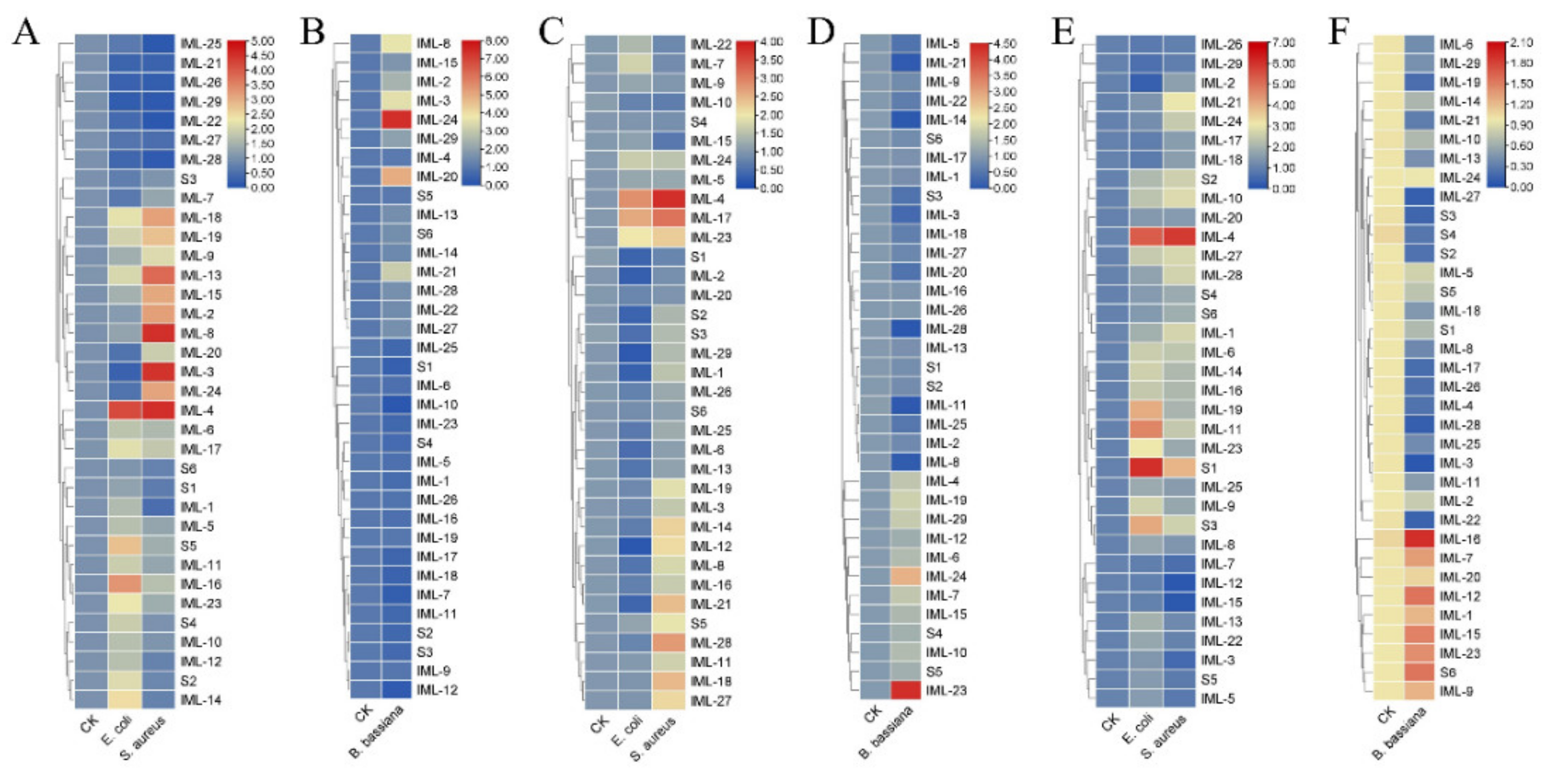
3.5. Expression of CTLs Responding to Bacterial Cells and Fungal Spores

C-type lectins, especially immulectins, are important for immune responses against bacteria and fungi [17,38]. The expression of CTLs in larval hemocytes, fat body, and midgut responding to E. coli, S. aureus, and B. bassiana were analyzed 6 h and 24 h post-challenge. At 6 hpi, in hemocytes, IML-20, IML-24, IML-21, and IML-16 were induced by E. coli; IML-21, IML-1, IML-29, and IML-26 were induced by S. aureus (Figure 8A); CTL-S1, CTL-S6, IML-1, and IML-11 were induced by B. bassiana (Figure 8B). In the fat body, IML-17, IML-10, IML-21, IML-6, IML-4, and IML-12 were induced by E. coli; IML-4, IML-8, IML-9, and IML-12 were induced by S. aureus (Figure 8C); IML-8 and IML-19 were induced by B. bassiana (Figure 8D). In the midgut, IML-3, IML-4, IML-25, IML-13, IML-17, IML-14, IML-22, IML-10, and IML-16 were induced by E. coli; IML-20 and IML-8 were induced by S. aureus (Figure 8E); IML-2, IML-16, and IML-23 were induced by B. bassiana (Figure 8F). At 24 hpi, in hemocytes, IML-4 and IML-16 were induced by E. coli; IML-18, IML-19, IML-13, IML-15, IML-2, IML-8, IML-3, IML-24, and IML-4 were induced by S. aureus (Figure 9A); IML-24 and IML-20 were induced by B. bassiana (Figure 9B). In the fat body, IML-4 and IML-17 were induced by E. coli; IML-4, IML-17, IML-21, IML-28, and IML-18 were induced by S. aureus (Figure 9C); IML-23 and IML-24 were induced by B. bassiana (Figure 9D). In the midgut, IML-4, IML-19, IML-11, CTL-S1, and CTL-S3 were induced by E. coli; IML-4 and CTL-S1 were induced by S. aureus (Figure 9E); IML-16, IML-12, IML-15, IML-23, and CTL-S6 were induced by B. bassiana (Figure 9F).

4. Discussion

With the advancement of the next-generation sequencing (NGS) technique, RNA-seq has become an indispensable tool for studying the transcriptome of non-model organisms, including some agricultural pests [39]. In this study, the transcriptome generated 44,966,148 clean reads and 45,888 unigenes with a mean length of 910 bp, which was comparable to the previous M. separata transcriptomes [40,41,42,43,44,45,46,47]. Most homologs of M. separata transcripts were found in lepidopteran species, especially in A. transitella and B. mori. By Gene Ontology (GO) classification and KEGG pathway annotation, unigenes were classified into a variety of biological processes, cellular components, molecular functions, and pathways.
Animal C-type lectin-like domain-containing proteins can be classified into 16 groups based on domain architecture and phylogenetic relationships [48]. In insects, CTLs can be classified based on domain architecture into CTL-S, IML, and CTL-X. CTL-S exist in several insect orders: Lepidoptera, Coleoptera, Hymenoptera, Diptera, and Hemiptera. The numbers of CTL-S vary in different species: Bombyx mori (12), Manduca sexta (8), Tribolium castaneum (10), Drosophila melanogaster (30), Anopheles gambiae (21), Aedes aegypti (37), Acyrthosiphon pisum (2), and Plutella xylostella (5). Immulectins were almost entirely found in Lepidoptera: Bombyx mori (6) and Manduca sexta (19) [5]. Here, we identified 6 CTL-S and 29 immulectins from the unigenes of M. separata larvae. The phylogenetic analysis showed that CTL-S genes were duplicated in the common ancestor before speciation (Figure 5A), while most IML genes were duplicated after speciation (Figure 5B). Similar phylogenetic relationships were also found in B. mori and M. sexta [7,8]. Since immulectins broadly participate in regulating the innate immune responses, the expansion of immulectins may greatly improve the survival rates of lepidopteran pests in the natural environment.
The expression profile of developmental stages shows that most IMLs express in larvae and pupae. Only three IMLs (IML-4, IML-5, and IML-18) express in adults. Interestingly, most CTL-S express in eggs and early-stage larvae (Figure 7A). These results suggest that CTL-S may be important for the development of and immunity in embryos and early stage larvae, while IMLs are critical for immunity in larvae, pupae, and adults. A Periplaneta lectin participates in the organization or stabilization of the epidermis during leg regeneration [49]. H. armigera CTL3 maintains normal larval growth and development by maintaining ecdysone and juvenile hormone signaling and suppressing the abundance of Enterocuccus mundtii [50]. CTLs also show a specific spatial expression pattern in naïve larval tissues: hemocytes, fat body, and epidermis are responsible for expressing CTLs (Figure 7B). These are the major larval tissues generating immune molecules. Bacterial and fungal infections induced dramatic changes in the expression of some CTLs (Figure 8 and Figure 9). Our findings are similar to previous transcriptomic studies. In the cotton bollworm H. armigera, a transcriptome-based analysis showed that most CTL genes did not undergo any significant changes in the second instar larvae after B. bassiana infection, while most of them were upregulated in the fat body of the fifth instar larvae [17]. Although we did not compare the induction of CTLs between early stage and late-stage M. separata larvae, we found that most IMLs were expressed in mid-late-stage larvae and pupae. These data suggest that IMLs are important for immune responses in these stages. In the Japanese pine sawyer beetle, Monochamus alternatus infected with the entomopathogenic fungus Metarhizium anisopliae, several differentially expressed unigenes were CTLs [51]. Twelve CTL genes were identified in Adelphocoris suturalis (Hemiptera: Miridae) immune responsive genes against fungal and bacterial pathogens [52]. Fourteen CTLs were identified in immunity-related genes in Ostrinia furnacalis against entomopathogenic fungi [53].
A simple rule to predict the ligand specificity of CRDs is based on some key residues: ‘EPN’-motif CRDs can recognize mannose-type ligands; ‘QPD’-motif CRDs usually recognize galactose-type ligands. Mutating ‘EPN’ in MBP-A to ‘QPD’ caused a shift from mannose to galactose ligands [54]. Conversely, mutating ‘QPD’ in sea cucumber CEL-I to ‘EPN’ led to a weak binding affinity for mannose [55]. Some surrounding residues or structures also can affect ligand selection. An additional mutation of Trp105 to His in MBP-A further increased the affinity to mannose [55]. A glycine-rich loop helps to exclude mannose and accommodate galactose in Gal-type CTLs [56,57]. However, there are exceptions to this rule. CEL-IV, a CTL in sea cucumber, Cucumaria echinate, contains the ‘EPN’ motif but binds galactose [58]. TC14, a CTL from the tunicate Polyandrocarpa misakiensis contains ‘EPS’ but binds galactose [59]. Some CRDs bind carbohydrates in the absence of the ‘EPN/QPD’ motif or Ca2+. The CRD of eosinophil major basic protein (EMBP) binds to heparin and heparan sulfate at a different contact site through electrostatic interactions and hydrogen bonds [60]. Bivalve lectins SPL-1 and SPL-2, which contained ‘RPD’ and ‘KPD’ motifs, showed Ca2+-independent binding affinity for GlcNAc or GalNAc [61]. Structural studies have elucidated how these residues interact with Ca2+ and carbohydrates. For monosaccharide ligands, steric restrictions are imposed by the coordination bonds and hydrogen bonds formed between 3-OH/4-OH, Ca2+, and ‘EPN/QPD’ motifs. Mannose has equatorial 3-OH and equatorial 4-OH, while galactose has equatorial 3-OH and axial 4-OH. In addition, the hydrogen donors and acceptors are reversed between E(acceptor)-P-N(donor) and Q(donor)-P-D(acceptor) (Figure 6D,E). Therefore, the predicted ligand specificity needs to be verified experimentally.
To sum up, this study built a de novo transcriptome assembly of M. separata larvae, from which 6 ‘S-type’ and 29 ‘IML-type’ CTLs were identified. Sequence features, phylogenetic relationships, ligand specificity, and expression profiles were studied. Further studies are required to explore the function of each CTL in the oriental armyworm.

Supplementary Materials

The following are available online at https://www.mdpi.com/article/10.3390/insects12060559/s1, Figure S1: Sequence alignment of three types of CRDs; Table S1: List of Primers.

Author Contributions

Conceptualization, X.-J.R.; methodology, H.L.; investigation, H.L., F.-F.L., L.-Q.F., Z.L., W.-T.Z. and Q.W.; data curation, H.L. and X.-J.R.; writing—original draft preparation, H.L. and X.-J.R.; writing—review and editing, X.-J.R.; supervision, X.-J.R.; project administration, X.-J.R.; funding acquisition, X.-J.R. All authors have read and agreed to the published version of the manuscript.

Funding

This study was supported by grants from the National Natural Science Foundation of China (31872284) and the Natural Science Foundation of Anhui Province (2008085J17) to X.-J.R.

Institutional Review Board Statement

Not applicable.

Data Availability Statement

Not applicable.

Conflicts of Interest

The authors declare no conflict of interest.

Abbreviations

PRR: pattern recognition receptor; PAMP: pathogen-associated molecular pattern; CTL: C-type lectin; PGRP: peptidoglycan-recognition protein; βGRP: β-1,3-glucan recognition protein; CRD: carbohydrate-recognition domain; CTLD: C-type lectin-like domain; IML: immulectin; PBS, phosphate-buffered saline; RT-qPCR: quantitative reverse transcription PCR; SMART: Simple Modular Architecture Research Tool; I-TASSER: Iterative Threading Assembly Refinement.

References

  1. Janeway, C.A., Jr.; Medzhitov, R. Innate immune recognition. Annu. Rev. Immunol. 2002, 20, 197–216. [Google Scholar] [CrossRef] [Green Version]
  2. Lin, Z.; Wang, J.L.; Cheng, Y.; Wang, J.X.; Zou, Z. Pattern recognition receptors from lepidopteran insects and their biological functions. Dev. Comp. Immunol. 2020, 108, 103688. [Google Scholar] [CrossRef] [PubMed]
  3. Imler, J.L. Overview of Drosophila immunity: A historical perspective. Dev. Comp. Immunol. 2014, 42, 3–15. [Google Scholar] [CrossRef]
  4. Rao, X.J.; Zhan, M.Y.; Pan, Y.M.; Liu, S.; Yang, P.J.; Yang, L.L.; Yu, X.Q. Immune functions of insect βGRPs and their potential application. Dev. Comp. Immunol. 2018, 83, 80–88. [Google Scholar] [CrossRef] [PubMed]
  5. Xia, X.; You, M.; Rao, X.J.; Yu, X.Q. Insect C-type lectins in innate immunity. Dev. Comp. Immunol. 2018, 83, 70–79. [Google Scholar] [CrossRef]
  6. Zelensky, A.N.; Gready, J.E. The C-type lectin-like domain superfamily. FEBS J. 2005, 272, 6179–6217. [Google Scholar] [CrossRef]
  7. Rao, X.J.; Shahzad, T.; Liu, S.; Wu, P.; He, Y.T.; Sun, W.J.; Fan, X.Y.; Yang, Y.F.; Shi, Q.; Yu, X.Q. Identification of C-type lectin-domain proteins (CTLDPs) in silkworm Bombyx mori. Dev. Comp. Immunol. 2015, 53, 328–338. [Google Scholar] [CrossRef]
  8. Rao, X.J.; Cao, X.; He, Y.; Hu, Y.; Zhang, X.; Chen, Y.R.; Blissard, G.; Kanost, M.R.; Yu, X.Q.; Jiang, H. Structural features, evolutionary relationships, and transcriptional regulation of C-type lectin-domain proteins in Manduca sexta. Insect Biochem. Mol. Biol. 2015, 62, 75–85. [Google Scholar] [CrossRef] [Green Version]
  9. Nagae, M.; Yamaguchi, Y. Structural Aspects of Carbohydrate Recognition Mechanisms of C-Type Lectins. In C-Type Lectins in Immune Homeostasis, 2019/11/30 ed.; Yamasaki, S., Ed.; Springer: Cham, Switzerland, 2019; Volume 429, pp. 147–176. [Google Scholar]
  10. Weis, W.I.; Drickamer, K.; Hendrickson, W.A. Structure of a C-type mannose-binding protein complexed with an oligosaccharide. Nature 1992, 360, 127–134. [Google Scholar] [CrossRef]
  11. Yu, X.Q.; Gan, H.; Kanost, M.R. Immulectin, an inducible C-type lectin from an insect, Manduca sexta, stimulates activation of plasma prophenol oxidase. Insect Biochem. Mol. Biol. 1999, 29, 585–597. [Google Scholar] [CrossRef]
  12. Yu, X.Q.; Kanost, M.R. Immulectin-2, a lipopolysaccharide-specific lectin from an insect, Manduca sexta, is induced in response to gram-negative bacteria. J. Biol. Chem. 2000, 275, 37373–37381. [Google Scholar] [CrossRef] [PubMed] [Green Version]
  13. Yu, X.Q.; Ling, E.; Tracy, M.E.; Zhu, Y. Immulectin-4 from the tobacco hornworm Manduca sexta binds to lipopolysaccharide and lipoteichoic acid. Insect Mol. Biol. 2006, 15, 119–128. [Google Scholar] [CrossRef] [PubMed]
  14. Ao, J.; Ling, E.; Yu, X.Q. Drosophila C-type lectins enhance cellular encapsulation. Mol. Immunol. 2007, 44, 2541–2548. [Google Scholar] [CrossRef] [Green Version]
  15. Bi, J.; Ning, M.; Li, J.; Zhang, P.; Wang, L.; Xu, S.; Zhong, Y.; Wang, Z.; Song, Q.; Li, B. A C-type lectin with dual-CRD from Tribolium castaneum is induced in response to bacterial challenge. Pest Manage. Sci. 2020, 76, 3965–3974. [Google Scholar] [CrossRef]
  16. Wang, J.L.; Zhang, Q.; Tang, L.; Chen, L.; Liu, X.S.; Wang, Y.F. Involvement of a pattern recognition receptor C-type lectin 7 in enhancing cellular encapsulation and melanization due to its carboxyl-terminal CRD domain in the cotton bollworm, Helicoverpa armigera. Dev. Comp. Immunol. 2014, 44, 21–29. [Google Scholar] [CrossRef] [PubMed]
  17. Cheng, Y.; Lin, Z.; Wang, J.M.; Xing, L.S.; Xiong, G.H.; Zou, Z. CTL14, a recognition receptor induced in late stage larvae, modulates anti-fungal immunity in cotton bollworm Helicoverpa armigera. Dev. Comp. Immunol. 2018, 84, 142–152. [Google Scholar] [CrossRef]
  18. Koizumi, N.; Imamura, M.; Kadotani, T.; Yaoi, K.; Iwahana, H.; Sato, R. The lipopolysaccharide-binding protein participating in hemocyte nodule formation in the silkworm Bombyx mori is a novel member of the C-type lectin superfamily with two different tandem carbohydrate-recognition domains. FEBS Lett. 1999, 443, 139–143. [Google Scholar] [CrossRef] [Green Version]
  19. Watanabe, A.; Miyazawa, S.; Kitami, M.; Tabunoki, H.; Ueda, K.; Sato, R. Characterization of a novel C-type lectin, Bombyx mori multibinding protein, from the B. mori hemolymph: Mechanism of wide-range microorganism recognition and role in immunity. J. Immunol. 2006, 177, 4594–4604. [Google Scholar] [CrossRef] [PubMed] [Green Version]
  20. Fang, Q.; Wang, F.; Gatehouse, J.A.; Gatehouse, A.M.; Chen, X.X.; Hu, C.; Ye, G.Y. Venom of parasitoid, Pteromalus puparum, suppresses host, Pieris rapae, immune promotion by decreasing host C-type lectin gene expression. PLoS ONE 2011, 6, e26888. [Google Scholar] [CrossRef] [Green Version]
  21. Cheng, G.; Cox, J.; Wang, P.; Krishnan, M.N.; Dai, J.; Qian, F.; Anderson, J.F.; Fikrig, E. A C-type lectin collaborates with a CD45 phosphatase homolog to facilitate West Nile virus infection of mosquitoes. Cell 2010, 142, 714–725. [Google Scholar] [CrossRef] [PubMed] [Green Version]
  22. Jiang, X.; Luo, L.; Zhang, L.; Sappington, T.W.; Hu, Y. Regulation of migration in Mythimna separata (Walker) in China: A review integrating environmental, physiological, hormonal, genetic, and molecular factors. Environ. Entomol. 2011, 40, 516–533. [Google Scholar] [CrossRef]
  23. Wang, C.; Wang, S. Insect Pathogenic Fungi: Genomics, Molecular Interactions, and Genetic Improvements. Annu. Rev. Entomol. 2017, 62, 73–90. [Google Scholar] [CrossRef] [PubMed]
  24. Qu, S.; Wang, S.B. Interaction of entomopathogenic fungi with the host immune system. Dev. Comp. Immunol. 2018, 83, 96–103. [Google Scholar] [CrossRef] [PubMed]
  25. Liu, F.F.; Li, H.; Yang, P.J.; Rao, X.J. Structure-function analysis of PGRP-S1 from the oriental armyworm, Mythimna separata. Arch. Insect Biochem. Physiol. 2021, 106, e21763. [Google Scholar] [CrossRef]
  26. Liu, F.F.; Ding, C.; Yang, L.L.; Li, H.; Rao, X.J. Identification and analysis of two lebocins in the oriental armyworm Mythimna separata. Dev. Comp. Immunol. 2021, 116, 103962. [Google Scholar] [CrossRef] [PubMed]
  27. Yang, P.-J.; Zhan, M.-Y.; Yang, L.-L.; Liu, Q.-Q.; Xu, Y.; Pan, Y.-M.; Rao, X.-J. Characterization of PGRP-S1 from the oriental armyworm, Mythimna separata. Dev. Comp. Immunol. 2019, 90, 121–129. [Google Scholar] [CrossRef] [PubMed]
  28. Martin, M. Cutadapt removes adapter sequences from high-throughput sequencing reads. EMBnet.journal 2011, 17, 3. [Google Scholar] [CrossRef]
  29. Haas, B.J.; Papanicolaou, A.; Yassour, M.; Grabherr, M.; Blood, P.D.; Bowden, J.; Couger, M.B.; Eccles, D.; Li, B.; Lieber, M.; et al. De novo transcript sequence reconstruction from RNA-seq using the Trinity platform for reference generation and analysis. Nat. Protoc. 2013, 8, 1494–1512. [Google Scholar] [CrossRef]
  30. Altschul, S.F.; Madden, T.L.; Schäffer, A.A.; Zhang, J.; Zhang, Z.; Miller, W.; Lipman, D.J. Gapped BLAST and PSI-BLAST: A new generation of protein database search programs. Nucleic Acids Res. 1997, 25, 3389–3402. [Google Scholar] [CrossRef] [Green Version]
  31. Waterhouse, A.M.; Procter, J.B.; Martin, D.M.A.; Clamp, M.; Barton, G.J. Jalview Version 2—A multiple sequence alignment editor and analysis workbench. Bioinformatics 2009, 25, 1189–1191. [Google Scholar] [CrossRef] [PubMed] [Green Version]
  32. Kumar, S.; Stecher, G.; Li, M.; Knyaz, C.; Tamura, K. MEGA X: Molecular Evolutionary Genetics Analysis across Computing Platforms. Mol. Biol. Evol. 2018, 35, 1547–1549. [Google Scholar] [CrossRef]
  33. Roy, A.; Kucukural, A.; Zhang, Y. I-TASSER: A unified platform for automated protein structure and function prediction. Nat. Protoc. 2010, 5, 725–738. [Google Scholar] [CrossRef] [Green Version]
  34. Pettersen, E.F.; Goddard, T.D.; Huang, C.C.; Couch, G.S.; Greenblatt, D.M.; Meng, E.C.; Ferrin, T.E. UCSF Chimera—A visualization system for exploratory research and analysis. J. Comput. Chem. 2004, 25, 1605–1612. [Google Scholar] [CrossRef] [PubMed] [Green Version]
  35. Crooks, G.E.; Hon, G.; Chandonia, J.M.; Brenner, S.E. WebLogo: A sequence logo generator. Genome Res. 2004, 14, 1188–1190. [Google Scholar] [CrossRef] [PubMed] [Green Version]
  36. Chen, C.; Chen, H.; Zhang, Y.; Thomas, H.R.; Frank, M.H.; He, Y.; Xia, R. TBtools: An Integrative Toolkit Developed for Interactive Analyses of Big Biological Data. Mol. Plant 2020, 13, 1194–1202. [Google Scholar] [CrossRef] [PubMed]
  37. Lu, Y.; Su, F.; Zhu, K.; Zhu, M.; Li, Q.; Hu, Q.; Zhang, J.; Zhang, R.; Yu, X.-Q. Comparative genomic analysis of C-type lectin-domain genes in seven holometabolous insect species. Insect Biochem. Mol. Biol. 2020, 126, 103451. [Google Scholar] [CrossRef]
  38. Koizumi, N.; Imai, Y.; Morozumi, A.; Imamura, M.; Kadotani, T.; Yaoi, K.; Iwahana, H.; Sato, R. Lipopolysaccharide-binding protein of Bombyx mori participates in a hemocyte-mediated defense reaction against gram-negative bacteria. J. Insect Physiol. 1999, 45, 853–859. [Google Scholar] [CrossRef]
  39. Stark, R.; Grzelak, M.; Hadfield, J. RNA sequencing: The teenage years. Nat. Rev. Genet. 2019, 20, 631–656. [Google Scholar] [CrossRef] [PubMed]
  40. Liu, Y.; Qi, M.; Chi, Y.; Wuriyanghan, H. De Novo Assembly of the Transcriptome for Oriental Armyworm Mythimna separata (Lepidoptera: Noctuidae) and Analysis on Insecticide Resistance-Related Genes. J. Insect Sci. 2016, 16. [Google Scholar] [CrossRef] [PubMed] [Green Version]
  41. Wang, J.D.; Wang, W.Z.; Wang, Y.R.; Gao, S.J.; Elzaki, M.; Wang, R.; Wang, R.; Wu, M. Response of detoxification and immune genes and of transcriptome expression in Mythimna separata following chlorantraniliprole exposure. Comp. Biochem. Physiol. Part D Genom. Proteom. 2018, 28, 90–98. [Google Scholar] [CrossRef]
  42. Duan, Y.; Gong, Z.; Wu, R.; Miao, J.; Jiang, Y.; Li, T.; Wu, X.; Wu, Y. Transcriptome analysis of molecular mechanisms responsible for light-stress response in Mythimna separata (Walker). Sci. Rep. 2017, 7, 45188. [Google Scholar] [CrossRef] [PubMed]
  43. Liu, Z.; Wang, X.; Lei, C.; Zhu, F. Sensory genes identification with head transcriptome of the migratory armyworm, Mythimna separata. Sci. Rep. 2017, 7, 46033. [Google Scholar] [CrossRef]
  44. Chang, X.Q.; Nie, X.P.; Zhang, Z.; Zeng, F.F.; Lv, L.; Zhang, S.; Wang, M.Q. De novo analysis of the oriental armyworm Mythimna separata antennal transcriptome and expression patterns of odorant-binding proteins. Comp. Biochem. Physiol. Part D Genom. Proteom. 2017, 22, 120–130. [Google Scholar] [CrossRef] [PubMed]
  45. He, Y.Q.; Feng, B.; Guo, Q.S.; Du, Y. Age influences the olfactory profiles of the migratory oriental armyworm mythimna separate at the molecular level. BMC Genom. 2017, 18, 32. [Google Scholar] [CrossRef] [Green Version]
  46. Du, L.; Zhao, X.; Liang, X.; Gao, X.; Liu, Y.; Wang, G. Identification of candidate chemosensory genes in Mythimna separata by transcriptomic analysis. BMC Genom. 2018, 19, 518. [Google Scholar] [CrossRef] [Green Version]
  47. Huang, Y.; Yin, H.; Zhu, Z.; Jiang, X.; Li, X.; Dong, Y.; Sheng, C.; Liao, M.; Cao, H. Expression and functional analysis of cytochrome P450 genes in the integument of the oriental armyworm, Mythimna separata (Walker). Pest Manage. Sci. 2021, 77, 577–587. [Google Scholar] [CrossRef]
  48. Cummings, R.; McEver, R. C-Type Lectins. In Essentials of Glycobiology [Internet], 3rd ed.; Varki, A., Cummings, R.D., Esko, J.D., Stanley, P., Hart, G.W., Aebi, M., Darvill, A.G., Kinoshita, T., Packer, N.H., Prestegard, J.H., et al., Eds.; Cold Spring Harbor Laboratory Press: Cold Spring Harbor, NY, USA, 2017. [Google Scholar]
  49. Kubo, T.; Kawasaki, K.; Natori, S. Transient appearance and localization of a 26-kDa lectin, a novel member of the Periplaneta lectin family, in regenerating cockroach leg. Dev. Biol. 1993, 156, 381–390. [Google Scholar] [CrossRef]
  50. Wang, W.; Wang, G.; Zhuo, X.; Liu, Y.; Tang, L.; Liu, X.; Wang, J. C-type lectin-mediated microbial homeostasis is critical for Helicoverpa armigera larval growth and development. PLoS Pathog. 2020, 16, e1008901. [Google Scholar] [CrossRef]
  51. Kim, J.C.; Lee, M.R.; Kim, S.; Park, S.E.; Lee, S.J.; Shin, T.Y.; Kim, W.J.; Kim, J. Transcriptome Analysis of the Japanese Pine Sawyer Beetle, Monochamus alternatus, Infected with the Entomopathogenic Fungus Metarhizium anisopliae JEF-197. J. Fungi 2021, 7. [Google Scholar] [CrossRef] [PubMed]
  52. Ma, M.; Guo, L.; Tu, C.; Wang, A.; Xu, L.; Luo, J. Comparative Analysis of Adelphocoris suturalis Jakovlev (Hemiptera: Miridae) Immune Responses to Fungal and Bacterial Pathogens. Front. Physiol. 2021, 12. [Google Scholar] [CrossRef] [PubMed]
  53. Shen, D.X.; Liu, Y.; Zhou, F.; Wang, G.R.; An, C.J. Identification of Immunity-Related Genes in Ostrinia furnacalis against Entomopathogenic Fungi by RNA-Seq Analysis. PLoS ONE 2014, 9. [Google Scholar] [CrossRef] [Green Version]
  54. Drickamer, K. Engineering galactose-binding activity into a C-type mannose-binding protein. Nature 1992, 360, 183–186. [Google Scholar] [CrossRef]
  55. Moriuchi, H.; Unno, H.; Goda, S.; Tateno, H.; Hirabayashi, J.; Hatakeyama, T. Mannose-recognition mutant of the galactose/N-acetylgalactosamine-specific C-type lectin CEL-I engineered by site-directed mutagenesis. Biochim. Biophys. Acta 2015, 1850, 1457–1465. [Google Scholar] [CrossRef] [PubMed] [Green Version]
  56. Iobst, S.T.; Drickamer, K. Binding of sugar ligands to Ca(2+)-dependent animal lectins. II. Generation of high-affinity galactose binding by site-directed mutagenesis. J. Biol. Chem. 1994, 269, 15512–15519. [Google Scholar] [CrossRef]
  57. Kolatkar, A.R.; Weis, W.I. Structural basis of galactose recognition by C-type animal lectins. J. Biol. Chem. 1996, 271, 6679–6685. [Google Scholar] [CrossRef] [PubMed] [Green Version]
  58. Hatakeyama, T.; Kamiya, T.; Kusunoki, M.; Nakamura-Tsuruta, S.; Hirabayashi, J.; Goda, S.; Unno, H. Galactose recognition by a tetrameric C-type lectin, CEL-IV, containing the EPN carbohydrate recognition motif. J. Biol. Chem. 2011, 286, 10305–10315. [Google Scholar] [CrossRef] [Green Version]
  59. Poget, S.F.; Legge, G.B.; Proctor, M.R.; Butler, P.J.; Bycroft, M.; Williams, R.L. The structure of a tunicate C-type lectin from Polyandrocarpa misakiensis complexed with D -galactose. J. Mol. Biol. 1999, 290, 867–879. [Google Scholar] [CrossRef] [Green Version]
  60. Swaminathan, G.J.; Myszka, D.G.; Katsamba, P.S.; Ohnuki, L.E.; Gleich, G.J.; Acharya, K.R. Eosinophil-granule major basic protein, a C-type lectin, binds heparin. Biochemistry 2005, 44, 14152–14158. [Google Scholar] [CrossRef] [PubMed]
  61. Unno, H.; Itakura, S.; Higuchi, S.; Goda, S.; Yamaguchi, K.; Hatakeyama, T. Novel Ca2+-independent carbohydrate recognition of the C-type lectins, SPL-1 and SPL-2, from the bivalve Saxidomus purpuratus. Protein Sci. 2019, 28, 766–778. [Google Scholar] [CrossRef] [PubMed] [Green Version]
Figure 1. The top 10 species distribution in the NR database. Unigenes were aligned with the NCBI NR protein database. The top 10 species with numbers of matches were shown.
Insects 12 00559 g001
Figure 2. Gene Ontology (GO) classification analysis. Numbers of matched unigenes were plotted against each category for three main categories: biological process, cellular component, and molecular function.
Insects 12 00559 g002
Figure 3. KEGG pathway annotation. KEGG Orthology (KO) and pathway annotations were completed by KEGG Automatic Annotation Server (KAAS) online automated annotation system. Numbers of matched unigenes were plotted against different KEGG pathways.
Insects 12 00559 g003
Figure 4. Domain architecture and PCR amplification of M. separata CTLs: (A) domain architecture of CTLs was analyzed by SMART (http://smart.embl-heidelberg.de/) (accessed on 10 May 2021); and (B) CTL genes were amplified using primers in Table S1 and analyzed on 1% agarose gel.
Insects 12 00559 g004
Figure 5. Phylogenetic trees of CTL-S and IML. Protein sequences of 57 CTL-S (A) and 90 IML (B) were aligned by MUSCLE (https://www.ebi.ac.uk/Tools/msa/muscle/) (accessed on 10 May 2021). The neighbor-joining tree was constructed in MEGA X. The percentage (>70%) of replicate trees in which the associated taxa clustered together in the bootstrap test (1000 replicates) were shown next to the branches as decimals. The evolutionary distances were computed using the p-distance method and are in the units of the number of amino acid differences per site. All ambiguous positions were removed for each sequence pair (pairwise deletion option). M. separata CTLs are shown in red. Msex: Manduca sexta; Bm: Bombyx mori; Ame: Apis mellifera; Sl: Spodoptera litura; Har: Helicoverpa armigera.
Insects 12 00559 g005
Figure 6. Sequence logos and structures of CRDs. Sequence logos of the ‘EPN-type’ (A); ‘QPD-type’ (B); and ‘noncanonical-type’ (C) CRDs were generated using WebLogo. Amino acids were colored according to chemical properties: polar, green; neutral, purple; basic, blue; acidic, red; hydrophobic, black. The sugar-binding sites of rat MBP-A (PDB: 2MSB) (D) and the MBP-A (QPDWG) mutant (PDB: 1AFA) (E). The coordination bonds and hydrogen bonds were shown as purple and black dashed lines, respectively. Ca2+ ions were shown as green dots. Blue arrows indicate the direction of hydrogen bonds from the hydrogen donors to acceptors. The predicted structures of ‘EPN’ IML-10-CRD1 (F), ‘QPD’ IML-14-CRD1 (G), and ‘EPD’ IML-17-CRD1 (H). Hydrogen bonds were shown as blue lines with distance (Å).
Insects 12 00559 g006
Figure 7. Temporal and spatial expression patterns of M. separata CTLs: (A) the expression profile of 35 CTLs in eggs, early stage larvae (E), mid-stage larvae (M), late-stage larvae (L), pupa, and adults; (B) the expression profile of 35 CTLs in larval tissues. HC, hemocytes; FB, fat body; MG, midgut; MT, Malpighian tube; EP, epidermis. Values (log2) representing the relative expression levels were normalized for each gene (row) and mapped to the color scale on the bottom. Dendrograms were generated by the hierarchical clustering analysis of rows. Genes displaying similar expression patterns were recursively merged into clusters.
Insects 12 00559 g007
Figure 8. Expression of CTLs responding to E. coli, S. aureus, and B. bassiana conidia after 6 h. Formaldehyde-killed E. coli, S. aureus, and B. bassiana conidia were injected into fifth instar larvae. The expression of CTLs in hemocytes (A,B), fat body (C,D), and midgut (E,F) were analyzed by RT-qPCR. Values (log2) representing the relative expression levels were directly mapped to the color scale without normalization. Dendrograms were generated by the hierarchical clustering analysis of rows.
Insects 12 00559 g008
Figure 9. Expression of CTLs responding to E. coli, S. aureus, and B. bassiana conidia after 24 h. Formaldehyde-killed E. coli, S. aureus, and B. bassiana conidia were injected into fifth instar larvae. The expression of CTLs in hemocytes (A,B), fat body (C,D), and midgut (E,F) were analyzed by RT-qPCR. Values (log2) representing the relative expression levels were directly mapped to the color scale without normalization. Dendrograms were generated by the hierarchical clustering analysis of rows.
Insects 12 00559 g009
Table 1. Functional annotation of unigenes in databases.
DatabaseNumberPercentage
NR18,15739.57
GO803517.51
KEGG883619.26
Pfam952720.76
eggNOG17,12537.32
Swiss-Prot11,14224.28
In all database35647.77
Table 2. M. separata CTL genes identified from unigenes.
NameAccession NumberORF (aa)BLASTX Best Hit
Reference OrganismAccession NumberE ValueIdentity (%)
CTL-S1MW658722217Manduca sextaXP_030027697.24 × 10−14799
CTL-S2MW658723306Spodoptera lituraXP_022830124.12 × 10−17986
CTL-S3MW658724207Helicoverpa armigeraXP_021189783.15 × 10−143100
CTL-S4MW658725221Manduca sextaXP_030021432.11 × 10−14995
CTL-S5MW658726387Spodoptera lituraXP_022815888.1089
CTL-S6MW658727223Spodoptera lituraXP_022821058.13 × 10−15299
IML-1MW658728339Spodoptera frugiperdaXP_035436562.16 × 10−13158
IML-2MW658729321Spodoptera frugiperdaXP_035436562.1077
IML-3MW658730325Spodoptera exiguaKAF9421847.16 × 10−12254
IML-4MW658731330Mythimna separataBBC20960.12 × 10−16571
IML-5MW658732277Spodoptera exiguaAQX37248.16 × 10−10654
IML-6MW658733291Mythimna separataBBC20960.13 × 10−11862
IML-7MW658734281Spodoptera frugiperdaXP_035436562.12 × 10−10453
IML-8MW658735307Trichoplusia niXP_026724737.19 × 10−15065
IML-9MW658736340Spodoptera frugiperdaXP_035436562.13 × 10−11051
IML-10MW658737331Spodoptera exiguaAQX37248.12 × 10−11051
IML-11MW658738327Spodoptera frugiperdaXP_035436562.11 × 10−11250
IML-12MW658739334Spodoptera frugiperdaXP_035436562.18 × 10−11154
IML-13MW658740318Spodoptera frugiperdaXP_035436562.14 × 10−11454
IML-14MW658741320Spodoptera frugiperdaXP_035436562.12 × 10−12157
IML-15MW658742316Spodoptera frugiperdaXP_035436562.13 × 10−11956
IML-16MW658743330Spodoptera frugiperdaXP_035436562.13 × 10−11652
IML-17MW658744320Mythimna separataBAV01248.1098
IML-18MW658745325Mythimna separataBBC20960.11 × 10−14663
IML-19MW658746320Mythimna separataBAV01248.19 × 10−12759
IML-20MW658747317Spodoptera frugiperdaXP_035436562.14 × 10−12156
IML-21MW658748329Spodoptera frugiperdaXP_035436562.11 × 10−12054
IML-22MW658749326Trichoplusia niXP_026732761.11 × 10−11253
IML-23MW658750317Mythimna separataBAV01248.12 × 10−12057
IML-24MW658751326Spodoptera frugiperdaXP_035436562.18 × 10−12655
IML-25MW658752330Spodoptera frugiperdaXP_035436562.11 × 10−11959
IML-26MW658753289Mythimna separataBBC20960.13 × 10−1262
IML-27MW658754324Mythimna separataBBC20960.1099
IML-28MW658755326Mythimna separataBBC20960.12 × 10−13559
IML-29MW658756312Trichoplusia niXP_026732761.11 × 10−14264
Table 3. Sequence features of CRDs.
CRDMotifPDB HitCa2+ Coordinators 2Ligand 1Ligand Binding Residues 2Disulfide Bridges
CTL-S1QPD1dv8A38,40,44,158 [0.14]GQ259,115,117,118,132,145,146,147,153 [0.48]70–198; 171–190
CTL-S2EPN4kzvA62,66,90,99,100 [0.39]MAN87,89,99,115,116,118 [0.87]197–301; 275–293
CTL-S3VPQ1htnA36,38,42,160 [0.13]1SL5A104,105,106,107,108,109,110,111,116,117,119,120,121,122,123,124,125,126,148,153 [0.24]55–187; 164–179
CTL-S4QPD4kzvA35,37,41,154 [0.13]4RS56,112,114,124,130,141,142,147 [0.51]75–202; 175–194
CTL-S5-1wmyA31,33,37,121 [0.12]GQ253,85,87,88,98,108,109,110,116 [0.35]69–167; 143–159
CTL-S6QPD4kzvA38,40,44,163 [0.10]BM3120,122,124,133,150,151,152 [0.50]71–204; 177–196
IML-1-CRD1QPD4kzvA56,60,85,90,91 [0.44]NGA49,82,84,86,90,94,96,105,106 [0.57]57–157; 132–149
IML-1-CRD2QPD4yliA31,33,37,126 [0.22]29193,95,101,107,113,114,115 [0.32]203–306; 284–298
IML-2-CRD1EPN4yliA55,59,62,84,89,90 [0.27]MAN81,83,89,101,102,104 [0.73]58–154; 132–146
IML-2-CRD2QPD1wmyA31,33,37,126 [0.13]4RS61,94,96,101,107,113,114,119 [0.66]200–303; 281–295
IML-3-CRD1QPD4yliA56,60,85,90,91 [0.38]GQ249,82,84,86,94,102,103,104,110 [0.58]58–155; 133–147
IML-3-CRD2QPE4yliA31,33,37,125 [0.16]4RS61,93,95,100,106,112,113,118 [0.51]202–304; 282–296
IML-4-CRD1EPD4yliA24,26,30,114 [0.03]MAN81,83,89,101,102,104 [0.81]51–149; 127–141
IML-4-CRD2EPN4yliA31,33,37,131 [0.11]4RS64,98,100,106,112,118,119,124 [0.64]195–303; 281–295
IML-5-CRD1QPD4yliA56,60,63,85,90,91 [0.10]TRE49,82,84,86,90,96,102,103,108 [0.67]17–114; 92–106
IML-5-CRD2KPA4yliA29,31,35,123 [0.23]BM392,94,96,98,110,111,112 [0.21]160–262; 240–254
IML-6-CRD1EPD4yliA37,41,44,66,71,72 [0.15]NAG63,65,71,83,84,89 [0.79]19–114; 91–105
IML-6-CRD2EPN4yliA31,33,37,131 [0.11]MAN61,98,100,106,118,119,121 [0.67]158–266; 244–258
IML-7-CRD1QPS4yliA61,65,90,95,96 [0.38]MAN87,89,95,107,108,110 [0.65]58–155; 133–147
IML-7-CRD2LPQ4yliA66,70,72,95,98,99 [0.15]MAN92,94,96,98 [0.39]201-?; 281-?
IML-8-CRD1NTD1htnA40,42,46,122 [0.16]NGA63,89,90,91,92,109,110,111 [0.23]57–147; 125–139
IML-8-CRD2EPN1jznA27,29,33,127 [0.10]MAN54,93,95,101,114,115,117 [0.72]193–301; 278–293
IML-9-CRD1HPD4yliA31,33,37,117 [0.16]4RS54,87,89,92,98,104,105,110 [0.45]58–152; 130–144
IML-9-CRD2QAD4yliA31,33,37,125 [0.22]GQ261,92,94,95,104,112,113,114,120 [0.34]198–300; 278–292
IML-10-CRD1EPN4yliA56,60,85,90,91 [0.22]MAN82,84,90,103,104,106 [0.71]57–155; 132–147
IML-10-CRD2QPD4yliA29,31,35,125 [0.20]NGA59,92,94,96,100,104,106,112,113 [0.20]201–305; 283–297
IML-11-CRD1TPD4yliA56,60,85,90,91 [0.24]MAN82,84,90,102,103,105 [0.62]57–154; 132–146
IML-11-CRD2KPD4yliA31,33,37,124 [0.14]GAL94,96,99,111,112 [0.21]200–301; 279–293
IML-12-CRD1TPD4kzvA56,60,85,90,91 [0.27]NGA49,82,84,86,90,94,96,105,106 [0.55]57–157; 132–149
IML-12-CRD2EPD4yliA31,33,37,126 [0.19]GQ261,93,95,96,105,113,114,115,121 [0.52]203–306; 284–298
IML-13-CRD1EPN4yliA57,61,86,90,91 [0.23]MAN83,85,90,102,103,105 [0.69]59–155; 133–147
IML-13-CRD2QPD4yliA31,33,37,125 [0.21]4RS61,93,95,100,106,112,113,118 [0.47]201–303; 281–295
IML-14-CRD1QPD4yliA56,60,63,86,91,92 [0.35]MAN83,85,91,103,104,106 [0.67]57–155; 133–147
IML-14-CRD2QPD4yliA31,33,37,127 [0.32]GQ261,94,96,97,106,114,115,116,122 [0.09]201–305; 283–297
IML-15-CRD1EPN4yliA56,60,63,85,89,90 [0.29]MAN82,84,89,101,102,104 [0.70]58–154; 132–146
IML-15-CRD2SRL4yliA31,33,37,124 [0.24]GQ261,91,93,94,103,111,112,113,119 [0.30]200–301; 279–293
IML-16-CRD1EPN4yliA56,60,85,90,91 [0.23]MAN82,84,90,102,103,105 [0.70]57–154; 132–146
IML-16-CRD2QPV4yliA31,33,37,124 [0.13]MMA94,96,98,99,111,112,113 [0.18]200–301; 279–293
IML-17-CRD1EPD4yliA18,20,24,109 [0.05]MAN76,78,84,96,97,99 [0.79]53–152; 130–144
IML-17-CRD2EPN4yliA31,33,37,128 [0.11]TRE61,95,97,99,103,109,115,116,121 [0.63]198–303; 281–295
IML-18-CRD1EPD4yliA18,20,24,106 [0.04]MAN73,75,81,93,94,96 [0.78]51–147; 125–139
IML-18-CRD2EPT4yliA-4RS63,97,99,104,110,116,117,122 [0.68]193–299, 277–291
IML-19-CRD1EPD4yliA26,28,32,117 [0.04]TRE51,84,86,88,92,98,104,105,110 [0.79]57–156; 134–148
IML-19-CRD2NPD1b08A31,33,37,127 [0.18]GQ261,95,97,98,106,114,115,116,122 [0.56]202–306; 284–298
IML-20-CRD1QPN4yliA26,28,32,113 [0.07]MAN82,84,89,100,101,103 [0.66]58–153; 132–145
IML-20-CRD2QPI4yliA31,33,37,126 [0.15]NGA94,96,98,101,113,114,115 [0.41]198–301; 279–293
IML-21-CRD1IPD4yliA56,60,86,91,92 [0.23]TRE49,83,85,87,91,97,103,104,109 [0.62]57–155; 133–147
IML-21-CRD2QPV1wmyA31,33,37,124 [0.21]NAG91,93,100,111,112,113 [0.35]201–302; 280–294
IML-22-CRD1EPD4yliA26,28,32,114 [0.04]TRE48,81,83,85,89,95,101,102,107 [0.81]51–147; 125–139
IML-22-CRD2KPT4yliA31,33,37,133 [0.17]GQ267,101,103,104,112,120,121,122,128 [0.54]193–303; 281–295
IML-23-CRD1EPD4yliA26,28,32,114 [0.04]MAN81,83,89,101,102,104 [0.81]51–147; 125–139
IML-23-CRD2DPN4yliA31,33,37,129 [0.10]4RS61,95,97,104,110,116,117,122 [0.64]192–298; 276–290
IML-24-CRD1EPN4yliA56,60,85,90,91 [0.36]MAN82,84,90,102,103,105 [0.68]58–155; 133–147
IML-24-CRD2VPT4yliA31,33,37,126 [0.13]GQ261,94,96,97,105,113,114,115,121 [0.27]201–304; 282–296
IML-25-CRD1EPN4yliA57,61,64,86,91,92 [0.16]MAN83,85,91,103,104,106 [0.69]58–156; 134–148
IML-25-CRD2QPG4yliA29,31,35,124 [0.19]GQ259,92,94,95,103,111,112,113,119 [0.31]202–305; 283–297
IML-26-CRD1EPD4kzvA34,36,40,122 [0.04]NGA56,89,91,93,97,101,103,109,110 [0.78]51–147; 125–139
IML-26-CRD2EPT3zhgA-MMA97,99,101,104 [0.23]194-?; 278-?
IML-27-CRD1EPD4yliA24,26,30,113 [0.04]MAN80,82,88,100,101,103 [0.80]51–148; 126–140
IML-27-CRD2EPN4yliA31,33,37,130 [0.14]FUC98,100,102,105,117,118,119 [0.32]194–301; 279–293
IML-28-CRD1EPD4yliA24,26,30,112 [0.04]MAN79,81,87,99,100,102 [0.81]51–147; 125–139
IML-28-CRD2EPN4yliA31,33,37,133 [0.14]4RS67,101,103,108,114,120,121,126 [0.59]192–302; 280–294
IML-29-CRD1EPD4yliA26,28,32,114 [0.04]NGA48,81,83,85,89,93,95,101,102 [0.80]51–147; 125–139
IML-29-CRD2EPN4yliA68,72,75,98,103,104 [0.03]TRE61,95,97,99,103,109,115,116,121 [0.65]193–298; 276–290
1 GQ2: 6-o-alpha-d-glucopyranosyl-4-o-sulfo-alpha-d-glucopyranose; MAN: alpha-d-mannose; 1SL5A: alpha-l-fucopyranose-(1-3)-[beta-d-galactopyranose-(1-4)]2-acetamido-2-deoxy-beta-d-glucopyranose-(1-3)-beta-d-galactopyranose; 4RS: 6-butanoyl-trehalose; BM3: n-acetyl-alpha-d-mannosamine; NGA: n-acetyl-d-galactosamine; 291: prop-2-en-1-yl 7-o-carbamoyl-l-glycero-alpha-d-manno-heptopyranoside; TRE: trehalose; NAG: aldehydo-n-acetyl-d-glucosamine; GAL: beta-d-galactose; MMA: methyl alpha-d-mannopyranoside; FUC: alpha-l-fucopyranose. 2 C-scores of predicted Ca2+ and sugar ligands are shown in []. C-score is the confidence score of the prediction [0–1], where a higher score indicates a more reliable prediction.
Publisher’s Note: MDPI stays neutral with regard to jurisdictional claims in published maps and institutional affiliations.

