Article

Antibiotic Treatment Decrease the Fitness of Honeybee (Apis mellifera) Larvae

1
College of Animal Science (Bee Science), Fujian Agriculture and Forestry University, Fuzhou 350002, China
2
Fujian Honey Bee Biology Observation Station, Ministry of Agriculture and Rural Affairs, Fuzhou 350002, China
*
Author to whom correspondence should be addressed.
These authors contributed equally to this work.
Insects 2021, 12(4), 301; https://doi.org/10.3390/insects12040301
Submission received: 5 February 2021 / Revised: 11 March 2021 / Accepted: 15 March 2021 / Published: 30 March 2021
(This article belongs to the Section Insect Physiology, Reproduction and Development)

Abstract

:

Simple Summary

To determine the biologic function of gut bacteria with no host specificity in honeybee larvae, honeybee larvae were treated with antibiotics for disrupting the gut bacteria. Then, the body weight, development time, and expression of nutrient metabolism genes and immune genes of honeybee larvae were investigated. The results demonstrated that the disruption of gut microbiota by antibiotics weakened the nutrient metabolism, decreased the body weight, extended the development process, and decrease the immune competence of honeybee larvae, indicating the vital roles of gut bacteria in bee larvae fitness.

Abstract

Symbiotic bacteria could increase the nutrient provision, regulate the physiological state, and promote immunity in their insect host. Honeybee larvae harbor plenty of bacteria in their gut, but their functions are not well studied. To determine their effect on honeybee larvae, the 1-day-old larvae were grafted on to 24-well plates from the comb and artificially reared in the lab. They were treated with penicillin–streptomycin to remove the gut symbiotic bacteria. Then, the 5-day-old larvae and the newly emerged adults were weighted. The developmental periods to pupae and eclosion were investigated, respectively. The bacterial amount, expression of developmental regulation genes (ecr and usp), nutrient metabolism genes (ilp1, ilp2, hex 70a, hex 70b, hex 70c, and hex 110), and immunity genes (apidaecin, abaecin, defensin-1, and hymenoptaecin) were determined by qRT-PCR. The result showed that the antibiotics-treated larvae have significantly lower body weights in the 5-day-old larvae and the emerged bees. The expression of ilp2 and hex 70c in 5-day-old larvae was down-regulated. The usp was down-regulated in 5-day-old larvae, but increased in 7-day-old larvae, which disturbed the normal developmental process and caused the extension of eclosion. Moreover, antibiotics treatment significantly decreased the expression of apidaecin and abaecin in 5-day-old larvae, and defensin-1 and hymenoptaecin in 7-day-old larvae, respectively. These results showed that antibiotics could weaken the nutrient metabolism, disturb the development process, and decrease the immune competence of honeybee larvae, indicating the vital roles of gut bacteria in bee larvae fitness, so the antibiotics should be avoided to control microbial disease in honeybee larvae.

1. Introduction

The honeybee is among the most economically important insects; it not only pollinates agricultural crops and wild plants, which account for EUR 153 billion per year worldwide, but also provide abundant bee products [1,2,3]. In recent years, the dramatic reductions in bee colonies, causing significant economic losses, were reported from all over the world [4,5,6,7,8]. The main reasons were diverse agrochemicals, parasites, viruses, changes of planting structure and distribution, or their interaction factors [5].
In animals, from humans to insects, the gut-dwelling microbial communities are indispensable for their host’s health [9,10]. The gut symbionts could shape the physiology, behavior, and fitness traits of the host. In honeybees, a distinctive community of bacterial species was found in the guts of honeybee workers [11]. Five core microbiotas (Snodgrassella alvi, Gilliamella apicola, Lactobacillus Firm-4, Lactobacillus Firm-5, and Bifidobacterium) and other four bacterial strains (Frischella perrara, Bartonella apis, Parasaccharibacter apium, and Alpha 2.1) constitute the main gut microbiota of honeybees [12]. The microbial profile was affected by the in-hive tasks of honeybees [13]. It was also influenced by many factors such as veterinary drugs, dietary supplements, antibiotics, and non-protein amino acids [14]. These gut microbiota could promote the food digestion, nutrition provision, and body weight of adult bees [15]. The nutrient level of an adult bee in a colony determines its labor division: nurse bees with a high level of nutrient state and foragers with a low level of nutrient state [16]. Low levels of the nutrient will lead to precocious foraging [17,18]. Adult bees with gut bacteria removed or disrupted by antibiotics have low nutrition levels, which increase the gustatory responses for food requirements [19]. These bees also have an earlier transition from a nurse bee to a forager, which indicates the gut bacteria can participate in the regulation of the honeybee behavioral development process [20].
Moreover, the microbiota could protect the hosts against pathogens either through indirect ways of increasing the host’s immunity or direct ways of the antagonist [21,22,23]. So, the host microbiota was considered to be an “extended immune phenotype” in addition to the host immune system itself [24]. Compared with the great achievements in the structure and function of gut microbiota in adult bees, the function of gut microbiota in larva bees has not been fully determined. The gut microbial communities in larva bees were simple and could be easily influenced by food and the environment [23]. Acetobacteraceae Alpha 2.2 could live in as early as 1st and 2nd instar larval guts; L. kunkeei and L. Firm-5 were detected in later instars. These bacteria could contribute to larval immunity during the early stages of honeybee development [25]. The lactic acid bacteria (LAB) in food could significantly reduce the honeybee larvae infected by the pathogen of American foul brood [26]. Thus, gut bacteria play a positive role in honeybee larvae [27,28].
Honeybee larvae are sensitive to nutrient provision. Queen larvae feeding on royal jelly have a better nutrient state, shorter developmental period, heavier body weight, and longer life span, which are all opposite to the worker larvae with a low nutrient level [29]. Considering the comprehensive function of gut bacteria in adult bees in nutrient provision and the regulation of physiological states, behavior, and immunity, we hypothesize a similar role of gut bacteria in honeybee larvae. To verify this, antibiotics were used to disturb the symbiotic bacteria of honeybee larva guts in this study; then, the development parameters of larvae were investigated. Moreover, the expression levels of two development-regulation genes (ecr and usp), six nutrient metabolism-related genes (ilp1, ilp2, hex 70a, hex 70b, hex 70c, and hex 110), and four antimicrobial peptide genes (apidaecin, abaecin, defensin-1, and hymenoptaecin) were determined. The results showed that the disruption of gut bacteria by antibiotics weakened the nutrition metabolism, extended the development process, and inhibited host larva immunity, which verified the vital roles of gut bacteria in honeybee larvae and provide a theoretical basis for related research and application in the beekeeping industry.

2. Materials and Methods

2.1. Honeybee

The experimental honeybees were collected from the healthy colonies kept in the experimental apiary of the College of Animal Science (College of Bee Science), Fujian Agriculture and Forestry University (Fuzhou, China). The test larvae were obtained by the following method. Three normal egg-laying queens were separately confined on empty combs which were placed in a queen egg-laying controller for laying eggs for 8 h; then, the combs with new-laid eggs were moved to a separated place in the same colony by a queen excluder. Four days later, the comb with the 2-day-old larvae was swiftly transferred to the laboratory and kept in an incubator at 34 ± 1 °C.

2.2. Antibiotic Treatment of A. mellifera Larvae

The larvae were reared according to the method of Jensen et al. [30] with a few modifications. In detail, the 2-day-old larvae in the three combs kept in the incubator were randomly transferred into six 48-well tissue culture plates with 10 μL artificial diet in each well by using the Chinese grafting tool. Three plates were taken as the control group, and the left three plates were taken as the treatment group. The artificial diet formula referred to that in Vojvodic et al. [31]. These larvae were fed once per day based on the quantity, as described by Nie et al. [32]. The plates were kept in a dark incubator at 34 ± 1 °C, relative humanity 95 ± 2%. To avoid the negative influence of some antibiotic-resistant bacteria formed in the beekeeping industry by the use of antibiotics, such as tetracycline or tylosin, for controlling honeybee bacterial diseases, such as American Foulbrood Diseases (AFB) and European Foulbrood Diseases (EFB), the antibiotic combination, penicillin–streptomycin, which is rarely used in beekeeping, was used for inhibiting the bacteria in larvae. For the treatment group, larvae were fed a diet containing penicillin–streptomycin (1/50, v/w, Sangon Biotech Co., Ltd., Shanghai, China). The control group larvae were fed the normal diet without penicillin–streptomycin. To determine the effect of antibiotics on larva development, the time (hours) of pupation and eclosion in each group was recorded through observation from 6 h before the standard developmental time of pupation and emergence. The weight of new pupae and emerged bees was measured by an electronic balance (Sartorius, BS124S) individually for investigating the effect of gut bacteria on larva nutrition. Moreover, four 3-, 5-, and 7-day-old larvae, respectively, were randomly sampled from each culture plate; a total of 72 larvae were sampled from the treatment and control groups. These samples were immediately stored at −80 °C for RNA extraction.

2.3. RNA Isolation and cDNA Synthesis

Total RNA was extracted from the larvae stored in −80 °C using TRIzol® Reagent (Invitrogen, Carlsbad, CA, USA), according to the bench protocol. The quality and concentration of the total RNA were determined by NanoDrop One (Thermo Fisher Scientific, Waltham, MA, USA); the qualified RNA samples were stored at −80 °C. cDNA was synthesized using the Hifair® II 1st Strand cDNA Synthesis SuperMix for qPCR (gDNA digester plus) based on the protocol (Yeasen Biotech Co., Ltd., Shanghai, China). The qualified cDNA samples were stored at −20 °C.

2.4. Quantitative Real-Time PCR (qPCR)

The qPCR assay was performed to examine the quantity of gut bacteria using the universal bacteria primer (unibac), and the expression levels of two development regulation genes, ecdysone receptor (ecr) and ultraspiracle protein (usp); six nutrient metabolism-related genes, insulin-like peptides 1 (ilp1), insulin-like peptides 2 (ilp2), hexamerin 70a (hex70a), hexamerin 70b (hex70b), hexamerin 70c (hex70c), and hexamerin 110 (hex110); and four immunity genes, apidaecin, abaecin, hymenoptaecin, and defensin-1 were investigated. β-actin was used as the reference gene. The primers of these genes are shown in Table 1. qPCR was performed in a 10 μL reaction volume containing 5 μL qPCR SYBR Green Master Mix (Yeasen Biotech Co., Ltd., Shanghai, China), 0.25 μL each gene-specific primer (10 μM) (Table 1), 0.5 μL cDNA template, and 4 μL RNase-free water. The reactions were performed with an ABI QuantStudio 6 Flex System (Thermo Fisher Scientific, Waltham, MA, USA). Thermal cycling conditions were 95 °C for 2 min, followed by 40 cycles of denaturation at 95 °C for 15 s, annealing at 60 °C for 65 s, and elongation at 72 °C for 20 s. The melting curve was obtained by raising the temperature from 65 to 95 °C for 10 s in 0.5 °C increments. Both technical and biological triplicates were performed in all experiments.

2.5. Statistical Analysis

Ct values from the upper qPCR were firstly normalized by the corresponding Ct value of the reference gene, β-actin, and then the relative expression levels were calculated using the 2−△△CT method [39]. All statistical analyses were performed using SPSS 20.0 software (IBM, Armonk, NY, USA). All data are presented as mean ± standard error (S.E.). The significance of the differences in gene expression and the quantity of gut bacteria between the control groups and treatment groups was determined using a t-test. One-way analysis of variance followed by Tukey’s test (with homogeneity of variance) or Dunnett T3 test (with the heterogeneity of variance) was used for multiple comparisons of the number of gut bacteria among larvae of different ages. The significance level was set at a value of p < 0.05 and p < 0.01.

3. Results

3.1. Penicillin–Streptomycin Inhibited Symbiotic Bacteria

The average expression levels of bacteria 16S rRNA in 5- and 7-day-old larvae were significantly inhibited by penicillin–streptomycin compared with the corresponding control (for 5-day-old larvae: t = 2.196, p < 0.05; for 7-day-old larvae: t = 2.235, p < 0.05), but not significantly influenced in 3-day-old larvae between the treatment groups and the control groups (t = 0.395, p > 0.05) (Figure 1). Thereby, we did not perform further analysis on the gene expression in 3-day-old larvae between the treatment groups and the control groups. Meanwhile, the amount of symbiotic bacteria in 5-day-old larvae was significantly more than that in 3- and 7-day-old larvae (F = 4.299, p < 0.05), but no significant difference was found between the 3 and 7-day-old larvae (p > 0.05) from the control groups.

3.2. Antibiotics Treatment Extended Eclosion Time and Decreased Body Weight

Based on our investigation, the pupation time of the larvae since our test established in treatment groups was 213.19 ± 0.72 h, which were statically identical to the 212.79 ± 0.90 h of larvae from the control groups (t = −0.35, p > 0.05); But the eclosion time of larvae from treatment groups was 413.69 ± 1.59 h, which were significantly longer than the 406.94 ± 1.60 h from the control group larvae (t = −2.966, p < 0.01) (Table 2). Thereby, antibiotic treatment extended the eclosion time of honeybee; furthermore, the body weights of 5-day-old larvae and newly emerged adults in the control group were 128.68 and 113.46 mg, respectively, which were also significantly higher than the 114.62 and 102.28 mg from the treated group, respectively (5-day-old larvae, t = 3.132, p < 0.01; newly emerged adults, t = 3.129, p < 0.01).

3.3. Antibiotics Treatment Disturbed the Expression of Development-Regulation Genes

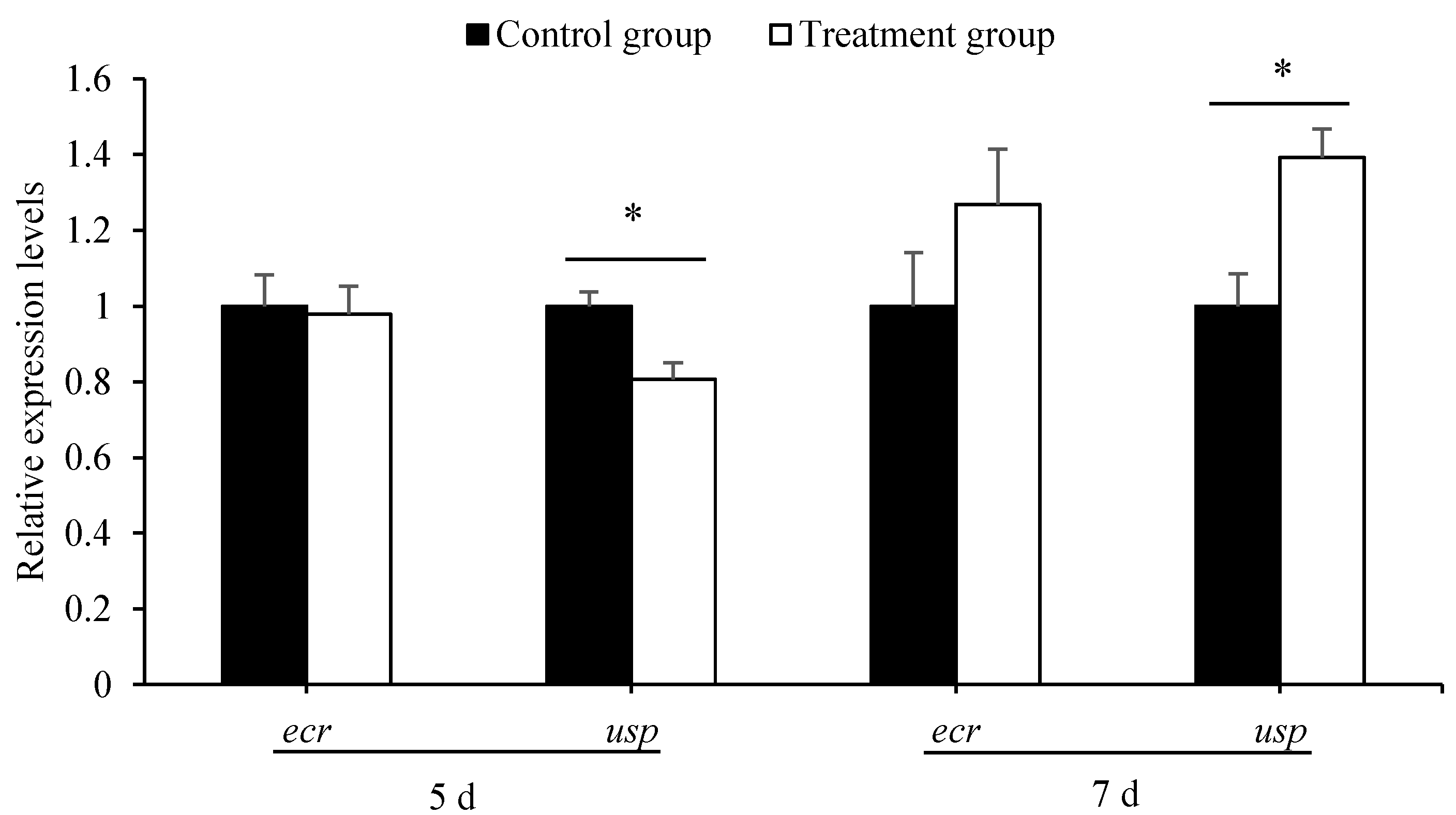
qPCR showed that the expression level of usp in 5-day-old larvae from the treatment group was significantly lower than that in larvae from the control groups (t = 3.359, p < 0.01). However, it was significantly higher in 7-day-old larvae from the treatment group than that from the control groups (t = −3.439, p < 0.01). However, the expression of ecr, in both 5- and 7-day-old larvae, was the same between the treatment groups and the control groups (for 5-day-old larvae, t = 0.201, p > 0.05; for 7-day-old larvae, t = −1.392, p > 0.05) (Figure 2).

3.4. Antibiotics Treatment Down-Regulated the Expression of Nutrient Metabolism Genes

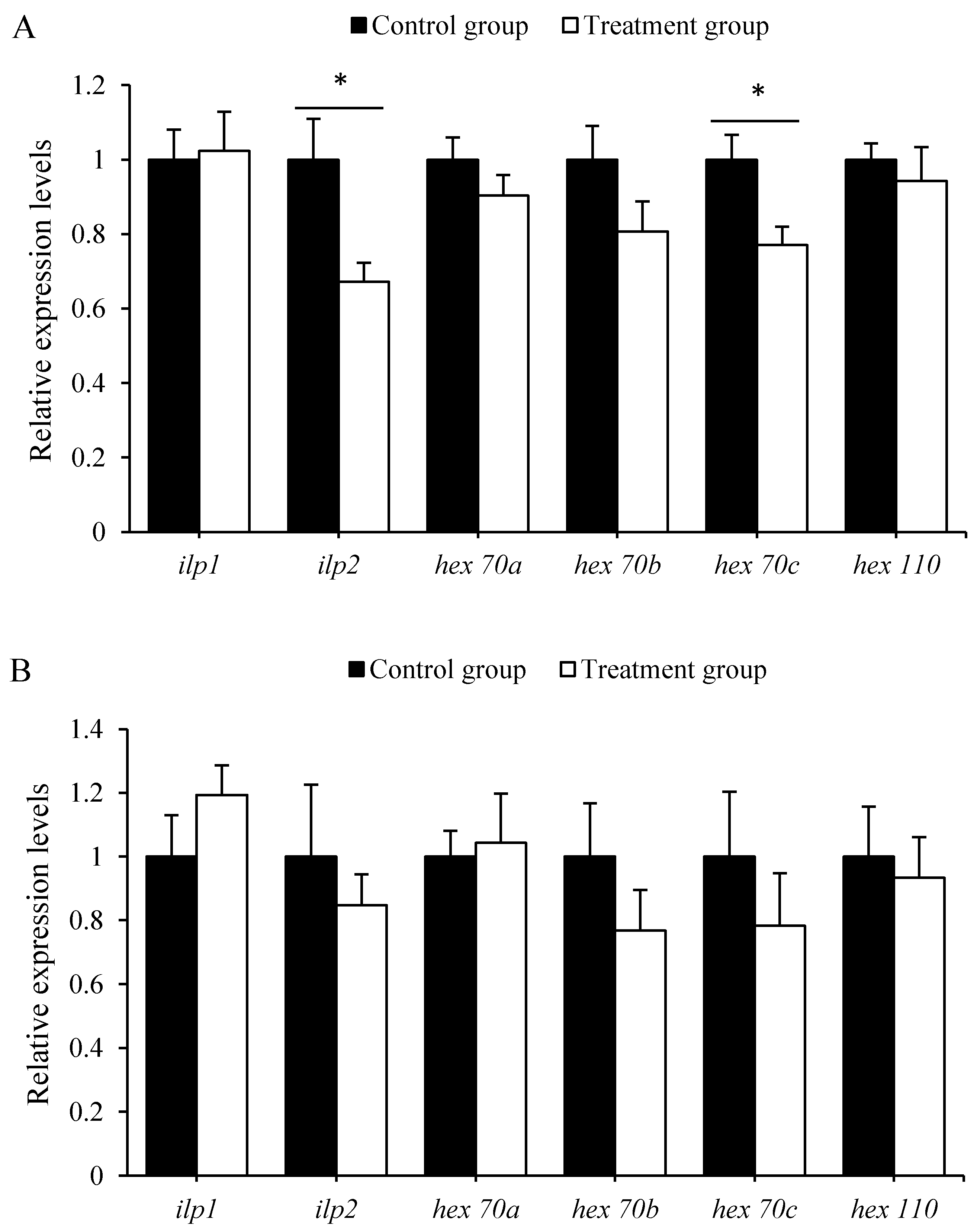
The qRT-PCR results showed that antibiotics can significantly down-regulate the expression of ilp2 (t = 2.726, p < 0.01) and hex 70c (t = 2.776, p <0.01) in 5-day-old larvae (Figure 3A). However, the expression levels of ilp1 (t = −0.181, p > 0.05), hex 70a (t = 1.188, p > 0.05), hex70b (t = 1.587, p > 0.05), and hex110 (t = 0.571, p > 0.05) in 5-day-old honeybee larvae, and ilp1 (t = −1.199, p > 0.05), ilp2 (t = 0.623, p > 0.05), hex 70a (t = −0.248, p > 0.05), hex70b (t = 1.102, p > 0.05), hex 70c (t = 0.83, p > 0.05), and hex110 (t = 0.33, p > 0.05) in 7-day-old honeybee larvae, were not statistically different between the treated and control groups (Figure 3B).

3.5. Antibiotics Treatment Decreased Immunity Genes Expression

The expression levels of apidaecin (t = 2.523, p < 0.05) and abaecin (t = 2.578, p < 0.05) in 5-day-old larvae, and defencin-1 (t = 2.444, p < 0.05) and hymenoptaecin (t = 2.278, p < 0.05) in 7-day-old larvae, were significantly decreased in treated groups (Figure 4). Therefby, antibiotics treatment down-regulated the expression of the immune genes in honeybee larvae.

4. Discussion

Many studies involving gut microbiota have provided a deeper understanding of the ecological and functional dynamics of gut environments and their benefit in the health of host honeybees [19,25,28,40,41]. Compared with the microbiota in mammals, the gut microbiota in honeybees is more simple and host-specific [19,42]. In recent years, the adult honeybee of A. mellifera has increased as a valuable model system for researching the relationship between its gut microbiota and the host [43]. However, the microbiota in honeybee larvae (primarily the Acetobacteraceae (Alpha2.2) and Lactobacillus) is generally considered less important due to its absence of host specificity and it being easily affected by the food [19,24]. Previous reports have shown that the microbiota in honeybee larvae also play a positive role in nutrient uptake and defense against pathogens [25,41,44]. In this study, to determine the effect of gut microbiota on the whole fitness of honeybee larvae, we disrupted the gut bacteria by using antibiotics and found that the treated larvae showed a lower fitness in development, nutrient metabolism, and immune competence than the control larvae.
Gut microbiota can supply amino acids, proteins and vitamins, and other essential materials, and regulate some pathways for host growth, development, and metabolism [45,46]. This result is consistent with previous reports that antibiotics cause rapid and profound alterations of gut microbiota [40,47]. Meanwhile, the decrease in gut microbiota leads to a reduction in nutrient metabolism and body weight, and extended the growth time of larvae. The body weight of honeybee larvae was determined by the nutrient intake and related metabolism. Our investigation showed that the body weights of 5-day-old larvae and newly emerged adults from the antibiotics-treated larvae were both significantly lower than the control. Moreover, the expression levels of nutrient metabolism-related genes, such as ilp2, hex 70a, hex 70b, hex70c, and hex 110, in 5- and 7-day-old antibiotics-treated larvae were lower than those of the controls. Nutrient-sensitive insulin-like peptides (ILPs) have profound effects on invertebrate metabolism, nutrient storage, fertility, and aging [48]. Hexamerins are the most abundant proteins in larval hemolymph and serve as storage proteins for gonad development, egg production, and foraging activity [49]. The expression levels of both ILPs and hexamerins reflected the nutritional status of larvae [50,51]. Our data indicate that the disruption of gut bacteria by antibiotics could decrease the expression of nutrient metabolism-related genes, which in turn, decreased the body weights of 5-day-old larvae and newly emerged adults. Thereby, gut microbiota in larvae could to some extent determine the host larvae’s nutrition state.
As a complete metamorphosis insect, the development rhythm was coordinately regulated by the juvenile hormone (JH) and molting hormone (20-hydroxyecdysone, 20E) [52,53]. The ecdysteroid receptor (EcR) and ultraspiracle protein (USP) were the key genes responsible for the transduction of the JH/20E signals during metamorphosis development. Our results showed that the antibiotics-treated larvae have significantly longer developmental periods than the control larvae, and the expression levels of usp in 5- and 7-day-old antibiotics-treated larvae were altered, which might disturb the normal development process leading to the prolonged development period of adult honeybees (Table 2). Such extension in the developmental periods of treated larvae might come from either the decreased hexamerins for building the pupae tissues or some metabolites for the synthesis of JH/20E by gut microbiota, which were already disrupted by antibiotics [49].
Successful immunity is indispensable for the survivorship of all organisms. Like other insects, honeybees have evolved innate immunity through synthesizing a variety of antimicrobial peptides (AMPs) by their fat bodies to defend against microbial infection [54], which are regulated by Toll, Imd, JNK, and JAK/STAT signaling pathways [47,55,56]. The vital immune regulating roles of gut bacteria have been well reported in adult honeybees [47]. We found here that the expression levels of apidaecin, abaecin in 5-day-old larvae and defensing-1, hymenoptaecin in 7-day-old larvae were significantly down-regulated in antibiotics-treated larvae (Figure 4). Therefore, gut bacteria in honeybee larvae, despite their lower host specificity, were essential in regulating larval immune response, which was similar to what happened in adult honeybees. Such a result verified that the antibiotics could cause a huge negative impact on larva bees as well. In practice, antibiotics were also used to control honeybee larval diseases, such as American Foulbrood Diseases (AFB) and European Foulbrood Diseases (EFB), caused by pathogenic bacteria [48]. However, the benefit of the control of the disease might not counteract the loss caused by using the antibiotic. The beekeeper should avoid using antibiotics for honeybee disease control.

5. Conclusions

Despite their variability and low host specificity, the gut microbiota in honeybee larvae could affect the host’s nutrient metabolism, increase the larval weight, and participate in the developmental regulation and immune response, which were similar to the roles of the gut microbiota played in adult bees. They increased the fitness of host larvae and were, therefore, indispensable for the development of honeybee larvae. The disruption of gut microbiota by antibiotics could decrease the nutrient metabolism, decrease the body weight, postpone the development, and lower the immunity of larvae. These results fulfill the biological functions of gut microbiota in honeybee larvae and provide fundamental information for future research and application in this field.

Author Contributions

Conceived and designed the experiments: X.D., B.Z., S.H. and J.L.; Performed the experiments: B.Z., X.J. and X.C.; Analyzed the data: B.Z., X.J., X.C. and S.H.; Wrote the paper: X.D., B.Z. and J.L. All authors have read and agreed to the published version of the manuscript.

Funding

This research was funded by the National Natural Science Foundation of China (31472156), the Special Foundation of Investigation on Basic Resources of Ministry of Science and Technology of China (2018FY100402), the Science and technology innovation special fund project of Fujian Agriculture and Forestry University (KFA17244A), and the Training Program of Innovation and Entrepreneurship for Undergraduates of Fujian province (201710389068).

Institutional Review Board Statement

Not applicable.

Data Availability Statement

Not applicable.

Acknowledgments

We are grateful for the assistance of all staff and students in the Bee health Group, Fujian Agriculture and Forestry University at Fuzhou, Fujian, China.

Conflicts of Interest

The authors declare no conflict of interest.

References

  1. Gallai, N.; Salles, J.M.; Settele, J.; Vaissiere, B.E. Economic valuation of the vulnerability of world agriculture confronted with pollinator decline. Ecol. Econ. 2009, 68, 810–821. [Google Scholar] [CrossRef]
  2. Champetier, A.; Sumner, D.A.; Wilen, J.E. The Bioeconomics of Honey Bees and Pollination. Environ. Resour. Econ. 2015, 60, 143–164. [Google Scholar] [CrossRef]
  3. Zheng, B.Y.; Zhao, B.A.; Jin, X.; Duan, X.L.; Huang, S.K.; Li, J.H. Effect of Sacbrood virus infection on nutritional and immune responses of Apis cerana cerana (Hymenoptera: Apidae). Acta Entomol. Sin. 2019, 62, 1054–1064. [Google Scholar]
  4. Van Der Zee, R.; Pisa, L.; Andonov, S.; Brodschneider, R.; Charriere, J.D.; Chlebo, R.; Coffey, M.F.; Crailsheim, K.; Dahle, B.; Gajda, A.; et al. Managed honey bee colony losses in Canada, China, Europe, Israel and Turkey, for the winters of 2008–9 and 2009–10. J. Apic. Res. 2012, 51, 100–114. [Google Scholar] [CrossRef]
  5. Van Dooremalen, C.; Cornelissen, B.; Poleij-Hok-Ahin, C.; Blacquière, T. Single and interactive effects of Varroa destructor, Nosema spp., and imidacloprid on honey bee colonies (Apis mellifera). Ecosphere 2018, 9, e02378. [Google Scholar] [CrossRef] [Green Version]
  6. Calatayud-Vernich, P.; Calatayud, F.; Simó, E.; Aguilar, J.A.P.; Picó, Y. A two-year monitoring of pesticide hazard in-hive: High honey bee mortality rates during insecticide poisoning episodes in apiaries located near agricultural settings. Chemosphere 2019, 232, 471–480. [Google Scholar] [CrossRef]
  7. Kablaua, A.; Eckertb, J.H.; Pistoriusb, J.; Sharbatia, S.; Einspanier, R. Effects of selected insecticidal substances on mRNA transcriptome in larvae of Apis mellifera. Pestic. Biochemi. Phys. 2020, 170, 104703. [Google Scholar] [CrossRef] [PubMed]
  8. Duan, X.L.; Xiong, M.Q.; Liu, W.B.; Zhao, B.A.; Huang, S.K.; Li, J.H. Effects of three fungicides on the activities of protective enzymes and detoxifying enzymes in Apis mellifera. Acta Pratac. Sin. 2020, 29, 74–82. [Google Scholar]
  9. Ley, R.E.; Hamady, M.; Lozupone, C.; Turnbaugh, P.J.; Ramey, R.R.; Bircher, J.S.; Schlegel, M.L.; Tucker, T.A.; Schrenzel, M.D.; Knight, R.; et al. Evolution of Mammals and Their Gut Microbes. Science 2008, 320, 1647–1651. [Google Scholar] [CrossRef] [Green Version]
  10. Clemente, J.C.; Ursell, L.K.; Parfrey, L.W.; Knight, R. The Impact of the Gut Microbiota on Human Health: An Integrative View. Cell 2012, 148, 1258–1270. [Google Scholar] [CrossRef] [Green Version]
  11. Nancy, A.M. Genomics of the honey bee microbiome. Curr. Opin. Insect Sci. 2015, 10, 22–28. [Google Scholar]
  12. Li, C.Y.; Zhou, X.; Zheng, H. Diversity and function of insect gut microbiome. Acta Microbiol. Sin. 2018, 58, 1016–1024. [Google Scholar]
  13. Cilia, G.; Fratini, F.; Tafi, E.; Mancini, S.; Turchi, B.; Mancini, S.; Sagona, S.; Nanetti, A.; Cerri, D.; Felicioli, A. Microbial Profile of the Ventriculum of Honey Bee (Apis mellifera ligustica Spinola, 1806) Fed with Veterinary Drugs, Dietary Supplements and Non-Protein Amino Acids. Vet. Sci. 2020, 7, 76. [Google Scholar] [CrossRef] [PubMed]
  14. Cilia, G.; Fratini, F.; Tafi, E.; Mancini, S.; Turchi, B.; Sagona, S.; Cerri, D.; Felicioli, A.; Nanetti, A. Changes of Western honey bee Apis mellifera ligustica (Spinola, 1806) ventriculus microbial profile related to their in-hive tasks. J. Apic. Res. 2020, 60, 198–202. [Google Scholar] [CrossRef]
  15. Kwong, W.K.; Mancenido, A.L.; Moran, N.A. Immune system stimulation by the native gut microbiota of honey bees. R. Soc. Open Sci. 2017, 4, 170003. [Google Scholar] [CrossRef] [Green Version]
  16. Ament, S.A.; Wang, Y.; Robinson, G.E. Nutritional regulation of division of labor in honey bees: Toward a systems biology perspective. WIREs Syst. Biol. Med. 2010, 2, 566–576. [Google Scholar] [CrossRef]
  17. Li, W.F.; Chen, Y.P.; Cook, S.C. Chronic Nosema ceranae infection inflicts comprehensive and persistent immuno-suppression and accelerated lipid loss in host Apis mellifera honey bees. Int. J. Parasitol. 2018, 48, 433–444. [Google Scholar] [CrossRef]
  18. Antonio, D.S.M.; Guidugli-Lazzarini, K.R.; do Nascimento, A.M.; Simoes, Z.L.P.; Hartfelder, K. RNAi-mediated silencing of vitellogenin gene function turns honeybee (Apis mellifera) workers into extremely precocious foragers. Naturwissenschaften 2008, 95, 953–961. [Google Scholar] [CrossRef]
  19. Zheng, H.; Powell, J.E.; Steele, M.I.; Dietrich, C.; Moran, N.A. Honeybee gut microbiota promotes host weight gain via bacterial metabolism and hormonal signaling. Proc. Natl. Acad. Sci. USA 2017, 114, 4775–4780. [Google Scholar] [CrossRef] [Green Version]
  20. Ortiz-Alvarado, Y.; Clark, D.R.; Vega-Melendez, C.J.; Flores-Cruz, Z.; Domingez-Bello, M.G.; Giray, T. Antibiotics in hives and their effects on honey bee physiology and behavioral development. Biol. Open 2020, 9, bio053884. [Google Scholar] [CrossRef] [PubMed]
  21. Evans, J.D.; Armstrong, T.N. Inhibition of the American foulbrood bacterium, Paenibacillus larvae larvae, by bacteria isolated from honey bees. J. Apic. Res. 2005, 44, 168–171. [Google Scholar] [CrossRef]
  22. McFrederick, Q.; Wcislo, W.T.; Taylor, D.R.; Ishak, H.D.; Dowd, S.E.; Mueller, U. Environment or kin: Whence do bees obtain acidophilic bacteria? Mol. Ecol. 2012, 21, 1754–1768. [Google Scholar] [CrossRef]
  23. Emery, O.; Schmidt, K.; Engel, P. Immune system stimulation by the gut symbiont Frischella perrara in the honey bee (Apis mellifera). Mol. Ecol. 2017, 26, 2576–2590. [Google Scholar] [CrossRef] [PubMed]
  24. Koch, H.; Schmid-Hempel, P. Socially transmitted gut microbiota protect bumble bees against an intestinal parasite. Proc. Natl. Acad. Sci. USA 2011, 108, 19288–19292. [Google Scholar] [CrossRef] [Green Version]
  25. Vojvodic, S.; Rehan, S.M.; Anderson, K.E. Microbial Gut Diversity of Africanized and European Honey Bee Larval Instars. PLoS ONE 2013, 8, e72106. [Google Scholar] [CrossRef] [PubMed] [Green Version]
  26. Forsgren, E.; Olofsson, T.C.; Váasquez, A.; Fries, I. Novel lactic acid bacteria inhibiting Paenibacillus larvae in honey bee larvae. Apidologie 2010, 41, 99–108. [Google Scholar] [CrossRef] [Green Version]
  27. Evans, J.D.; Lopez, D.L. Bacterial probiotics induce an immune response in the honey bee (Hymenoptera: Apidae). J. Econ. Entomol. 2004, 97, 752–756. [Google Scholar] [CrossRef] [PubMed]
  28. Janashia, I.; Alaux, C. Specific immune stimulationby endogenous bacteria in honey bees (Hymenoptera: Apidae). J. Econ. Entomol. 2016, 109, 1474–1477. [Google Scholar] [CrossRef]
  29. Mutti, N.S.; Dolezal, A.G.; Wolschin, F.; Mutti, J.S.; Gill, K.S.; Amdam, G.V. IRS and TOR nutrient-signaling pathways act via juvenile hormone to influence honey bee caste fate. J. Exp. Biol. 2011, 214, 3977–3984. [Google Scholar] [CrossRef] [Green Version]
  30. Jensen, A.B.; Pedersen, B.V.; Eilenberg, J. Differential susceptibility across honey bee colonies in larval chalk-brood resistance. Apidologie 2009, 40, 524–534. [Google Scholar] [CrossRef] [Green Version]
  31. Vojvodic, S.; Jensen, A.B.; James, R.R.; Boomsma, J.J.; Eilenberg, J. Temperature dependent virulence of obligate and facultative fungal pathogens of honeybee brood. Vet. Microbiol. 2011, 149, 200–205. [Google Scholar] [CrossRef] [PubMed]
  32. Nie, H.Y.; Wang, X.Y.; Gao, Y.; Lin, Y.; Zhu, Y.N.; Yang, D.L.; Li, Z.G.; Su, S.K. Changes in the gene expression of chalkbrood resistance in Apis mellifera larvae infected by Ascosphaera apis. Apidologie 2020, 51, 35–47. [Google Scholar] [CrossRef]
  33. Simone, M.; Evans, J.D.; Spivak, M. Resin collection and social immunity in honey bees. Evolution 2009, 63, 3016–3022. [Google Scholar] [CrossRef] [PubMed]
  34. Schwarz, R.Z.; Moran, N.A.; Evans, J.D. Early gut colonizers shape parasite susceptibility and microbiota composition in honey bee workers. Proc. Natl. Acad. Sci. USA 2016, 113, 9345–9350. [Google Scholar] [CrossRef] [Green Version]
  35. Liu, C.D.; Wang, Q.; Yu, H.; Zhang, Z.B.; Zheng, B.Y.; Li, J.H. Expression and functional analysis of early-response factors ecr and usp of ecdysteroid in different castes of Apis mellifera. Genom. Appl. Biol. 2018, 37, 686–692. [Google Scholar]
  36. De Azevedo, S.V.; Hartfelder, K. The insulin signaling pathway in honey bee (Apis mellifera) caste development-differential expression of insulin-like peptides and insulin receptors in queen and worker larvae. J. Insect Physiol. 2008, 54, 1064–1071. [Google Scholar] [CrossRef]
  37. Zhang, G.D. Transcription Analysis of Hexamerin in Different Development Stage of Honeybee (Apis mellifera). Master’s Thesis, Fujian Agriculture and Forestry University, Fuzhou, China, 2014. [Google Scholar]
  38. Evans, J.D.; Aronstein, K.; Chen, Y.P.; Hetru, C.; Imler, J.L.; Jiang, H.; Kanost, M.; Thompson, G.J.; Zou, Z.; Hultmark, D. Immune pathways and defence mechanisms in honey bees Apis mellifera. Insect Mol. Biol. 2006, 15, 645–656. [Google Scholar] [CrossRef] [Green Version]
  39. Livak, K.J.; Schmittgen, T.D. Analysis of relative gene expression data using realtime quantitative PCR and the 2−ΔΔCT method. Methods 2001, 25, 402–408. [Google Scholar] [CrossRef]
  40. Raymann, K.; Bobay, L.M.; Moran, N.A. Antibiotics reduce genetic diversity of core species in the honeybee gut microbiome. Mol. Ecol. 2018, 27, 2057–2066. [Google Scholar] [CrossRef] [PubMed]
  41. Khan, K.A.; Al-Ghamdi, A.A.; Ghramh, H.A.; Ansari, M.J.; Ali, H.; Alamri, S.A.; Kahtani, S.N.A.; Adgaba, N.; Qasim, M.; Hafeez, M. Structural diversity and functional variability of gut microbial communities associated with honey bees. Microb. Pathog. 2019, 138, 103793. [Google Scholar] [CrossRef]
  42. Kwong, W.K.; Moran, N.A. Gut microbial communities of social bees. Nat. Rev. Microbiol. 2016, 14, 374–384. [Google Scholar] [CrossRef] [PubMed]
  43. Romero, S.; Nastasa, A.; Chapman, A.; Kwong, W.K.; Foster, L.J. The honey bee gut microbiota: Strategies for study and characterization. Insect Mol. Biol. 2019, 28, 455–472. [Google Scholar] [CrossRef] [Green Version]
  44. Martinson, V.G.; Moy, J.; Moran, N.A. Establishment of Characteristic Gut Bacteria during Development of the Honeybee Worker. Appl. Environ. Microbiol. 2012, 78, 2830–2840. [Google Scholar] [CrossRef] [PubMed] [Green Version]
  45. Singh, A.S.; Shah, A.; Brockmann, A. Honey bee foraging induces upregulation of early growth response protein 1, hormone receptor 38 and candidate downstream genes of the ecdysteroid signalling pathway. Insect Mol. Biol. 2017, 27, 90–98. [Google Scholar] [CrossRef] [Green Version]
  46. Lu, C.Y.; Huang, P.J.; Hsu, C.Y. The cholesterol-hydroxyecdysone-vitellogenin pathway is involved in the longevity of trophocytes and oenocytes of queen honey bees (Apis mellifera). Apidologie 2018, 49, 721–733. [Google Scholar] [CrossRef] [Green Version]
  47. Li, J.H.; Evans, J.D.; Li, W.F.; Zhao, Y.Z.; De Grandi-Hoffman, G.; Huang, S.K.; Li, Z.G.; Hamilton, M.; Chen, Y.P. New evidence showing that the destruction of gut bacteria by antibiotic treatment could increase the honey bee’s vulnerability to Nosema infection. PLoS ONE 2017, 12, e0187505. [Google Scholar] [CrossRef]
  48. Flatt, T.; Tu, M.P.; Tatar, M. Hormonal pleiotropy and the juvenile hormone regulation of Drosophila development and life history. BioEssays 2005, 27, 999–1010. [Google Scholar] [CrossRef]
  49. Martins, J.R.; Nunes Francis, M.F.; Cristino, A.S.; Simes Zilá, L.P.; Bitondi Márcia, M.G. The four hexamerin genes in the honey bee: Structure, molecular evolution and function deduced from expression patterns in queens, workers and drones. BMC Mol. Bio. 2010, 11, 23. [Google Scholar] [CrossRef] [Green Version]
  50. Kanost, M.R. Encyclopedia of Insects, 2nd ed.; Elsevier: Amsterdam, The Netherlands, 2009; Chapter 17; pp. 446–449. ISBN 978-0-12-374144-8. [Google Scholar]
  51. Nilsen, K.A.; Ihle, K.E.; Frederick, K.; Fondrk, M.K.; Smedal, B.; Hartfelder, K.; Amdam, G.V. Insulin-like peptide genes in honey bee fat body respond differently to manipulation of social behavioral physiology. J. Exp. Biol. 2011, 214, 1488–1497. [Google Scholar] [CrossRef] [Green Version]
  52. Ryoo, H.D.; Baehrecke, E.H. Distinct death mechanisms in Drosophila development. Curr. Opin. Cell Biol. 2010, 22, 889–895. [Google Scholar] [CrossRef] [PubMed] [Green Version]
  53. Liu, H.; Wang, J.; Li, S. E93 predominantly transduces 20-hydroxyecdysone signaling to induce autophagy and caspase activity in Drosophila fat body. Insect Biochem. Mol. Biol. 2014, 45, 30–39. [Google Scholar] [CrossRef] [PubMed]
  54. Danihlík, J.; Aronstein, K.; Petrivalsky, M. Antimicrobial peptides: A key component of honey bee innate immunity physiology, biochemistry, and chemical ecology. J. Apic. Res. 2015, 54, 123–136. [Google Scholar] [CrossRef]
  55. Bullet, P.; Hetru, C.; Dimarcq, J.L.; Hoffmann, D. Antimicrobial peptides in insects; structure and function. Dev. Comp. Immunol. 1999, 23, 329–344. [Google Scholar] [CrossRef]
  56. Lemaitre, B.; Hoffmann, J. The Host Defense of Drosophila melanogaster. Annu. Rev. Immunol. 2007, 25, 697–743. [Google Scholar] [CrossRef] [PubMed] [Green Version]
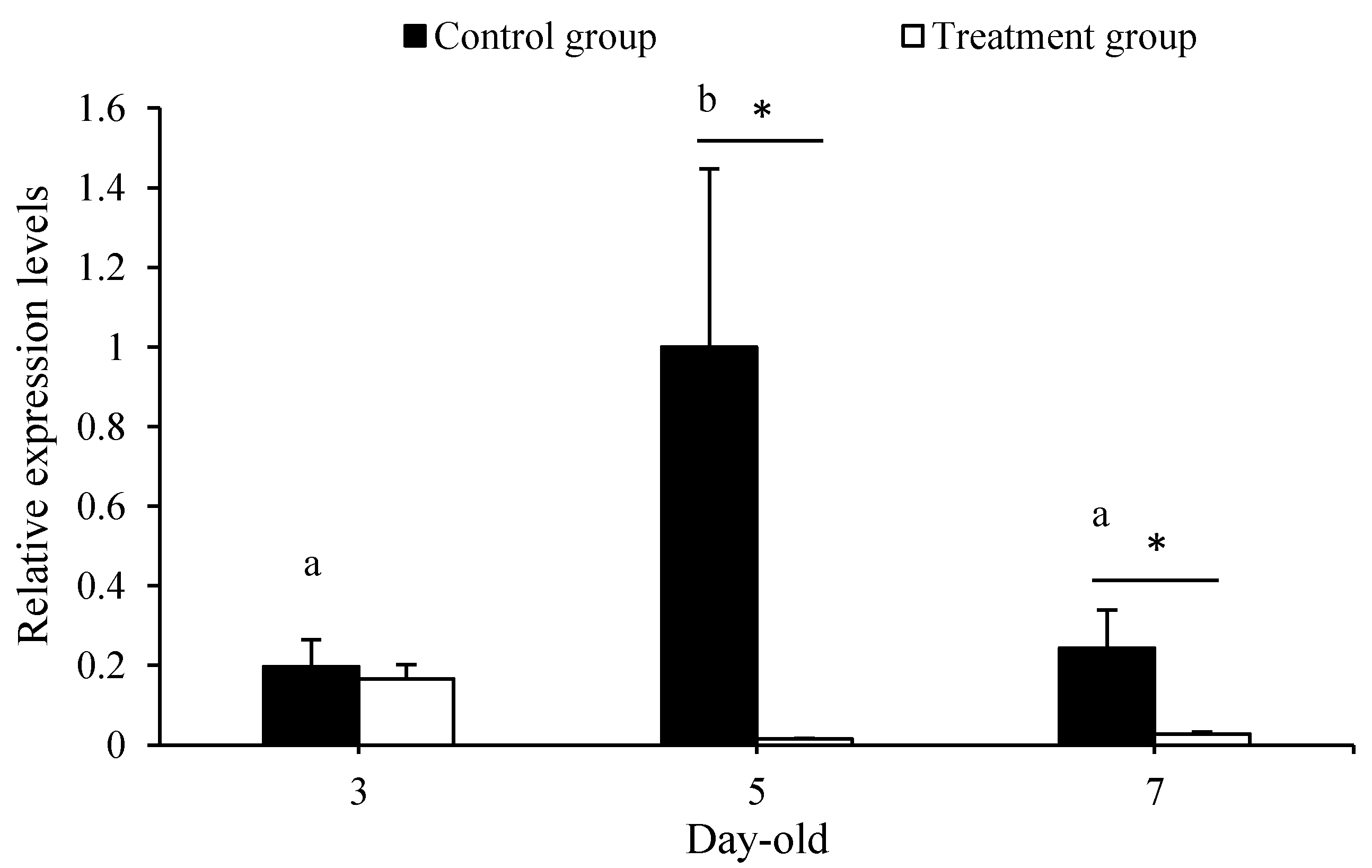
Figure 1. Antibiotics treatment effect on the amount of gut bacteria in larvae of different ages. The different letters on the error bars indicate the significant differences in gut bacteria amount among the 3-, 5-, and 7-day-old larvae at the 0.05 level. (Tukey’s test). The asterisk above the bars indicates a significant difference in gut bacteria amount between the treatment and corresponding control (* for p < 0.05).
Insects 12 00301 g001
Figure 2. Antibiotics treatment effect on the expression of two development-regulation genes in 5- and 7-day-old A. mellifera larvae. The asterisk above the bars indicates a significant difference in gene expression level between the treatment and corresponding control (* for p < 0.05).
Insects 12 00301 g002
Figure 3. Antibiotics treatment effect on the expression of six nutrient metabolism-related genes in 5-(A) and 7-day-old (B) A. mellifera larvae. The asterisk above the bars indicates a significant difference in gene expression level between the treatment and corresponding control (* for p < 0.05).
Insects 12 00301 g003
Figure 4. Antibiotics treatment effect on the expression of four innate immunity genes in 5- (A) and 7-day-old (B) A. mellifera larvae. The asterisk above the bars indicates a significant difference in gene expression level between the treatment and corresponding control (* for p < 0.05 and ** for p < 0.01).
Insects 12 00301 g004
Table 1. Primers used for quantitative PCR.
GenesPrimer Sequences (5′-3′)Gene IDReference
Reference geneβ-actinF: TTGTATGCCAACACTGTCCTTT
R: TGGCGCGATGATCTTAATTT
NM_001185145.1Simone et al. [33]
16S rRNA geneunibacF: AGAGTTTGATCCTGGCTCAG
R: CTGCTGCCTCCCGTAGGAGT
Schwarz et al. [34]
Development-related genesecrF: TGCCAACACTGTCCTTTCTG
R: AAAGAGCCAGGCTGCGACAA
XM_016913298.2Liu et al. [35]
Liu et al. [35]
uspF: GCCAAGATGATGAAGAAGGAGA
R: GGTTGATGAGGCTTGCTGTGTC
NM_001011634.2
Nutrient metabolism-related genesilp1F: CGATAGTCCTGGTCGGTTTG
R: CAAGCTGAGCATAGCTGCAC
XM_026442143.1De Azevedo et al. [36]
ilp2F: TTCCAGAAATGGAGATGGATG
R: TAGGAGCGCAACTCCTCTGT
NM_001177903.1
hex 70aF: GCCTTCAGTTTGGTCGGTGC
R: GCTGGTTGAGCGACGCGATA
NM_001110764.1Zhang [37]
hex 70bF: AACAGCCACGAATCCGTCTT
R: CAGGCTTGTCCAGAGGGAAG
NM_001011600.1Zheng et al. [3]
Zheng et al. [3]
Zheng et al. [3]
hex 70cF: TAAGGCAGGCAGACTTGAGC
R: AAACGCTGTGGTGAACATGC
NM_001098717.1
hex 110F: ACGGACAATACCCGAACACC
R: AGCATGCTGATGCCTCTGTT
NM_001101023.1
Innate virus immunity genesabaecinF: CAGCATTCGCATACGTACCA
R: GACCAGGAAACGTTGGAAAC
NM_001011617.1Simone et al. [33]
Evans et al. [38]
Evans et al. [38]
hymenoptaecinF: CTCTTCTGTGCCGTTGCATA
R: GCGTCTCCTGTCATTCCATT
NM_001011615.1
defencin-1F: TGCGCTGCTAACTGTCTCAG
R: AATGGCACTTAACCGAAACG
NM_001011616.2
apidaecinF: TTTTGCCTTAGCAATTCTTGTTG
R: GTAGGTCGAGTAGGCGGATCT
NM_001011613.1Evans et al. [38]
Table 2. Effect of antibiotics on development period and body weight of honeybee larvae.
TreatmentDevelopment Period (h)Body Weight (mg)
PupationEclosion5-Day-Old LarvaeNewly Emerged Adults
Control group212.79 ± 0.90406.94 ± 1.60128.68 ± 2.95113.46 ± 2.69
Treatment group213.19 ± 0.72413.69 ± 1.59 **114.62 ± 3.39 **102.38 ± 2.30 **
The asterisk above the bars indicates a significant difference in development period and body weight between the treatment and corresponding control (** for p < 0.01).
Publisher’s Note: MDPI stays neutral with regard to jurisdictional claims in published maps and institutional affiliations.