Article

Variant A of the Deformed Wings Virus Alters the Olfactory Sensitivity and the Expression of Odorant Binding Proteins on Antennas of Apis mellifera

1
Laboratorios de Virología y Patologías en Abejas, Facultad de Agronomía, Universidad de Concepción, Av. Vicente Méndez 595, Chillán 3780000, Chile
2
Laboratorio de Ecología Química, Instituto de Investigaciones Agropecuarias, INIA Quilamapu, Av. Vicente Méndez 515, Chillán 3780000, Chile
3
Centro de Investigación Austral Biotech, Facultad de Ciencias, Universidad Santo Tomás, Av. Picarte 1130–1160, Valdivia 5090000, Chile
4
Unité de Recherche Abeilles et Environnement, INRAE, F-84000 Avignon, France
*
Author to whom correspondence should be addressed.
Insects 2021, 12(10), 895; https://doi.org/10.3390/insects12100895
Submission received: 15 July 2021 / Revised: 7 September 2021 / Accepted: 23 September 2021 / Published: 1 October 2021
(This article belongs to the Section Insect Behavior and Pathology)

Abstract

:

Simple Summary

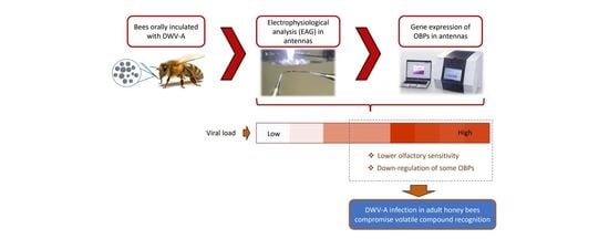
Honey bees, Apis melllifera, are the most commonly managed bee in the world for pollination services. However, worldwide continuous colony losses have been reported for almost a decade. One factor of these losses is associated to pathogens being the virus one of the most important problems in honey bee health. One of the known viruses that affect the honey bee population is deformed wing virus (DWV). DWV causes physical malformation and behavioral disturbances, but also, this virus can be found in the antenna affecting the anatomical integrity of infected areas, which could compromise normal antennal functioning associated to aroma perception. Thus, we evaluate olfactory sensitivity and the expression of antenna-specific odorant-binding proteins (OBP) genes in honey bees inoculated with variant A of the DWV. We performed olfactory sensitivity analysis using the essential oils Eucalyptus globulus and Mentha piperita, but also, and molecular analysis of gene expression of nine OBPs. We found that the high level of replication of DWV-A in the antennae decreased the olfactory sensitivity and led to a down-regulation of some OBPs in middle- and forager-age worker bees. Thus, DWV-A infection in adults of honey bees could compromise volatile compound recognition inside the hive and outside the hive.

Abstract

Insects have a highly sensitive sense of smell, allowing them to perform complex behaviors, such as foraging and peer recognition. Their sense of smell is based on the recognition of ligands and is mainly coordinated by odorant-binding proteins (OBPs). In Apis mellifera, behavior can be affected by different pathogens, including deformed wing virus (DWV) and its variants. In particular, it has been shown that variant A of DWV (DWV-A) is capable of altering the ultra-cellular structure associated with olfactory activity. In this study was evaluated olfactory sensitivity and the expression of OBP genes in honey bees inoculated with DWV-A. Electroantennographic analyses (EAG) were carried out to determine the olfactory sensitivity to the essential oils Eucalyptus globulus and Mentha piperita. The expression of nine antenna-specific OBP genes and DWV-A load in inoculated bees was also quantified by qPCR. We observed an inverse relationship between viral load and olfactory sensitivity and the expression of some OBP proteins. Thus, high viral loads reduced olfactory sensitivity to essential oils and the gene expression of the OBP2, OBP5, OBP11, and OBP12 proteins on the antennas of middle- and forager-age bees. These results suggest that DWV-A could have negative effects on the processes of aroma perception by worker bees, affecting their performance in tasks carried out in and outside the colony.

Graphical Abstract

1. Introduction

Pollination is one of the key processes in ecosystem services and agricultural production [1,2,3]. However, a significant decrease in the population of pollinating insects has been observed, particularly in Apis mellifera L. [4,5,6,7], attributed in part to pathogens that affect this species [4,8]. Among these pathogens, viruses are considered one of the most important problems in honey bee health [9,10]. In fact, compared to other pathogens, viruses can go unnoticed in honey bees because they often do not present clinical symptoms that allow their rapid detection [11]. One of the known viruses that affect the honey bee population is deformed wing virus (DWV). To date, three variants of DWV with epidemiological importance have been identified (A, B, and C); types A and C have been associated with winter colony losses in Europe [12,13,14,15].
DWV causes wing deformities, abdominal swelling, paralysis, and behavioral disturbances [15,16,17]. The DWV infection has been reported to alter molecular learning mechanisms, impairing the memorization process and altering the central and peripheral nervous systems [18]. Furthermore, DWV can replicate in the brain and alter the processing of sensory information [19]. Additionally, viral particles have been found in the optic and antennal lobes of the brain, which could compromise the vision and olfactory senses of honey bees [19,20]. More recently, it has been reported that variant-A (DWV-A) is capable of replicating in the epithelial cells of antennas, affecting the anatomical integrity of infected areas, which could compromise normal antennal functioning [21].
In fact, the olfactory system located mainly in the sensilla of the antennas plays a crucial role for insects, coordinating behaviors such as looking for and recognizing food sources, avoiding and identifying predators, and partner selection. Additionally, for social insects, such as bees, the olfactory system is essential in coordinating social events, beehive management, maintaining internal cohesion through the perception of pheromones, and defense-related behaviors [22,23]. The sensitivity, perception, and interpretation of aromas in the environment involve a great diversity of molecules that interact in the olfactory sensory cells; odor binding proteins (OBPs) are among these [24,25,26]. These small water-soluble molecules, located in the sensory dendrite, present selective binding to different compounds [25,27], such as hydrophobic odorants and pheromones, characteristic of environmental aromas [24,28]. The amount and diversity of OBPs vary among insect species, but in A. mellifera, a total of 21 OBPs have been identified, with only nine of the specific expression in the antennas [24]. Thus, the presence of DWV in the antenna could intervene in aroma recognition processes, possibly due to alterations in the expression of OBPs. For these reasons, the aim of this study was to evaluate the olfactory perception and gene expression of OBPs in antennas of worker bees that were inoculated with variant A of the deformed wing virus (DWV-A), with non-inoculated worker bees as a control. This study furthers our understanding of the magnitude of harm caused by the DWV-A infection in the antennas of honey bees and how this infection affects olfactory perception and could possibly alter bee behavior at the individual and colony level.

2. Materials and Methods

2.1. Inoculum Preparation

An isolate of deformed wing virus variant-A (DWV-A) was obtained from infected colonies, according to Gusachenko et al. [29], and was subsequently used. A group of 20 bees was homogenized in a Stomacher bag with phosphate-buffered saline (1X PBS) for 90 s at high speed in a Stomacher 80 Lab Blender (Seward, London, UK). The samples were centrifuged twice at 1500× g for 10 min, followed by 10,000× g for 10 min, both at 4 °C. The supernatant was purified by filtration with a 0.22 µM filter (PES, Merck Millipore, Darmstadt, Germany) and treated with RNase to destroy all unencapsidated RNA, according to Skubnik et al. [30]. Later, 200 µL of the supernatant was used for RNA extraction, cDNA synthesis, and the subsequent viral load quantification of the inoculum, according to the methodology detailed in the section “RNA extraction, cDNA synthesis, and real-time PCR quantification (qPCR)”. The rest of the extracted inoculum was stored at −80 °C until later use.

2.2. Bee Inoculation

Adult A. mellifera for the assay were supplied by the experimental apiary located in the Experimental Center “El Nogal” (36°35′58.25″ S–72°04′51.77″ W), Universidad de Concepción, Chillán, Chile. Before starting the assay, the health status of the apiary was determined by identifying the viral level in each colony (n = 54 beehives). Thus, the pathogen level was determined by molecular techniques, according to Vargas et al. [31]. Brood combs with capped worker bees were then removed from colonies with low DWV (1.0 × 10² copy number per bee), considering that no colonies were detected DWV-free. The brood combs were maintained under controlled conditions in a rearing room (30 °C ± 1; 60% ± 3 RH). Afterwards, newly emerged worker bees (24 h old) were carefully collected from the brood combs and randomly confined in plastic cages (base = 8 cm diameter, mouth = 10 cm diameter, and height = 15 cm). Two treatments were then established. One group of bees were inoculated (I-DWV treatment) orally with 5 µL of a viral suspension (1.0 × 10⁹ copy number per bee) in a 50% sucrose solution, according to the methodology reported by Porrini et al. [32]. Another group of bees were not inoculated with the viral suspension (N-DWV treatment), as a control. Those bees that did not consume the total amount of viral inoculum were discarded from the assay. Then, 60 worker bees were maintained in plastic cages (3 replicates per treatment) containing 3 g of pollen substitute and supplied with 60% sucrose syrup ad libitum, according to Arismendi et al. [33]. Five bees were removed from each cage of each treatment every 3 days, up to a maximum of 21 days post-inoculation (23-day-old bees).

2.3. Stimulus Preparation

As a stimulus, the volatile fraction of the essential oils of Mentha piperita and Eucalyptus globulus leaves, both described as forage species of A. mellifera, were used [34,35,36,37]. In the summer of 2019, we collected leaves from ornamental specimens of both species in Chillán, Ñuble Region, and Chile and obtained 270 and 365 g from M. piperita and E. globulus, respectively. The leaves were air-dried at room temperature for 48 h and then boiled for 8 h to obtain the essential oil by hydro-distillation using a Clevenger apparatus [38]. After this extraction, we added sodium sulfate anhydrous (Na2SO4) to remove water from the samples, after which they were stored at 8 °C in complete darkness. We prepared four diluted concentrations in hexane (≥99% GC, Sigma-Aldrich, Munich, Germany) at 0.01, 0.1, 1.0, and 10.0 mg/mL as stimuli for the electrophysiological test.

2.4. Antennal Response

The olfactory sensitivity of adults of A. mellifera was evaluated by electroantennography (EAG). The antennas, stimulus receptor organs, were used for the experiment once they were removed from the sampled individuals. One antenna was immediately stored at −80 °C, and the other was used in the EAG record. After testing, this antenna was returned and stored at −80 °C for further molecular analyses. The antennas were extirpated on the base of the scape with a scalpel, and the first segment of the distal end was cut with dissecting scissors to improve the connection. The excised antennas were mounted between glass electrodes filled with a conductive saline solution of KCl (0.1 M) and 0.1% polyvinylpyrrolidone [39]. A volume of 10 µL of stimuli, at different concentrations, was deposited on filter paper (Whatman N°1, Whatman®, Sigma-Aldrich, Darmstadt, Germany) and was then delivered to the antenna at 40 mL/s; thus, the duration of each stimulation was 0.2 s using a stimulus controller (CS-55, Syntech, Hilversum, The Netherlands), with a period of at least 30 s between each stimulation to permit the recovery of the antennas. Each antenna was stimulated 3 times for each concentration of essential oil; additionally, each antenna was stimulated with a target (air) and a control (Hexane ≥99% GC, Sigma-Aldrich, Munich, Germany). The data were recorded using the EAG 2014 Software (Syntech, Hilversum, The Netherlands) for the acquisition and analysis of the depolarizations obtained during the electroantennography.

2.5. RNA Extraction and cDNA Synthesis

For the RNA extraction and subsequent quantification of viral load (DWV) and OBP gene expression, a pool of 10 antennas from 5 bees (including antennas used in the electroantennography assay) were used. The RNA extraction was carried out according to Kim et al. [21] and Mondet et al. [20]. In brief, the antennas were cooled with liquid nitrogen and then, were ground by adding 500 µL of TrizolTM (Invitrogen, Thermo Fisher Scientific, Waltham, MA, USA) and 5 µL of Carrier RNA, according to the manufacturer’s recommendations (Invitrogen, Thermo Fisher Scientific, Waltham, MA, USA). The homogenate was incubated in the cold for 5 min, then it was transferred to a 1.7 mL tube, and 100 µL of chloroform was added and incubated at 4 °C for 3 min. It was then centrifuged for 15 min at 10,300× g at 4 °C, and finally, the supernatant was collected for the RNA extraction. The RNA extraction was carried out following the instructions provided by E.Z.N.A. Total RNA kit I (Omega Bio-Tek, Norcross, GA, USA). RNA quality and yield were determined using a spectrometer (Infinite 200 PRO NanoQuant, Tecan Group, Männedorf, Switzerland), which was stored at −80 °C. The extracted RNA was subsequently used for first-strand cDNA synthesis, by using the enzyme reverse transcriptase M-MLV (Invitrogen, Life Technologies, Carlsbad, CA, USA), according to the manufacturer’s instructions. The cDNA samples were stored at −20 °C for later use.

2.6. Real-Time PCR Quantification (qPCR)

To quantify the viral load and expression of OBP genes, specific primers were used (Table 1). The PCR reaction was carried out using 1× of KAPA SYBR FAST Universal 2× qPCR Master Mix (Kapa Biosystems, Wilmington, MA, USA), following the supplier’s instructions. Samples were brought to a reaction volume of 15 µL, including 20 ng of cDNA, 530 nM of each primer, and sterile filtered molecular-grade water to reach 15 µL. The thermal reaction conditions used were 96 °C for 10 min, followed by 40 cycles at 95 °C for 15 s, 60 °C for 15 s, and 72 °C for 30 s. Real-time PCR assays were performed on a Stratagene Mx3000P thermal cycler (Agilent Technologies, Santa Clara, CA, USA), and the data were analyzed using MxPro software (Stratagene, Agilent Technologies, Santa Clara, CA, USA). The relative expression of each OBP gene was calculated after normalization with an endogen gene (β-actin), as described by Pfaffl [40]. Similarly, to determine the viral load in the antennas, absolute quantification of DWV-A was performed. In brief, a standard curve using purified PCR product (Wizard®VR SV gel and PCR clean-up system, Promega, Madison, WI, USA), belonging to the viral target sequence, was used [41]. The purified amplicon was then quantified by spectrophotometry (EpochTM Microplate Spectrophotometer, BioTek, Winooski, VT, USA) to calculate the copy number, according to Wu et al. [42]. Thus, linear standard curves (95–100% efficiency) were then generated using a serial dilution (1.0 × 101 to 1.0 × 10⁹) of viral copy numbers of purified cDNA. Afterwards, the Ct values were plotted against copy number values (log10). Thus, the sample copy numbers were estimated using the Ct values and comparisons with the linear equation of the standard curve and normalizing values of the housekeeping β -actin gene [43]. Data were then expressed as the copy number of DWV per bee by considering the dilutions that were performed in the cDNA synthesis and qPCR reaction.

2.7. Data Analysis

To meet the assumptions for normality and homogeneity of variances, the EAG values were transformed to square roots (√x). A general linear model (GLM) was then used to design a split-split-plot ANOVA with data of the EAG response. Thus, honey bee age (2, 5, 8, 11, 14, 17, 20, and 23 days old) was considered as the main factor (and levels), and the (1) essential oil concentrations (0.01, 0.1, 1.0, and 10.0 mg/mL) and (2) viral status (bees inoculated and non-inoculated with DWV-A) were considered as split factors in function to the EAG response (response variable) to stimuli elicited by the volatile fractions of essential oils from E. globulus and M. piperita. The Bonferroni test (p < 0.05) was then run to separate means between treatments.
Statistical differences in the viral loads in honey bees according to (1) viral status (non-inoculated bees collected from frames that were detected with a basal low level of DWV-A (N-DWV) and those that were additionally inoculated with DWV-A (I-DWV)) and (2) bee ages (honey bees 2 to 23 days old) were estimated by Factorial ANOVA. Tukey’s HSD test (p < 0.05) was then used to separate means between treatments.
Considering that OBPs expression values were correlated in bees that were inoculated and non-inoculated with DWV-A, a multivariable analysis (MANOVA) was carried out. Thus, honey bee age and viral status were considered independent variables in function to OBPs relative expression (response variable). After this, means were separated between treatments with a Bonferroni test (p < 0.05). In cases that detected statistical differences, the results were graphically represented on a heat-map illustrating the intensity of the OBPs gene expression in the antennae of worker honey bees that were inoculated with DWV-A in contrast with non-inoculated bees at different ages.
Additionally, Spearman’s correlations were performed to estimate relationships among the viral load, the relative expression of OBP genes, and EAG values in DWV-A inoculated bees. For the Spearman correlation, the data from worker bees that were 8 days old or more were considered because, from this period on, the viral load was significantly higher in inoculated bees compared to those that were not inoculated. All analyses were carried out with STATISTICA 7.0 software (StatSoft, Tulsa, OK, USA).

3. Results

3.1. Electroantennography Responses and Viral Load in Antennas

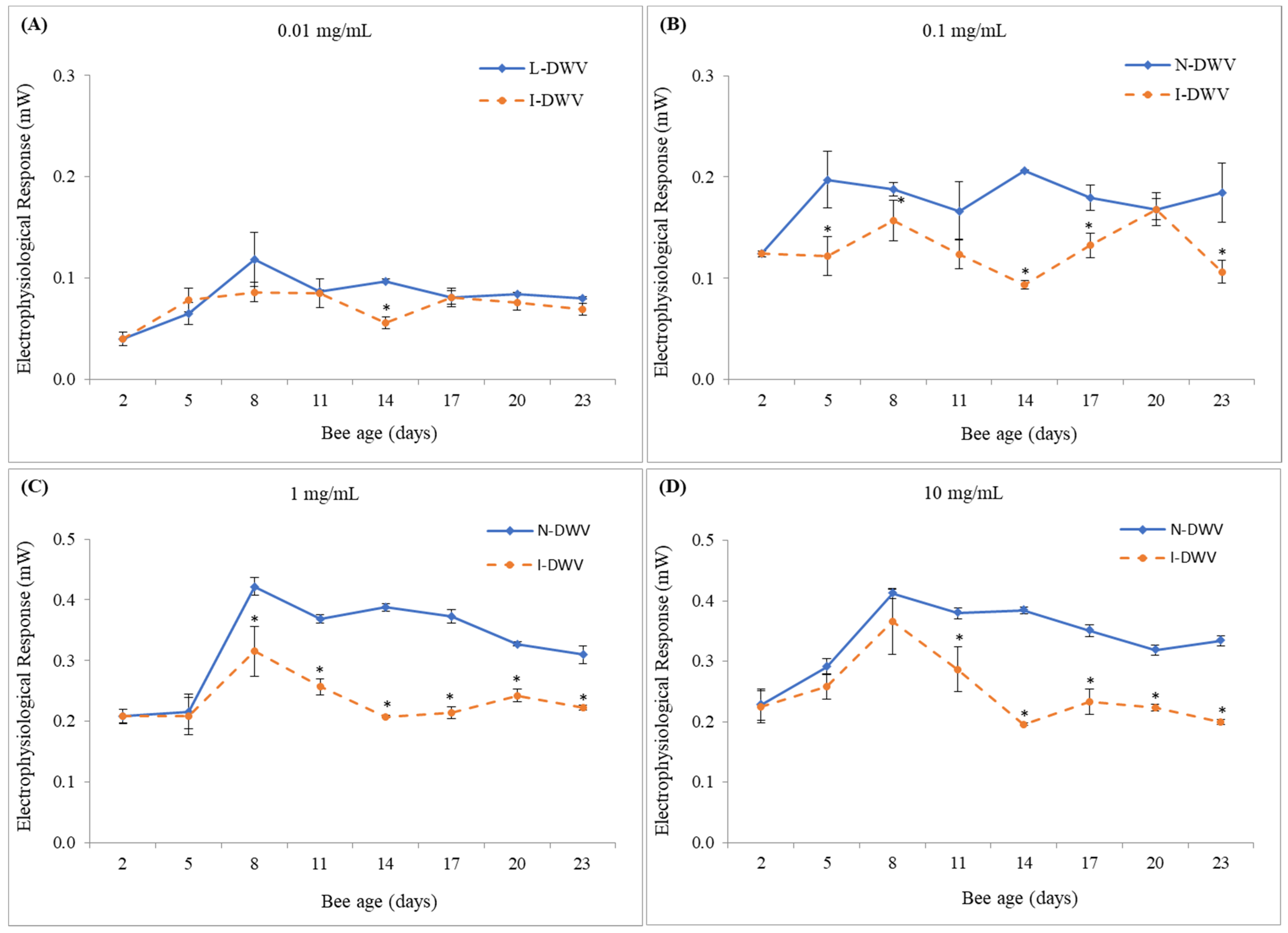
When inoculated worker bees (I-DWV) and non-inoculated bees (N-DWV) of different ages post-emergence (2, 5, 8, 11, 14, 17, 20, and 23 days old) were exposed to volatile stimuli of essential oils of E. globulus and M. piperita at different concentrations, a significant interaction between the viral status of worker bees and the essential oil concentration was found (E. globulus F = 5.73; df = 3, 64; p = 0.002 and M. piperita F = 4.25; df = 3, 64; p = 0.008). On the other hand, there was no significant interaction between bee age, viral status, and the essential oil concentration (E. globulus F = 0.62; df = 21, 64; p = 0.892 and M. piperita F = 0.77; df = 21, 64; p = 0.743). Bees that were inoculated with DWV-A (I-DWV treatment) responded less to stimuli of essential oils of E. globulus at concentrations of 0.1, 1.0, and 10.0 mg/mL, especially in bees that were 11 to 17 days old (Figure 1B–D), than bees that were not inoculated with DWV-A (N-DWV treatment). In fact, inoculated bees (I-DWV) from 14 to 17 days old, responded between 47% and 53% less compared to non-inoculated bees when exposed to essential oils at concentrations of 1.0 mg/mL (Figure 1C). Furthermore, this negative response in olfactory sensibility was also observed in older bees (20 and 23 days old), especially at concentrations of 1.0 and 10.0 mg/mL (Figure 1C,D). On the other hand, no significant differences were observed in the electrophysiological responses of antennas exposed to the essential oil of E. globulus at a concentration of 0.01 mg/mL (Figure 1A).
Similarly, antennas stimulated with the volatile fraction of the essential oil from M. piperita also showed significant differences in bees that were inoculated with DWV-A (I-DWV) compared to those that were not inoculated (N-DWV) (Figure 2). However, these differences were mostly observed at 0.1, 1.0, and 10.0 mg/mL of essential oil from M. piperita, where worker bees that were inoculated with DWV-A showed a lower response to volatile compounds than non-inoculated bees, especially bees from 8–11 to 23 days old (Figure 2B–D). In some cases, these reduced responses to essential oils of M. piperita were, on average, 50% less than bees that were not inoculated with DWV-A (N-DWV treatment), especially in 14 day-old bees (Figure 2B–D). A significant difference in the electrophysiological responses of antennas exposed to the essential oil of M. piperita at a concentration of 0.01 mg/mL was also observed (Figure 2A). However, this difference was punctual and only observed in DWV-A inoculated 14 day-old bees, which showed a lower electroanemographic response to the volatile compounds of the essential oil of M. piperita (Figure 2A).
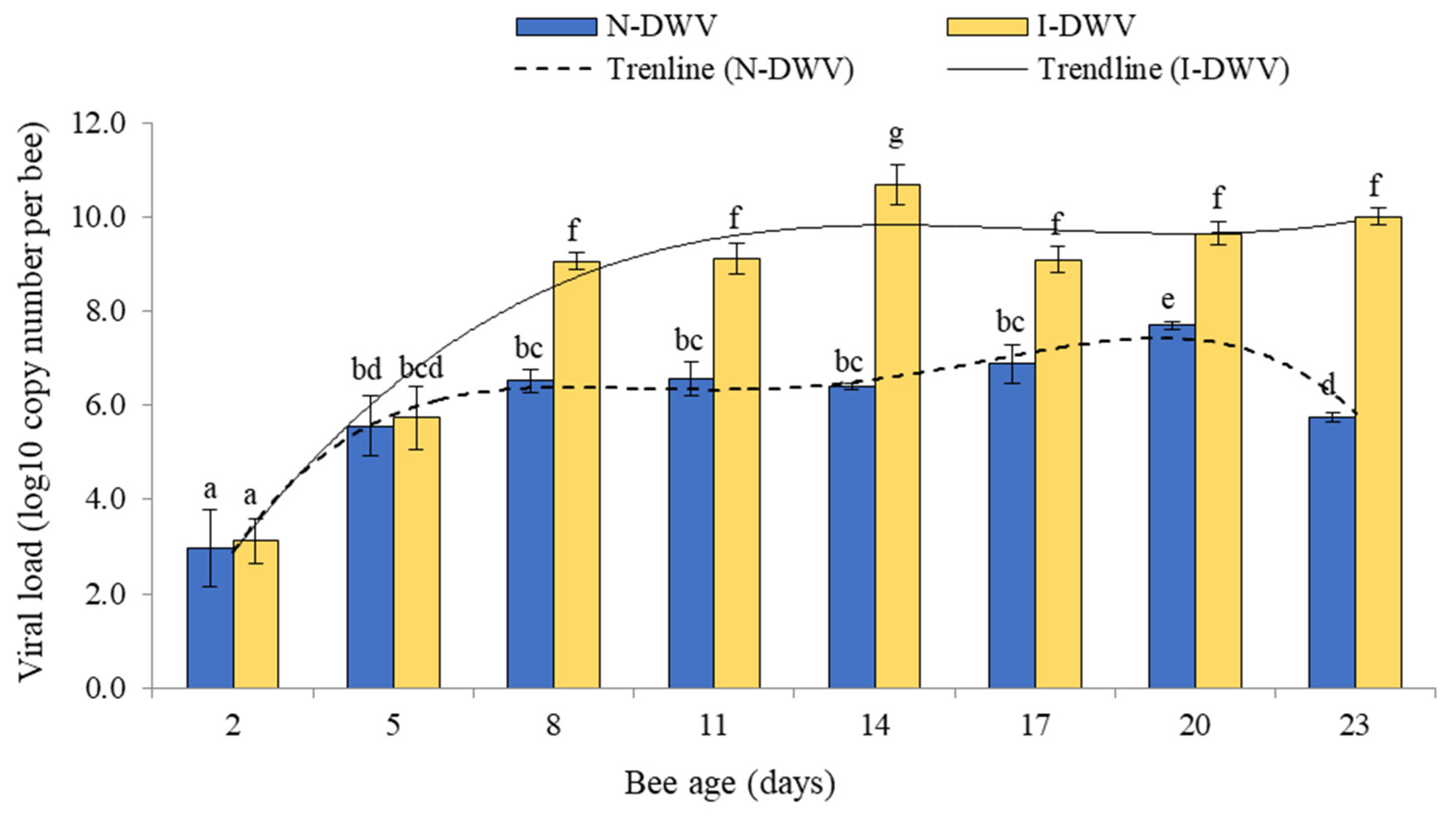
The viral load detected in antennas varied significantly between bees that were inoculated and non-inoculated; these changes were influenced by bee ages (Factorial ANOVA F = 7.78; df = 7, 32; p < 0.001). These differences were observed in 8 to 23-day-old bees, when bees inoculated with DWV-A (I-DWV treatment) showed a higher viral load (1.0 × 10⁹ to 1.0 × 10¹¹ copy number per bee) compared to bees that were not inoculated (N-DWV treatment) (1.0 × 10⁶ to 1.0 × 10⁷ copy number per bee) (Figure 3).

3.2. Expression of OBP Genes

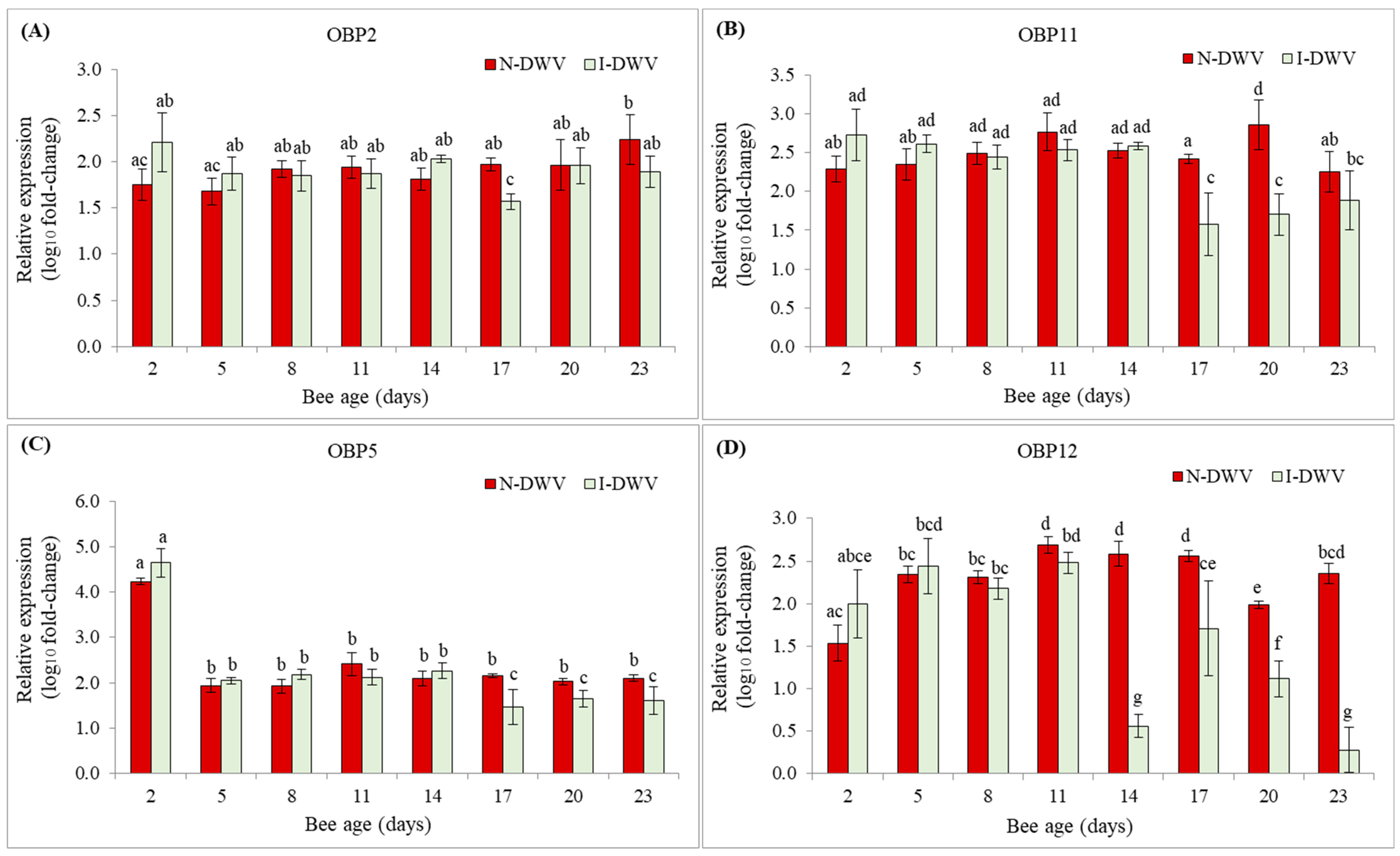
Regarding the relative expression of the nine antenna-specific OBP genes of worker bees of A. mellifera, we observed significant differences in four of them; OBP2, OBP 5, OBP11, and OBP12 (Figure 4). However, changes in these gene expressions were an effect of the interaction between bee age and viral status (MANOVA Wilks λ = 0.01; F = 2.53; df = 70, 141; p < 0.001). In bees that were inoculated with DWV-A, OBP5, and OPB11, gene expression was significantly down-regulated compared with non-inoculated 17-day-old bees, a trend that continued until the end of the experiment (Figure 4B,C and Figure 5). Similarly, the OBP12 gene expression was down-regulated in inoculated bees that were 14 to 23 days old (Figure 4D and Figure 5). The remaining OBPs that were tested were not significantly altered by the viral level in the antenna (Figure S1, Supplementary Materials).
Correlations among the viral load, OBPs gene relative expression (OBP2, OBP5, OBP11, and OBP12), and EAG values (M. piperita and E. globulus at 0.1, 1.0, and 10.0 mg/mL) in DWV-A inoculated bees were identified using the Spearman correlation test (Table 2). Significant negative correlations were detected among the viral load and OBP12 expression (rs = −0.69, p = 0.001). Similarly, the increase of the viral load was negatively correlated with the olfactory response of bees inoculated with DWV-A to M. piperita volatile compounds at different concentrations (0.1 mg/mL rs = −0.76, p < 0.001; 1.0 mg/mL rs = −0.70, p = 0.001; 10.0 mg/mL rs = −0.83, p < 0.001). On the other hand, there were no detected significant correlations among viral load, the expression of OBP5 and OBP11, or the response of bees that were inoculated with DWV-A to E. globulus volatiles (Table 2).

4. Discussion

To the best of our knowledge, this is the first evidence demonstrating that the olfactory sensitivity, evaluated by EAG, is negatively altered in in the antenna of bees inoculated with DWV-A. However, this alteration in the olfactory sensibility was dependent on the DWV-A titer in the antenna and the concentration of the stimuli component. Thus, the olfactory responses could be associated with the volatile compounds present in the essential oils from the plant species used in the study. Previous reports have shown that the presence of compounds, such as α-pinene, myrcene, linalool, and α-terpineol, in both essential oils [45,46,47,48] could be responsible for the EAG response since these compounds have been reported to stimulate the olfactory response in A. mellifera when individually exposed [49,50,51].
The antennae of bees were more sensitive to volatile compounds of essential oils of M. piperita than to those of E. globulus. In fact, the increase in the viral load of DWV-A in the antennae was negatively and significantly correlated with the electrophysiological response (EAG) of honey bees exposed to volatile essential oils of M. piperita (Table 2). This olfactory response was visible in 8-day-old bees and older, when the viral concentrations were also significantly higher (1.2 × 10⁹ copy number per bee) in bees that were inoculated with DWV-A (Figure 1 and Figure 2). We observed that, in general, the olfactory sensitivity of DWV-A inoculated and non-inoculated bees to essential oil concentrations of 0.1 and 1.0 mg/mL showed clearly differential responses. At high concentrations of essential oils, the olfactory response may be more irregular and could depend on the source from which it was obtained. For instance, essential oils of E. globolus at 10.0 mg/mL caused an irregular response in bees that were inoculated with DWV-A, although some cases were significant compared to non-inoculated bees. This irregular response could be associated with a saturation of the environment by the volatiles present in the essential oils, generating nonspecific stimulation [52]. It has been proposed that, when faced with a high concentration of stimulus, a nonspecific stimulation can be generated; on the other hand, exposure to low concentrations of the stimulus may not generate a complete stimulation of the antenna [52]. We observed that very low concentrations of the stimulus did not induce olfactory responses, although, exceptionally, 14-day-old bees inoculated with DWV-A and exposed to 0.01 mg/mL of M. piperita essential oil showed a significant decrease in the EAG response; bee age also coincided with a higher DWV-A load (1.0 × 10¹¹ copy number per bee) detected in the antennas (Figure 2A and Figure 3). In fact, we observed that with an increase in the DWV-A load in the antennae, even at high concentrations of stimulus, the olfactory response decreased substantially (Figure 2D and Figure 3).
There exists the possibility that this negative effect on the olfactory response could be associated with damage to specialized structures in the antenna. In fact, Kim et al. [21] observed an alteration in the cellular ultrastructure of the antennas in bees with classic symptoms (e.g., deformed wings) and high viral loads (1.0 × 10¹⁴ copy number per bee). In contrast, non-symptomatic bees showed no damage to the cellular ultrastructure of the antennas, although the viral load found in this sensorial organ could also be considered high at 1.0 × 10¹¹ copy number per bee. We did not observe common symptoms of the DWV in the bees used in the experiment; even so, we quantified considerably high viral particle levels in DWV-A inoculated bees (Figure 3), which caused significant negative sensorial effects; effects that could be even greater in symptomatic bees, as suggested by Kim et al. [21]. Thus, high levels of DWV in the antenna may affect the performance of this organ in terms of the perception of environmental ligands. Mondet et al. [20] showed that DWV infection in antennas compromised the development of Varroa Sensitive Hygiene (VSH) behavior, possibly due to the deterioration of the olfactory signal and damage in cognitive functions, which suggests that DWV may seriously compromise the processes involved in the perception of volatile compounds.
The negative effect on the olfactory response could also be associated with the gene expression of olfactory sensory proteins, such as OBPs. We found that the DWV-A infection significantly affected the gene expression of some OBPs located in the antenna. High levels (>1.0 × 10⁹ copy number per bee) of DWV-A in the antennas of middle-aged bees (8–23 days old) generated a decrease in the expression of OBP2, OBP5, OBP11, and OBP12, which could then cause the decrease in olfactory sensitivity. Previous studies have reported on the fundamental role of these proteins in the transport and solubility of environmental ligands that enter the lymph and, through the action of OBPs, are transported to chemosensitive neurons, especially due to their high concentration in the lymphatic zone [26,53,54,55]. This guarantees the detection and discrimination of chemical signals, as well as the sensitivity of the olfactory system [54,56,57]. In particular, the affinity and selectivity of the OBP2 protein in plant odors has been demonstrated, showing its physiological role in the recognition of forage species, especially in those that present compounds such as 1–8 cineole in their aroma [58]. Additionally, Zhao et al. [59] reported the crucial role of the OBP5 protein in the larval care processes within the hive. This protein has an affinity for volatile compounds released by larvae when they are affected by different pathogens, suggesting that the recognition of these volatile compounds by nurse bees could aid in recognizing diseased larvae and caring for health larvae, being key in the sanitary maintenance of the brood within the hive. Previously, Zhao et al. [60] also showed that OBP11 protein is produced in greater quantity in worker bees that perform tasks within the hive, reaching their peak gene expression in bees 10 to 15 days old, suggesting that this OBP is related to the olfactory detection of volatile compounds in the brood. However, Nie et al. [61] found that OBP2, OBP5, and OBP11 are expressed continuously from mid- to high levels in newly emerged worker bees, nurse bees, and forager bees. It is well known that 4- to 12-day-old worker bees feed larvae, while 12- to 20-day-old workers have many tasks, ranging from nest building and maintenance to nectar receiving and processing, and other labors associated with colony defense (e.g., guardian bees) [62]. Many of these tasks are mediated by pheromones and other secreted volatile compounds by queen and worker bees [63]. We observed that OBP5 and OBP11 were down-regulated significantly in bees 17 days old and older (Figure 4) when they were inoculated with DWV-A. This suggests that high levels of DWV-A infection in the antennae may affect many worker bees’ tasks, not only those described by Zhao et al. [60], but also other tasks associated with middle- and older-age bees [62], since these OBPs were expressed continuously in bees more than 15 days of age (Figure 4). On the other hand, there is little information on the specific olfactory role of the OBP12 protein. However, Nie et al. [61] also found that OBP12 is expressed continuously in antennae of newly emerged worker, nurse, and forage bees, which suggests that this protein could play an important role in the worker bees’ performance in the perception of aromas in and outside the hive. Furthermore, it was the only OBP in which gene expression was negatively and significantly correlated with the increasing DWV-A load (Table 2), which suggests that there is a strong relationship between DWV-A infection and the performance of aroma perception mediated by OBP12 in the antennae. In fact, the OBP12 was the most sensitive to DWV-A inoculation; its expression was down-regulated strongly in 14-day-old bees, until the end of the experiment (23-day-old bees) (Figure 5). This evidence suggests that this OBP may play key roles in middle-aged bees associated with maintenance tasks within a colony, but also with foraging bee activities outside the hive, such as locating and selecting food sources. However, new studies are required to support this hypothesis since there is no evidence of the specific functions of OBP12 in worker bees of different ages. Furthermore, we could be underestimating the effect of DWV-A on the gene expressions of OBP2, OBP5, OBP11, and OBP12, considering that the down-regulation of these OBPs was significantly observed, in most cases, in bees 17 days old and older. But these effects could also be related to the methodological procedure, in the sense that, in this study, newly emerged adult bees were inoculated orally, which reflects only one possible route of viral infection (e.g., trophallaxis). If the viral inoculation occurred in the pupa stage, simulating a transmission route via Varroa destructor, the results could be different. Therefore, viral infection of bees in immature stages could lead to a low expression of these OBPs (OBP2, OBP5, OBP11, and OBP12), much lower than the levels detected here; the expression of other OBPs that showed no significant changes in this study could also be affected in this case (Figure S1, Supplementary Materials). Hence, further studies are required to test this hypothesis, including inoculation with DWV-A in immature bee stages in order to more precisely measure OBPs expression and other odorant-associated genes present in the antennae of newly emerged, middle-aged, and forager bees.
It has been hypothesized that the presence of DWV-A may compromise the maturation process of worker bees from nurse to forager bees, particularly due to the change that this virus generates in the brain transcriptome. A recent study reported that nurse bees infected with DWV-A showed a brain transcriptome similar to forager bees and this change in behavioral maturation could be generating younger bees with foraging behavior [64]. However, this early foraging activity in middle-age bees could be precarious and deficient [65]. Thus, an DWV-A infection could impair worker bees’ olfactory sensitivity to aromas, as shown in this study, and a neurogenomic spoilage [64] could result in learning deficits in worker bees [18] that could lead to the loss of honey bees in the field (e.g., forage bees) and affect the efficiency of bees’ labors within the hive, producing a gradual decline in the colony, as has been reported with this DWV variant [66].
An interesting result in our study was that the difference in DWV-A load in the antennae between inoculated and non-inoculated bees was only observed in bees that were 8 days old or more (Figure 3). We expected that 3 days after (5-day-old bees) the DWV-A inoculation, the viral load in the antennae would be higher in inoculated bees than non-inoculated bees; however, no significant difference was observed at that time (Figure 3). Notwithstanding, when we analyzed and quantified the viral load in the rest of the body tissues (including head, thorax, and abdomen) of worker honey bees from which the antennas were removed, we found that the DWV-A load was significantly higher in 5 day-old inoculated bees (1.0 × 10⁸ copy number per bee) than non-inoculated bees (1.0 × 10⁶ copy number per bee) (Figure S2, Supplementary Materials). This shows that the DWV-A establishment and replication occurred only a few days after the inoculation. This result suggests that the DWV-A distribution process towards the antenna and its replication there occurs a little later than in other tissues or organs that have immediate contact (e.g., gut tissues) with the virus when it is inoculated orally. It was also interesting to observe that the DWV load in non-inoculated bees, which reflects the viral infection of the colony, was, on average, over time (1.0 × 10⁶ copy number per bee) in antennae and the rest of the analyzed bee body (Figure 3, Figure S2, Supplementary Materials); values that are considered low are in [64]. Although no DWV-free colonies were found in our experimental apiary, we used frames from colonies with very low DWV-A, without varroa or other pathogens for the experiment in this study, a strategy that has also been used in other related studies [67,68,69]. In fact, DWV is widespread in low levels in almost all honey bees, including Varroa-free colonies [70]. Honey bees can live with these basal low levels of DWV, which are apparently not harmful for them [71], although other stressful factors (e.g., ectoparasite V. destructor, gut parasite Nosema ceranae, poor nutrition, etc.) can eventually induce an increase in these levels and cause damage to the bees [69,72,73,74].
Our study provides novel information that aids in understanding the magnitude of harm generated by the DWV-A infection in middle- and forager-age worker bees. The presence of this virus in A. mellifera antennas could generate deterioration in crucial tasks inside the hive, such as larval care, cleaning, and defense activities. This damage could also extend outside the hive, affecting the forager bees and their ability to recognize food sources. However, further studies are required to understand and quantify the damage caused by DWV-A on behavioral responses of honey bees at the field level, considering the other DWV variants in the study model.

5. Conclusions

In summary, the high level of replication of DWV-A in the antennae decreased the olfactory sensitivity to the evaluated volatile compounds in middle- and forager-age worker bees. DWV-A also led to a down-regulation of some OBPs that are specifically expressed in antennas. Thus, DWV-A infection could compromise volatile compound recognition inside the hive and outside the hive in bee foraging behaviors, affecting bee survival at the individual and colony levels.

Supplementary Materials

The following are available online at https://www.mdpi.com/article/10.3390/insects12100895/s1, Figure S1: Gene expression of other OBPs analyzed in antennae of worker bees of different ages that were inoculated (I-DWV) and non-inoculated (N-DWV) with DWV-A. Figure S2: DWV-A load measured in worker honey bees (without antennas) of different ages that were inoculated (I-DWV) and non-inoculated (N-DWV).

Author Contributions

M.V. conceived of this research; M.V., R.C. and D.S. designed experiments; D.S. performed the experiments and the analysis in labs; D.S., N.A., R.C., A.D. and M.V. wrote the paper and participated in the revisions of it. All authors have read and agreed to the published version of the manuscript.

Funding

This study was supported by Grants FONDECYT N° 1211688 and N° 1210942 from the National Research and Development Agency (ANID, Ex-CONICYT), Chile.

Institutional Review Board Statement

Not Applicable.

Informed Consent Statement

Not Applicable.

Data Availability Statement

Not Applicable.

Conflicts of Interest

The authors declare that they have no conflict of interest. The funders had no role in the design of the study; in the collection, analyses, or interpretation of data; in the writing of the manuscript, or in the decision to publish the results.

References

  1. Aguilar, R.; Ashworth, L.; Galetto, L.; Aizen, M.A. Plant reproductive susceptibility to habitat fragmentation: Review and synthesis through a meta-analysis. Ecol. Lett. 2006, 9, 968–980. [Google Scholar] [CrossRef]
  2. Ricketts, T.H.; Regetz, J.; Steffan-Dewenter, I.; Cunningham, S.A.; Kremen, C.; Bogdanski, A.; Gemmill-Herren, B.; Greenleaf, S.S.; Klein, A.M.; Mayfield, M.M.; et al. Landscape effects on crop pollination services: Are there general patterns? Ecol. Lett. 2008, 11, 499–515. [Google Scholar] [CrossRef]
  3. Potts, S.G.; Imperatriz-Fonseca, V.; Ngo, H.T.; Aizen, M.A.; Biesmeijer, J.C.; Breeze, T.D.; Dicks, L.V.; Garibaldi, L.A.; Hill, R.; Settele, J.; et al. Safeguarding pollinators and their values to human well-being. Nature 2016, 540, 220–229. [Google Scholar] [CrossRef]
  4. vanEngelsdorp, D.; Evans, J.D.; Saegerman, C.; Mullin, C.; Haubruge, E.; Nguyen, B.K.; Frazier, M.; Frazier, J.; Cox-Foster, D.; Chen, Y.; et al. Colony collapse disorder: A descriptive study. PLoS ONE 2009, 4, e6481. [Google Scholar] [CrossRef] [PubMed]
  5. Goulson, D.; Nicholls, E.; Botias, C.; Rotheray, E.L. Bee declines driven by combined stress from parasites, pesticides, and lack of flowers. Science 2015, 347, 1255957. [Google Scholar] [CrossRef]
  6. Semmens, B.X.; Semmens, D.J.; Thogmartin, W.E.; Wiederholt, R.; Lopez-Hoffman, L.; Diffendorfer, J.E.; Pleasants, J.M.; Oberhauser, K.S.; Taylor, O.R. Quasi-extinction risk and population targets for the Eastern, migratory population of monarch butterflies (Danaus plexippus). Sci. Rep. 2016, 6, 23265. [Google Scholar] [CrossRef] [PubMed]
  7. Hall, D.M.; Steiner, R. Insect pollinator conservation policy innovations at subnational levels: Lessons for lawmakers. Environ. Sci. Policy 2019, 93, 118–128. [Google Scholar] [CrossRef]
  8. vanEngelsdorp, D.; Caron, D.; Hayes, J.; Underwood, R.; Henson, M.; Rennich, K.; Spleen, A.; Andree, M.; Snyder, R.; Lee, K.; et al. A national survey of managed honey bee 2010–11 winter colony losses in the USA: Results from the Bee Informed Partnership. J. Apic. Res. 2015, 51, 115–124. [Google Scholar] [CrossRef] [Green Version]
  9. Ullah, A.; Tlak Gajger, I.; Majoros, A.; Dar, S.A.; Khan, S.; Kalimullah; Haleem Shah, A.; Nasir Khabir, M.; Hussain, R.; Khan, H.U.; et al. Viral impacts on honey bee populations: A review. Saudi J. Biol. Sci. 2021, 28, 523–530. [Google Scholar] [CrossRef] [PubMed]
  10. Beaurepaire, A.; Piot, N.; Doublet, V.; Antunez, K.; Campbell, E.; Chantawannakul, P.; Chejanovsky, N.; Gajda, A.; Heerman, M.; Panziera, D.; et al. Diversity and global distribution of viruses of the western Honey Bee, Apis. mellifera. Insects 2020, 11, 239. [Google Scholar] [CrossRef] [PubMed]
  11. Gauthier, L.; Tentcheva, D.; Tournaire, M.; Dainat, B.; Cousserans, F.; Colin, M.E.; Bergoin, M. Viral load estimation in asymptomatic honey bee colonies using the quantitative RT-PCR technique. Apidologie 2007, 38, 426–435. [Google Scholar] [CrossRef] [Green Version]
  12. Mordecai, G.J.; Wilfert, L.; Martin, S.J.; Jones, I.M.; Schroeder, D.C. Diversity in a honey bee pathogen: First report of a third master variant of the deformed Wing Virus quasispecies. ISME J. 2016, 10, 1264–1273. [Google Scholar] [CrossRef] [Green Version]
  13. Kevill, J.L.; Highfield, A.; Mordecai, G.J.; Martin, S.J.; Schroeder, D.C. ABC Assay: Method development and application to quantify the role of three DWV master variants in overwinter colony losses of European honey bees. Viruses 2017, 9, 314. [Google Scholar] [CrossRef] [Green Version]
  14. Ongus, J.R.; Peters, D.; Bonmatin, J.M.; Bengsch, E.; Vlak, J.M.; van Oers, M.M. Complete sequence of a picorna-like virus of the genus Iflavirus replicating in the mite Varroa destructor. J. Gen. Virol. 2004, 85, 3747–3755. [Google Scholar] [CrossRef]
  15. Lanzi, G.; de Miranda, J.R.; Boniotti, M.B.; Cameron, C.E.; Lavazza, A.; Capucci, L.; Camazine, S.M.; Rossi, C. Molecular and biological characterization of deformed wing virus of honeybees (Apis mellifera L.). J. Virol. 2006, 80, 4998–5009. [Google Scholar] [CrossRef] [Green Version]
  16. Benaets, K.; Van Geystelen, A.; Cardoen, D.; De Smet, L.; de Graaf, D.C.; Schoofs, L.; Larmuseau, M.H.; Brettell, L.E.; Martin, S.J.; Wenseleers, T. Covert deformed wing virus infections have long-term deleterious effects on honeybee foraging and survival. Proc. Biol. Sci. 2017, 284. [Google Scholar] [CrossRef] [PubMed]
  17. Grozinger, C.M.; Flenniken, M.L. Bee Viruses: Ecology, Pathogenicity, and Impacts. Annu. Rev. Entomol. 2019, 64, 205–226. [Google Scholar] [CrossRef] [PubMed]
  18. Iqbal, J.; Mueller, U. Virus infection causes specific learning deficits in honeybee foragers. Proc. Biol. Sci. 2007, 274, 1517–1521. [Google Scholar] [CrossRef] [PubMed] [Green Version]
  19. Shah, K.S.; Evans, E.C.; Pizzorno, M.C. Localization of deformed wing virus (DWV) in the brains of the honeybee, Apis mellifera Linnaeus. Virol. J. 2009, 6, 182. [Google Scholar] [CrossRef] [Green Version]
  20. Mondet, F.; Alaux, C.; Severac, D.; Rohmer, M.; Mercer, A.; Le Conte, Y. Antennae hold a key to Varroa-sensitive hygiene behaviour in honey bees. Sci. Rep. 2015, 5, 10454. [Google Scholar] [CrossRef] [PubMed] [Green Version]
  21. Kim, S.H.; Mercer, A.; Mitchell, A.; de Miranda, J.; Ward, V.; Mondet, F.; Bostina, M. Viral infections alter antennal epithelium ultrastructure in honey bees. J. Invertebr. Pathol. 2019, 168, 107252. [Google Scholar] [CrossRef]
  22. Groot, A.T.; Dekker, T.; Heckel, D.G. The genetic basis of pheromone evolution in moths. Annu. Rev. Entomol. 2016, 61, 99–117. [Google Scholar] [CrossRef] [PubMed]
  23. Li, Q.; Liberles, S.D. Aversion and attraction through olfaction. Curr. Biol. 2015, 25, R120–R129. [Google Scholar] [CrossRef] [PubMed] [Green Version]
  24. Foret, S.; Maleszka, R. Function and evolution of a gene family encoding odorant binding-like proteins in a social insect, the honey bee (Apis mellifera). Genome Res. 2006, 16, 1404–1413. [Google Scholar] [CrossRef] [PubMed] [Green Version]
  25. Pelosi, P.; Calvello, M.; Ban, L. Diversity of odorant-binding proteins and chemosensory proteins in insects. Chem. Senses 2005, 30 (Suppl. 1), i291–i292. [Google Scholar] [CrossRef] [Green Version]
  26. Leal, W.S. Odorant reception in insects: Roles of receptors, binding proteins, and degrading enzymes. Annu. Rev. Entomol. 2013, 58, 373–391. [Google Scholar] [CrossRef]
  27. Zhou, J.-J. Odorant-binding proteins in insects. In Pheromones; Vitamins & Hormones: Harpenden, Hertfordshire, UK, 2010; pp. 241–272. [Google Scholar]
  28. Deyu, Z.; Leal, W.S. Conformational isomers of insect Odorant-Binding Proteins. Arch. Bioche. Biophys. 2002, 397, 99–105. [Google Scholar] [CrossRef]
  29. Gusachenko, O.N.; Woodford, L.; Balbirnie-Cumming, K.; Ryabov, E.V.; Evans, D.J. Evidence for and against deformed wing virus spillover from honey bees to bumble bees: A reverse genetic analysis. Sci. Rep. 2020, 10, 16847. [Google Scholar] [CrossRef]
  30. Skubnik, K.; Novacek, J.; Fuzik, T.; Pridal, A.; Paxton, R.J.; Plevka, P. Structure of deformed wing virus, a major honey bee pathogen. Proc. Natl. Acad. Sci. USA 2017, 114, 3210–3215. [Google Scholar] [CrossRef] [Green Version]
  31. Vargas, M.; Arismendi, N.; Riveros, G.; Zapata, N.; Bruna, A.; Vidal, M.; Rodríguez, M.; Gerding, M. Viral and intestinal diseases detected in Apis mellifera in Central and Southern Chile. Chil. J. Agric. Res. 2017, 77, 243–249. [Google Scholar] [CrossRef] [Green Version]
  32. Porrini, M.P.; Garrido, P.M.; Eguaras, M.J. Individual feeding of honey bees: Modification of the Rinderer technique. J. Apic. Res. 2013, 52, 194–195. [Google Scholar] [CrossRef]
  33. Arismendi, N.; Caro, S.; Castro, M.P.; Vargas, M.; Riveros, G.; Venegas, T. Impact of mixed infections of gut parasites Lotmaria passim and Nosema ceranae on the lifespan and immune-related biomarkers in Apis mellifera. Insects 2020, 11, 420. [Google Scholar] [CrossRef]
  34. Bobis, O.; Moise, A.R.; Ballesteros, I.; Reyes, E.S.; Duran, S.S.; Sanchez-Sanchez, J.; Cruz-Quintana, S.; Giampieri, F.; Battino, M.; Alvarez-Suarez, J.M. Eucalyptus honey: Quality parameters, chemical composition and health-promoting properties. Food Chem. 2020, 325, 126870. [Google Scholar] [CrossRef]
  35. Eliyahu, A.; Duman, Z.; Sherf, S.; Genin, O.; Cinnamon, Y.; Abu-Abied, M.; Weinstain, R.; Dag, A.; Sadot, E. Vegetative propagation of elite Eucalyptus clones as food source for honeybees (Apis mellifera); adventitious roots versus callus formation. Isr. J. Plant. Sci. 2020, 67, 83–97. [Google Scholar] [CrossRef] [Green Version]
  36. Terrab, A.; Valdés, B.; Díez, M.J. Study of plants visited by honeybees (Apis mellifera L.) in the Central Rif Region (N. Morocco) using pollen analysis. Grana 2005, 44, 209–215. [Google Scholar] [CrossRef] [Green Version]
  37. Ciotlaus, I.; Balea, A.; Pojar-Fenesan, M.; Petean, I. Cromathographic profile of volatiles of multifloral and unifloral honey collected by Apis Mellifera from Transilvania, Romania. Rev. Chim. 2020, 71, 91–98. [Google Scholar] [CrossRef]
  38. Silva, D.; Curkovic, T.; Ceballos, R. Behavioral and antennal responses of Lobesia botrana (Lepidoptera: Tortricidae) to volatiles from the non-host plant Schinus molle L. (Anacardiaceae). Chil. J. Agric. Res. 2019, 79, 165–171. [Google Scholar] [CrossRef] [Green Version]
  39. Ceballos, R.; Fernández, N.; Zúñiga, S.; Zapata, N. Electrophysiological and behavioral responses of pea weevil Bruchus pisorum L. (Coleóptera: Bruchidae) to volatiles collected from its host Pisum sativum L. Chil. J. Agric. Res. 2015, 75, 202–209. [Google Scholar] [CrossRef] [Green Version]
  40. Pfaffl, M.W. A new mathematical model for relative quantification in real-time RT-PCR. Nucleic Acids Res. 2001, 29, 2003–2007. [Google Scholar] [CrossRef] [PubMed]
  41. Riveros, G.; Arismendi, N.; Zapata, N.; Evans, D.; Pérez, I.; Aldea, P.; Vargas, M. Occurrence, prevalence and viral load of deformed wing virus variants in Apis mellifera colonies in Chile. J. Apic. Res. 2019, 59, 63–68. [Google Scholar] [CrossRef]
  42. Wu, Y.; Dong, X.; Kadowaki, T. Characterization of the copy number and variants of deformed wing virus (DWV) in the pairs of honey bee pupa and infesting Varroa destructor or Tropilaelaps mercedesae. Front. Microbiol. 2017, 8, 1558. [Google Scholar] [CrossRef] [PubMed]
  43. Yang, X.; Cox-Foster, D. Impact of an ectoparasite on the immunity and pathology of an invertebrate: Evidence for host immunosuppression and viral amplification. Proc. Natl. Acad. Sci. USA 2005, 102, 7470–7475. [Google Scholar] [CrossRef] [Green Version]
  44. McMahon, D.P.; Natsopoulou, M.E.; Doublet, V.; Furst, M.; Weging, S.; Brown, M.J.; Gogol-Doring, A.; Paxton, R.J. Elevated virulence of an emerging viral genotype as a driver of honeybee loss. Proc. Biol. Sci. 2016, 283. [Google Scholar] [CrossRef] [PubMed]
  45. Salem, N.; Kefi, S.; Tabben, O.; Ayed, A.; Jallouli, S.; Feres, N.; Hammami, M.; Khammassi, S.; Hrigua, I.; Nefisi, S.; et al. Variation in chemical composition of Eucalyptus globulus essential oil under phenological stages and evidence synergism with antimicrobial standards. Ind. Crop. Prod. 2018, 124, 115–125. [Google Scholar] [CrossRef]
  46. Jerbi, A.; Derbali, A.; Elfeki, A.; Kammoun, M. Essential oil composition and biological activities of Eucalyptus globulus leaves extracts from Tunisia. J. Essent Oil. Bear Plants 2017, 20, 438–448. [Google Scholar] [CrossRef]
  47. Moghaddam, M.; Pourbaige, M.; Tabar, H.K.; Farhadi, N.; Hosseini, S.M.A. Composition and antifungal activity of Peppermint (Mentha piperita) essential oil from Iran. J. Essent. Oil Bear. Plants 2013, 16, 506–512. [Google Scholar] [CrossRef]
  48. Yong, W.T.L.; Ades, P.K.; Goodger, J.Q.D.; Bossinger, G.; Runa, F.A.; Sandhu, K.S.; Tibbits, J.F.G. Using essential oil composition to discriminate between myrtle rust phenotypes in Eucalyptus globulus and Eucalyptus obliqua. Ind. Crop. Prod. 2019, 140, 111595. [Google Scholar] [CrossRef]
  49. Wiese, N.; Fischer, J.; Heidler, J.; Lewkowski, O.; Degenhardt, J.; Erler, S. The terpenes of leaves, pollen, and nectar of thyme (Thymus vulgaris) inhibit growth of bee disease-associated microbes. Sci. Rep. 2018, 8, 14634. [Google Scholar] [CrossRef] [Green Version]
  50. Jung, J.W.; Park, K.W.; Oh, H.-W.; Kwon, H.W. Structural and functional differences in the antennal olfactory system of worker honey bees of Apis mellifera and Apis cerana. J. Asia-Pac. Entomol. 2014, 17, 639–646. [Google Scholar] [CrossRef]
  51. Larson, N.R.; O’Neal, S.T.; Bernier, U.R.; Bloomquist, J.R.; Anderson, T.D. Terpenoid-induced feeding deterrence and antennal response of honey bees. Insects 2020, 11, 83. [Google Scholar] [CrossRef] [Green Version]
  52. Holt, H.L.; Villar, G.; Cheng, W.; Song, J.; Grozinger, C.M. Molecular, physiological and behavioral responses of honey bee (Apis mellifera) drones to infection with microsporidian parasites. J. Invertebr. Pathol. 2018, 155, 14–24. [Google Scholar] [CrossRef]
  53. Vieira, F.G.; Rozas, J. Comparative genomics of the odorant-binding and chemosensory protein gene families across the Arthropoda: Origin and evolutionary history of the chemosensory system. Genome Biol. Evol. 2011, 3, 476–490. [Google Scholar] [CrossRef] [PubMed]
  54. Pelosi, P.; Iovinella, I.; Zhu, J.; Wang, G.; Dani, F.R. Beyond chemoreception: Diverse tasks of soluble olfactory proteins in insects. Biol. Rev. Camb. Philos. Soc. 2018, 93, 184–200. [Google Scholar] [CrossRef] [PubMed] [Green Version]
  55. Pelosi, P.; Iovinella, I.; Felicioli, A.; Dani, F.R. Soluble proteins of chemical communication: An overview across arthropods. Front. Physiol. 2014, 5, 320. [Google Scholar] [CrossRef] [PubMed] [Green Version]
  56. Qiao, H.; Tuccori, E.; He, X.; Gazzano, A.; Field, L.; Zhou, J.J.; Pelosi, P. Discrimination of alarm pheromone (E)-beta-farnesene by aphid odorant-binding proteins. Insect Biochem. Mol. Biol. 2009, 39, 414–419. [Google Scholar] [CrossRef] [PubMed]
  57. Pelletier, J.; Guidolin, A.; Syed, Z.; Cornel, A.J.; Leal, W.S. Knockdown of a mosquito odorant-binding protein involved in the sensitive detection of oviposition attractants. J. Chem. Ecol. 2010, 36, 245–248. [Google Scholar] [CrossRef] [Green Version]
  58. Briand, L.; Nespoulous, C.; Huet, J.C.; Takahashi, M.; Pernollet, J.C. Ligand binding and physico-chemical properties of ASP2, a recombinant odorant-binding protein from honeybee (Apis mellifera L.). Eur. J. Biochem. 2001, 268, 752–760. [Google Scholar] [CrossRef]
  59. Zhao, H.; Zeng, X.; Liang, Q.; Zhang, X.; Huang, W.; Chen, H.; Luo, Y. Study of the obp5 gene in Apis mellifera ligustica and Apis cerana cerana. Genet. Mol. Res. 2015, 14, 6482–6494. [Google Scholar] [CrossRef]
  60. Zhao, H.; Luo, Y.; Lee, J.; Zhang, X.; Liang, Q.; Zeng, X. The Odorant-binding protein gene obp11 shows different spatiotemporal roles in the olfactory system of Apis mellifera ligustica and Apis cerana cerana. Sociobiology 2013, 60, 429–435. [Google Scholar] [CrossRef] [Green Version]
  61. Nie, H.; Xu, S.; Xie, C.; Geng, H.; Zhao, Y.; Li, J.; Huang, W.F.; Lin, Y.; Li, Z.; Su, S. Comparative transcriptome analysis of Apis mellifera antennae of workers performing different tasks. Mol. Genet. Genomics 2018, 293, 237–248. [Google Scholar] [CrossRef]
  62. Johnson, B.R. Division of labor in honeybees: Form, function, and proximate mechanisms. Behav. Ecol. Sociobiol. 2010, 64, 305–316. [Google Scholar] [CrossRef] [PubMed] [Green Version]
  63. Bortolotti, L.; Costa, C. Chemical communication in the honey bee society. In Neurobiology of Chemical Communication; Mucignat-Caretta, C., Ed.; CRC Press/Taylor & Francis: Boca Raton, FL, USA, 2014. [Google Scholar]
  64. Traniello, I.M.; Bukhari, S.A.; Kevill, J.; Ahmed, A.C.; Hamilton, A.R.; Naeger, N.L.; Schroeder, D.C.; Robinson, G.E. Meta-analysis of honey bee neurogenomic response links deformed wing virus type A to precocious behavioral maturation. Sci. Rep. 2020, 10, 3101. [Google Scholar] [CrossRef] [Green Version]
  65. Wells, T.; Wolf, S.; Nicholls, E.; Groll, H.; Lim, K.S.; Clark, S.J.; Swain, J.; Osborne, J.L.; Haughton, A.J. Flight performance of actively foraging honey bees is reduced by a common pathogen. Environ. Microbiol. Rep. 2016, 8, 728–737. [Google Scholar] [CrossRef] [PubMed] [Green Version]
  66. Kevill, J.L.; de Souza, F.S.; Sharples, C.; Oliver, R.; Schroeder, D.C.; Martin, S.J. DWV-A lethal to honey bees (Apis mellifera): A colony level survey of DWV variants (A, B, and C) in England, Wales, and 32 States across the US. Viruses 2019, 11, 426. [Google Scholar] [CrossRef] [Green Version]
  67. Ryabov, E.V.; Childers, A.K.; Lopez, D.; Grubbs, K.; Posada-Florez, F.; Weaver, D.; Girten, W.; van Engelsdorp, D.; Chen, Y.; Evans, J.D. Dynamic evolution in the key honey bee pathogen deformed wing virus: Novel insights into virulence and competition using reverse genetics. PLoS Biol. 2019, 17, e3000502. [Google Scholar] [CrossRef] [PubMed] [Green Version]
  68. Ryabov, E.V.; Wood, G.R.; Fannon, J.M.; Moore, J.D.; Bull, J.C.; Chandler, D.; Mead, A.; Burroughs, N.; Evans, D.J. A virulent strain of deformed wing virus (DWV) of honey bees (Apis mellifera) prevails after Varroa destructor-mediated, or in vitro, transmission. PLoS Pathog. 2014, 10, e1004230. [Google Scholar] [CrossRef] [PubMed] [Green Version]
  69. Tesovnik, T.; Zorc, M.; Ristanic, M.; Glavinic, U.; Stevanovic, J.; Narat, M.; Stanimirovic, Z. Exposure of honey bee larvae to thiamethoxam and its interaction with Nosema ceranae infection in adult honey bees. Environ. Pollut. 2020, 256, 113443. [Google Scholar] [CrossRef]
  70. Martin, S.J.; Highfield, A.C.; Brettell, L.; Villalobos, E.M.; Budge, G.E.; Powell, M.; Nikaido, S.; Schroeder, D.C. Global honey bee viral landscape altered by a parasitic mite. Science 2012, 336, 1304–1306. [Google Scholar] [CrossRef] [PubMed]
  71. Martin, S.J.; Brettell, L.E. Deformed wing virus in boneybees and other insects. Annu. Rev. Virol. 2019, 6, 49–69. [Google Scholar] [CrossRef] [Green Version]
  72. Wilfert, L.; Long, G.; Leggett, H.C.; Schmid-Hempel, P.; Butlin, R.; Martin, S.J.M.; Boots, M. Deformed wing virus is a recent global epidemic in honeybees driven by Varroa mites. Science 2016, 351, 594–597. [Google Scholar] [CrossRef] [PubMed] [Green Version]
  73. Tritschler, M.; Vollmann, J.J.; Yanez, O.; Chejanovsky, N.; Crailsheim, K.; Neumann, P. Protein nutrition governs within-host race of honey bee pathogens. Sci. Rep. 2017, 7, 14988. [Google Scholar] [CrossRef] [PubMed] [Green Version]
  74. Barroso-Arevalo, S.; Fernandez-Carrion, E.; Goyache, J.; Molero, F.; Puerta, F.; Sanchez-Vizcaino, J.M. High load of deformed wing virus and Varroa destructor infestation are related to weakness of honey bee colonies in southern Spain. Front. Microbiol. 2019, 10, 1331. [Google Scholar] [CrossRef] [PubMed]
Figure 1. Electrophysiological response of inoculated and non-inoculated worker bees exposed to essential oil of Eucalyptus globulus. Uppercase letter in each subfigure represent the essential oil at concentration of (A) 0.01, (B) 0.1, (C) 1.0 and (D) 10.0 mg/mL. Asterisks indicate significant differences for the same sampling time in bees that were inoculated (I-DWV) and non-inoculated (N-DWV) with DWV-A, according to the Bonferroni test (p < 0.05). The bar in each age indicates standard error.
Insects 12 00895 g001
Figure 2. Electrophysiological response of inoculated and non-inoculated worker bees exposed to essential oil of Mentha piperita. Uppercase letter in each subfigure represent the essential oil at concentration of (A) 0.01, (B) 0.1, (C) 1.0 and (D) 10.0 mg/mL. Asterisks indicate significant differences for the same sampling time in bees that were inoculated (I-DWV) and non-inoculated (N-DWV) with DWV-A, according to the Bonferroni test (p < 0.05). The bar in each age indicates standard error.
Insects 12 00895 g002
Figure 3. DWV-A load measured in antennae of worker honey bees of different ages that were inoculated (I-DWV) and non-inoculated (N-DWV). Means (±SE) with different letters indicate significant difference according to the Tukey HSD test (p < 0.05). Values of DWV-A load in 2-day old bees indicate the basal level of viral infection before the inoculation.
Insects 12 00895 g003
Figure 4. Gene expression of OBPs in worker honey bees of different ages and that were inoculated (I-DWV) and non-inoculated (N-DWV) with DWV-A. Means (±SE) with different letters indicate significant differences according to the Bonferroni test (p < 0.05) in (A) OBP2, (B) OBP11, (C) OBP5 and (D) OBP12.
Insects 12 00895 g004
Figure 5. Heat-map illustrating the intensity of down-regulation of odorant binding proteins (OBPs) in the antennae of worker honey bees of different ages that were inoculated with DWV-A. Asterisks indicate significant differences (Bonferroni test, p < 0.05). Gene expression of OBPs in antennae of inoculated bees with DWV-A compared to non-inoculated bees.
Insects 12 00895 g005
Table 1. Primers used in the qPCR amplification for the odorant binding proteins (OBPs), the β- Actin gene, and specific primer to variant A of deformed wing virus (DWV-A).
Primer Sequence (5′–3′)References
AmelOBP1FACCTGGTAAACGAACCGTCCA[23]
RTCAACACAGCCTGTTCTCGA
AmelOBP2FTCTGACCGTTGTACGTGGCA[23]
RTGGCATTCTCGATGCACTCA
AmelOBP4FTGCGCTGGTTCACGCAGACA[23]
RATGCATTCGTCTTCGTCTGCA
AmelOBP5FATGCGGAAATCGTGCTTGCA[23]
RTGCCATTACTCACGGGAAGA
AmelOBP8FGTTGCGTGGCAATTTGGCAAATG[23]
RTACGTTGTTTCGCCCTTCCAGT
AmelOBP11FTGAGGATGTCGAAGCTACGGAA[23]
RCACGGAGCAATAAACGCTATGG
AmelOBP12FTGCGTGGATCGATCAAACATGA[23]
RACGTTAACGCGATCTTATGGA
AmelOBP15FTTGCATGGCAAAAACTGGCA[23]
RTCTCTGGATACGTGTTCGTTGA
β-ActinFATGCCAACACTGTCCTTTCTGG[43]
RGACCCACCAATCCATACGGA
DWV-AFTATCTTCATTAAAGCCACCTGGAA[44]
RTTTCCTCATTAACTGTGTCGTTGAT
Table 2. Spearman correlation analysis between the viral load, OBPs gene relative expression, and EAG values in DWV-A inoculated worker honey bees.
First VariableSecond Variablerst-Valuep-Value
DWV-A loadRelative expression
 OBP20.452.030.059
 OBP50.251.040.316
 OBP12−0.693.860.001
 OBP110.210.860.404
EAG values
 Mp 0.1 mg/mL−0.764.72<0.001
 Mp 1.0 mg/mL−0.703.880.001
 Mp 10.0 mg/mL−0.835.88<0.001
 Eg 0.1 mg/mL0.050.220.829
 Eg 1.0 mg/mL−0.281.170.261
 Eg 10.0 mg/mL−0.110.440.663
Mp = Mentha piperita; Eg = Eucalyptus globulus; rs = Spearman’s coefficient correlation.
Publisher’s Note: MDPI stays neutral with regard to jurisdictional claims in published maps and institutional affiliations.