Article

Unexpected Diversity of Wolbachia Associated with Bactrocera dorsalis (Diptera: Tephritidae) in Africa

1
International Centre of Insect Physiology and Ecology (icipe), Kasarani, Nairobi 00100, Kenya
2
Unit for Environmental Sciences and Management, North-West University, Potchefstroom 2520, South Africa
3
MRC-University of Glasgow Centre for Virus Research, Henry Wellcome Building, Glasgow G61 1QH, UK
*
Author to whom correspondence should be addressed.
Insects 2019, 10(6), 155; https://doi.org/10.3390/insects10060155
Submission received: 8 February 2019 / Revised: 17 May 2019 / Accepted: 20 May 2019 / Published: 31 May 2019

Abstract

:
Bactrocera dorsalis (Hendel) is an important pest of fruit-bearing plants in many countries worldwide. In Africa, this pest has spread rapidly and has become widely established since the first invasion report in 2003. Wolbachia is a vertically transmitted endosymbiont that can significantly influence aspects of the biology and, in particular, the reproduction of its host. In this study, we screened B. dorsalis specimens collected from several locations in Africa between 2005 and 2017 for Wolbachia using a PCR-based assay to target the Wolbachia surface protein wsp. Of the 357 individuals tested, 10 were positive for Wolbachia using the wsp assay. We identified four strains of Wolbachia infecting two B. dorsalis mitochondrial haplotypes. We found no strict association between the infecting strain and host haplotype, with one strain being present in two different host haplotypes. All the detected strains belonged to Super Group B Wolbachia and did not match any strains reported previously in B. dorsalis in Asia. These findings indicate that diverse Wolbachia infections are present in invasive populations of B. dorsalis.

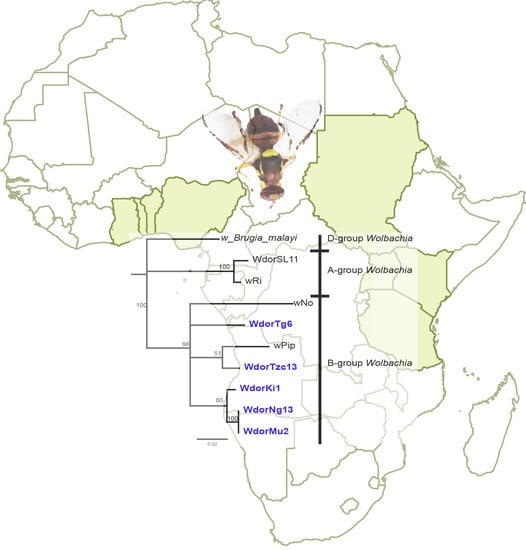
Graphical Abstract

1. Introduction

Bactrocera dorsalis (Hendel) (Diptera: Tephritidae) is amongst the most serious pests of cultivated fruits across Asia and Africa owing to its high adaptation, polyphagy, fecundity and the extent to which it causes yield and revenue losses [1]. Many other regions in the world are at risk of invasion and establishment of B. dorsalis [2,3]. Notably, B. dorsalis has been intercepted on more than 50 occasions since the 1980s in Florida and California, indicating that North America is at constant risk for the establishment of this pest [1,4]. In addition, there has been a recent report of B. dorsalis invading Europe, where is has been found in Italy [5]. In Africa, B. dorsalis was first detected in 2003 in Kenya and since then the pest has rapidly spread and established in most African countries often displacing the native Ceratitis cosyra (Walker) (Diptera: Tephritidae) as the primary fruit fly pest of mango [6,7]. The invasion of Africa by this pest had major consequences for fruit production, causing major losses in yield [8] as well as revenues [9].
Wolbachia is an intracellular bacterial parasite known to infect many arthropod species [10,11,12]. Wolbachia are maternally-transmitted in the egg cytoplasm, and therefore, have evolved a number of reproductive manipulations to increase the fitness of Wolbachia-infected matrilines. In many cases, Wolbachia cause cytoplasmic incompatibility between uninfected females and infected males. This ability to cause cytoplasmic incompatibility can result in Wolbachia-infected lineages rapidly increasing in frequency in a host population. The release of Wolbachia-infected incompatible males is potentially a very effective mechanism for decreasing pest insect populations (incompatible insect technique, IIT) [13]. IIT may have a number of benefits relative to the sterile insect technique (SIT) because radiation is not required. Notably, a symbiont-based pest management technique utilizing a Wolbachia strain that causes cytoplasmic incompatibility in fruit flies has been demonstrated in the Mediterranean fruit fly, Ceratitis capitata (Wiedemann) (Diptera: Tephritidae) [14] and evaluated for the olive fruit fly Bactrocera oleae (Rossi) (Diptera: Tephritidae) [15]. In addition, Wolbachia-induced cytoplasmic incompatibility can be used to spread symbionts and transgenes through target insect populations, which could be useful for controlling pests and blocking the capacity of vectors to transmit diseases [16,17,18,19]. Some Wolbachia strains have also been found to modify their host’s susceptibility to parasitoids [20]; therefore, knowledge of Wolbachia infection status can be of relevance to optimizing integrated pest control strategies employing parasitoid wasps.
Wolbachia are a diverse bacterial clade that has been broadly categorized into several super groups. Currently, there are at least 16 recognized super groups, designated A–F and H–Q [21,22,23,24,25,26,27,28,29,30]. Some strains show strong associations with certain host species, while others infect more than one host species and observations of multiple infections of same species or even same individuals are commonly reported [31,32,33,34,35,36,37]. This pattern indicates that over evolutionary timescales, horizontal transmission of Wolbachia is commonplace [38]. At the population level, Wolbachia are transmitted vertically and since mitochondria are co-inherited, this can establish a linkage disequilibrium between Wolbachia and the host mitochondrial haplotype [39].
In the Tephritidae family, several studies have detected Wolbachia strains in the genera Rhagoletis [32,40,41], Anastrepha [35,42,43,44,45,46], Ceratitis [13,47,48] Dacus [49,50,51] and Bactrocera [49,50,51,52,53,54]. In B. dorsalis, Wolbachia has been reported at low prevalence in populations from China [53] and Thailand [52,54]. The objective of the current study was to investigate the presence and diversity of Wolbachia strains in B. dorsalis populations in Africa and to evaluate the infection patterns of Wolbachia and associations with mtDNA haplotypes in populations of this host sampled between 2005 and 2017.

2. Materials and Methods

Bactrocera dorsalis male flies were collected using attract-and-kill bait stations with methyl eugenol as attractant and malathion as killing agent, placed in Lynfield traps. Trapping was done in mango farms in 2017 from Mwanza (S 2°43′01.3″ E 33°01′20.4″) and Morogoro (S 06°57′38.5″ E 037°31′59.1″) in Tanzania, Bunamwaya (N 0°16′17.8752″ E 32°33′25.6284″) in Uganda and Kassala (N 15°28′39.1728″, E 36°21′57.9204″), Gezira (N 14°36′29.4″ E 33°47′27.5″) and Singa (N 12°47′46.6″ E 33°11′51.5″) in Sudan. Bactrocera dorsalis female flies were retrieved from infested mango collected from mango farms in 2017 from Nguruman (S 01°48′32″ E 36°03′35″), Kitui (S 01°21′ E 38°00′), Muranga (S 0°42′50.0″ E 37°07′03.4″) and Embu (S 0°28′56.6″ E 37°34′55.5″) in Kenya. Infested fruit were dissected for third stage larvae to emerge and pupate in fine sterile sand. Puparia were sieved from the sand and maintained in ventilated perspex cages until eclosion. All samples were stored in absolute ethanol at −20 °C. DNA was extracted from each individual using the ISOLATE II Genomic DNA Kit (Bioline, London, UK). Voucher specimens collected between 2005 and 2009 in African sites as well as in Sri Lanka from an earlier study of B. dorsalis were obtained from the molecular biology laboratory at the International Centre of Insect Physiology and Ecology, icipe [55,56]. Wolbachia infections were initially screened by PCR using the wsp primers 81F and 691R [57] and subsequently all positives were screened using the 16S rRNA primers for Wolbachia pipientis [58] and the Wolbachia MLST gene primer sets [59]. Reactions were set up in total volumes of 10 µL each, containing 5 × MyTaq reaction buffer (5 mM dNTPs, 15 mM MgCl2, stabilizers and enhancers) (Bioline, London, UK), 2 µM of each primer, 0.25 mM MgCl2 (Thermo Fischer Scientific, Waltham, MA, USA), 0.125 µL MyTaq DNA polymerase (Bioline, London, UK), and 7.5 ng/µL of DNA template. These reactions were set up in a Master cycler Nexus gradient thermo-cycler (Thermo Fischer Scientific, MA, USA). Cycling conditions for the 16S rRNA primers included an initial denaturation for 2 min at 95 °C, followed by 30 cycles of 1 min at 95 °C, 1 min at 52 °C and 1 min at 72 °C, then a final elongation step of 10 min at 72 °C. For the MLST and wsp primers, an initial denaturation for 2 min at 94 °C was used followed by 40 cycles of denaturation of 30 s at 94 °C, 45 s at annealing temperature (55 °C –wsp, 54 °C –hcpA, gatB, ftsZ, coxA and 59 °C –fbpA), 1 min 30 s at 72 °C followed by a final extension step of 10 min at 72 °C. Host mitochondrial DNA was amplified by PCR in similar reaction volumes and cycling conditions as the wsp and MLST genes, using the primer LCO1490 and HCO2198 [60] at an annealing temperature of 50.6 °C. PCR products were run through 1% agarose gel electrophoresis and visualized by ethidium bromide staining and UV trans-illumination. Direct sequencing was done for all host COI and Wolbachia positive samples. Sequencing was carried out in both directions (F and R) for Wolbachia and host COI. Wolbachia sequences and representative host haplotype sequences were submitted to the GenBank. Sequence alignments were performed using Clustal W in Geneious 8.1.9 software (www.geneious.com) [61]. Phylogenetic trees were constructed by the neighbor-joining method with the Tamura-Nei model in Geneious 8.1.9 software. Support for tree topology was assessed by bootstrap resampling. A haplotype map was generated using median-joining network algorithm in the population analysis with reticulate trees (popART) software (http://popart.otago.ac.nz) [62,63]. Allelic profiles for wsp and MLST sequences obtained were inferred using the Wolbachia MLST database (https://pubmlst.org) [59].

3. Results

Using a PCR-based assay to amplify the Wolbachia wsp gene, we found that out of the 357 individuals tested, 10 were positive for Wolbachia (Table 1). These include 6 samples collected between 2005 and 2009 from African sites and 2 collected in Sri Lanka in 2007. In the samples collected in 2017, only 2 were found positive, which corresponds to an overall Wolbachia prevalence of 3.6% in the period between 2005 and 2009 and 1.1% in 2017 for the sampled African populations of B. dorsalis. Two sites, Muranga and Nguruman, had Wolbachia positives in the 2005 to 2009 sample set but not in the 2017 set, whereas Kitui had no positives in the period between 2005 and 2009 but one positive in 2017.
For a total of 6 samples (H-Ng13, Ki1, H-Mu2, Tzc13, H-Tg6 and H-Sl11), we amplified the Wolbachia coxA gene in addition to wsp, whereas in fewer samples (Ki1, Ng13, Mu2 and Sl11) other MLST genes were amplified and sequenced (Table 2). A full MLST profile and 16SrDNA was achieved for one sample (Tzc13), which had identical allelic profiles in their wsp hypervariable regions and coxA locus to strains in the Wolbachia pubMLST database, however, none of Tzc13’s other loci were identical to those in previously characterized Wolbachia strains (Table S1). The rest of the positive samples had incomplete profiles and partial similarities to those in the pubMLST database.
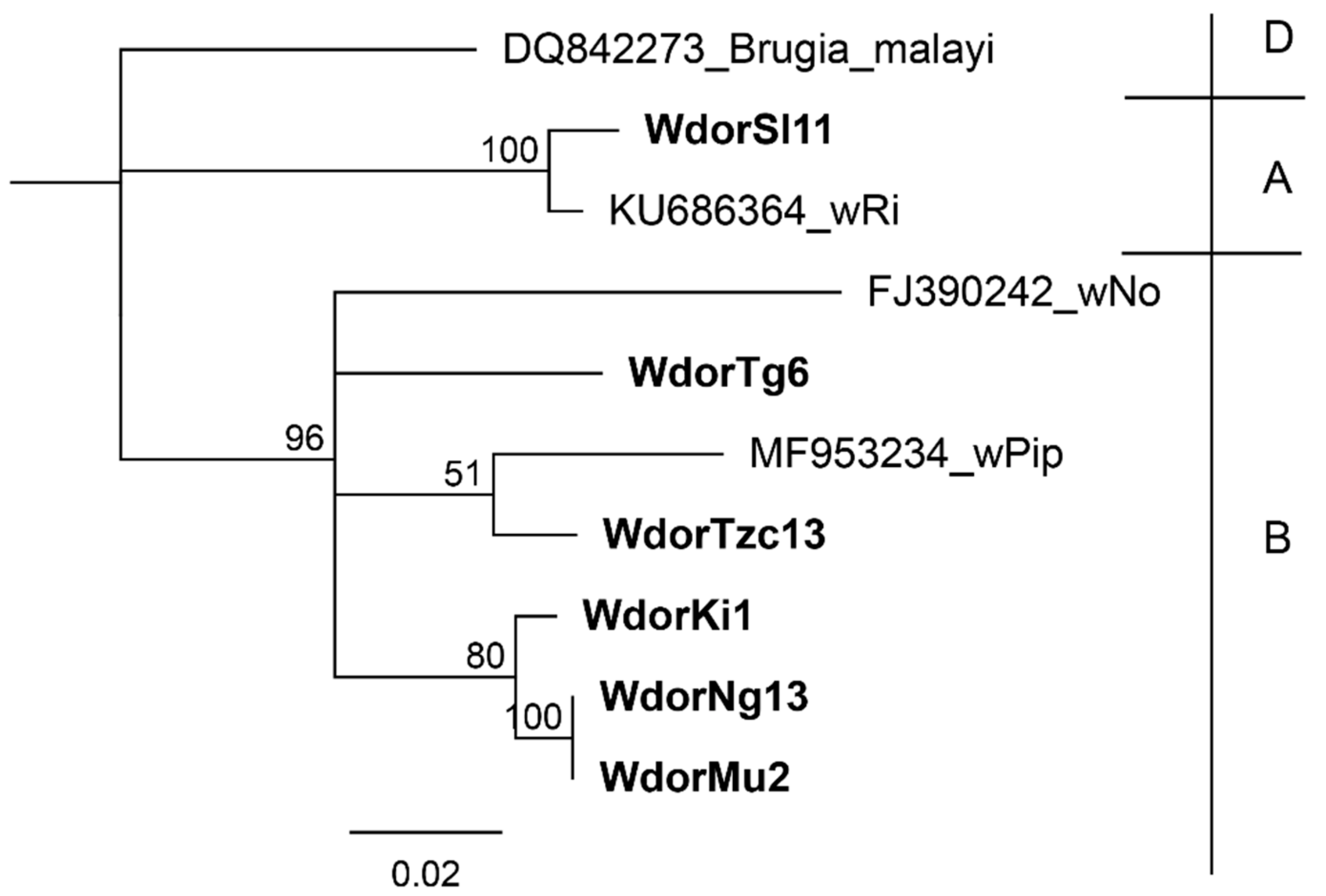
To investigate the phylogenetic relationship between the detected Wolbachia, we constructed a phylogenetic tree with 41 sequences available in GenBank. We used wsp sequences for 5 African samples and 1 Sri Lankan sample for which we were able to amplify at least one additional MLST gene. None of these Wolbachia were identical to previously detected Wolbachia strains in B. dorsalis or other Bactrocera species. The wsp sequences clustered with other Wolbachia strains in super group B (Figure 1). Two samples collected in Kenya in the period between 2005 and 2009 (H-Ng3 and H-Mu2) were found to have identical wsp sequences, whereas all other samples had wsp gene sequences that were unique within B. dorsalis.
Amongst the MLST loci, the coxA was amplified in the greatest number of samples. The phylogenetic relationships between strains as inferred by coxA gene sequence are similar to those inferred using the wsp gene with one notable exception; the coxA gene sequence for H-Sl11 clustered with Wolbachia group A as opposed to its wsp gene sequence, which clusters with group B (Figure 2). We observed that samples H-Ng3 and H-Mu2, which had identical wsp sequences, also had identical coxA sequences.
Sequences from other common Wolbachia strains (wPip, wNo and wRi) are included, while a sequence from Wolbachia endosymbiont of Brugia Malayi is included as an outgroup. Wolbachia super groups are indicated in higher case letters on the right. The detected Wolbachia are denoted by W followed by the host denoted as dor (B. dorsalis), population (Tg-Togo, Tzc-Tanzania, Ki-Kitui, Sl-Sri Lanka, Ng-Nguruman and Mu-Muranga) and population sample ID.
In addition to coxA, the MLST genes fbpA, hcpA, gatB and ftsZ were amplified in a number of Wolbachia positive B. dorsalis samples. Phylogenetic inferences based on these genes largely supported the inferences based on wsp; H-Mu2 and H-Ng13, which were found to have identical wsp and coxA sequences were also found identical at the fbpA locus (Figure S1). Similarly, the fbpA sequence for Ki1 indicated, in agreement with both wsp and coxA, that this strain belongs to super group B. However, there were a few notable exceptions; in samples H-Sl11 and Tzc13, which were B super group Wolbachia based on the wsp locus, one or more MLST genes clustered with super group A Wolbachia (Figure S1).
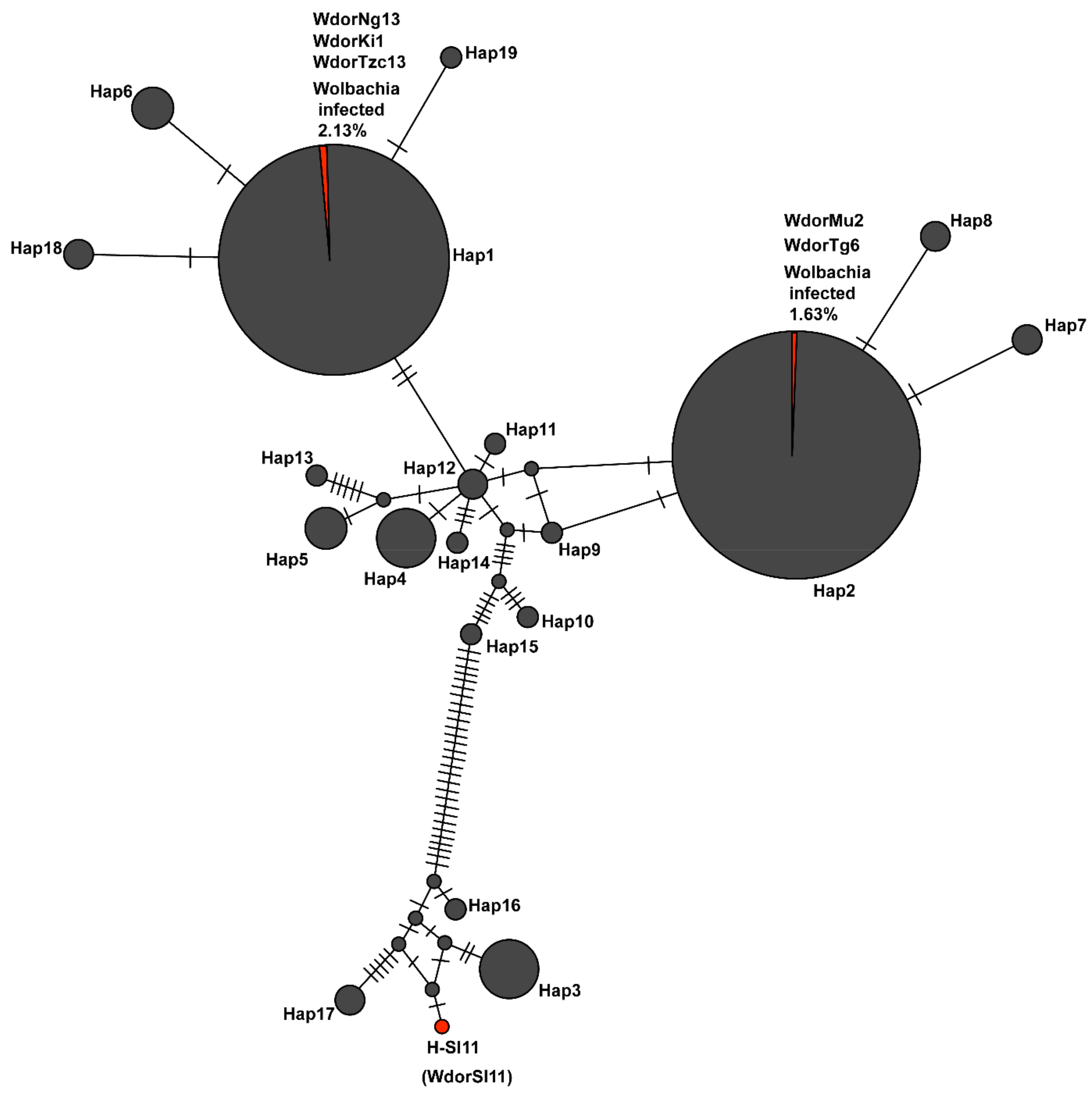
The samples collected in 2017 clustered into 7 mitochondrial COI haplotypes (Hap1-Hap7), whereas samples collected between 2005 and 2009 clustered into five of the aforementioned haplotypes (with the exception of Hap5 and 7) and an additional 12 smaller haplotypes (Figure 3). The COI gene sequences of the Wolbachia-infected B. dorsalis from African populations indicated that all were either Hap1 (H-Ng13, Ki1 and Tzc13) or Hap2 (H-Mu2 and H-Tg6). The infected Sri Lankan sample (H-Sl11) did not cluster into any of the 7 major and 12 minor mitochondrial COI haplotypes known from Africa. The two samples that had identical wsp and coxA gene sequences (H-Ng13 and H-Mu2) were found to have different mitochondrial COI haplotypes.
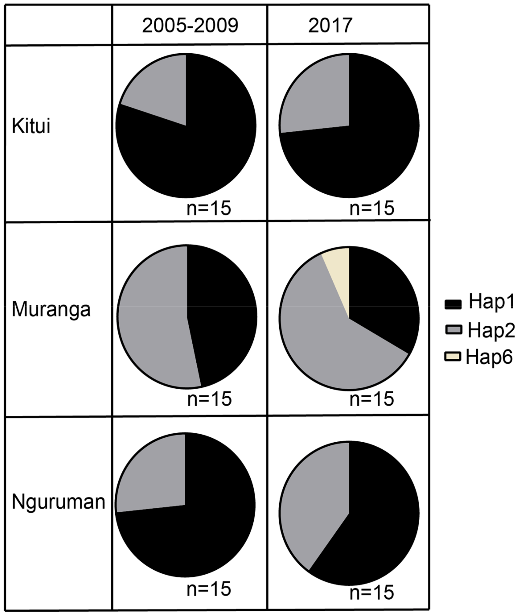
In sites that were sampled in both the 2005 to 2009 period and 2017, the most dominant haplotype in the 2005 to 2009 population was also dominant in the 2017 populations (Figure 4), inferring minimal change in the population structures as inferred by mitochondrial haplotype.

4. Discussion

We investigated Wolbachia infections in B. dorsalis in Africa and one location outside of Africa. Wolbachia sequences were detected in 10 samples. Overall, this indicates a low rate of Wolbachia infection across the African populations, although infection rates appear to be marginally higher than found in Asia [52,53]. It is notable that a higher prevalence of Wolbachia infection was observed in Sri Lanka, which is within the native range for this species. Based on their wsp and coxA sequence, four distinct variants of Wolbachia: WdorTg6, WdorTzc13, WdorKi1 and WdorNg3/WdorMu2 were detected in the African populations of B. dorsalis. A fifth variant, WdorSl11, was detected in one of the Sri Lankan samples. All of these variants were distinct from those previously observed in B. dorsalis [52,53]; this therefore suggests that this species has a high diversity of low prevalence Wolbachia.
The WdorNg3/WdorMu2 variant was detected in two individuals from two different sampling sites, Nguruman and Muranga, which are within geographically-separated agro-ecological zones in Kenya. It was notable that this variant was observed in two different mitochondrial haplotypes (Hap1 and Hap2). Wolbachia strains only transmitted from an infected female to her offspring (strict vertical transmission) tend to associate strictly with the same host haplotypes while strains that are occasionally transmitted horizontally (from one individual to another unrelated individual) do not. Altogether, the non-concordance with mitochondrial DNA, suggests that some of the Wolbachia strains infecting African populations of B. dorsalis may have an appreciable rate of horizontal transmission.
In four out of the ten wsp positive samples, we were not able to amplify any of the MLST or the 16S rRNA genes. This may be indicative of transient infections that could have spilled over from an unknown source, or less likely they could be related to a partial transfer of genes (more common than the transfer of a full Wolbachia genome) from the symbiont to the host genome. This phenomenon has been observed in previous studies [49], where Wolbachia pseudogenes have been detected in other tephritid fruit flies. The nature of these infections could be examined by monitoring vertical transmission rates and tissue localization patterns. We also cannot rule out the possibility that sample trapping and storage may have caused DNA degradation, which could have led to some false Wolbachia negative samples and an inability to amplify all loci for the Wolbachia positive samples.
At the majority of the loci investigated, the Wolbachia variants detected among the African populations clustered with super group B Wolbachia, which differs from the majority of super group A strains as found in B. dorsalis species in China [53] and Thailand [52]. For sample Tzc13, one MLST locus (fbpA) segregated with group A Wolbachia, suggesting a possible recombination event between an A and B group strains, a phenomenon that has been reported in previous studies [64,65]. For another sample, H-Sl11, we observed that the wsp gene segregated with super group B Wolbachia in contrast to the coxA and hcpA loci which segregated as super group A Wolbachia. However, sequencing results of the 16S rRNA, gatB and ftsZ loci of H-Sl11 (data not shown) revealed an interfering secondary chromatogram in few segments suggesting that the sample is likely to have been infected by two Wolbachia strains (A and B group).
In addition, none of the detected Wolbachia had complete identity of allelic profiles to Wolbachia strains in the Wolbachia MLST database. Full length sequences of each of the MLST and wsp marker could not be retrieved for all the samples, and this limited our ability to confirm their designation as novel strain types through allelic profiles.
Some strains of Wolbachia are known to cause male killing, and in insect populations with these strains, infected male specimens are generally not observed or observed only very rarely. To avoid bias against the detection of male-killing Wolbachia, female specimens are generally screened. Wolbachia are also known to reach high densities in insect ovaries and therefore may be easier to detect in females. Male lures are among the most widely used attractants in trapping of tephritid fruit flies. Therefore, the majority of the samples we screened, in particular from the 2005 to 2009 collections, were male. It is possible that if female flies had been screened, a higher Wolbachia prevalence rate would have been observed. However, it is noteworthy that the female samples from the 2017 collections did not have a higher rate of Wolbachia infection. Similar observations have been reported among B. dorsalis populations in Asia where the sexual differences in this host does not influence the detection rate of Wolbachia [53]. The Wolbachia variants that were found in males (WdorTg6, WdorTzc13, WdorNg3/WdorMu2 and WdorSl11) are unlikely to have a male-killing phenotype, whereas we cannot rule out the possibility that WdorKi1, which was detected in a female fly, causes male killing.
The population structure of B. dorsalis in the African region was observed to be largely unchanged between the 2005 to 2009 and 2017 samples, with two major mtCOI haplotypes (Hap1 and Hap2) dominating in both [56]. This suggests that based on mitochondrial DNA diversity, the current population structure has been largely unchanged since the initial B. dorsalis invasion in the Eastern Africa region, and that no significant new reintroductions with new mtCOI haplotypes have occurred in this region since the first invasion. To confirm this, however, it would be necessary to use nuclear DNA markers as mitochondrial DNA introgression could have occurred. The two main haplotypes, Hap1 and Hap2, in addition to Hap3, Hap4 and Hap7 were observed to be fairly distributed across the East and West of Africa. Only two haplotypes: Hap5 and Hap6 were observed in Eastern Africa only while the rest of the smaller mtCOI haplotypes (Hap8 to Hap18) were observed in West Africa, particularly in Nigeria, except for Hap8 that was observed exclusively in Benin. Previous microsatellite genotyping data also revealed differences between the Eastern Africa populations with that of Nigeria, which were more closely allied to the diverse Sri Lankan populations of B. dorsalis [56]. It has been suggested that contemporary gene flow may have contributed to this diversity in West Africa [56].

5. Conclusions

We detected four different Wolbachia variants in African populations of the oriental fruit fly. Analysis of the host mtCOI haplotypes did not reveal a link between a particular Wolbachia variant and host haplotypes. Only the two dominant haplotypes were found to be infected with Wolbachia in Africa. These Wolbachia should be investigated for their capacity to manipulate host reproduction and to confer hosts with differential susceptibility to parasitoids and pathogens. A comprehensive understanding of the role of Wolbachia in this species could improve the effectiveness of integrated control strategies and eventually play a role in the sustainable pest management of B. dorsalis.

Supplementary Materials

The following is available online at https://www.mdpi.com/2075-4450/10/6/155/s1, Figure S1: Neighbour joining trees showing relationship between Wolbachia in B. dorsalis (in bold) to strains in known super groups. Trees were generated from sequences of 16S rRNA (A), fbpA (B), ftsZ (C), gatB (D) and hcpA (E) genes. Sequences of Wolbachia endosymbiont of Brugia malayi are included as out-groups in all the trees and respective super groups are indicated to the right of each tree. The detected Wolbachia are denoted by W followed by the host denoted as dor (B. dorsalis), population (Tg-Togo, Tzc-Tanzania, Ki-Kitui, Sl-Sri Lanka, Ng-Nguruman and Mu-Muranga) and population sample ID. Bootstrap values are indicated above branches. Branches with bootstrap support lower than 50% are collapsed; Table S1: Wolbachia MLST and wsp gene allelic profiles. Exact matches to alleles present in the database are shown whereas instances where no exact matches were found are represented as N. Dashes represent the genes for each strain that could not be amplified. GenBank accession numbers for respective sequences are shown in brackets.

Author Contributions

Conceptualization: F.M.K, S.E. and J.K.H.; Investigation, data analysis and original draft preparation: J.G.; Supervision: F.M.K., S.E., J.K.H and J.V.B.; Writing-review and editing of final draft: all authors; Funding acquisition: S.E.

Funding

The authors gratefully acknowledge support for this research by the following organizations and agencies: the European Union Integrated Biological Control Applied Research Program (IBCARP)-Fruit Fly Component; the Wellcome trust [107372]; the UK’s Department for International Development (DFID); Swedish International Development Cooperation Agency (Sida); the Swiss Agency for Development and Cooperation (SDC); Federal Democratic Republic of Ethiopia and the Kenyan Government. The views expressed herein do not necessarily reflect the official opinion of the donors.

Acknowledgments

The authors acknowledge Ivan Rwomushana, Abdullah Mkiga, Idriss Gamal, Peterson Nderitu and Emmanuel Mlato for assistance during sampling.

Conflicts of Interest

The authors declare no conflict of interest. The funders had no role in the design of the study; in the collection, analyses, or interpretation of data; in the writing of the manuscript, or in the decision to publish the results.

References

  1. Dohino, T.; Hallman, G.J.; Grout, T.G.; Clarke, A.R.; Follett, P.A.; Cugala, D.R.; Minh Tu, D.; Murdita, W.; Hernandez, E.; Pereira, R.; et al. Phytosanitary treatments against Bactrocera dorsalis (Diptera: Tephritidae): Current situation and future prospects. J. Econ. Entomol. 2017, 110, 67–79. [Google Scholar]
  2. De Villiers, M.; Hattingh, V.; Kriticos, D.J.; Brunel, S.; Vayssières, J.; Sinzogan, A.; Billah, M.K.; Mohamed, S.A.; Mwatawala, M.; Abdelgader, H.; et al. The potential distribution of Bactrocera dorsalis: Considering phenology and irrigation patterns. Bull. Entomol. Res. 2016, 106, 19–33. [Google Scholar] [CrossRef]
  3. Stephens, A.E.A.; Kriticos, D.J.; Leriche, A. The current and future potential geographical distribution of the oriental fruit fly, Bactrocera dorsalis (Diptera: Tephritidae). Bull. Entomol. Res. 2007, 97, 369–378. [Google Scholar] [CrossRef] [PubMed]
  4. USDA-APHIS. Oriental Fruit Fly Cooperative Eradication Program Los Angeles and Orange Counties, Califonia; U.S. Department of Agriculture Health Animal and Plant Inspection Service: Riverdale, MD, USA, 2014.
  5. Nugnes, F.; Russo, E.; Viggiani, G.; Bernardo, U. First record of an invasive fruit fly belonging to Bactrocera dorsalis complex (Diptera: Tephritidae) in Europe. Insects 2018, 9, 182. [Google Scholar] [CrossRef]
  6. Lux, S.A.; Copeland, R.S.; White, I.M.; Manrakhan, A.; Billah, M.K. A new invasive fruit fly species from the Bactrocera dorsalis (Hendel) group detected in east Africa. Int. J. Trop. Insect Sci. 2003, 23, 355–361. [Google Scholar] [CrossRef]
  7. Ekesi, S.; Billah, M.K.; Nderitu, P.W.; Lux, S.A.; Rwomushana, I. Evidence for competitive displacement of Ceratitis cosyra by the invasive fruit fly Bactrocera invadens (Diptera: Tephritidae) on mango and mechanisms contributing to the displacement. J. Econ. Entomol. 2009, 102, 981–991. [Google Scholar] [CrossRef] [PubMed]
  8. Nankinga, C.M.; Isabirye, B.E.; Muyinza, H.; Rwomushana, I.; Stevenson, P.C.; Mayamba, A.; Aool, W.; Akol, A.M. Fruit fly infestation in mango: A threat to the horticultural sector in Uganda. Uganda J. Agric. Sci. 2014, 15, 1–14. [Google Scholar]
  9. Ekesi, S.; De Meyer, M.; Mohamed, S.A.; Virgilio, M.; Borgemeister, C. Taxonomy, ecology and management of native and exotic fruit fly species in Africa. Annu. Rev. Entomol. 2016, 61, 219–238. [Google Scholar] [CrossRef] [PubMed]
  10. Zug, R.; Hammerstein, P. Still a host of hosts for Wolbachia: Analysis of recent data suggests that 40% of terrestrial arthropod species are infected. PLoS ONE 2012, 7, 38544. [Google Scholar] [CrossRef]
  11. Hilgenboecker, K.; Hammerstein, P.; Schlattmann, P.; Telschow, A.; Werren, J.H. How many species are infected with Wolbachia?—A statistical analysis of current data. FEMS Microbiol. Lett. 2008, 281, 215–220. [Google Scholar] [CrossRef]
  12. Weinert, L.A.; Araujo-Jnr, E.V.; Ahmed, M.Z.; Welch, J.J. The incidence of bacterial endosymbionts in terrestrial arthropods. Proc. R. Soc. B Biol. Sci. 2015, 282, 20150249. [Google Scholar] [CrossRef]
  13. Zabalou, S.; Riegler, M.; Theodorakopoulou, M.; Stauffer, C.; Savakis, C.; Bourtzis, K. Wolbachia-induced cytoplasmic incompatibility as a means for insect pest population control. Proc. Natl. Acad. Sci. USA 2004, 101, 15042–15045. [Google Scholar] [CrossRef] [PubMed]
  14. Zabalou, S.; Apostolaki, A.; Livadaras, I.; Franz, G.; Robinson, A.S.; Savakis, C.; Bourtzis, K. Incompatible insect technique: Incompatible males from a Ceratitis capitata genetic sexing strain. Entomol. Exp. Appl. 2009, 132, 232–240. [Google Scholar] [CrossRef]
  15. Apostolaki, A.; Livadaras, I.; Saridaki, A.; Chrysargyris, A.; Savakis, C.; Bourtzis, K. Transinfection of the olive fruit fly Bactrocera oleae with Wolbachia: Towards a symbiont-based population control strategy. J. Appl. Entomol. 2011, 135, 546–553. [Google Scholar] [CrossRef]
  16. Iturbe-Ormaetxe, I.; Walker, T.; O’Neill, S.L. Wolbachia and the biological control of mosquito-borne disease. EMBO Rep. 2011, 12, 508–518. [Google Scholar] [CrossRef] [PubMed]
  17. McMeniman, C.J.; Roxanna, L.V.; Bodil, C.N.; Amy, F.W.C.; Manpreet, S.; Wang, Y.F.; O’Neill, S.L. Stable introduction of a life-shortening Wolbachia infection into the mosquito Aedes aegypti. Science 2009, 323, 141–145. [Google Scholar] [CrossRef] [PubMed]
  18. Kambris, Z.; Cook, P.E.; Phuc, H.K.; Sinkins, S.P. Immune activation by life-shortening Wolbachia and reduced filarial competence in mosquitoes. Science 2009, 326, 134–136. [Google Scholar] [CrossRef]
  19. Sinkins, S.P. Wolbachia and cytoplasmic incompatibility in mosquitoes. Insect Biochem. Mol. Biol. 2004, 34, 723–729. [Google Scholar] [CrossRef]
  20. Van Nouhuys, S.; Kohonen, M.; Duplouy, A. Wolbachia increases the susceptibility of a parasitoid wasp to hyperparasitism. J. Exp. Biol. 2016, 219, 2984–2990. [Google Scholar] [CrossRef]
  21. Lo, N.; Casiraghi, M.; Salati, E.; Bazzocchi, C.; Bandi, C. How many Wolbachia supergroups exist? Mol. Biol. Evol. 2002, 19, 341–346. [Google Scholar] [CrossRef]
  22. Bordenstein, S.; Rosengaus, R.B. Discovery of a novel Wolbachia supergroup in isoptera. Curr. Microbiol. 2005, 51, 393–398. [Google Scholar] [CrossRef] [PubMed]
  23. Lo, N.; Paraskevopoulos, C.; Bourtzis, K.; O’Neill, S.L.; Werren, J.H.; Bordenstein, S.R.; Bandi, C. Taxonomic status of the intracellular bacterium Wolbachia pipientis. Int. J. Syst. Evol. Microbiol. 2007, 57, 654–657. [Google Scholar] [CrossRef] [PubMed]
  24. Bordenstein, S.R.; Paraskevopoulos, C.; Dunning Hotopp, J.C.; Sapountzis, P.; Lo, N.; Bandi, C.; Tettelin, H.; Werren, J.H.; Bourtzis, K. Parasitism and mutualism in Wolbachia: What the phylogenomic trees can and cannot say. Mol. Biol. Evol. 2009, 26, 231–241. [Google Scholar] [CrossRef] [PubMed]
  25. Ros, V.I.D.; Fleming, V.M.; Feil, E.J.; Breeuwer, J.A.J. How diverse is the genus Wolbachia? Multiple-gene sequencing reveals a putatively new Wolbachia supergroup recovered from spider mites (Acari: Tetranychidae). Appl. Environ. Microbiol. 2009, 75, 1036–1043. [Google Scholar] [CrossRef]
  26. Haegeman, A.; Vanholme, B.; Jacob, J.; Vandekerckhove, T.T.M.; Claeys, M.; Borgonie, G.; Gheysen, G. An endosymbiotic bacterium in a plant-parasitic nematode: Member of a new Wolbachia supergroup. Int. J. Parasitol. 2009, 39, 1045–1054. [Google Scholar] [CrossRef] [PubMed]
  27. Augustinos, A.A.; Diego, S.G.; Dionyssopoulou, E.; Moreira, M.; Papapanagiotou, A.; Scarvelakis, M.; Doudoumis, V.; Ramos, S.; Aguiar, A.F.; Borges, P.A.V.; et al. Detection and characterization of Wolbachia infections in natural populations of aphids: Is the hidden diversity fully unraveled? PLoS ONE 2011, 6, e28695. [Google Scholar] [CrossRef] [PubMed]
  28. Bing, X.L.; Xia, W.Q.; Gui, J.D.; Yan, G.H.; Wang, X.W.; Liu, S.S. Diversity and evolution of the Wolbachia endosymbionts of Bemisia (Hemiptera: Aleyrodidae) whiteflies. Ecol. Evol. 2014, 4, 2714–2737. [Google Scholar] [CrossRef]
  29. Glowska, E.; Dragun-Damian, A.; Dabert, M.; Gerth, M. New Wolbachia supergroups detected in quill mites (Acari: Syringophilidae). Infect. Genet. Evol. 2015, 30, 140–146. [Google Scholar] [CrossRef]
  30. Wang, G.H.; Jia, L.Y.; Xiao, J.H.; Huang, D.W. Discovery of a new Wolbachia supergroup in cave spider species and the lateral transfer of phage WO among distant hosts. Infect. Genet. Evol. 2016, 41, 1–7. [Google Scholar] [CrossRef]
  31. Pascar, J.; Chandler, C.H. A bioinformatics approach to identifying Wolbachia infections in arthropods. PeerJ 2018, 6, e5486. [Google Scholar] [CrossRef]
  32. Schuler, H.; Arthofer, W.; Riegler, M.; Bertheau, C.; Krumböck, S.; Köppler, K.; Vogt, H.; Teixeira, L.A.F.; Stauffer, C. Multiple Wolbachia infections in Rhagoletis pomonella. Entomol. Exp. Appl. 2011, 139, 138–144. [Google Scholar] [CrossRef]
  33. Hughes, G.L.; Allsopp, P.G.; Brumbley, S.M.; Woolfit, M.; McGraw, E.A.; O’Neill, S.L. Variable infection frequency and high diversity of multiple strains of Wolbachia pipientis in Perkinsiella planthoppers. Appl. Environ. Microbiol. 2011, 77, 2165–2168. [Google Scholar] [CrossRef] [PubMed]
  34. Roehrdanz, R.L.; Wichmann, S.S. Wolbachia multilocus sequence typing of singly infected and multiply infected populations of northern corn rootworm (Coleoptera: Chrysomelidae). Ann. Entomol. Soc. Am. 2014, 107, 832–841. [Google Scholar] [CrossRef]
  35. Mascarenhas, R.O.; Prezotto, L.F.; Perondini, A.L.P.; Marino, C.L.; Selivon, D. Wolbachia in guilds of Anastrepha fruit flies (Tephritidae) and parasitoid wasps (Braconidae). Genet. Mol. Biol. 2016, 39, 600–610. [Google Scholar] [CrossRef] [PubMed]
  36. Yang, X.H.; Zhu, D.H.; Liu, Z.; Zhao, L.; Su, C.Y. High levels of multiple infections, recombination and horizontal transmission of Wolbachia in the Andricus mukaigawae (Hymenoptera; Cynipidae) communities. PLoS ONE 2013, 8, e78970. [Google Scholar] [CrossRef]
  37. Valette, V.; Bitome Essono, P.Y.; Le Clec’h, W.; Johnson, M.; Bech, N.; Grandjean, F. Multi-infections of feminizing Wolbachia strains in natural populations of the terrestrial isopod Armadillidium vulgare. PLoS ONE 2013, 8, e82633. [Google Scholar] [CrossRef] [PubMed]
  38. White, P.M.; Pietri, J.E.; Debec, A.; Russell, S.; Patel, B.; Sullivan, W. Mechanisms of horizontal cell-to-cell transfer of Wolbachia spp. in Drosophila melanogaster. Appl. Environ. Microbiol. 2017, 83. [Google Scholar] [CrossRef] [PubMed]
  39. Yeap, H.L.; Rašić, G.; Endersby-Harshman, N.M.; Lee, S.F.; Arguni, E.; Le Nguyen, H.; Hoffmann, A.A. Mitochondrial DNA variants help monitor the dynamics of Wolbachia invasion into host populations. Heredity 2016, 116, 265. [Google Scholar] [CrossRef] [PubMed]
  40. Schuler, H.; Bertheau, C.; Egan, S.P.; Feder, J.L.; Riegler, M.; Schlick-Steiner, B.C.; Steiner, F.M.; Johannesen, J.; Kern, P.; Tuba, K.; et al. Evidence for a recent horizontal transmission and spatial spread of Wolbachia from endemic Rhagoletis cerasi (Diptera: Tephritidae) to invasive Rhagoletis cingulata in Europe. Mol. Ecol. 2013, 22, 4101–4111. [Google Scholar] [CrossRef] [PubMed]
  41. Riegler, M.; Stauffer, C. Wolbachia infections and superinfections in cytoplasmically incompatible populations of the European cherry fruit fly Rhagoletis cerasi (Diptera, Tephritidae). Mol. Ecol. 2002, 11, 2425–2434. [Google Scholar] [CrossRef]
  42. Coscrato, V.E.; Braz, A.S.K.; André, A.L.; Selivon, D.; Marino, C.L. Wolbachia in Anastrepha fruit flies (Diptera: Tephritidae). Curr. Microbiol. 2009, 59, 295–301. [Google Scholar] [CrossRef]
  43. Mateos, M.; Martinez, H.; Lanzavecchia, S.B.; Conte, C.; Morán-aceves, B.M.; Toledo, J.; Liedo, P.; Asimakis, E.D.; Doudoumis, V.; Kyritsis, G.A.; et al. Wolbachia pipientis associated to tephritid fruit fly pests: From basic research to applications. BioRxiv 2018. [Google Scholar]
  44. Martınez, H.; Toledo, J.; Liedo, P.; Mateos, M. Survey of heritable endosymbionts in Southern Mexico populations of the fruit fly species Anastrepha striata. Curr. Microbiol. 2012, 65, 711–718. [Google Scholar] [CrossRef]
  45. Jeyaprakash, A.; Hoy, M.A. Long PCR improves Wolbachia DNA amplification: Wsp sequences found in 76% of sixty-three arthropod species. Insect Mol. Biol. 2000, 9, 393–405. [Google Scholar] [CrossRef] [PubMed]
  46. Selivon, D.; Perondini, A.P.; Ribeiro, A.F.; Marino, C.L.; Lima, M.M.A.; Coscrato, V.E. Wolbachia endosymbiont in a species of the Anastrepha fraterculus complex (diptera: Tephritidae). Invertebr. Reprod. Dev. 2002, 42, 121–127. [Google Scholar] [CrossRef]
  47. Rocha, L.S.; Mascarenhas, R.O.; Perondini, A.L.P.; Selivon, D. Occurrence of Wolbachia in Brazilian samples of Ceratitis capitata (Wiedemann) (Diptera: Tephritidae). Neotrop. Entomol. 2005, 34, 1013–1015. [Google Scholar] [CrossRef]
  48. Sarakatsanou, A.; Diamantidis, A.D.; Papanastasiou, S.A.; Bourtzis, K.; Papadopoulos, N.T. Effects of Wolbachia on fitness of the Mediterranean fruit fly (Diptera: Tephritidae). J. Appl. Entomol. 2011, 135, 554–563. [Google Scholar] [CrossRef]
  49. Morrow, J.L.; Frommer, M.; Royer, J.E.; Shearman, D.C.A.; Riegler, M. Wolbachia pseudogenes and low prevalence infections in tropical but not temperate Australian tephritid fruit flies: Manifestations of lateral gene transfer and endosymbiont spillover? BMC Evol. Biol. 2015, 15, 202. [Google Scholar] [CrossRef] [PubMed]
  50. Morrow, J.L.; Frommer, M.; Shearman, D.C.A.A.; Riegler, M. Tropical tephritid fruit fly community with high incidence of shared Wolbachia strains as platform for horizontal transmission of endosymbionts. Environ. Microbiol. 2014, 16, 3622–3637. [Google Scholar] [CrossRef]
  51. Morrow, J.L. Molecular Studies of Wolbachia and Sex-Determination Genes in Australian Bactrocera Species—Complementary Approaches to Improved Fruit Fly Control. Ph.D. Thesis, University of Western Sydney, Sydney, NSW, Australia, 2014. [Google Scholar]
  52. Jamnongluk, W.; Kittayapong, P.; Baimai, V.; O’Neill, S.L. Wolbachia infections of tephritid fruit flies: Molecular evidence for five distinct strains in a single host species. Curr. Microbiol. 2002, 45, 255–260. [Google Scholar] [CrossRef]
  53. Sun, X.; Cui, L.; Li, Z. Diversity and phylogeny of Wolbachia infecting Bactrocera dorsalis (Diptera: Tephritidae) populations from China. Environ. Entomol. 2007, 36, 1283–1289. [Google Scholar] [CrossRef]
  54. Kittayapong, P.; Milne, J.R.; Tigvattananont, S.; Baimai, V. Distribution of the reproduction-modifying bacteria, Wolbachia, in natural populations of tephritid fruit flies in Thailand. Sci. Asia 2000, 26, 93–103. [Google Scholar] [CrossRef]
  55. Khamis, F.M.; Masiga, D.K.; Mohamed, S.A.; Salifu, D.; de Meyer, M.; Ekesi, S. Taxonomic identity of the invasive fruit fly pest, Bactrocera invadens: Concordance in morphometry and DNA barcoding. PLoS ONE 2012, 7, 44862. [Google Scholar] [CrossRef]
  56. Khamis, F.M.; Karam, N.; Ekesi, S.; De Meyer, M.; Bonomi, A.; Gomulski, L.M.; Scolari, F.; Gabrieli, P.; Siciliano, P.; Masiga, D.; et al. Uncovering the tracks of a recent and rapid invasion: The case of the fruit fly pest Bactrocera invadens (Diptera: Tephritidae) in Africa. Mol. Ecol. 2009, 18, 4798–4810. [Google Scholar] [CrossRef] [PubMed]
  57. Braig, H.R.; Zhou, W.; Dobson, S.L.; O’Neill, S.L. Cloning and characterization of a gene encoding the major surface protein of the bacterial endosymbiont Wolbachia pipientis. J. Bacteriol. 1998, 180, 2373–2378. [Google Scholar] [PubMed]
  58. O’Neill, S.L.; Giordano, R.; Colbert, A.M.; Karr, T.L.; Robertson, H.M. 16S rRNA phylogenetic analysis of the bacterial endosymbionts associated with cytoplasmic incompatibility in insects. Proc. Natl. Acad. Sci. USA 2006, 89, 2699–2702. [Google Scholar] [CrossRef]
  59. Baldo, L.; Hotopp, J.C.D.; Jolley, K.A.; Bordenstein, S.R.; Biber, S.A.; Choudhury, R.R.; Hayashi, C.; Maiden, M.C.J.; Tettelin, H.; Werren, J.H. Multilocus sequence typing system for the endosymbiont Wolbachia pipientis. Appl. Environ. Microbiol. 2006, 72, 7098–7110. [Google Scholar] [CrossRef]
  60. Folmer, O.; Black, M.; Hoeh, W.; Lutz, R.; Vrijenhoek, R. DNA primers for amplification of mitochondrial cytochrome c oxidase subunit I from diverse metazoan invertebrates. Mol. Mar. Biol. Biotechnol. 1994, 3, 294–299. [Google Scholar]
  61. Kearse, M.; Moir, R.; Wilson, A.; Stones-Havas, S.; Cheung, M.; Sturrock, S.; Buxton, S.; Cooper, A.; Markowitz, S.; Duran, C.; et al. Geneious basic: An integrated and extendable desktop software platform for the organization and analysis of sequence data. Bioinformatics 2012, 28, 1647–1649. [Google Scholar] [CrossRef]
  62. Leigh, J.W.; Bryant, D. POPART: Full-feature software for haplotype network construction. Methods Ecol. Evol. 2015, 6, 1110–1116. [Google Scholar] [CrossRef]
  63. Bandelt, H.J.; Forster, P.; Rohl, A. Median-joining networks for inferring intraspecific phylogenies. Mol. Biol. 1994, 16, 37–48. [Google Scholar] [CrossRef] [PubMed]
  64. Malloch, G.; Fenton, B. Super-infections of Wolbachia in byturid beetles and evidence for genetic transfer between A and B super-groups of Wolbachia. Mol. Ecol. 2005, 14, 627–637. [Google Scholar] [CrossRef] [PubMed]
  65. Baldo, L.; Werren, J.H. Revisiting Wolbachia supergroup typing based on WSP: Spurious lineages and discordance with MLST. Curr. Microbiol. 2007, 55, 81–87. [Google Scholar] [CrossRef] [PubMed]
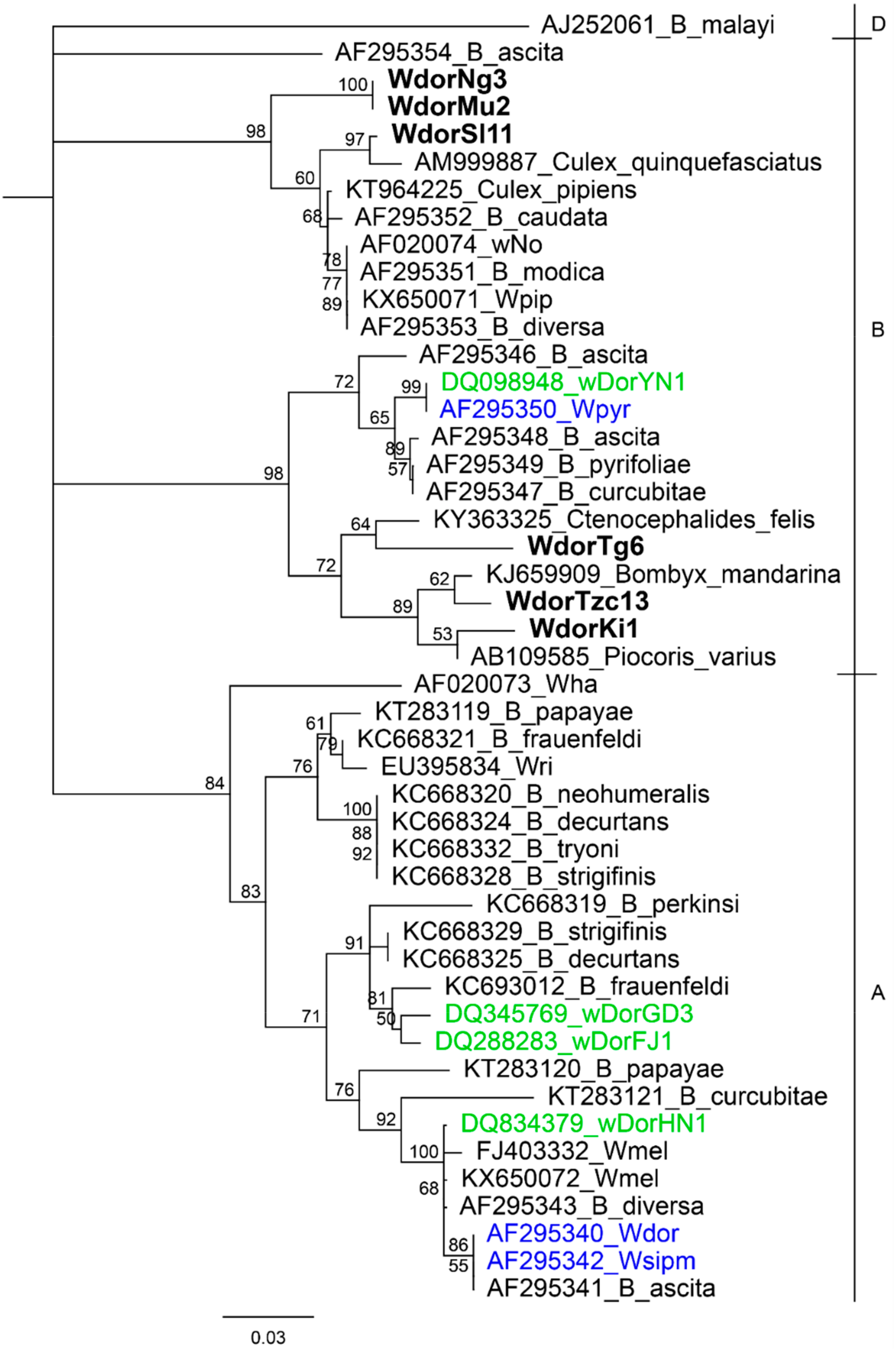
Figure 1. Neighbour joining tree based on Wolbachia surface protein (wsp) gene sequences of Wolbachia detected from B. dorsalis in this study (in bold), from B. dorsalis in China (labelled in blue), from B. dorsalis in Thailand (labelled in green) and from other Bactrocera species. Sequences from closest homology matches to Wolbachia detected in this study are also included. Other common Wolbachia strains (wMel, wRi, wHa, wNo and wPip) are also included. Wsp sequence from Wolbachia endosymbiont of Brugia malayi is included as an outgroup. Sequences are labelled with genbank accession numbers followed by strain name or host organism, or strain name for sequences from this study. Wolbachia super groups are indicated in higher case letters on the right. The detected Wolbachia are denoted by W followed by the host denoted as dor (B. dorsalis), population (Tg-Togo, Tzc-Tanzania, Ki-Kitui, Sl-Sri Lanka, Ng-Nguruman and Mu-Muranga) and population sample ID. Bootstrap values are indicated above branches. Branches with bootstrap support lower than 50% are collapsed.
Insects 10 00155 g001
Figure 2. Neighbour joining tree based on the cytochrome c oxidase subunit I (coxA) gene sequences from this study. The detected Wolbachia are denoted by W followed by the host denoted as dor (B. dorsalis), population (Tg-Togo, Tzc-Tanzania, Ki-Kitui, Sl-Sri Lanka, Ng-Nguruman and Mu-Muranga) and population sample ID. Bootstrap values are indicated above branches. Branches with bootstrap support lower than 50% are collapsed.
Insects 10 00155 g002
Figure 3. Haplotype map of Bactrocera dorsalis mtCOI sequences from African populations. Node size is proportional to number of samples while mutations are represented as hatchmarks. Proportions of Wolbachia infected samples (H-Ng13, Ki1 and Tzc13 in Haplotype1 and H-Mu2 and H-Tg6 in Haplotype 2) in their respective haplotypes are shaded in red. Sequences of represented haplotypes are accessible at Genbank using the accessions MK314052-MK31452 for Haplotypes: 3, 2, 4, 6, 7, 5 and 1 respectively, JQ692656, JQ692727, JQ692777, JQ692863, JQ692731, JQ692684, JQ692812, JQ692698, JQ692723, JQ692816, JQ692816, and JQ692691 for haplotypes 8-19 respectively. The infected Sri Lanka sample (labelled H-Sl11, Genbank accession: JQ692764) is not numbered to distinguish it from haplotypes detected in Africa.
Insects 10 00155 g003
Figure 4. Pie charts depicting haplotype compositions in sites sampled during the period between 2005 to 2009 (left column) and in 2017 (right column).
Insects 10 00155 g004
Table 1. B. dorsalis populations screened for Wolbachia using wsp. Samples collected between 2005 and 2009 are preceded with “H” to distinguish them from those collected in 2017.
LocalitySpecimen CodesCollection Year/Collection Sex/(n)wsp+
Nguruman, KenyaH-Ng2008m (15)1
Nguruman, KenyaNg2017f (15)0
Kitui, KenyaH-Ki2005m (15)0
Kitui, KenyaKi2017f (15)1
Muranga, KenyaH-Mu2005m (15)1
Muranga, KenyaMu2017f (15)0
Embu, KenyaEm2017f (15)0
Dar es Salaam, TanzaniaH-Tz2009m (15)0
Mwanza, TanzaniaTz-ab2017m (30)0
Morogoro, TanzaniaTz-c2017m (15)1
Kawanda, UgandaH-Ug2007m (15)0
Bunamwaya, UgandaUg-b2017m (30)0
Khartoum, SudanH-Su2007m (15)1
Kassala, SudanSu-a2017m (15)0
Gezira, SudanSu-b2017m (15)0
Singa, SudanSu-c2017m (15)0
Zaria, NigeriaH-Zr2005m (15)0
Monts Kouffe, BeninH-Be2009m (15)1
Lome, TogoH-Tg2009m (15)1
UBG, GhanaH-Gh2009m (15)1
Ibadan, NigeriaH-Ib2009m (15)0
Ranbukpitiya, Sri LankaH-Sl2007m (15)2
Table 2. Amplification of 16S rRNA and MLST genes for wsp + B. dorsalis samples. Numbers represent the number of samples for which there was successful gene amplification.
Sampleswsp
+
16S
+
coxA
+
fbpA
+
gatB
+
hcpA
+
ftsZ
+
H-Ng13 1-11---
Ki1 1-1-1--
H-Mu2 1-11---
Tzc13 1111111
H-Su6 1------
H-Be3 1------
H-Gh4 1------
H-Tg6 1-1----
H-Sl6 1------
H-Sl11 1-1----