Article

Surgical Ventricular Restoration for Ischemic Heart Failure: A Glance at a Real-World Population

by
Serenella Castelvecchio
1,*,
Valentina Milani
2,
Federico Ambrogi
2,3,
Marianna Volpe
4,
Lucia Ramputi
1,
Giovanni Soletti, Jr.
5 and
Lorenzo Menicanti
1
1
Department of Cardiac Surgery, I.R.C.C.S. Policlinico San Donato, 20097 San Donato Milanese, Italy
2
Laboratory of Biostatistics and Data Management, Scientific Directorate, I.R.C.C.S. Policlinico San Donato, 20097 San Donato Milanese, Italy
3
Department of Clinical Sciences and Community Health, University of Milan, 20122 Milan, Italy
4
Department of Cardiac Rehabilitation, I.R.C.C.S. Policlinico San Donato, 20097 San Donato Milanese, Italy
5
Cardiothoracic Surgery Department, Weill Cornell Medicine, NewYork-Presbyterian Hospital, New York, NY 10065, USA
*
Author to whom correspondence should be addressed.
J. Pers. Med. 2022, 12(4), 567; https://doi.org/10.3390/jpm12040567
Submission received: 17 February 2022 / Revised: 28 March 2022 / Accepted: 29 March 2022 / Published: 2 April 2022
(This article belongs to the Special Issue Diagnosis, Treatment and Prognosis of Cardiovascular Disease)

Abstract

:
Surgical ventricular restoration (SVR) has repeatedly been suggested as a viable therapeutic strategy for ischemic heart failure (HF) patients, although the survival benefit is still debated. We investigated a real-world population treated with SVR in a single center with high case volumes. From July 2001 to June 2017, 648 patients (111 females) underwent SVR; coronary surgery was performed in 582 patients. Data were analyzed by dividing the population into two groups: Group I (371 patients operated between July 2001 and December 2007) and Group II (277 patients operated between January 2008 and June 2017). At baseline, Group I patients were more symptomatic for angina (47.4% versus 19.4%, p < 0.0001) and less symptomatic for HF (NYHA class III/IV, 46.3% versus 57%, p = 0.0071). The end-diastolic volume (106 mL/m2 versus 118.3 mL/m2, p < 0.0001) and the end-systolic volume (70.5 mL/m2 versus 81.5 mL/m2, p < 0.0001) were lower in Group I. The presence of 3-vessel coronary artery disease (CAD) was higher in Group I (73.3% versus 59.2%, p < 0.0001). Thirty-day mortality (6.64%) was similar in the two groups (p = 0.4475). The Kaplan–Meier estimate for all-cause mortality for the entire population was 13% at 2 years, 19.2% at 4 years and 36.6% at 8 years, and the probability was not different between groups (Log-rank = 0.11). In a real-world ischemic HF population, SVR may be carried out with favorable results; in patients with worse LV remodeling and less extensive CAD, SVR showed a trend toward a better outcome.

Graphical Abstract

1. Introduction

Heart failure (HF) remains a lethal condition with a mortality rate of up to 60% within five years of diagnosis [1,2], driven mostly by the causal role of coronary artery disease (CAD) in the development of left ventricular (LV) remodeling and dysfunction. The increasing prevalence of this poor condition is related to the longevity and the improved survival after a previous myocardial infarction (MI) [3]. Coronary artery revascularization (CABG) is the first strategy in patients with chronic HF and systolic LV dysfunction (class I, level of evidence B) [4]. Surgical ventricular restoration (SVR) at the time of CABG may be performed in selected patients treated in centers with expertise (class IIb, level of evidence B) [4]. Both recommendations stem from the results of the STICH (Surgical Treatment of Ischemic Heart Failure) trial, which is more robust for the comparison of CABG versus optimal medical therapy in patients with ischemic LV dysfunction and HF [5,6]. However, it is less evident for the additional role of SVR to CABG [7]. After more than ten years after the publication of the STICH- Hypothesis 2 results, it has been well recognized that this trial enrolled a heterogeneous population of patients with CAD and low ejection fraction (EF), with moderate symptoms of either angina and HF and relative small volumes, for which the preference between different therapeutic strategies (i.e., CABG or SVR) was not so relevant [8]. However, no effort has been made to understand which group of patients would actually benefit from this procedure in the long term, leaving uncertainties among the scientific community.
The objectives of this study were (i) to assess the clinical and imaging features, surgical treatment and prognosis of a real-world ischemic HF population referred to our center over sixteen years; and (ii) to evaluate the long-term outcome. The outcome of interest of the study was all-cause mortality, including death within 30 days.

2. Materials and Methods

2.1. Study Design

This was a retrospective study based on the database of the I.R.C.C.S. Policlinico San Donato for patients with ischemic HF undergoing cardiac surgery. The study protocol was approved by the Local Ethics Committee, according to the Italian regulatory law for observational retrospective studies. Patient consent was waived due to the processing of data in anonymized form.

2.2. Study Population

From July 2001 to June 2017, 648 patients (111 females (17%), median age 66 [IC 58–72]) with previous MI and LV remodeling were referred to our center for cardiac surgery. Follow-up continued through June 2018. All patients underwent SVR (carried out by a single surgeon—LM); CABG was performed in 582 patients (89.8%) and mitral valve surgery in 200 patients (30.1%). Indications for surgery were heart failure and/or angina.
Given changes in clinical practice over time that could have resulted in differences in the baseline profile, data were analyzed by dividing the population into two groups: Group I (patients operated between July 2001 and December 2007) and Group II (patients operated between January 2008 and June 2017). Time intervals were chosen in order to have two comparable groups by number and length of average follow-up of at least 5 years.

2.3. Echocardiography

A comprehensive echocardiographic assessment was performed using a GE Vivid 7 (GE Healthcare, Waukesha, WI, USA) instrument, and all 4-chamber, 2-chamber, long-axis and short-axis views were analyzed before and after surgery at a median time of 12 months from the operation. The following parameters were collected: LV diastolic diameter (mm), LV systolic diameter (mm), end-diastolic volume (EDV) index (mL/m2), end-systolic volume (ESV) index (mL/m2), EF (%), stroke volume (SV) index (mL/m2), LV mass index (g/m2), tricuspid annular plane systolic excursion (TAPSE) (mm), systolic pulmonary artery pressure (PAPs) (mmHg), left atrial diameter (mm), E wave velocity (cm/sec), E/A ratio, deceleration time (DT) (mm), mitral annulus dimension (mm), inter-papillary distance (IPD) (mm) at the end of diastole and systole, diastolic and systolic sphericity index (SI) (calculated as the short- to long-axis ratio in apical 4ch-view) and systolic and diastolic conicity index (CI) (calculated as the ratio between the apical and the short axis to assess the shape of the apex) [9]. All measurements were done in triplicate and displayed as an average value.

2.4. Follow-Up

The outcome of the study was considered all-cause mortality, including death within 30 days, during the follow-up period. Follow-up was 100% complete and was conducted either at the hospital during a routine clinical evaluation or by telephone contact with the patients, their relatives or their family doctors. If a patient was not seen in the hospital or a telephone interview was not possible, the National Registry of Death was contacted.

2.5. Surgical Technique

The surgical technique has been previously reported in detail [8]. Briefly, the procedure was performed under total cardiac arrest with antegrade crystalloid cardioplegia. After completion of coronary grafting when indicated, the left ventricle was opened with an incision parallel to the left anterior descending artery, starting at the middle scarred region and ending at the apex. Surgical ventricular reconstruction was performed using a mannequin (TRISVR, Chase Medical Richardson, TX, USA) filled at 50–60 mL/m2 to optimize the size and shape of the new ventricle. When needed, the mitral valve was repaired through the ventricular opening.

2.6. Statistical Analysis
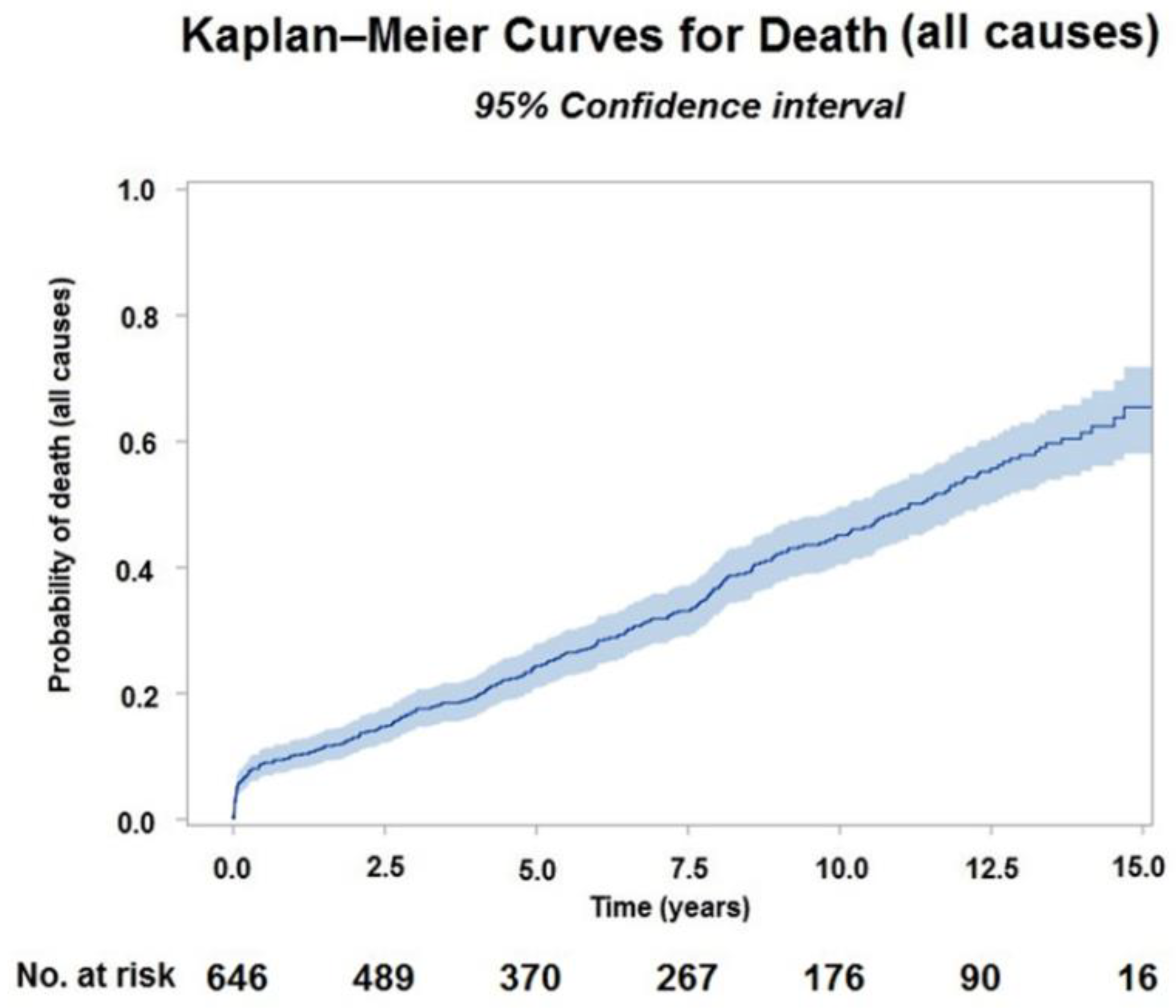
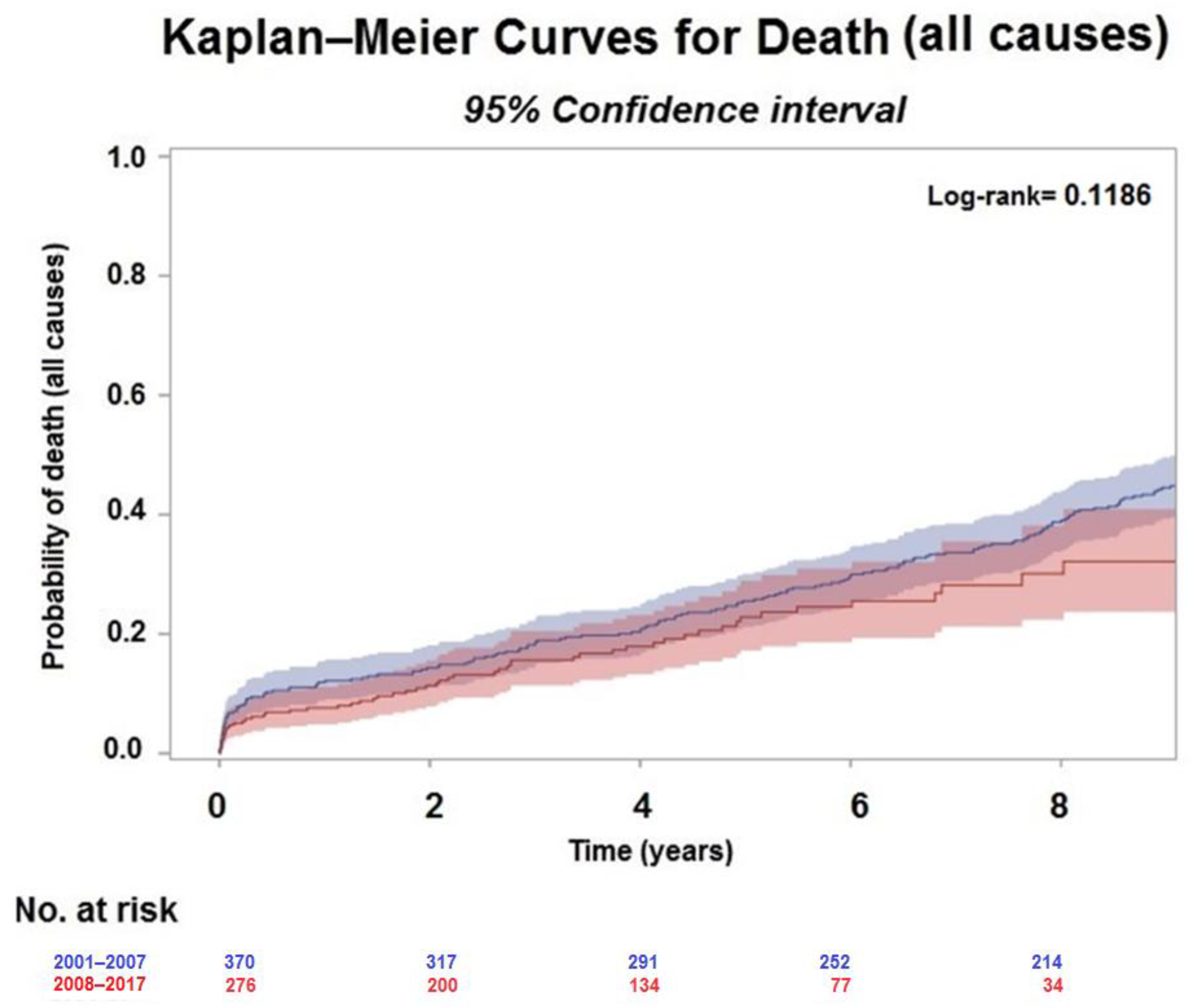
Categorical variables were presented as number (%) and continuous variables as median [interquartile range]. Changes over time between preoperative and postoperative echocardiographic characteristics were compared with the McNemar paired test.
The differences in baseline demographic and clinical characteristics between Group I and Group II were compared with the Chi-square test; continuous variables were compared by the non-parametric Kruskal–Wallis test for non-normally distributed data.
The association between demographic, preoperative echocardiographic variables and the time to all-cause death was investigated by the use of a univariate and multivariable Cox proportional hazard regression analysis.
A first screening of potential predictors was performed by a univariate analysis. For this method, the model considered baseline demographic and preoperative echocardiographic characteristics significantly associated with the outcome and defined by a backward selection. The internal validity of the final models was partially assessed by the bootstrap resampling technique [10]. For each of the 200 bootstrap samples, the model was refitted and tested on the original sample to obtain estimates of predictive accuracy. The bootstrap resampling did not include the first stage of variable selection.
The proportionality and linearity of hazards were evaluated by graphic inspection and testing for Martingale residuals. Median follow-up time was calculated according to the reverse Kaplan–Meier method.
All p values were two-tailed and considered significant if <0.05. Statistical analyses were done with SAS software, version 9.4 (SAS Institute, Inc., Cary, NC, USA) and with the R program (http://CRAN.R-project.org (accessed on 28 March 2022)) with the RMS package.

3. Results

The demographics and clinical profile data of the patient population are shown in Table 1, based on the two different periods.
At baseline, although patients in Group I had less hypertension (203 [54.7%] versus 178 [64.2%], p = 0.0146), hyperlipidemia (188 [50.6%] versus 193 [69.7%], p < 0.0001) and tabagic habit (222 [59.8%] versus 217 [78.3%], p < 0.0001), they were more symptomatic for angina (176 [47.4%] versus 54 [19.5%], p < 0.0001), but less symptomatic for HF (NYHA class III/IV, 172 [46.3%] versus 158 [57%], p = 0.0071).
Medical treatment at the time of admission is reported in Table 1. The prescription of ACE-inhibitors was similar between Group I and Group II (83.20% versus 81.82%, p = 0.6467), while β–blockers (64.23% versus 89.09%, p < 0.0001), aspirin (72.63 % versus 93.45%, p < 0.0001), diuretics (75.88 % versus 93.12%, p < 0.0001), statins (47.70% versus 93.48%, p < 0.0001) and oral anticoagulants (6.79% versus 11.39, p < 0.0001) were prescribed significantly more frequently in Group II. Conversely, Group I received more nitrates (42.82% versus 17.09%, p < 0.0001). PCI prior to surgery was performed less frequently in Group I (79 [21.2%] versus 104 [37.6%], p < 0.0001), while the arrhythmogenic burden was greater in Group II compared with Group I, with more ventricular arrhythmias (75 [27.1%] versus 31 [8.3%], p < 0.0001) and implanted ICD (25 [9%] versus 15 [4.0%], p < 0.0001), (Table 1).
At baseline echocardiography (Table 2), the EDV index (106 [88.8–128.1] mL/m2 versus 118.3 [88.7–140.8] mL/m2, p < 0.0001) and the ESV index (70.5 [55.7–92.0] mL/m2 versus 81.5 [65.8–101.9] mL/m2, p < 0.0001) were lower in Group I, but not the EF (33 [26–38] % versus 31 [25–36] %, p = 0.010). Group I patients had a higher LV mass (168.26 [141.9–203.8] g/m2 versus 160.5 [131.2–193.7] g/m2, p = 0.0060), a lower left atrial dimension (46 [40.5–50] mm versus 47 [43–52] mm, p = 0.0083), a lower E/A ratio (0.82 [0.64–1.40] versus 1.15 [0.72–2.09], p = 0.0003) and more anterior remodeling (316 [85.2%] versus 210 [75.8%], p = 0.0100), while the rate of posterior remodeling was higher (44 [18.7%] versus 52 [11.8%], p = 0.0100) in Group II. Grade 3/4 of mitral regurgitation was higher in Group II (40.4% versus 28.2%, p < 0.0001), which showed a higher SI.
The presence of 3-vessel CAD was higher in Group I (272 [73.3%] versus 164 [59.2%], p < 0.0001), resulting in more coronary grafts combined with SVR (347 [93.53%] versus 235 [84.84%], p = 0.0003) as well as a higher percentage of two or more distal anastomosis (278 [74.9%] versus 160 [57.7%], p < 0.0001) (Table 2).
Figure 1 shows the changes after surgery in echocardiographic parameters in Group I and Group II in respect to baseline differences.
Postoperative variables by period: diastolic diameter (mm) n total = 454, n = 230 in [2001–2007] and n = 224 in [2008–2017]—EF (%) n total = 478, n = 239 in [2001–2007] and n = 239 in [2008–2017]—EDV index (mL/m2) n total = 466, n = 227 in [2001–2007] and n = 239 in [2008–2017]—ESV index (mL/m2) n total = 466, n = 227 in [2001–2007] and n = 239 in [2008–2017]—E/A ratio n total = 359, n = 173 in [2001–2007] and n = 186 in [2008–2017]—MR grade n total = 495, n = 252 in [2001–2007] and n = 243 in [2008–2017]—sphericity index, diastole n total = 397, n = 175 in [2001–2007] and n = 222 in [2008–2017]—sphericity index, systole n total = 396, n = 174 in [2001–2007] and n = 222 in [2008–2017].
Overall, operative mortality defined as mortality within 30 days from surgery was 6.6%, corresponding to 43 patients. Of these, 27 (7.3%) were in Group 1 and 16 (5.7%) in Group 2 (p = 0.4475).
The mean follow-up time was 10 years (range, 0 days to 16 years) for the entire cohort; 13 years (range, 0 days to 16 years) in Group I and 5 years (range, 0 days to 10 years) in Group II.
The Kaplan–Meier estimate for all-cause mortality of the entire population was 13% (10.5–15.8%) at 2 years, 19.2% (16.2–22.5%) at 4 years and 36.6% (32.4–40.9%) at 8 years (Figure 2).
The Kaplan–Meier estimate for mortality was 14.6% (11.2–18.4%) and 11.8% (8.2–16%) at 2 years, 20.6% (16.6–24.9%) and 18.5% (13.8–23.9%) at 4 years and 39% (34–44%) and 32.2% (23.7–40.9%) at 8 years (Figure 3), in Group I and Group II, respectively. There was no evidence of a difference in all-cause death between the two groups (Log-rank = 0.11).
In the multivariable Cox regression analysis, age (HR 1.069, 95% CI: 1.046–1.093, p < 0.0001), baseline EF (HR 0.97, 95% CI: 0.95–0.99, p = 0.0151) and E/A ratio (HR 1.61, 95% CI: 1.31–1.99, p < 0.0001) were significantly associated with mortality in Group I. Conversely, baseline NYHA class (HR 4.282, 95% CI: 1.474–12.441, p = 0.0075) and diastolic sphericity index (HR 1.03, 95% CI: 1.00–1.07, p = 0.0379) were found to be independent predictors of mortality in Group II (Table 3).

4. Discussion

This study reports the largest single-center experience on SVR over 16 years of observation. The major findings of this study include: (1) SVR, mostly combined with CABG in patients with ischemic systolic LV dysfunction, was carried out with a risk of death acceptably low at long-term follow-up; (2) in a real world ischemic HF population, the volume reduction was feasible even in patients with more extensive CAD (Group I); (3) in patients with worse LV remodeling and less extensive CAD (Group II), SVR showed a trend toward a better outcome, though not statistically significant.
Although the comparison between populations of different studies is always hazardous because of differences in baseline characteristics or study design, our results portend an excellent prognosis for patients with ischemic HF and systolic LV dysfunction in terms of all-cause mortality, with a 4-year risk of death of 19.2% in the overall population, lower than other rates reported in the literature, including the STICH trial [5,7].
The surgical treatment of patients with ischemic HF has been a challenge for many years because of the paucity of data from randomized clinical trials mainly regarding patients with angina and significant CAD. The landmark STICH trial was designed to determine the benefit of CABG plus SVR in patients with ischemic LV dysfunction (Hypothesis 2 [7]). The primary outcome analysis failed to show a superiority of the combined procedure compared with CABG alone, calling into question the role of SVR in this high-risk population. Indeed, the STICH trial enrolled only a small percentage (about 20%) of the eligible population, limiting the generalizability of the results, especially in the absence of a concomitant registry. Furthermore, few non-pre-specified retrospective analyses of the STICH trial –Hypothesis 2 have been conducted due to the poor confidence of many surgeons with this procedure [11].
The results of our study share some insights with the STICH trial and, at the same time, focus attention on the selection of patients that could best benefit from this procedure.
Firstly, the Group I population had very similar results to the STICH population (enrollment period 2002–2005). As most patients were more symptomatic for angina and less for HF, they received less PCI before surgery and had more CAD that required a higher number of coronary grafts combined with SVR (Figure 3), resulting in a favorable post-surgical volume (median Δ ESVI equal to 28.7%). Although we cannot discern the weight of the individual procedures on the percentage of volume reduction, it is noteworthy that in this population the decrease in LV size was more important than that reported in the STICH trial [7] (ranging between 6% in the CABG group and 19% in the CABG plus SVR group), supporting a major role of SVR in inducing LV reverse remodeling compared to medical therapy and/or devices [12]. On the other side, despite the evident benefit in all-cause death at 2, 4 and 8 years, the complex ischemic burden related to the presence of extensive CAD and angina in this group of patients might have been the driver for the negative impact of the diastolic dysfunction on the outcome. This implies that an increased E/A ratio along with a low EF may confer a higher risk of death at follow-up, in agreement with previous observations [13,14].
Conversely, the Group II population presented more symptoms of HF, a higher arrhythmogenic burden likely driven by the LV dilatation and a less ischemic profile, apart from the aetiology, with a more extensive LV remodeling and more secondary mitral regurgitation causing a less favorable geometry (Figure 3 and Figure 4).
In this group, a higher baseline LV diastolic SI, with the ventricle being even more spherical after surgery, was associated with worse survival (Figure 1). This observation is in agreement with the results from the STICH trial reported by Oh et al. [15]. However, the link between baseline SI, changes after surgery and survival is not completely clear because the STICH investigators ascribed the worsening of diastolic dysfunction and/or mitral regurgitation to an increase in SI. Conversely, our results show that, although SI increases after surgery, there is a significant improvement in mitral regurgitation (Figure 1). Moreover, changes in E/A ratio do not necessarily indicate a deterioration in diastolic function, meaning that the degree of dysfunction can be the same (grade II) beyond the statistical significance (Figure 1) [16]. Moreover, although the survival was not statistically different between groups, a trend toward a better outcome was observed in Group II, supporting a more robust role of SVR in patients with greater remodeling. Therefore, to better understand the link between changes in LV remodeling phenotypes, surgical technique and outcomes, a new analysis is needed.
This study has several limitations. First, the observation time was extremely long and included changes in patients’ profile, medical and percutaneous therapies; surgical techniques; and length of follow-up. The division used in this study, partially arbitrary, was an attempt to take into account, in some way, how the population has changed over the years. Although our center has a large amount of experience in this field, data collection suffered from missing information, including the total number of previous PCI, which could have affected changes in LV remodeling as well as the presentation of the patients. On the other side, the released STICH results in March 2009 have undoubtedly affected the decision to refer a patient for SVR, but the same results pushed expert surgeons to reconsider the indications [17].
However, despite these potential confounders, the overall number of patients and the length of the follow-up make the survival analysis fairly consistent.
Lastly, our findings should be applied to other populations with ischemic HF with caution because of many differences in baseline characteristics, data collection and surgical expertise.

5. Conclusions

The patient population has consistently changed over time, making a shift from patients mainly symptomatic for angina and suitable for CABG to patients with prevalent symptoms of HF and worse LV remodeling. In the latter group, the therapeutic target might be the ventricle rather than coronary arteries. Suggested indications for SVR include: a previous anterior or posterior MI; a preoperative LV ESV index > 60 mL/m2; regional LV asynergy, either dyskinetic or akinetic; predominant HF symptoms [NYHA functional class III/IV] or in the presence of ventricular arrhythmias and/or angina needing surgical revascularization if the previous conditions are present. The procedure is controindicated in the presence of severe dysfunction of the right ventricle and/or severe diastolic dysfunction [8]. These criteria should guide the patient selection, making SVR still a possible therapeutic strategy for ischemic HF, along with the operator experience and the volume of treated patients [18]. Of course, further studies are required in order to establish the real benefits of SVR at the time of CABG.

Author Contributions

Conceptualization, S.C. and F.A.; methodology, V.M. and F.A.; software, V.M. and F.A.; validation, V.M. and F.A.; formal analysis, V.M. and F.A.; investigation, M.V. and L.R.; resources, L.M.; data curation, L.R.; writing—original draft preparation, S.C.; writing—review and editing, S.C., G.S.J. and L.M.; visualization, V.M. and S.C.; supervision, L.M.; project administration, V.M. and F.A.; funding acquisition, L.M. All authors have read and agreed to the published version of the manuscript.

Funding

This research was funded by Italian Ministry of Health grant number Ricerca Corrente 2022 and The APC was funded by Italian Ministry of Health.

Institutional Review Board Statement

The study was conducted in accordance with the Declaration of Helsinki, and approved by the Ethics Committee of IRCCS Ospedale San Raffaele (REMODEL-HF Study, Protocol No 179/int/2019).

Informed Consent Statement

Patient consent was waived due to the processing of data in anonymized form.

Data Availability Statement

The data presented in this study are available on request from the corresponding author. The data are not publicly available due to the “Restricted Access” policy, as stated by Zenodo (the multi-disciplinary open repository maintained by CERN).

Conflicts of Interest

The authors declare no conflict of interest.

References

  1. Go, A.S.; Mozaffarian, D.; Roger, V.L.; Benjamin, E.J.; Berry, J.D.; Borden, W.B.; Bravata, D.M.; Dai, S.; Ford, E.S.; Fox, C.S.; et al. Heart disease and stroke statistics–2013 update: A report from the American Heart Association. Circulation 2013, 127, e6–e245. [Google Scholar] [CrossRef] [PubMed]
  2. Gheorghiade, M.; Sopko, G.; De Luca, L.; Velazquez, E.J.; Parker, J.D.; Binkley, P.F.; Sadowski, Z.; Golba, K.S.; Prior, D.L.; Rouleau, J.L.; et al. Navigating the crossroads of coronary artery disease and heart failure. Circulation 2006, 114, 1202–1213. [Google Scholar] [CrossRef]
  3. Ezekowitz, J.A.; Kaul, P.; Bakal, J.A.; Armstrong, P.W.; Welsh, R.C.; McAlister, F.A. Declining in-hospital mortality and increasing heart failure incidence in elderly patients with first myocardial infarction. J. Am. Coll. Cardiol. 2009, 53, 13–20. [Google Scholar] [CrossRef] [PubMed] [Green Version]
  4. Neumann, F.J.; Sousa-Uva, M.; Ahlsson, A.; Alfonso, F.; Banning, A.P.; Benedetto, U.; Byrne, R.A.; Collet, J.P.; Falk, V.; Head, S.J.; et al. 2018 ESC/EACTS Guidelines on myocardial revascularization. Eur. Heart J. 2018, 40, 87–165. [Google Scholar] [CrossRef] [PubMed]
  5. Velazquez, E.J.; Lee, K.L.; Jones, R.H.; Al-Khalidi, H.R.; Hill, J.A.; Panza, J.A.; Michler, R.E.; Bonow, R.O.; Doenst, T.; Petrie, M.C.; et al. STICHES Investigators. Coronary-Artery Bypass Surgery in Patients with Ischemic Cardiomyopathy. N. Engl. J. Med. 2016, 374, 1511–1520. [Google Scholar] [CrossRef] [PubMed]
  6. Panza, J.A.; Velazquez, E.J.; She, L.; Smith, P.K.; Nicolau, J.C.; Favaloro, R.R.; Gradinac, S.; Chrzanowski, L.; Prabhakaran, D.; Howlett, J.G.; et al. Extent of coronary and myocardial disease and benefit from surgical revascularization in ischemic LV dysfunction [Corrected]. J. Am. Coll. Cardiol. 2014, 64, 553–561. [Google Scholar] [CrossRef] [PubMed] [Green Version]
  7. Jones, R.H.; Velazquez, E.J.; Michler, R.E.; Sopko, G.; Oh, J.K.; O’Connor, C.M.; Hill, J.A.; Menicanti, L.; Sadowski, Z.; Desvigne-Nickens, P.; et al. STICH Hypothesis 2 Investigators. Coronary bypass surgery with or without surgical ventricular reconstruction. N. Engl. J. Med. 2009, 360, 1705–1717. [Google Scholar] [CrossRef] [PubMed]
  8. Castelvecchio, S.; Pappalardo, O.A.; Menicanti, L. Myocardial reconstruction in ischaemic cardiomyopathy. Eur. J. Cardiothorac. Surg. 2019, 55, i49–i56. [Google Scholar] [CrossRef] [PubMed] [Green Version]
  9. Di Donato, M.; Dabic, P.; Castelvecchio, S.; Santambrogio, C.; Brankovic, J.; Collarini, L.; Joussef, T.; Frigiola, A.; Buckberg, G.; Menicanti, L.; et al. Left ventricular geometry in normal and post-anterior myocardial infarction patients: Sphericity index and ‘new’ conicity index comparisons. Eur. J. Cardiothorac. Surg. 2006, 29, S225–S230. [Google Scholar] [CrossRef] [PubMed]
  10. Frank, E.; Harrel, J.R. Regression Modeling Strategies with Applications to Linear Models, Logistic and Ordinal Regression and Survival Analysis; Springer: New York, NY, USA, 2001. [Google Scholar]
  11. Michler, R.E.; Rouleau, J.L.; Al-Khalidi, H.R.; Bonow, R.O.; Pellikka, P.A.; Pohost, G.M.; Holly, T.A.; Oh, J.K.; Dagenais, F.; Milano, C.; et al. STICH Trial Investigators. Insights from the STICH trial: Change in left ventricular size after coronary artery bypass grafting with and without surgical ventricular reconstruction. J. Thorac. Cardiovasc. Surg. 2013, 146, 1139–1145. [Google Scholar] [CrossRef] [PubMed] [Green Version]
  12. Castelvecchio, S.; Moroni, F.; Menicanti, L. The matter of reverse ventricular remodeling after acute myocardial infarction between fiction and reality. J. Cardiovasc. Med. (Hagerstown) 2018, 19, 397–398. [Google Scholar] [CrossRef] [PubMed]
  13. Meta-analysis Research Group in Echocardiography (MeRGE) Heart Failure Collaborators; Doughty, R.N.; Klein, A.L.; Poppe, K.K.; Gamble, G.D.; Dini, F.L.; Møller, J.E.; Quintana, M.; Yu, C.M.; Whalley, G.A. Independence of restrictive filling pattern and LV ejection fraction with mortality in heart failure: An individual patient meta-analysis. Eur. J. Heart Fail. 2008, 10, 786–792. [Google Scholar] [CrossRef] [PubMed]
  14. Rigolli, M.; Rossi, A.; Quintana, M.; Klein, A.L.; Yu, C.M.; Ghio, S.; Dini, F.L.; Prior, D.; Troughton, R.W.; Temporelli, P.L.; et al. The prognostic impact of diastolic dysfunction in patients with chronic heart failure and post-acute myocardial infarction: Can age-stratified E/A ratio alone predict survival? Int. J. Cardiol. 2015, 181, 362–368. [Google Scholar] [CrossRef] [PubMed]
  15. Choi, J.O.; Daly, R.C.; Lin, G.; Lahr, B.D.; Wiste, H.J.; Beaver, T.M.; Iacovoni, A.; Malinowski, M.; Friedrich, I.; Rouleau, J.L.; et al. Impact of surgical ventricular reconstruction on sphericity index in patients with ischemic cardiomyopathy: Follow-up from the STICH trial. Eur. J. Heart Fail. 2015, 17, 453–463. [Google Scholar] [CrossRef] [PubMed] [Green Version]
  16. Castelvecchio, S.; Menicanti, L.; Ranucci, M.; Di Donato, M. Impact of surgical ventricular restoration on diastolic function: Implications of shape and residual ventricular size. Ann. Thorac. Surg. 2008, 86, 1849–1854. [Google Scholar] [CrossRef] [PubMed]
  17. Fatehi Hassanabad, A.; Wiebe, K.; Ali, I.S. Clinical and hemodynamic outcomes of the Dor procedure in adults with ischemic cardiomyopathy. J. Card. Surg. 2021, 36, 4345–4436. [Google Scholar] [CrossRef] [PubMed]
  18. Gaudino, M.; Castelvecchio, S.; Rahouma, M.; Robinson, N.B.; Audisio, K.; Soletti, G.J.; Garatti, A.; Benedetto, U.; Girardi, L.N.; Menicanti, L. Results of surgical ventricular reconstruction in a specialized center and in comparison to the STICH trial: Rationale and study protocol for a patient-level pooled analysis. J. Card. Surg. 2021, 36, 689–692. [Google Scholar] [CrossRef] [PubMed]
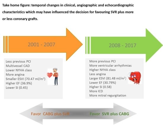
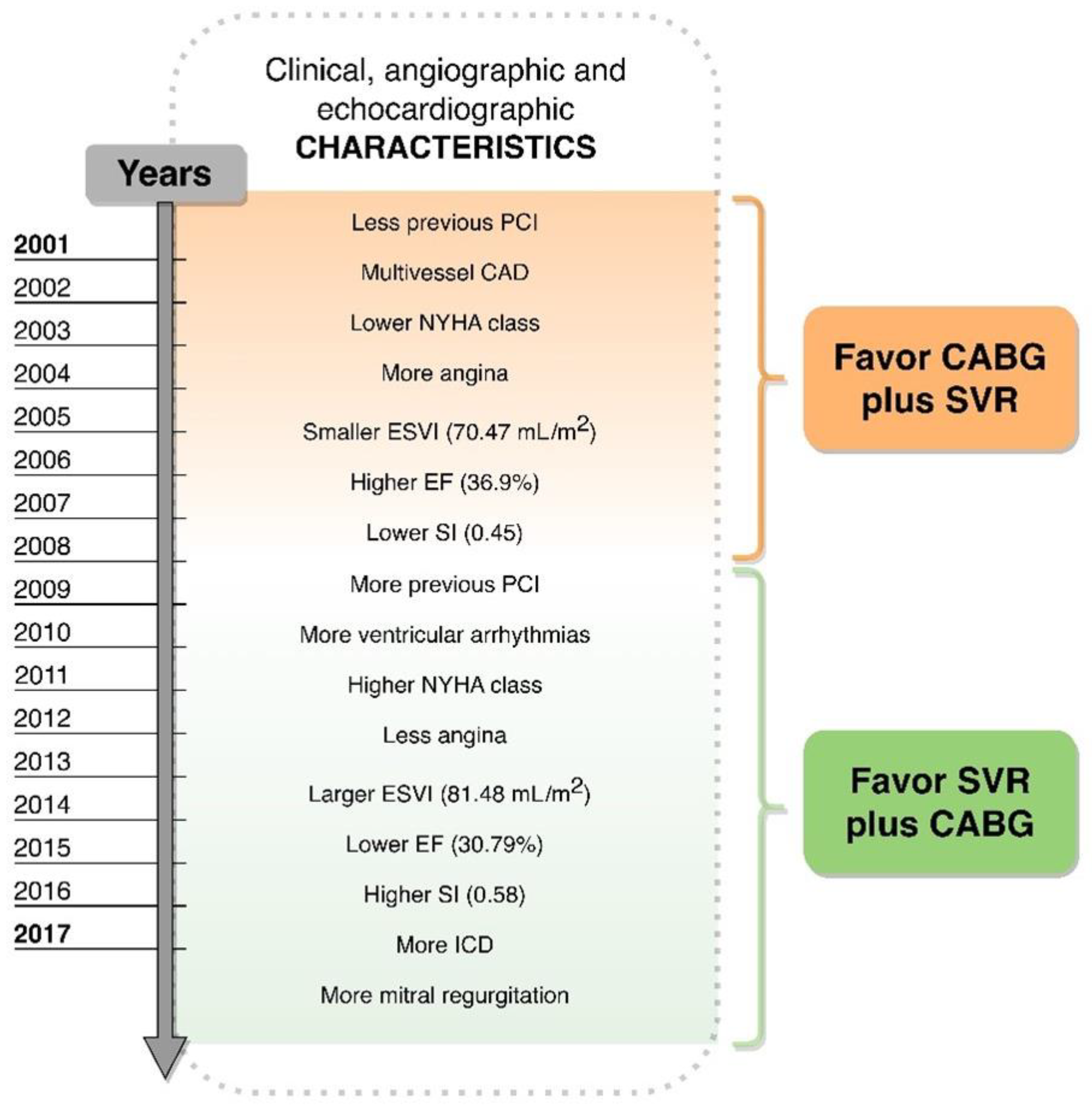
Figure 1. Boxplot for baseline and postoperative echocardiographic variables by period. *, p < 0.05; **, p < 0.001.
Jpm 12 00567 g001
Figure 2. Kaplan-Meier curve for all-cause mortality for the total study cohort.
Jpm 12 00567 g002
Figure 3. Kaplan-Meier curves for all-cause mortality by period.
Jpm 12 00567 g003
Figure 4. Temporal changes in clinical, angiographic and echocardiographic characteristics which may have influenced the decision for favoring SVR plus more or less coronary grafts.
Jpm 12 00567 g004
Table 1. Baseline characteristics of the total study cohort and by period.
nTotaln2001–2007 (n = 371)n2008–2017p-Value
Age, years 64866 [58–72]37167 [58–72]27764 [58–71]0.1365
BSA6481.83 [1.74–1.92]3711.83 [1.73–1.94]2771.85 [1.75–1.94]0.0689
Creatinine6481.10 [0.91–1.39]3711.14 [0.96–1.44]2771.05 [0.88–1.30]0.0020
Family history of CAD648271 (41.82) 132 (35.58) 139 (50.18)0.0002
Smokers or ex-smokers648439 (67.75) 222 (59.84) 217 (78.34)<0.0001
Hypertension648381 (58.80) 203 (54.72) 178 (64.26)0.0146
Atrial fibrillation64891 (14.04) 49 (13.21) 42 (15.16)0.4786
Stroke64855 (8.49) 42 (11.32) 13 (4.69)0.0027
Angina648230 (35.49) 176 (47.44) 54 (19.49)<0.0001
Ventricular arrhythmias648106 (16.36) 31 (8.36) 75 (27.08)<0.0001
Chronic renal failure64847 (7.25) 24 (6.47) 23 (8.30)0.3731
Diabetes mellitus648166 (25.66) 97 (26.15) 69 (25.00)0.7414
Hypercholesterolemia648381 (58.80) 188 (50.67) 193 (69.68)<0.0001
NYHA class III/IV648330 (50.93) 172 (46.36) 158 (57.04)0.0071
Previous PCI 183 (28.24) 79 (21.29) 104 (37.55)<0.0001
        PCI + ICD 37 (5.21) 3 (0.81) 34 (12.27)
        ICD 40 (6.17) 15 (4.04) 25 (9.03)
ACE inhibitor644532 (82.61) 304 (83.20) 225 (81.82)0.6477
β-blockers644482 (74.84) 237 (64.23) 245 (89.09)<0.0001
Aspirin644525 (81.52) 268 (72.63) 257 (93.45)<0.0001
Digoxin64446 (7.14) 37 (10.03) 9 (3.27)0.0010
Statins645434 (67.29) 176 (47.70) 258 (93.48)<0.0001
Diuretic645537 (83.26) 280 (75.88) 257 (93.12)<0.0001
Oral anticoagulant64473 (11.34) 25 (6.79) 48 (11.39)<0.0001
Amiodarone644146 (22.67) 85 (23.04) 61 (22.18)0.7981
Nitrates644205 (31.83) 158 (42.82) 47 (17.09)<0.0001
Data are median [Q1–Q3] or number (%); BSA = body surface area; CAD = coronary artery disease; PCI = percutaneous coronary intervention; NYHA = New York Heart Association.
Table 2. Baseline echocardiographic, angiographic and operative variables of the total study cohort and by period.
NTotal n2001–2007 (n = 371)n2008–2017 (n = 277)p-Value
Diastolic diameter (mm)63065 [58–71]35665 [58–70.50]27464 [59–71]0.8698
Systolic diameter (mm)62551 [44–59]35551 [44–59]27051 [45–59]0.6611
EDV index (mL/m2)645112.12 [92.34–134.30]368106.68 [88.77–128.06]277118.27 [98.86–140.83]<0.0001
ESV index (mL/m2)64676.08 [59.17–95.81]36970.47 [55.65–92.02]27781.48 [65.79–101.85]<0.0001
EF (%)64832 [26–37]37133 [26–38]27731 [25–36]0.0100
SV index (mL/m2)64435.20 [29.38–41.69]36734.69 [24.14–40.61]27736.57 [29.63–42.55]0.1186
TAPSE (mm)60920 [18–23]33520 [17–23]27421 [18–24]0.0158
PAPs (mmHg) 55638 [30.5–48]28238 [32–46]27438 [30–49]0.4370
LVMI (g/m2)561164.29 [137.88–199.48]336168.26 [141.93–203.81]225160.51 [131.22–193.68]0.0060
RWT5970.32 [0.27–0.38]3470.33 [0.28–0.40]2500.29 [0.25–0.36]<0.0001
Left atrial diameter (mm)59446 [41–51]34046 [40.50–50]25447 [43–52]0.0083
E/A ratio4580.96 [0.66–1.71]2200.82 [0.64–1.40]2381.15 [0.72–2.09]0.0003
DT (mm)435185 [149–239]197190 [155–231]238182 [144–254]0.9478
Mitral annulus (mm)36734 [30–37]16235 [31–39]20532 [29–37]0.0017
IPD, diastole (mm)2543 [2.5–3.5]1242.60 [2.20–3.1]1323.20 [2.8–3.6]<0.0001
IPD, systole (mm)2542.10 [1.7–2.6]1241.77 [1.4–2.6]1322.30 [2.00–2.7]<0.0001
Sphericity index, diastole 3950.60 [0.50–0.68]1520.60 [0.50–0.68]2430.64 [0.57–0.72]<0.0001
Sphericity index, systole3940.53 [0.43–0.62]1520.45 [0.37–0.54]2430.58 [0.49–0.65]<0.0001
Conicity index, diastole3850.88 [0.80–0.96]1420.81 [0.73–0.95]2430.89 [0.83–0.97]<0.0001
Conicity index, systole3850.94 [0.83–1.09]1420.95 [0.80–1.11]2430.93 [0.84–1.08]0.8379
MR grade
0 80 (12.35) 74 (19.95) 6 (2.17)
1 209 (32.25) 126 (33.96) 83 (29.96)
2 142 (21.91) 66 (17.79) 76 (27.44)<0.0001
3 119 (18.36) 58 (15.63) 61 (22.02)
4 98 (15.12) 47 (12.67) 51 (18.41)
Site of remodeling
Posterior 96 (14.81) 44 (11.86) 52 (18.77)
Anterior 526 (81.17) 316 (85.18) 210 (75.81)0.0100
Anterior & posterior 26 (4.01) 11 (2.96) 15 (5.42)
Coronary angiography
Single vessel disease 159 (24.54) 85 (22.91) 74 (26.71)
Multivessel disease 436 (67.28) 272 (73.32) 164 (59.21)<0.0001
No residual stenosis 53 (8.18) 14 (3.77) 39 (14.08)
Mitral valve surgery 200 (30.86) 96 (25.88) 104 (37.55)0.0015
CABG + SVR 582 (89.81) 347 (93.53) 235 (84.84)0.0003
SVR 66 (10.19) 24 (6.47) 42 (15.16)
Number of distal anastomosis
0 66 (10.19) 24 (6.47) 42 (15.16)
1 144 (22.22) 69 (18.60) 75 (27.08)<0.0001
>=2 438 (67.59) 278 (74.93) 160 (57.76)
Data are median [Q1–Q3] or number (%). EDV = end-diastolic volume; ESV = end-systolic volume; EF= ejection fraction; SV = stroke volume; TAPSE = tricuspid annular plane systolic excursion; PAPs = systolic pulmonary artery pressure; LVMI = left ventricular mass index; RWT = relative wall thickness; DT = deceleration time; IPM = inter-papillary distance; MR = mitral regurgitation.
Table 3. Univariate and multivariable Cox models for all-cause mortality.
2001–2007 (n = 371) 2008–2017 (n = 277)
Univariate Multivariate Univariate Multivariate
Death (n = 212) Death Death (n = 57) Death
Risk Category Hazard Ratio
(95% IC)
p-ValueHazard Ratio
(95% IC)
p-ValueHazard Ratio
(95% IC)
p-ValueHazard Ratio
(95% IC)
p-Value
SexM vs. F0.83 [0.60–1.17]0.2899 0.94 [0.44–1.99]0.8688
Site of remodeling
Ant/postAnt/post vs. Ant1.82 [0.89–3.71]0.0991 2.61 [1.17–5.85]0.0194
PostPost vs. Ant1.52 [1.03–2.24]0.0359 1.56 [0.79–3.07]0.1996
Previous MIYes vs. No1.10 [0.76–1.59]0.5986 1.48 [0.73–3.03]0.2761
HypertensionYes vs. No1.04 [0.79–1.36]0.7783 1.01 [0.60–1.73]0.9739
DyslipidemiaYes vs. No0.66 [0.50–0.86]0.0024 1.06 [0.60–1.87]0.8449
DiabetesYes vs. No1.24 [0.92–1.66]0.1578 1.96 [1.15–3.34]0.01361.96 [0.94–4.08]0.0728
Smoke Yes vs. No1.18 [0.90–1.56]0.2340 0.92 [0.50–1.71]0.7928
Previous procedures
PCI vs. No0.87 [0.61–1.24]0.4436 0.57 [0.30–1.09]0.0908
PCI + ICD vs. No2.27 [0.56–9.20]0.2493 1.33 [0.60–2.96]0.4868
ICD vs. No1.01 [0.49–2.05]0.9849 1.79 [0.81–3.99]0.1516
Other vs. No1.48 [0.92–2.40]0.1071 0.98 [0.14–7.22]0.9831
Ventricular arrhythmiasYes vs. No1.29 [0.81–2.04]0.2842 0.758 [0.40–1.43]0.3858
Atrial fibrillationYes vs. No1.47 [1.02–2.11]0.0384 3.14 [1.79–5.49]<0.0001
StrokeYes vs. No1.32 [0.88–1.99]0.1864 1.63 [0.59–4.50]0.3496
Chronic renal failureYes vs. No2.92 [1.87–4.56]<0.0001 3.36 [1.74–6.50]0.0003
Angiography
Single vesselsSingle vessels vs. 0.67 [0.33–1.37]0.27500.96 [0.35–2.59]0.93170.91 [0.31–2.67]0.8617
No significant stenosis
Multivessel diseaseMultivessels disease vs.0.95 [0.48–1.86]0.87611.63 [0.62–4.28]0.32191.68 [0.66–4.27]0.2728
No significant stenosis
AnginaYes vs. No1.20 [0.91–1.57]0.1885 0.83 [0.44–1.58]0.5709
NYHAIII-IV vs. I-II1.82 [1.38–2.39]<0.0001 3.12 [1.65–5.90]0.00054.28 [1.47–12.44]0.0075
MR grade>= 2 vs. <21.41 [1.08–1.85]0.0126 2.48 [1.25–4.92]0.0092
Age1 unit1.06 [1.04–1.07]<0.00011.07 [1.05–1.09]<0.00011.07 [1.03–1.10]<0.00011.04 [0.99–1.09]0.0815
BSA1 unit0.96 [0.43–2.14]0.9241 0.07 [0.01–0.54]0.0099
Haemoglobin1 unit0.86 [0.78–0.94]0.0005 0.83 [0.70–0.97]0.0205
Creatinine1 unit2.15 [1.78–2.59]<0.0001 1.62 [1.21–2.18]0.0012
Diastolic diameter (mm)1 unit1.02 [1.00–1.04]0.0126 1.03 [1.00–1.06]0.0511
Systolic diameter (mm)1 unit1.02 [1.00–1.03]0.0122 1.03 [1.00–1.06]0.0173
EDV index (mL/m2)1 unit1.00 [1.00–1.01]0.0906 1.00 [1.00–1.01]0.4235
ESV index (mL/m2)1 unit1.00 [1.00–1.01]0.0070 1.00 [1.00–1.01]0.1935
EF1 unit0.96 [0.94–0.98]<0.00010.97 [0.95–0.99]0.01510.95 [0.92–0.96]0.0051
SV index (mL/m2)1 unit0.98 [0.97–0.99]0.0421 0.98 [0.95–1.01]0.2228
RWT1 unit0.11 [0.03–0.53]0.0054 0.09 [0.01–1.29]0.0765
LVMI (g/m2)1 unit1.00 [1.00–1.00]0.2207 1.00 [1.00–1.01]0.1060
Left atrial diameter (mm)1 unit1.04 [1.02–1.06]<0.0001 1.05 [1.01–1.09]0.0146
E/A ratio 1 unit1.50 [1.23–1.84]<0.00011.61 [1.31–1.99]<0.00011.39 [1.09–1.77]0.0079
DT (m sec)1 unit0.99 [0.99–1.00]0.0604 1.00 [0.99–1.00]0.2290
TAPSE1 unit0.94 [0.91–0.98]0.0027 0.91 [0.84–0.97]0.0053
PAPs (mmHg) 1 unit1.02 [1.01–1.03]0.0006 1.02 [1.01–1.04]0.0082
Mitral annulus (mm)1 unit1.05 [1.02–1.09]0.0051 1.05 [0.99–1.11]0.0800
IPD, diastole (mm)1 unit1.36 [1.09–1.68]0.0058 1.34 [0.65–2.77]0.4312
IPD, systole (mm)1 unit1.47 [1.18–1.84]0.0007 1.68 [0.89–3.17]0.1061
MRYes vs. No1.73 [1.30–2.30]0.0002 2.64 [1.56–4.47]0.0003
Diastolic sphericity index1 unit1.01 [0.99–1.03]0.4265 1.04 [1.01–1.07]0.01041.03 [1.00–1.07]0.0379
Systolic sphericity index1 unit1.02 [1.00–1.03]0.0931 1.04 [1.01–1.07]0.0041
Diastolic conicity index1 unit0.99 [0.98–1.00]0.1062 0.98 [0.96–1.01]0.1311
Systolic conicity index1 unit0.99 [0.99–1.00]0.1099 0.99 [0.97–1.00]0.0552
MI = myocardial infarction; PCI = percutaneous coronary intervention; NYHA = New York Heart Association; MR = mitral regurgitation; BSA = body surface area; EDV = end-diastolic volume; ESV = end-systolic volume; EF = ejection fraction; SV = stroke volume; RWT = relative wall thickness; LVMI = left ventricular mass index; DT = deceleration time; TAPSE = tricuspid annular plane systolic excursion; PAPs = systolic pulmonary artery pressure; IPM = inter-papillary distance.
Publisher’s Note: MDPI stays neutral with regard to jurisdictional claims in published maps and institutional affiliations.

