
Does Neutrophil to Lymphocyte Ratio Have a Role in Identifying Cytokeratin 19-Expressing Hepatocellular Carcinoma?

 , ,
, ,  ,
,
Abstract
:1. Introduction
2. Materials and Methods
2.1. Patients
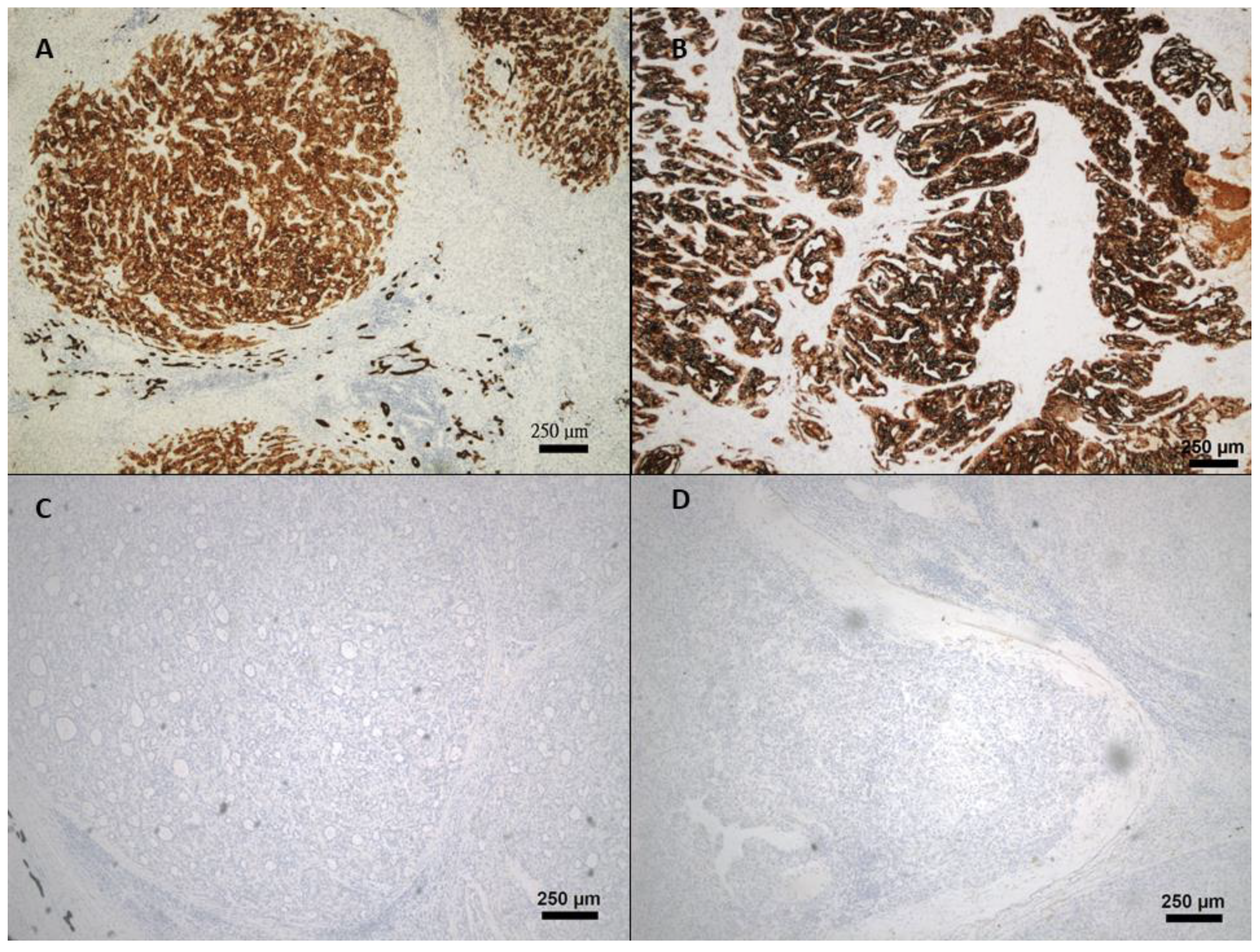
2.2. Immunohistochemistry (IHC)
2.3. Statistical Analysis
3. Results
3.1. Patient Demographics
3.2. Analysis of Biomarkers Predicting CK19 Expression
3.3. Performance of Biomarkers in Predicting CK19 Expression in HCC
4. Discussion
5. Conclusions
Author Contributions
Funding
Institutional Review Board Statement
Informed Consent Statement
Data Availability Statement
Acknowledgments
Conflicts of Interest
References
- Yang, J.D.; Hainaut, P.; Gores, G.J.; Amadou, A.; Plymoth, A.; Roberts, L.R. A global view of hepatocellular carcinoma: Trends, risk, prevention and management. Nat. Rev. Gastroenterol. Hepatol. 2019, 16, 589–604. [Google Scholar] [CrossRef]
- Forner, A.; Reig, M.; Bruix, J. Hepatocellular carcinoma. Lancet 2018, 391, 1301–1314. [Google Scholar] [CrossRef] [PubMed]
- Kim, J.; Kang, W.; Sinn, D.H.; Gwak, G.Y.; Paik, Y.H.; Choi, M.S.; Lee, J.H.; Koh, K.C.; Paik, S.W. Substantial risk of recurrence even after 5 recurrence-free years in early-stage hepatocellular carcinoma patients. Clin. Mol. Hepatol. 2020, 26, 516–528. [Google Scholar] [CrossRef]
- Nishida, N. Long-term prognosis and management of hepatocellular carcinoma after curative treatment. Clin. Mol. Hepatol. 2020, 26, 480–483. [Google Scholar] [CrossRef] [PubMed]
- Takano, M.; Shimada, K.; Fujii, T.; Morita, K.; Takeda, M.; Nakajima, Y.; Nonomura, A.; Konishi, N.; Obayashi, C. Keratin 19 as a key molecule in progression of human hepatocellular carcinomas through invasion and angiogenesis. BMC Cancer 2016, 16, 903. [Google Scholar] [CrossRef] [Green Version]
- Kawai, T.; Yasuchika, K.; Ishii, T.; Katayama, H.; Yoshitoshi, E.Y.; Ogiso, S.; Kita, S.; Yasuda, K.; Fukumitsu, K.; Mizumoto, M.; et al. Keratin 19, a Cancer Stem Cell Marker in Human Hepatocellular Carcinoma. Clin. Cancer Res. 2015, 21, 3081–3091. [Google Scholar] [CrossRef] [Green Version]
- Sun, D.W.; Zhang, Y.Y.; Sun, X.D.; Chen, Y.G.; Qiu, W.; Ji, M.; Lv, G.Y. Prognostic value of cytokeratin 19 in hepatocellular carcinoma: A meta-analysis. Clin. Chim. Acta 2015, 448, 161–169. [Google Scholar] [CrossRef]
- Shirasu, H.; Ono, A.; Omae, K.; Nakashima, K.; Omori, S.; Wakuda, K.; Kenmotsu, H.; Naito, T.; Murakami, H.; Endo, M.; et al. CYFRA 21-1 predicts the efficacy of nivolumab in patients with advanced lung adenocarcinoma. Tumour Biol. 2018, 40, 1010428318760420. [Google Scholar] [CrossRef] [PubMed] [Green Version]
- Rudhart, S.A.; Gehrt, F.; Birk, R.; Schultz, J.D.; Stankovic, P.; Georgiew, R.; Wilhelm, T.; Stuck, B.A.; Hoch, S. Clinical relevance of CYFRA 21-1 as a tumour marker in patients with oropharyngeal squamous cell carcinoma. Eur. Arch. Oto-Rhino-Laryngol. 2020, 277, 2561–2571. [Google Scholar] [CrossRef]
- Nakamura, T.; Ide, H.; Eguchi, R.; Hayashi, K.; Takasaki, K.; Watanabe, S. CYFRA 21-1 as a tumor marker for squamous cell carcinoma of the esophagus. Dis. Esophagus 2017, 11, 35–39. [Google Scholar] [CrossRef] [PubMed]
- Caviglia, G.P.; Ciruolo, M.; Olivero, A.; Carucci, P.; Rolle, E.; Rosso, C.; Abate, M.L.; Risso, A.; Ribaldone, D.G.; Tandoi, F.; et al. Prognostic Role of Serum Cytokeratin-19 Fragment (CYFRA 21-1) in Patients with Hepatocellular Carcinoma. Cancers 2020, 12, 2776. [Google Scholar] [CrossRef]
- Kawai, T.; Yasuchika, K.; Ishii, T.; Katayama, H.; Yoshitoshi, E.Y.; Ogiso, S.; Minami, T.; Miyauchi, Y.; Kojima, H.; Yamaoka, R.; et al. Identification of keratin 19-positive cancer stem cells associating human hepatocellular carcinoma using CYFRA 21-1. Cancer Med. 2017, 6, 2531–2540. [Google Scholar] [CrossRef] [PubMed] [Green Version]
- Wang, D.; Bai, N.; Hu, X.; OuYang, X.W.; Yao, L.; Tao, Y.; Wang, Z. Preoperative inflammatory markers of NLR and PLR as indicators of poor prognosis in resectable HCC. PeerJ 2019, 7, e7132. [Google Scholar] [CrossRef] [PubMed]
- Bruix, J.; Cheng, A.L.; Meinhardt, G.; Nakajima, K.; De Sanctis, Y.; Llovet, J. Prognostic factors and predictors of sorafenib benefit in patients with hepatocellular carcinoma: Analysis of two phase III studies. J. Hepatol. 2017, 67, 999–1008. [Google Scholar] [CrossRef] [PubMed] [Green Version]
- Mouchli, M.; Reddy, S.; Gerrard, M.; Boardman, L.; Rubio, M. Usefulness of neutrophil-to-lymphocyte ratio (NLR) as a prognostic predictor after treatment of hepatocellular carcinoma. “Review article. Ann. Hepatol. 2021, 22, 100249. [Google Scholar] [CrossRef]
- Wang, Y.; Liu, T.; Tang, W.; Deng, B.; Chen, Y.; Zhu, J.; Shen, X. Hepatocellular Carcinoma Cells Induce Regulatory T Cells and Lead to Poor Prognosis via Production of Transforming Growth Factor-β1. Cell. Physiol. Biochem. 2016, 38, 306–318. [Google Scholar] [CrossRef] [PubMed]
- Johnson, P.J.; Dhanaraj, S.; Berhane, S.; Bonnett, L.; Ma, Y.T. The prognostic and diagnostic significance of the neutrophil-to-lymphocyte ratio in hepatocellular carcinoma: A prospective controlled study. Br. J. Cancer 2021, 125, 714–716. [Google Scholar] [CrossRef]
- Uenishi, T.; Kubo, S.; Yamamoto, T.; Shuto, T.; Ogawa, M.; Tanaka, H.; Tanaka, S.; Kaneda, K.; Hirohashi, K. Cytokeratin 19 expression in hepatocellular carcinoma predicts early postoperative recurrence. Cancer Sci. 2003, 94, 851–857. [Google Scholar] [CrossRef]
- Govaere, O.; Komuta, M.; Berkers, J.; Spee, B.; Janssen, C.; de Luca, F.; Katoonizadeh, A.; Wouters, J.; van Kempen, L.C.; Durnez, A.; et al. Keratin 19: A key role player in the invasion of human hepatocellular carcinomas. Gut 2014, 63, 674–685. [Google Scholar] [CrossRef] [PubMed] [Green Version]
- Kim, H.; Choi, G.H.; Na, D.C.; Ahn, E.Y.; Kim, G.I.; Lee, J.E.; Cho, J.Y.; Yoo, J.E.; Choi, J.S.; Park, Y.N. Human hepatocellular carcinomas with “Stemness”-related marker expression: Keratin 19 expression and a poor prognosis. Hepatology 2011, 54, 1707–1717. [Google Scholar] [CrossRef] [PubMed]
- Kim, H.; Park, Y.N. Hepatocellular carcinomas expressing ’stemness’-related markers: Clinicopathological characteristics. Dig. Dis. 2014, 32, 778–785. [Google Scholar] [CrossRef]
- Yang, Z.Y.; Zhang, H.Y.; Wang, F.; Ma, Y.H.; Li, Y.Y.; He, H.L.; Wang, C.; Li, S.S. Expression of cytokeratin(CK)7, CK8/18, CK19 and p40 in esophageal squamous cell carcinoma and their correlation with prognosis. Chin. J. Pathol. 2018, 47, 834–839. [Google Scholar] [CrossRef]
- Gao, J.; Lv, F.; Li, J.; Wu, Z.; Qi, J. Serum cytokeratin 19 fragment, CK19-2G2, as a newly identified biomarker for lung cancer. PLoS ONE 2014, 9, e101979. [Google Scholar] [CrossRef]
- Bakaeean, B.; Gholamin, M.; Tabatabaee Yazdi, S.A.; Forghani, M.N. Novel Biomarkers Aim at Detecting Metastatic Sentinel Lymph Nodes in Breast Cancer. Iran. Biomed. J. 2020, 24, 183–191. [Google Scholar] [CrossRef] [PubMed] [Green Version]
- Haruna, Y.; Saito, K.; Spaulding, S.; Nalesnik, M.A.; Gerber, M.A. Identification of bipotential progenitor cells in human liver development. Hepatology 1996, 23, 476–481. [Google Scholar] [CrossRef]
- Roskams, T.A.; Libbrecht, L.; Desmet, V.J. Progenitor cells in diseased human liver. Semin. Liver Dis. 2003, 23, 385–396. [Google Scholar] [CrossRef] [PubMed]
- Roskams, T. Liver stem cells and their implication in hepatocellular and cholangiocarcinoma. Oncogene 2006, 25, 3818–3822. [Google Scholar] [CrossRef] [PubMed] [Green Version]
- Schobert, I.T.; Savic, L.J.; Chapiro, J.; Bousabarah, K.; Chen, E.; Laage-Gaupp, F.; Tefera, J.; Nezami, N.; Lin, M.; Pollak, J.; et al. Neutrophil-to-lymphocyte and platelet-to-lymphocyte ratios as predictors of tumor response in hepatocellular carcinoma after DEB-TACE. Eur. Radiol. 2020, 30, 5663–5673. [Google Scholar] [CrossRef] [PubMed]
- Estrade, F.; Lescure, C.; Muzellec, L.; Pedrono, M.; Palard, X.; Pracht, M.; Le Sourd, S.; Rolland, Y.; Uguen, T.; Garin, E.; et al. Lymphocytes and Neutrophil-to-Lymphocyte Ratio Variations After Selective Internal Radiation Treatment for HCC: A Retrospective Cohort Study. Cardiovasc. Interv. Radiol. 2020, 43, 1175–1181. [Google Scholar] [CrossRef]
- Zheng, J.; Cai, J.; Li, H.; Zeng, K.; He, L.; Fu, H.; Zhang, J.; Chen, L.; Yao, J.; Zhang, Y.; et al. Neutrophil to Lymphocyte Ratio and Platelet to Lymphocyte Ratio as Prognostic Predictors for Hepatocellular Carcinoma Patients with Various Treatments: A Meta-Analysis and Systematic Review. Cell. Physiol. Biochem. 2017, 44, 967–981. [Google Scholar] [CrossRef] [PubMed]
- Li, X.F.; Chen, D.P.; Ouyang, F.Z.; Chen, M.M.; Wu, Y.; Kuang, D.M.; Zheng, L. Increased autophagy sustains the survival and pro-tumourigenic effects of neutrophils in human hepatocellular carcinoma. J. Hepatol. 2015, 62, 131–139. [Google Scholar] [CrossRef]
- Kuang, D.M.; Zhao, Q.; Wu, Y.; Peng, C.; Wang, J.; Xu, Z.; Yin, X.Y.; Zheng, L. Peritumoral neutrophils link inflammatory response to disease progression by fostering angiogenesis in hepatocellular carcinoma. J. Hepatol. 2011, 54, 948–955. [Google Scholar] [CrossRef] [PubMed]
- Shen, Y.; Wei, Y.; Wang, Z.; Jing, Y.; He, H.; Yuan, J.; Li, R.; Zhao, Q.; Wei, L.; Yang, T.; et al. TGF-β regulates hepatocellular carcinoma progression by inducing Treg cell polarization. Cell. Physiol. Biochem. 2015, 35, 1623–1632. [Google Scholar] [CrossRef] [PubMed]
- Luo, C.; Xin, H.; Yin, D.; Zhao, T.; Hu, Z.; Zhou, Z.; Sun, R.; Yao, N.; Sun, Q.; Fan, J.; et al. Characterization of immune infiltration in sarcomatoid hepatocellular carcinoma. Aging 2021, 13, 15126–15138. [Google Scholar] [CrossRef]
- Unitt, E.; Marshall, A.; Gelson, W.; Rushbrook, S.M.; Davies, S.; Vowler, S.L.; Morris, L.S.; Coleman, N.; Alexander, G.J. Tumour lymphocytic infiltrate and recurrence of hepatocellular carcinoma following liver transplantation. J. Hepatol. 2006, 45, 246–253. [Google Scholar] [CrossRef] [PubMed]
- Hu, L.M.; Tsai, H.I.; Lee, C.W.; Chen, H.M.; Lee, W.C.; Yu, H.P. Uric Acid as a Predictor for Early Allograft Dysfunction after Living Donor Liver Transplantation: A Prospective Observational Study. J. Clin. Med. 2021, 10, 2729. [Google Scholar] [CrossRef]


| Variables | No. (%) | Variables | Mean ± SE e/Median (IQR) f |
|---|---|---|---|
| Age (≤65 year-old) | 19 (52.8) | Age (year-old) | 63.6 ± 1.60 |
| Gender (Male) | 26 (72.2) | ICG-15 (%) | 10.4 ± 0.88 |
| Comorbidity | Preoperative α-fetoprotein (ng/mL) | 18.05 (194.5) | |
| Diabetes Mellitus (Yes) | 12 (33.3) | Preoperative CEA d (ng/mL) | 1.99 (2.22) |
| Hypertension (Yes) | 22 (61.1) | Tumor size (cm) | 4.7 ± 0.53 |
| Smoking (Yes) | 12 (33.3) | Neutrophil to lymphocyte ratio | 1.93 (1.0) |
| Alcohol (Yes) | 6 (16.7) | Variables | No. (%) |
| Child-Pugh Classification (A) | 36 (100) | Vascular invasion (Yes) | 11 (30.6) |
| Preoperative α-fetoprotein (>200 ng/mL) | 9 (25.0) | Daughter nodules (Yes) | 3 (8.3) |
| ICG-15 a (≤10%) | 19 (52.8) | Resection margin (negative) | 36 (100) |
| Tumor size (>5 cm) | 12 (33.3) | Edmonson and Steiner grade (I/II vs. III/IV) | 16(44.4) vs. 20(55.6) |
| HBV infection b | 23 (63.9) | Liver cirrhosis (Yes) | 16 (44.4) |
| HCV infection c | 11 (30.6) | Tumor necrosis (Yes) | 11 (30.6) |
| Tumor encapsulation (Yes) | 29 (80.6) | AJCC stage (I/II/III/IV) g | 20(55.6)/9(25.0)/6(16.7)/1(2.8) |
| Tumor rupture (Yes) | 3 (8.3) | CK19 expression (Yes) | 9 (25.0) |
| Hepatocellular Carcinoma, n = 36 | |||||||
|---|---|---|---|---|---|---|---|
| CK19 a | CK19 a | ||||||
| Negative (%) b | Positive (%) c | p Value | Negative (%) b | Positive (%) c | p Value | ||
| Age(yr) | 0.706 | Vascular invasion | 0.409 | ||||
| >65 | 12 (44.4) | 5 (55.6) | Yes | 7 (25.9) | 4 (44.4) | ||
| Gender | 1.000 | Tumor rupture | 0.558 | ||||
| Male | 19 (70.4) | 7 (77.8) | Yes | 3 (11.1) | 0 (0) | ||
| Child-Pugh classification | 1.000 | Daughter nodules | 0.558 | ||||
| A | 27 (100) | 9 (100) | Yes | 3 (11.1) | 0 (0) | ||
| ICG-15 (%) | 0.118 | Resection margin | N/A d | ||||
| ≤10 | 10 (40.0) | 7 (77.8) | Positive | 0 (0) | 0 (0) | ||
| α-fetoprotein (ng/mL) | 1.000 | Edmonson Grade | 0.419 | ||||
| >200 | 7 (25.9) | 2 (22.2) | III/IV | 14 (51.9) | 6 (75.0) | ||
| Size (cm) | 0.443 | Tumor necrosis | 0.690 | ||||
| >5 | 8 (29.6) | 4 (44.4) | Yes | 9 (33.3) | 2 (22.2) | ||
| Lymph node metastasis | 0.002 | Tumor size (cm) e | 4.5 ± 0.62 | 5.2 ± 1.05 | 0.472 | ||
| Yes | 0 (0) | 4 (44.4) | α-fetoprotein (ng/mL) f | 8.9 (219.6) | 68.9 (179.5) | 0.109 | |
| Lung metastasis | 1.000 | PLR eg | 97.2 ± 7.11 | 114.2 ± 9.32 | 0.217 | ||
| Yes | 2 (7.4) | 1 (11.1) | NLR fh | 1.81 (0.73) | 2.32 (1.48) | 0.043 | |
| Intrahepatic metastasis | CEA fi (ng/mL) | 1.58 (2.53) | 2.45(1.015) | 0.599 | |||
| Yes | 8 (29.6) | 3 (33.3) | 1.000 | Triglyceride (mg/dL) f | 115 (83.5) | 49 (81) | 0.080 |
| Encapsulation | 1.000 | Cholesterol (mg/dL) e | 165.5 ± 7.71 | 158.3 ± 18.8 | 0.693 | ||
| Yes | 22 (81.5) | 7 (77.8) | Uric acid (mg/dL) e | 6.1 ± 0.39 | 4.3 ± 0.70 | 0.058 | |
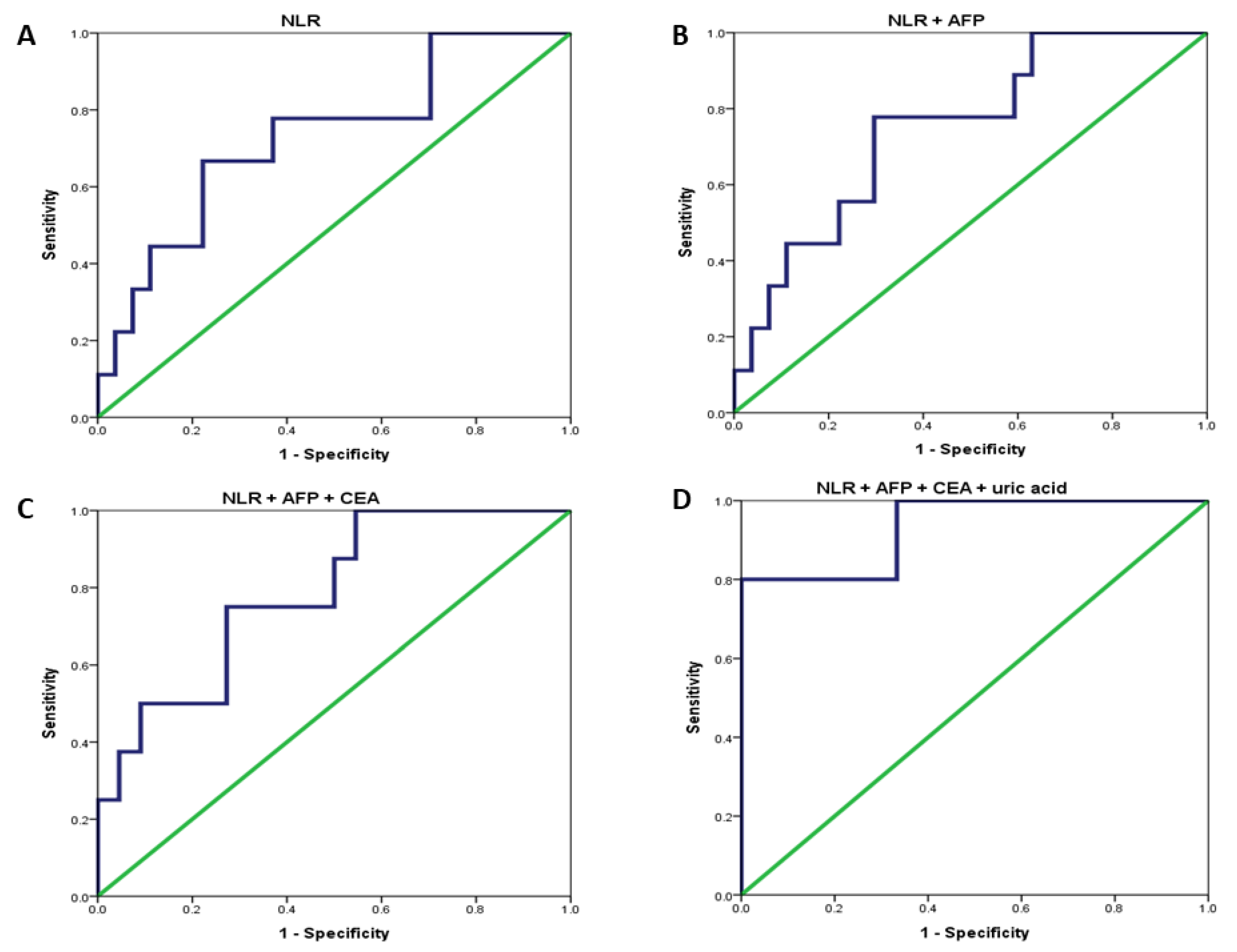
| Variables | Prediction of CK19 Expression | |||
|---|---|---|---|---|
| AUROC | Cutoff Value | Sensitivity/Specificity(%) | p Value | |
| Neutrophil to lymphocyte ratio (NLR) | 0.728 | 1.976 | 77.8/63.0 | 0.043 |
| α-fetoprotein (AFP, ng/mL) | 0.683 | 20.85 | 77.8/63.0 | 0.104 |
| CEA a (ng/mL) | 0.670 | 1.865 | 100/59.1 | 0.159 |
| Tumor size (cm) | 0.582 | 3.650 | 55.6/55.6 | 0.465 |
| Uric acid (mg/dL) | 0.750 | 4.50 | 86.4/50.0 | 0.065 |
| Triglyceride (mg/dL) | 0.767 | 53.5 | 94.460.0 | 0.074 |
| NLR + AFP | 0.749 | 0.027 | ||
| NLR + CEA | 0.756 | 0.035 | ||
| NLR + AFP + CEA | 0.784 | 0.019 | ||
| NLR + AFP + CEA + uric acid | 0.933 | 0.004 | ||
Publisher’s Note: MDPI stays neutral with regard to jurisdictional claims in published maps and institutional affiliations. |
© 2021 by the authors. Licensee MDPI, Basel, Switzerland. This article is an open access article distributed under the terms and conditions of the Creative Commons Attribution (CC BY) license (https://creativecommons.org/licenses/by/4.0/).
Share and Cite
Lee, C.-W.; Lin, S.-E.; Yu, M.-C.; Kou, H.-W.; Lee, C.-H.; Kuo, T.; Lee, K.-C.; Tsai, H.-I. Does Neutrophil to Lymphocyte Ratio Have a Role in Identifying Cytokeratin 19-Expressing Hepatocellular Carcinoma? J. Pers. Med. 2021, 11, 1078. https://doi.org/10.3390/jpm11111078
Lee C-W, Lin S-E, Yu M-C, Kou H-W, Lee C-H, Kuo T, Lee K-C, Tsai H-I. Does Neutrophil to Lymphocyte Ratio Have a Role in Identifying Cytokeratin 19-Expressing Hepatocellular Carcinoma? Journal of Personalized Medicine. 2021; 11(11):1078. https://doi.org/10.3390/jpm11111078
Chicago/Turabian StyleLee, Chao-Wei, Sey-En Lin, Ming-Chin Yu, Hao-Wei Kou, Cheng-Han Lee, Tony Kuo, Kuan-Chieh Lee, and Hsin-I Tsai. 2021. "Does Neutrophil to Lymphocyte Ratio Have a Role in Identifying Cytokeratin 19-Expressing Hepatocellular Carcinoma?" Journal of Personalized Medicine 11, no. 11: 1078. https://doi.org/10.3390/jpm11111078
APA StyleLee, C.-W., Lin, S.-E., Yu, M.-C., Kou, H.-W., Lee, C.-H., Kuo, T., Lee, K.-C., & Tsai, H.-I. (2021). Does Neutrophil to Lymphocyte Ratio Have a Role in Identifying Cytokeratin 19-Expressing Hepatocellular Carcinoma? Journal of Personalized Medicine, 11(11), 1078. https://doi.org/10.3390/jpm11111078