Review

Special Considerations in Pediatric Inflammatory Bowel Disease Pathology

1
Department of Pathology and Laboratory Medicine, University of Saskatchewan, Saskatoon, SK S7N 0W8, Canada
2
Department of Pathology, Boston Children’s Hospital and Harvard Medical School, Boston, MA 02115, USA
*
Author to whom correspondence should be addressed.
Diagnostics 2025, 15(7), 831; https://doi.org/10.3390/diagnostics15070831
Submission received: 31 January 2025 / Revised: 1 March 2025 / Accepted: 4 March 2025 / Published: 25 March 2025
(This article belongs to the Special Issue Pediatric Gastrointestinal Pathology)

Abstract

:
Inflammatory bowel disease (IBD) in the pediatric population presents distinct characteristics compared to adult cases. Pathology plays a critical role in its diagnosis, and this review underscores key considerations in the pathologic evaluation of pediatric IBD. Recognizing inflammatory patterns in the upper gastrointestinal tract can improve disease classification and aid in diagnosing IBD in certain scenarios, such as isolated upper gastrointestinal or small bowel involvement. Additionally, familiarity with distinctive subtypes, including IBD associated with primary sclerosing cholangitis and monogenic forms of IBD, supports early comorbidity detection, enhances patient management, and improves prognostication.

1. Introduction

Inflammatory bowel disease (IBD) is a chronic immune-mediated disorder of the gastrointestinal (GI) tract, characterized by alternating periods of flares and remission. Its development involves a complex interaction among host genetics, microbiome, immune system, and environmental factors [1]. A recent systematic review by Kuenzig et al. highlighted the global rise in the incidence and prevalence of pediatric IBD, with the highest rates reported in Northern Europe and North America and the lowest in Southern Europe, Asia, and the Middle East [2].
What distinguishes IBD in the pediatric population from adult-onset cases? Pediatric IBD is often diagnosed earlier in the disease course, before the development of complications. For example, children are more likely to present with Crohn disease (CD) prior to the onset of fibrostenotic or penetrating complications [2]. However, pediatric-onset IBD has been associated with a more extensive and severe disease trajectory [3,4,5]. This is due to a longer lifetime risk of accumulating inflammatory damage and its occurrence in individuals with the highest genetic and environmental susceptibility to IBD. Ultimately, these factors may lead to more severe disease activity over time. Additionally, pediatric IBD presents unique challenges, including the transition of care to adult services, psychosocial and financial burdens, and the substantial cost of managing younger patients, which often exceeds that of older individuals due to their lifelong need for healthcare [2].
This review highlights key considerations in the pathologic evaluation of pediatric IBD, focusing on disease classification, upper GI involvement, distinctive phenotypic presentations, such as IBD associated with primary sclerosing cholangitis, and monogenic forms of the disease. Additionally, it discusses important mimickers that should be considered during a pathologic assessment.

2. Pediatric Inflammatory Bowel Disease (IBD) Classification

The diagnosis of pediatric IBD is typically established through a comprehensive approach that includes clinical history, physical examination, laboratory tests, upper endoscopy, ileocolonoscopy, histologic evaluation, and small bowel imaging. Other potential diagnoses, particularly infections, should be carefully excluded. Once diagnosed, pediatric IBD is generally classified into ulcerative colitis (UC), CD, or IBD-unclassified (IBD-U). Accurate classification is essential for guiding the treatment and predicting disease outcomes. However, classification is not always straightforward and requires recognizing the typical features of UC and CD, identifying atypical phenotypes that still align with these diagnoses, and understanding factors that may preclude a definitive classification [6].
UC is characterized by continuous mucosal inflammation of the colon, originating distally from the rectum. It typically does not involve the small intestine, except in cases of backwash ileitis, and non-necrotizing granulomas are absent on biopsy. In 2014, the revised Porto criteria expanded the classification of UC to include typical and atypical forms [6]. Atypical UC encompasses cases with features that deviate from the classic UC, but occur frequently enough to rule out a CD diagnosis. These features include macroscopic rectal sparing, patchy disease or lack of chronicity in biopsies, left-sided colitis with a cecal patch (periappendiceal inflammation), upper GI involvement, and acute severe presentations that resemble CD.
Key features of CD include skip lesions, well-formed non-necrotizing granulomas distant from ruptured crypts, and characteristic macroscopic findings in the upper GI tract (e.g., serpentine ulcers and cobblestoning). Additional characteristics include bowel stenosis or stricturing, as well as cobblestoning and linear ulcerations in the ileum [6].
IBD-U refers to cases where differentiating between UC and CD remains uncertain despite clinical, endoscopic, and pathological evaluations. Over time, some patients are reclassified, more often as CD than UC; however, the majority retain the diagnosis of IBD-U [7,8]. The terms IBD-U and indeterminate colitis are often used interchangeably. According to the Montreal Working Party, IBD-U is diagnosed based on clinical and endoscopic findings, including biopsies, whereas indeterminate colitis is reserved for colectomy specimens [9].
The Pediatric IBD (PIBD) Porto Group of the European Society for Paediatric Gastroenterology, Hepatology and Nutrition (ESPGHAN) developed a classification algorithm (PIBD classes criteria) to facilitate the standardized and objective classification of IBD subtypes in children [10]. The scoring system includes 23 features of IBD, many of which are histopathologic findings, categorized into three classes: features incompatible with UC (Class 1), features rarely found in UC (Class 2), and features uncommon in UC (Class 3). The algorithm classifies IBD subtypes based on the number of features present in each class. In 2020, a simplified version of the algorithm with 19 features was introduced and is considered reliable [11,12]. The classification further differentiates CD into CD and isolated colonic CD, reflecting the full spectrum of pediatric IBD. The simplified items of PIBD classes are listed in Table 1; for details on the modified classification algorithm, please refer to the article by Ledder et al. [11]. Moreover, efforts to refine pediatric IBD classification using machine learning have been reported, though many require further validation [13,14,15].
Pediatric IBD can also be classified by the patient’s age into the following categories: very-early-onset IBD (VEO-IBD), diagnosed before 6 years of age; infantile IBD, diagnosed before 2 years of age; and neonatal-onset IBD, diagnosed within the first 28 days of life [16]. An important consideration in diagnosing IBD in these younger age groups is the potential for monogenic causes (see the Section 5 for further details).

3. Upper Gastrointestinal Tract Involvement in Pediatric Inflammatory Bowel Disease

Upper endoscopy is routinely performed during the initial evaluation of suspected IBD in pediatric patients to provide a more comprehensive assessment, contrary to adult clinical practice where it is not typically recommended [17,18]. In addition to aiding disease classification, macro- and microscopic evaluations of the upper gastrointestinal tract are particularly useful in patients with isolated upper or small bowel disease and a normal colonoscopy.
Upper GI involvement is more common in patients with CD than in those with UC. Although any part of the upper GI tract may be affected, the stomach is the most frequently involved site in these patients [19]. The histologic examination plays a crucial role, as the microscopic disease can be detected even in patients with a macroscopically normal upper GI endoscopy [20]. In most cases, the inflammatory pattern is non-specific; however, several distinct pathologic features may be observed in IBD, including lymphocytic esophagitis, focally enhanced gastritis, Brunner gland lobulitis, and non-necrotizing granulomas at any site. Notably, aside from non-necrotizing granulomas, no pathologic findings are specific enough to differentiate CD from UC [18].
Lymphocytic esophagitis (LE) is characterized by esophageal mucosal injury (e.g., spongiosis and dyskeratotic cells) resulting from an increased number of intraepithelial lymphocytes in the absence of significant granulocytes (Figure 1A). The threshold for intraepithelial lymphocytosis remains somewhat arbitrary and debated, as data on the normal range of intraepithelial lymphocytes in the pediatric esophagus are lacking. In clinical practice, the authors adopt the diagnostic criteria proposed by Rubio et al. (i.e., >20 intraepithelial lymphocytes) [21]. A recent review by international experts suggested a higher threshold of >40 intraepithelial lymphocytes for diagnosis [22]. Additionally, the presence of epithelial injury is crucial to avoid overdiagnosis. LE has been reported in up to 28% of children with CD and 7% of those with UC [23,24]. In a recent cohort of children with untreated CD, Spasic et al. identified an association between LE and an increased need for anti-tumor necrosis factor therapy [25]. Beyond IBD, LE has been linked to various conditions, including motility disorders, rheumatologic conditions, hypothyroidism, and lichen planus [22,26]. Moreover, Candida infection, a frequent cause of esophageal intraepithelial lymphocytosis, should be carefully excluded during an evaluation [27,28].
Focally enhanced gastritis (FEG) is histologically defined by focal inflammatory infiltrates involving gastric glands, accompanied by epithelial injury. The infiltrates predominantly consist of lymphohistiocytic cells, with variable presence of neutrophils and eosinophils, within an otherwise unremarkable background (Figure 1B,C) [29]. Although FEG is not specific for IBD in adults, its positive predictive value for IBD in the pediatric population is relatively high (75%) [30,31]. This finding has been observed in both children with CD (54–55%) and UC (21–30%); therefore, it should not be used to differentiate between the two conditions [32,33]. FEG is most commonly found in the antral mucosa, with endoscopic correlates including aphthoid ulcers and submucosal bleeding [29]. Histologically, multifocal involvement may be present, with a higher total number of affected glands in UC compared to CD [32].
Brunner gland lobulitis has been proposed by Abdullgaffar et al. [34] as a specific finding in patients with CD. Histologically, it is a distinct form of duodenitis marked by focal acute inflammation involving the Brunner glands (Figure 1D). However, the literature on this pathologic finding is limited, particularly in the pediatric population. Based on the senior author’s experience, Brunner gland lobulitis can also be observed in children with a confirmed diagnosis of UC. Therefore, its specificity should be interpreted with caution until further studies provide a more definitive evidence.
Non-necrotizing granulomas can occur at any site within the upper GI tract in patients with CD, with the stomach being the most commonly affected, followed by the duodenum and esophagus [35]. In untreated patients, non-necrotizing granulomas are observed in more than half of the cases, whereas they are less frequently seen in treated patients [35,36]. In some patients, granulomas are found exclusively in the upper GI tract, which can aid in disease classification. Histologically, non-necrotizing granulomas consist of aggregates of epithelioid histiocytes, with or without multinucleated giant cells, and without evidence of caseation (Figure 1E) [18]. The differential diagnosis includes immunologic disorders, such as chronic granulomatous disease and common variable immune deficiency, as well as infections and granulomas associated with crypt epithelial injury (cryptolytic granulomas). The latter may necessitate an examination of multiple tissue levels to detect crypt rupture. Cryptolytic granulomas can be observed in any subtype of IBD; in pediatric UC, their presence in the upper GI tract has been associated with severe colonic involvement [37].
Additionally, there are several caveats to consider in the pathologic evaluation of upper GI biopsies performed in children with IBD. Although chronic inflammation, with or without acute inflammation, is commonly observed in the gastric mucosa of these patients, pathologists should carefully assess for the presence of Helicobacter infection. While earlier studies suggested an inverse association between Helicobacter pylori gastritis and IBD, raising the possibility that the infection may play a protective role against IBD development, a recent prospective multicenter study in a pediatric population found no significant difference in the prevalence of Helicobacter pylori gastritis between patients with IBD and healthy controls [38,39,40]. Helicobacter organisms can be identified on routine H&E staining (Figure 1F) or, when necessary, with immunohistochemical or special stains. Interestingly, pediatric patients with Helicobacter infection, even without an IBD diagnosis, may exhibit elevated fecal calprotectin levels, which can mimic IBD-related laboratory findings and potentially lead to unnecessary colonoscopies [41].
The duodenal mucosa in children with IBD may exhibit intraepithelial lymphocytosis (>25 intraepithelial lymphocytes per 100 enterocytes on routine H&E staining) with preserved villous architecture, resembling celiac disease (type 1 Marsh) [42]. A recent systematic review and meta-analysis has shown that there is an increased risk of IBD in patients with celiac disease and an increased risk of celiac disease in patients with IBD [43]. Moreover, the coexisting celiac disease has been reported more frequently in pediatric-onset IBD cases compared to adults [44]. Therefore, in pathology reports, it is prudent to include a note clarifying that while duodenal mucosa with intraepithelial lymphocytosis could represent an upper GI manifestation of IBD, celiac disease should be clinically ruled out. Similar pathologic findings may also be observed in Helicobacter pylori gastritis, hypersensitivity to non-gluten alimentary proteins, or medication effects [42].

4. Inflammatory Bowel Disease with Primary Sclerosing Cholangitis (PSC-IBD)

PSC-IBD has been proposed as a distinct entity from IBD not associated with PSC, with differences observed in genetics, intestinal and fecal microbiomes, and intestinal transcriptomics [45,46]. Similar to adults, PSC-IBD in the pediatric population demonstrates unique clinical and endoscopic features. Ricciuto et al. reported that endoscopic findings such as backwash ileitis, pancolitis, rectal sparing, and more severe right-sided disease activity are more prevalent in children with PSC-IBD compared to those with IBD not associated with PSC [47]. However, the absence of symptoms in these patients does not necessarily indicate a lack of mucosal inflammation. Additionally, children with PSC-IBD have demonstrated greater growth impairments compared to their counterparts with non-PSC-associated IBD. Recognizing this subtype is essential for patient management and prognostication. For pathologists, understanding the disease distribution and salient pathologic features is crucial, especially for early PSC recognition.
Histologically, PSC-IBD correlates well with endoscopic findings. The common pathological features include pancolitis, more severe right-colon colitis, backwash ileitis, lamina propria-predominant neutrophils, prominent lamina propria eosinophilia in the left colon, and surface villiform changes in the right colon [48]. However, in a large cohort of children with PSC-IBD, Little et al. reported a discordance between endoscopic and histologic rectal sparing, noting a similar frequency and low rates in both children with PSC-IBD and those with IBD not associated with PSC [48].
Does age at diagnosis influence PSC-IBD outcomes? Catassi et al. evaluated patients diagnosed with PSC-IBD before the age of 6 years (very-early-onset PSC-IBD) [49]. Despite similar baseline characteristics between younger and older children, very-early-onset PSC-IBD is associated with a higher frequency of PSC overlap with autoimmune hepatitis, and lower rates of biliary complications, including infective cholangitis and biliary strictures. At the other end of the spectrum, in adults, PSC-IBD is linked to an increased risk of colorectal dysplasia and cancer despite its typically mild clinical course [50,51]. Zhang et al. reported that dysplasia in PSC-IBD is often endoscopically invisible and pathologically non-conventional (e.g., crypt cell dysplasia, hypermucinous dysplasia, and goblet cell-deficient dysplasia) [51]. Due to these risks, current clinical guidelines recommend that patients with PSC-IBD undergo annual colonoscopies starting at the time of PSC diagnosis, regardless of the duration of IBD [52].

5. Monogenic Inflammatory Bowel Disease (IBD)

“Classic” IBD is polygenic and may be influenced by numerous factors. However, IBD in some patients can be attributed to alterations in a single gene; these cases are termed Monogenic IBD (mIBD). To be classified as having mIBD, a patient should present with signs and symptoms of IBD and have a rare, damaging gene variant that is known to cause a monogenic disorder from previous reports and is consistent with the patient’s clinical phenotype [53]. Typically, mIBD presents early in life and tends to have increased morbidity and mortality when compared to classic IBD, in part due to the associated co-morbidities.
The first cases of IBD deemed to be associated with a specific gene mutation were reported in 2009 by Glocker et al. [54]. They identified four cases with mutations in genes encoding the interleukin-10 receptor [54]. Since then, numerous other genes have been identified. The literature on mIBD is heterogenous and lacking in prospective studies. Each individual monogenic disorder has its own characteristics, making generalization difficult. In 2021, ESPGHAN established a consensus list of 75 genes implicated in mIBD, which in addition to clinical applications for diagnosis, has been utilized as a template by other groups carrying out research on mIBD [53,55].
Monogenic IBD is typically considered to be associated with VEO-IBD (IBD in a patient aged less than 6 years) [55]. However, patients may be diagnosed with mIBD throughout childhood and into adulthood, with the average age at diagnosis varying widely for individual genes; from less than 2 years for mIBD associated with SCID to the fourth decade in cases associated with GUCY2C mutations [53]. In one cohort of 1000 Canadian children with IBD, presumed monogenic causative mutations were identified in 7.8% of children younger than 6 years and 2.3% of children aged 6–18 years [56].
Consanguinity and family history of autoimmune disease or suspected/confirmed monogenic disorders are associated with monogenic IBD [55]. Genetics of mIBD typically follow Mendelian patterns of inheritance. However, some mIBD disorders, such as those involving LRBA or CTLA4, present with diverse phenotypes that complicate data acquisition for pedigrees and mask their inheritance patterns [55]. The majority of mIBD cases show autosomal recessive inheritance [53]. The next most frequent inheritance pattern is X-linked recessive, followed by autosomal dominant [53]. Heterozygous-like cases in the context of symptomatic female persons with one copy of genes typically associated with X-linked recessive inheritance and persons with one copy of a gene typically associated with autosomal recessive inheritance may also occur [53]. The family history of IBD is most common in heterozygous-like cases (47%), and least common in X-linked recessive (20%) [53]. Consanguinity may be seen in up to 35% of autosomal recessive cases [53].
The genes implicated in mIBD can be classified into broad categories based on gene function and clinical presentation. Nambu et al. identified 29 distinct monogenic disorders and seven common clinical presentations: autoimmunity, infection, hemophagocytic lymphohistiocytosis (HLH), hematolymphoid organ involvement, malignancy, dysmorphic features, and metabolic disease [53]. Many of the 29 monogenic disorders exhibit more than one of the clinical presentations (Table 2). Bolton et al. developed a taxonomy for the genes implicated in mIBD using syndromic features, response to hematopoietic stem cell transplantation (HSCT), RNA-sequencing, and proteomes of immune subsets, which in addition to classifying genes, may guide future genomic testing, an understanding of shared mechanisms, and directed use of therapies [57].
In some cases, the diagnosis may be aided by the recognition of certain histological patterns of inflammation in biopsy specimens. Wilkins et al. have proposed a pattern-based approach in evaluating patients with VEO-IBD which categorized the pathologic findings into five patterns: chronic active enteritis (conventional pathologic pattern in IBD), apoptosis/epithelial injury pattern, eosinophil-rich pattern, lymphocytic patterns, and granulomatous pattern (Table 3) [58].
Current treatments for mIBD may include surgery, small molecules, biologics, and/or HSCT. Bowel surgery was undertaken in 27% of the cases with rates of surgery ranging from 0 to 100% for individual genes [53]. Stoma creation occurred in 21% of surgeries and bowel resection in 63% [53]. Biologics were used in 33% of cases, with an efficacy rate of 25.5% [53]. HSCT has been carried out in 23% of cases with some degree of improvement in IBD symptoms in 72% of treated patients [53]. In some cases, gastrointestinal symptoms and colitis do not resolve after HSCT, despite remission of other symptoms, especially when epithelium is the primary site of disease, such as in NF-kappa B essential modulator or TTC7A deficiency [59]. Targeted therapies for specific genes have been proposed but are not currently widely accessible and show variable efficacy. Uhlig et al. have proposed a reporting system for therapeutic responses of individual patients in hopes of streamlining clinical care and supporting future research including meta-analysis [59].
The five genetic alterations most commonly implicated in mIBD involve interleukin-10 and its receptor (IL-10/R), X-linked inhibitor of apoptosis protein (XIAP), cytochrome b-245 beta chain (CYBB), LPS responsive beige-like anchor protein (LRBA), and tetratricopeptide repeat domain 7A (TTC7A) [53]. Together, these alterations contribute over 40% of the reported cases of mIBD [53].

5.1. IL-10R/R

Mutations in genes encoding the IL-10RA and B subunit proteins involve hyperinflammatory immune responses in the intestine [54]. Cases associated with IL-10/IL-10R account for over 15% of mIBD, more than any other group of genes [53]. Clinical findings are similar to those seen in polygenic CD such as enteric fistulas, perianal disease, abscesses, and chronic folliculitis [54,60]. Histological findings also largely mimic polygenic CD and include multifocal colonic ulcerations and collections of polymorphic infiltrates, with inflammation variably present in the small bowel [54,60]. Pear-shaped intramural abscesses extending into the submucosa and muscularis propria have also been reported [54]. Multimodal therapy, including some combination of exclusive enteral nutrition, anti-inflammatory and immunomodulatory agents may not achieve remission [60]. HSCT has also been shown to be effective [54].

5.2. XIAP

Similar to the other inhibitor of apoptosis genes, XIAP is a component of both innate and adaptive immunity [61]. Up to 9% of mIBD cases are associated with XIAP [53]. Cases may present with severe bloody diarrhea clinically within the first few weeks of life, refractory to conventional therapies [61]. Intestinal and extraintestinal manifestations may resemble CD with histology showing severe pancolitis with crypt abscesses and epithelium apoptosis [61,62]. A subset of patients with XIAP deficiency show early-onset colitis, severe failure to thrive, and splenomegaly/hepatosplenomegaly an increased risk of medically refractory IBD phenotype and increased mortality [62]. In this subset, early HSCT with reduced intensity conditioning may result in both resolution of their colitis and reduction in their risk of developing HLH, which is the leading cause of mortality in XIAP-deficient patients [62].

5.3. CYBB

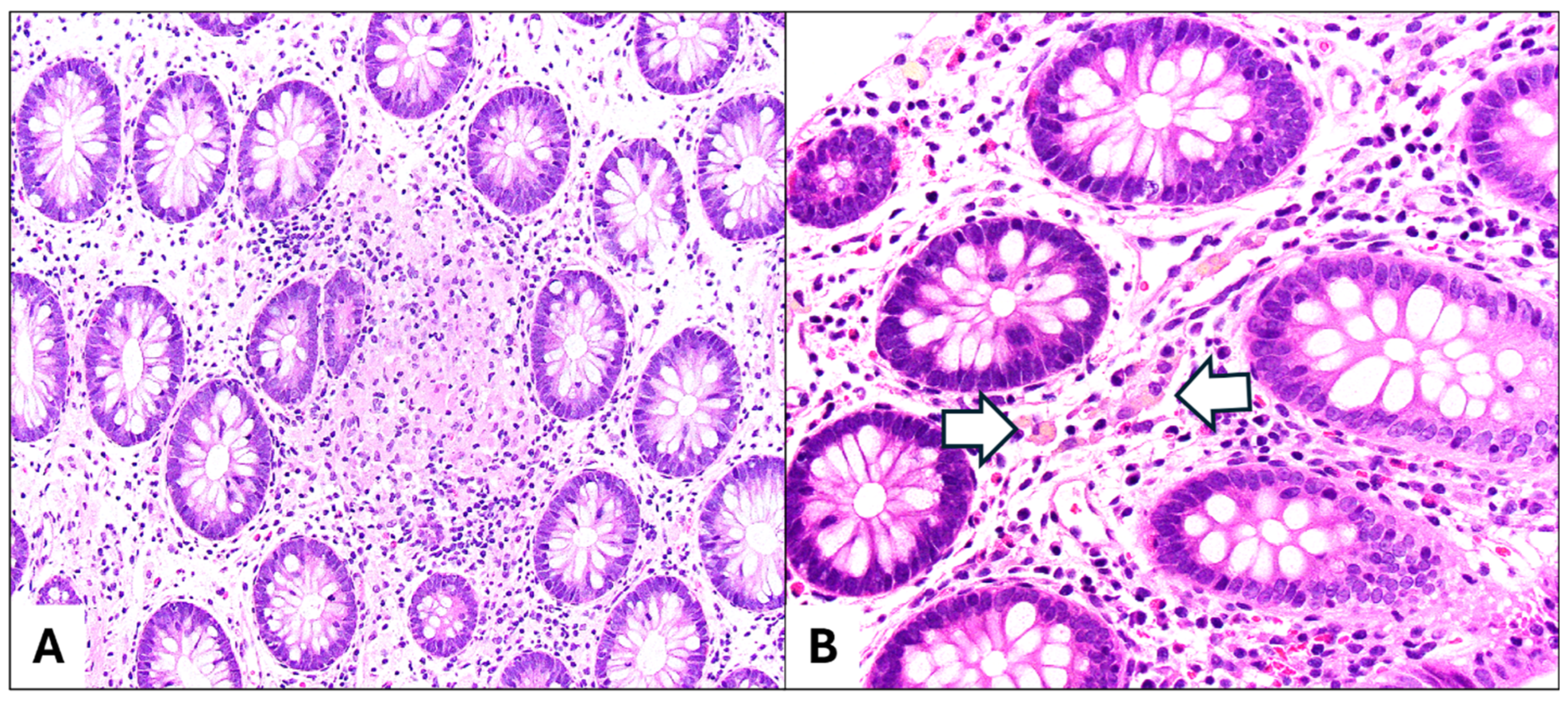
CYBB is one of the genes associated with Chronic Granulomatous Disease (CGD) [53]. CYBB alone is associated with 9% of mIBD cases [53]. Patients with CGD have a poor oxidative burst in their neutrophils resulting in the inability to clear certain infections and often present with a history of recurrent pneumonias, lymphadenopathy, upper GI involvement, and chronic colitis in early life [63]. Pathologic findings typically exhibit a granulomatous pattern and may also include mild chronic changes with increased lymphocytes and eosinophils in the lamina propria, and active colitis including crypt abscesses [63,64]. Granulomas in CGD are usually more confluent than in CD and may be accompanied by pigment-laden macrophages in the lamina propria (Figure 2A,B), which are thought to represent an accumulation of ceroid pigments due to the increased cell turnover [58]. The differentiation of CGD from CD is critical, because some IBD treatments, such as TNFα inhibitors, can lead to life-threatening infections in CGD patients [58]. A typical treatment of CGD includes subcutaneous interferon-gamma [63]. HSCT has shown good clinical responses including the normalization of oxidative burst and resolution of gastrointestinal symptoms [63,64]. Gene therapy with viral vectors has also been performed [59].

5.4. LRBA

Gene alterations in LRBA with resulting CTLA4 deficiency account for 4% of reported mIBD cases and fall under the umbrella of immunodysregulation polyendocrinopathy enteropathy X-linked (IPEX)-like syndrome [53]. They belong to the newly proposed functional category of “immune TOR-opathies” [65]. Clinically, the LRBA mutation may be associated with diarrhea, infections, and arthritis [58]. Histologically, a range of findings may be seen from a subtle lymphocytic pattern with surface and crypt intraepithelial lymphocytes and surface friability/denudation and normal mucosal architecture with slight increases in lamina propria cellularity to more classic patterns of chronic active colitis [58,65,66]. Targeted therapy directed to the mTOR pathway as well as individual genes with pathogenic variants has been proposed [65]. HSCT with reduced intensity conditioning has been used with good success [66].

5.5. TTC7A

Cases of mIBD associated with TTC7A represent another 4% of mIBD [53]. Patients with TTC7A mutations have disrupted intestinal and thymic epithelia and it was initially reported in children with multiple intestinal atresias and combined (T and B cell) immunodeficiency [67]. Small and large bowel biopsies in TTC7A-associated cases (with or without intestinal atresia) tend to show an epithelial injury-apoptosis pattern with predominantly neutrophilic infiltrates and numerous apoptotic bodies and diminished lymphocyte populations [58,67,68]. Additionally, atrophic gastritis has been described in a subset of patients [69]. HSCT in patients with TTC7A mutations may significantly increase their immune function, but as mentioned previously, it has not been shown to improve gastrointestinal disease [59,67]. Pharmacological targets in the PI4KIIIa-TTC7A-EFR3B pathway, with drugs such as leflunomide, represent a potential therapeutic approach [59,68].

6. Differential Diagnosis

Histologically, distinguishing IBD from its mimics remains a diagnostic challenge. A definitive diagnosis requires correlation with endoscopic findings, clinical history, and ancillary studies. Close communication between gastroenterologists and pathologists is essential and cannot be overemphasized. IBD should never be diagnosed solely on histologic grounds. When evaluating biopsies with a chronic active colitis pattern, two main alternative etiologies to consider are infection and medication effects. Other potential etiologies include vascular disorders, such as gastrointestinal manifestations of systemic vasculitis or drug-induced ischemia, as well as diversion proctocolitis, which affects nonfunctional bowel segments following surgery [70]. An accurate diagnosis is crucial, as treatment strategies often vary significantly depending on the underlying cause. A differential diagnosis for pattern-based injury (e.g., eosinophil-rich and apoptotic colitis) is summarized in Table 3.
Although infections typically present with an acute colitis pattern, some pathogens, such as Salmonella spp., Shigella spp., and Entamoeba histolytica, may exhibit features of chronicity that mimic IBD (e.g., architectural distortion, pyloric metaplasia, and Paneth cell hyperplasia) [71]. In older children and sexually active individuals, proctocolitis due to Treponema pallidum or Chlamydia trachomatis should also be considered, as these infections can closely resemble IBD both endoscopically and histologically [72]. However, the degree of chronicity in sexually transmitted infections is generally mild. Additionally, the presence of granulomas, particularly the necrotizing type, should prompt the consideration of infections caused by Mycobacterium tuberculosis, Yersinia spp., or Actinomyces spp. [71]. Stool cultures, PCR, and other clinical assays can aid in identifying infectious causes. In pediatric patients with infectious colitis, an underlying immunodeficiency should always be ruled out.
Drug-induced colitis can occur in all age groups, including pediatric patients, and presents with a wide spectrum of histologic patterns, ranging from active colitis and chronic active colitis (IBD-like) to microscopic colitis (lymphocytic/collagenous) and increased apoptosis [73]. Knowledge of the medication type, dosage, and the temporal relationship between drug exposure and symptom onset is critical for diagnosis. NSAIDs may induce a UC-like pattern with diffuse inflammation and ulceration or a CD-like pattern with granulomas [74]. Immune checkpoint inhibitors can cause acute colitis with or without chronicity, often with increased apoptotic bodies, a feature also seen in mycophenolate mofetil-associated colitis [73]. When an IBD-like injury pattern is present, the additional findings of intraepithelial lymphocytosis or prominent apoptosis should alert the pathologist to the possibility of drug-induced colitis.

7. Conclusions

In the pediatric population, the diagnosis and classification of IBD require an understanding of both classical and atypical presentations. Upper gastrointestinal manifestations are frequently observed in pediatric IBD, which is likely due to the routine inclusion of upper endoscopy in the diagnostic workup. Histopathological findings may reveal a non-specific inflammation; however, certain distinct patterns, such as LE, FEG, Brunner gland lobulitis, and non-necrotizing granulomas, should prompt the consideration of an IBD diagnosis. PSC-IBD and mIBD, though not exclusive to the pediatric age group, represent specific subtypes with important implications for prognosis and management. Early recognition of the clinical and pathological features of PSC-IBD can facilitate the timely detection of PSC. While PSC-IBD is generally associated with a milder disease course, it carries an increased risk for colorectal dysplasia and carcinoma. Furthermore, factors such as the patient’s age, family history, consanguinity, and comorbidities should be carefully considered to identify potential monogenic causes of IBD.

Author Contributions

A.R.A. drafted parts of the manuscript and edited as necessary; J.P. approved the final manuscript through substantial revisions and provided photomicrographs. All authors have read and agreed to the published version of the manuscript.

Funding

This research received no external funding.

Conflicts of Interest

The authors declare no conflicts of interest.

References

  1. Bouhuys, M.; Lexmond, W.S.; van Rheenen, P.F. Pediatric Inflammatory Bowel Disease. Pediatrics 2023, 151, e2022058037. [Google Scholar] [CrossRef] [PubMed]
  2. Kuenzig, M.E.; Fung, S.G.; Marderfeld, L.; Mak, J.W.Y.; Kaplan, G.G.; Ng, S.C.; Wilson, D.C.; Cameron, F.; Henderson, P.; Kotze, P.G.; et al. Twenty-First Century Trends in the Global Epidemiology of Pediatric-Onset Inflammatory Bowel Disease: Systematic Review. Gastroenterology 2022, 162, 1147–1159.e4. [Google Scholar] [CrossRef] [PubMed]
  3. Vuijk, S.A.; Camman, A.E.; de Ridder, L. Considerations in Paediatric and Adolescent Inflammatory Bowel Disease. J. Crohns Colitis 2024, 18 (Suppl. S2), ii31–ii45. [Google Scholar] [CrossRef]
  4. Herzog, D.; Fournier, N.; Buehr, P.; Rueger, V.; Koller, R.; Heyland, K.; Nydegger, A.; Braegger, C.P.; Swiss IBD Cohort Study Group. Prevalence of Intestinal Complications in Inflammatory Bowel Disease: A Comparison between Paediatric-Onset and Adult-Onset Patients. Eur. J. Gastroenterol. Hepatol. 2017, 29, 926–931. [Google Scholar] [CrossRef] [PubMed]
  5. Jakobsen, C.; Bartek, J.; Wewer, V.; Vind, I.; Munkholm, P.; Groen, R.; Paerregaard, A. Differences in Phenotype and Disease Course in Adult and Paediatric Inflammatory Bowel Disease—A Population-Based Study. Aliment. Pharmacol. Ther. 2011, 34, 1217–1224. [Google Scholar] [CrossRef]
  6. Levine, A.; Koletzko, S.; Turner, D.; Escher, J.C.; Cucchiara, S.; de Ridder, L.; Kolho, K.-L.; Veres, G.; Russell, R.K.; Paerregaard, A.; et al. ESPGHAN Revised Porto Criteria for the Diagnosis of Inflammatory Bowel Disease in Children and Adolescents. J. Pediatr. Gastroenterol. Nutr. 2014, 58, 795–806. [Google Scholar] [CrossRef]
  7. Rinawi, F.; Assa, A.; Eliakim, R.; Mozer-Glassberg, Y.; Nachmias Friedler, V.; Niv, Y.; Rosenbach, Y.; Silbermintz, A.; Zevit, N.; Shamir, R. The Natural History of Pediatric-Onset IBD-Unclassified and Prediction of Crohn’s Disease Reclassification: A 27-Year Study. Scand. J. Gastroenterol. 2017, 52, 558–563. [Google Scholar] [CrossRef]
  8. Malaty, H.M.; Mehta, S.; Abraham, B.; Garnett, E.A.; Ferry, G.D. The Natural Course of Inflammatory Bowel Disease-Indeterminate from Childhood to Adulthood: Within a 25 Year Period. Clin. Exp. Gastroenterol. 2013, 6, 115–121. [Google Scholar] [CrossRef]
  9. Satsangi, J.; Silverberg, M.S.; Vermeire, S.; Colombel, J.-F. The Montreal Classification of Inflammatory Bowel Disease: Controversies, Consensus, and Implications. Gut 2006, 55, 749–753. [Google Scholar] [CrossRef]
  10. Birimberg-Schwartz, L.; Zucker, D.M.; Akriv, A.; Cucchiara, S.; Cameron, F.L.; Wilson, D.C.; Lazowska, I.; Yianni, L.; Paul, S.P.; Romano, C.; et al. Development and Validation of Diagnostic Criteria for IBD Subtypes Including IBD-Unclassified in Children: A Multicentre Study From the Pediatric IBD Porto Group of ESPGHAN. J. Crohns Colitis 2017, 11, 1078–1084. [Google Scholar] [CrossRef]
  11. Ledder, O.; Sonnino, M.; Birimberg-Schwartz, L.; Escher, J.C.; Russell, R.K.; Orlanski-Meyer, E.; Matar, M.; Assa, A.; Tzion, R.L.; Shteyer, E.; et al. Appraisal of the PIBD-Classes Criteria: A Multicentre Validation. J. Crohns Colitis 2020, 14, 1672–1679. [Google Scholar] [CrossRef] [PubMed]
  12. Miura, Y.; Hagiwara, S.-I.; Hizuka, K.; Saura, R.; Hata, A.; Maeyama, T.; Etani, Y. Validation of the Simplified PIBD-Classes Criteria: A Single-Center Retrospective Study. Pediatr. Int. Off. J. Jpn. Pediatr. Soc. 2024, 66, e15846. [Google Scholar] [CrossRef] [PubMed]
  13. Dhaliwal, J.; Erdman, L.; Drysdale, E.; Rinawi, F.; Muir, J.; Walters, T.D.; Siddiqui, I.; Griffiths, A.M.; Church, P.C. Accurate Classification of Pediatric Colonic Inflammatory Bowel Disease Subtype Using a Random Forest Machine Learning Classifier. J. Pediatr. Gastroenterol. Nutr. 2021, 72, 262–269. [Google Scholar] [CrossRef]
  14. Schneider, N.; Sohrabi, K.; Schneider, H.; Zimmer, K.-P.; Fischer, P.; de Laffolie, J.; CEDATA-GPGE Study Group. Machine Learning Classification of Inflammatory Bowel Disease in Children Based on a Large Real-World Pediatric Cohort CEDATA-GPGE® Registry. Front. Med. 2021, 8, 666190. [Google Scholar] [CrossRef]
  15. Mossotto, E.; Ashton, J.J.; Coelho, T.; Beattie, R.M.; MacArthur, B.D.; Ennis, S. Classification of Paediatric Inflammatory Bowel Disease Using Machine Learning. Sci. Rep. 2017, 7, 2427. [Google Scholar] [CrossRef]
  16. Uhlig, H.H.; Schwerd, T.; Koletzko, S.; Shah, N.; Kammermeier, J.; Elkadri, A.; Ouahed, J.; Wilson, D.C.; Travis, S.P.; Turner, D.; et al. The Diagnostic Approach to Monogenic Very Early Onset Inflammatory Bowel Disease. Gastroenterology 2014, 147, 990–1007.e3. [Google Scholar] [CrossRef]
  17. Jevon, G.P.; Madhur, R. Endoscopic and Histologic Findings in Pediatric Inflammatory Bowel Disease. Gastroenterol. Hepatol. 2010, 6, 174–180. [Google Scholar]
  18. Abuquteish, D.; Putra, J. Upper Gastrointestinal Tract Involvement of Pediatric Inflammatory Bowel Disease: A Pathological Review. World J. Gastroenterol. 2019, 25, 1928–1935. [Google Scholar] [CrossRef]
  19. Repo, M.; Pessi, J.; Wirtanen, E.; Hiltunen, P.; Huhtala, H.; Kivelä, L.; Kurppa, K. Frequency and Clinical Significance of Histologic Upper Gastrointestinal Tract Findings in Children with Inflammatory Bowel Disease. Scand. J. Gastroenterol. 2022, 57, 1046–1050. [Google Scholar] [CrossRef]
  20. Chen, B.B.; Hamilton, Z.; Hasosah, M.; Zetler, P.; Popescu, O.; Bush, J.; Katz, R.; Smyth, M.; Jacobson, K. The Role of Upper Gastrointestinal Endoscopy in the Diagnosis of Pediatric Inflammatory Bowel Disease. Am. J. Clin. Pathol. 2024, 162, 597–603. [Google Scholar] [CrossRef]
  21. Rubio, C.A.; Sjödahl, K.; Lagergren, J. Lymphocytic Esophagitis: A Histologic Subset of Chronic Esophagitis. Am. J. Clin. Pathol. 2006, 125, 432–437. [Google Scholar] [PubMed]
  22. Coady, L.C.; Sheahan, K.; Brown, I.S.; Carneiro, F.; Gill, A.J.; Kumarasinghe, P.; Kushima, R.; Lauwers, G.Y.; Pai, R.K.; Shepherd, N.A.; et al. Esophageal Lymphocytosis: Exploring the Knowns and Unknowns of This Pattern of Esophageal Injury. Expert Rev. Gastroenterol. Hepatol. 2024, 18, 529–539. [Google Scholar] [CrossRef] [PubMed]
  23. Ebach, D.R.; Vanderheyden, A.D.; Ellison, J.M.; Jensen, C.S. Lymphocytic Esophagitis: A Possible Manifestation of Pediatric Upper Gastrointestinal Crohn’s Disease. Inflamm. Bowel Dis. 2011, 17, 45–49. [Google Scholar] [CrossRef]
  24. Sutton, L.M.; Heintz, D.D.; Patel, A.S.; Weinberg, A.G. Lymphocytic Esophagitis in Children. Inflamm. Bowel Dis. 2014, 20, 1324–1328. [Google Scholar] [CrossRef] [PubMed]
  25. Spasic, S.; Pankaj, A.; Kaplan, J.L.; Patil, D.; Moran, C.J.; Deshpande, V. Paediatric Crohn’s Disease: Histologic Findings at Initial Presentation. J. Clin. Pathol. 2024. [Google Scholar] [CrossRef]
  26. Pittman, M.E. Lymphocytic Esophagitis: Current Understanding and Controversy. Am. J. Surg. Pathol. 2022, 46, e55–e63. [Google Scholar] [CrossRef]
  27. Martin, I.W.; Atkinson, A.E.; Liu, X.; Suriawinata, A.A.; Lefferts, J.A.; Lisovsky, M. Mucosal Inflammation in Candida Esophagitis Has Distinctive Features That May Be Helpful Diagnostically. Mod. Pathol. 2018, 31, 1653–1660. [Google Scholar] [CrossRef]
  28. Abuquteish, D.; Siddiqui, I.; Putra, J. Lymphocytic Esophagitis: Inflammatory Pattern of Candida Esophagitis in a Patient with Ulcerative Colitis. Int. J. Surg. Pathol. 2019, 27, 762. [Google Scholar] [CrossRef]
  29. Putra, J.; Ornvold, K. Focally Enhanced Gastritis in Children with Inflammatory Bowel Disease: A Clinicopathological Correlation. Pathology 2017, 49, 808–810. [Google Scholar] [CrossRef]
  30. McHugh, J.B.; Gopal, P.; Greenson, J.K. The Clinical Significance of Focally Enhanced Gastritis in Children. Am. J. Surg. Pathol. 2013, 37, 295–299. [Google Scholar] [CrossRef]
  31. Kővári, B.; Pai, R.K. Upper Gastrointestinal Tract Involvement in Inflammatory Bowel Diseases: Histologic Clues and Pitfalls. Adv. Anat. Pathol. 2022, 29, 2–14. [Google Scholar] [CrossRef] [PubMed]
  32. Ushiku, T.; Moran, C.J.; Lauwers, G.Y. Focally Enhanced Gastritis in Newly Diagnosed Pediatric Inflammatory Bowel Disease. Am. J. Surg. Pathol. 2013, 37, 1882–1888. [Google Scholar] [CrossRef] [PubMed]
  33. Roka, K.; Roma, E.; Stefanaki, K.; Panayotou, I.; Kopsidas, G.; Chouliaras, G. The Value of Focally Enhanced Gastritis in the Diagnosis of Pediatric Inflammatory Bowel Diseases. J. Crohns Colitis 2013, 7, 797–802. [Google Scholar] [CrossRef]
  34. AbdullGaffar, B.; Quraishi, H. Histopathologic Manifestations of Crohn Disease in Duodenal Endoscopy Biopsy: The Value of Different Patterns of Involvement of Brunner Glands. Int. J. Surg. Pathol. 2021, 29, 710–715. [Google Scholar] [CrossRef]
  35. De Matos, V.; Russo, P.A.; Cohen, A.B.; Mamula, P.; Baldassano, R.N.; Piccoli, D.A. Frequency and Clinical Correlations of Granulomas in Children with Crohn Disease. J. Pediatr. Gastroenterol. Nutr. 2008, 46, 392–398. [Google Scholar] [CrossRef]
  36. Putniković, D.; Jevtić, J.; Ristić, N.; Milovanovich, I.D.; Đuknić, M.; Radusinović, M.; Popovac, N.; Đorđić, I.; Leković, Z.; Janković, R. Pediatric Crohn’s Disease in the Upper Gastrointestinal Tract: Clinical, Laboratory, Endoscopic, and Histopathological Analysis. Diagnostics 2024, 14, 877. [Google Scholar] [CrossRef]
  37. Queliza, K.; Ihekweazu, F.D.; Schady, D.; Jensen, C.; Kellermayer, R. Granulomatous Upper Gastrointestinal Inflammation in Pediatric Ulcerative Colitis. J. Pediatr. Gastroenterol. Nutr. 2018, 66, 620–623. [Google Scholar] [CrossRef]
  38. Dilaghi, E.; Felici, E.; Lahner, E.; Pilozzi, E.; Furio, S.; Lucchini, L.; Quatrale, G.; Piccirillo, M.; Parisi, P.; Curto, S.; et al. Helicobacter Pylori Infection in Children with Inflammatory Bowel Disease: A Prospective Multicenter Study. BMC Pediatr. 2024, 24, 417. [Google Scholar] [CrossRef]
  39. Roka, K.; Roubani, A.; Stefanaki, K.; Panayotou, I.; Roma, E.; Chouliaras, G. The Prevalence of Helicobacter Pylori Gastritis in Newly Diagnosed Children with Inflammatory Bowel Disease. Helicobacter 2014, 19, 400–405. [Google Scholar] [CrossRef]
  40. Shirzad-Aski, H.; Besharat, S.; Kienesberger, S.; Sohrabi, A.; Roshandel, G.; Amiriani, T.; Norouzi, A.; Keshtkar, A. Association Between Helicobacter Pylori Colonization and Inflammatory Bowel Disease: A Systematic Review and Meta-Analysis. J. Clin. Gastroenterol. 2021, 55, 380–392. [Google Scholar] [CrossRef]
  41. Villalba-Davila, P.; Aronson, S.; Lat, J.; Charles, C.; Schroeder, B.; Pittman, M.; Tang, V.; Wallach, T. Helicobacter Pylori Infection Is Associated with Significant Elevations to Fecal Calprotectin, Systemic Inflammatory Markers. J. Pediatr. Gastroenterol. Nutr. 2025. [Google Scholar] [CrossRef] [PubMed]
  42. Putra, J.; Goldsmith, J.D. Daily Dilemmas in Pediatric Gastrointestinal Pathology. Surg. Pathol. Clin. 2020, 13, 399–411. [Google Scholar] [CrossRef] [PubMed]
  43. Pinto-Sanchez, M.I.; Seiler, C.L.; Santesso, N.; Alaedini, A.; Semrad, C.; Lee, A.R.; Bercik, P.; Lebwohl, B.; Leffler, D.A.; Kelly, C.P.; et al. Association Between Inflammatory Bowel Diseases and Celiac Disease: A Systematic Review and Meta-Analysis. Gastroenterology 2020, 159, 884–903.e31. [Google Scholar] [CrossRef] [PubMed]
  44. Kori, M.; Zamir, Y.; Yermiyahu, S.O.; Ainbinder, I.; Daichman, S.; Pinto, G.D.; Loewenberg Weisband, Y.; Greenfeld, S.; Kariv, R.; Lederman, N.; et al. The Association of Inflammatory Bowel Disease with Coeliac Disease and Coeliac Autoimmunity in Children and Adults: A Nationwide Study from the Epi-IIRN. J. Crohns Colitis 2023, 17, 700–705. [Google Scholar] [CrossRef]
  45. Jiang, X.; Karlsen, T.H. Genetics of Primary Sclerosing Cholangitis and Pathophysiological Implications. Nat. Rev. Gastroenterol. Hepatol. 2017, 14, 279–295. [Google Scholar] [CrossRef]
  46. Quraishi, M.N.; Acharjee, A.; Beggs, A.D.; Horniblow, R.; Tselepis, C.; Gkoutos, G.; Ghosh, S.; Rossiter, A.E.; Loman, N.; van Schaik, W.; et al. A Pilot Integrative Analysis of Colonic Gene Expression, Gut Microbiota, and Immune Infiltration in Primary Sclerosing Cholangitis-Inflammatory Bowel Disease: Association of Disease with Bile Acid Pathways. J. Crohns Colitis 2020, 14, 935–947. [Google Scholar] [CrossRef]
  47. Ricciuto, A.; Hansen, B.E.; Ngo, B.; Aloi, M.; Walters, T.D.; Church, P.C.; Mazurek, A.; Khan, M.; Carman, N.; Siddiqui, I.; et al. Primary Sclerosing Cholangitis in Children with Inflammatory Bowel Diseases Is Associated with Milder Clinical Activity But More Frequent Subclinical Inflammation and Growth Impairment. Clin. Gastroenterol. Hepatol. Off. Clin. Pract. J. Am. Gastroenterol. Assoc. 2020, 18, 1509–1517.e7. [Google Scholar] [CrossRef]
  48. Little, R.; Putra, J.; Kamath, B.M.; Griffiths, A.M.; Ricciuto, A.; Siddiqui, I. Intestinal Histopathology in Pediatric PSC-IBD: Characterization of Phenotype and Assessment of the Nancy Index. J. Pediatr. Gastroenterol. Nutr. 2024, 80, 290–299. [Google Scholar] [CrossRef]
  49. Catassi, G.; D’Arcangelo, G.; Norsa, L.; Bramuzzo, M.; Hojsak, I.; Kolho, K.-L.; Romano, C.; Gasparetto, M.; Di Giorgio, A.; Hussey, S.; et al. Outcome of Very Early Onset Inflammatory Bowel Disease Associated With Primary Sclerosing Cholangitis: A Multicenter Study From the Pediatric IBD Porto Group of ESPGHAN. Inflamm. Bowel Dis. 2024, 30, 1662–1669. [Google Scholar] [CrossRef]
  50. de Vries, A.B.; Janse, M.; Blokzijl, H.; Weersma, R.K. Distinctive Inflammatory Bowel Disease Phenotype in Primary Sclerosing Cholangitis. World J. Gastroenterol. 2015, 21, 1956–1971. [Google Scholar] [CrossRef]
  51. Zhang, R.; Lauwers, G.Y.; Choi, W.-T. Increased Risk of Non-Conventional and Invisible Dysplasias in Patients with Primary Sclerosing Cholangitis and Inflammatory Bowel Disease. J. Crohns Colitis 2022, 16, 1825–1834. [Google Scholar] [CrossRef] [PubMed]
  52. Rubin, D.T.; Ananthakrishnan, A.N.; Siegel, C.A.; Sauer, B.G.; Long, M.D. ACG Clinical Guideline: Ulcerative Colitis in Adults. Am. J. Gastroenterol. 2019, 114, 384–413. [Google Scholar] [CrossRef] [PubMed]
  53. Nambu, R.; Warner, N.; Mulder, D.J.; Kotlarz, D.; McGovern, D.P.B.; Cho, J.; Klein, C.; Snapper, S.B.; Griffiths, A.M.; Iwama, I.; et al. A Systematic Review of Monogenic Inflammatory Bowel Disease. Clin. Gastroenterol. Hepatol. Off. Clin. Pract. J. Am. Gastroenterol. Assoc. 2022, 20, e653–e663. [Google Scholar] [CrossRef]
  54. Glocker, E.-O.; Kotlarz, D.; Boztug, K.; Gertz, E.M.; Schäffer, A.A.; Noyan, F.; Perro, M.; Diestelhorst, J.; Allroth, A.; Murugan, D.; et al. Inflammatory Bowel Disease and Mutations Affecting the Interleukin-10 Receptor. N. Engl. J. Med. 2009, 361, 2033–2045. [Google Scholar] [CrossRef]
  55. Uhlig, H.H.; Charbit-Henrion, F.; Kotlarz, D.; Shouval, D.S.; Schwerd, T.; Strisciuglio, C.; de Ridder, L.; van Limbergen, J.; Macchi, M.; Snapper, S.B.; et al. Clinical Genomics for the Diagnosis of Monogenic Forms of Inflammatory Bowel Disease: A Position Paper From the Paediatric IBD Porto Group of European Society of Paediatric Gastroenterology, Hepatology and Nutrition. J. Pediatr. Gastroenterol. Nutr. 2021, 72, 456–473. [Google Scholar] [CrossRef]
  56. Crowley, E.; Warner, N.; Pan, J.; Khalouei, S.; Elkadri, A.; Fiedler, K.; Foong, J.; Turinsky, A.L.; Bronte-Tinkew, D.; Zhang, S.; et al. Prevalence and Clinical Features of Inflammatory Bowel Diseases Associated with Monogenic Variants, Identified by Whole-Exome Sequencing in 1000 Children at a Single Center. Gastroenterology 2020, 158, 2208–2220. [Google Scholar] [CrossRef]
  57. Bolton, C.; Smillie, C.S.; Pandey, S.; Elmentaite, R.; Wei, G.; Argmann, C.; Aschenbrenner, D.; James, K.R.; McGovern, D.P.B.; Macchi, M.; et al. An Integrated Taxonomy for Monogenic Inflammatory Bowel Disease. Gastroenterology 2022, 162, 859–876. [Google Scholar] [CrossRef]
  58. Wilkins, B.J.; Kelsen, J.R.; Conrad, M.A. A Pattern-Based Pathology Approach to Very Early-Onset Inflammatory Bowel Disease: Thinking Beyond Crohn Disease and Ulcerative Colitis. Adv. Anat. Pathol. 2022, 29, 62. [Google Scholar] [CrossRef]
  59. Uhlig, H.H.; Booth, C.; Cho, J.; Dubinsky, M.; Griffiths, A.M.; Grimbacher, B.; Hambleton, S.; Huang, Y.; Jones, K.; Kammermeier, J.; et al. Precision Medicine in Monogenic Inflammatory Bowel Disease: Proposed mIBD REPORT Standards. Nat. Rev. Gastroenterol. Hepatol. 2023, 20, 810–828. [Google Scholar] [CrossRef]
  60. Kotlarz, D.; Beier, R.; Murugan, D.; Diestelhorst, J.; Jensen, O.; Boztug, K.; Pfeifer, D.; Kreipe, H.; Pfister, E.; Baumann, U.; et al. Loss of Interleukin-10 Signaling and Infantile Inflammatory Bowel Disease: Implications for Diagnosis and Therapy. Gastroenterology 2012, 143, 347–355. [Google Scholar] [CrossRef]
  61. Kelsen, J.R.; Dawany, N.; Martinez, A.; Grochowski, C.M.; Maurer, K.; Rappaport, E.; Piccoli, D.A.; Baldassano, R.N.; Mamula, P.; Sullivan, K.E.; et al. A de Novo Whole Gene Deletion of XIAP Detected by Exome Sequencing Analysis in Very Early Onset Inflammatory Bowel Disease: A Case Report. BMC Gastroenterol. 2015, 15, 160. [Google Scholar] [CrossRef]
  62. Lekbua, A.; Ouahed, J.; O’Connell, A.E.; Kahn, S.A.; Goldsmith, J.D.; Imamura, T.; Duncan, C.N.; Kelsen, J.R.; Worthey, E.; Snapper, S.B.; et al. Risk-Factors Associated with Poor Outcomes in VEO-IBD Secondary to XIAP Deficiency: A Case Report and Literature Review. J. Pediatr. Gastroenterol. Nutr. 2019, 69, e13–e18. [Google Scholar] [CrossRef] [PubMed]
  63. Zurro, N.B.; Tavares de Albuquerque, J.A.; França, T.T.; Vendramini, P.; Arslanian, C.; Tavares-Scancetti, F.; Condino-Neto, A. A Novel Mutation in CYBB Gene in a Patient with Chronic Colitis and Recurrent Pneumonia Due to X-Linked Chronic Granulomatous Disease. Pediatr. Blood Cancer 2018, 65, e27382. [Google Scholar] [CrossRef] [PubMed]
  64. Li, Z.; Chen, H.; Feng, X.; Ruan, Y.; Yang, M. Hematopoietic Stem Cell Transplantation for CYBB Heterozygous Mutation Resulting in Very Early Onset Inflammatory Bowel Disease in Children: A Case Report. BMC Pediatr. 2023, 23, 348. [Google Scholar] [CrossRef]
  65. He, M.; Wong, A.; Sutton, K.; Gondim, M.J.B.; Samson, C. Very-Early Onset Chronic Active Colitis with Heterozygous Variants in LRBA1 and CARD11, a Case of “Immune TOR-Opathies”. Fetal Pediatr. Pathol. 2023, 42, 297–306. [Google Scholar] [CrossRef]
  66. Okur, F.V.; Kuskonmaz, B.; Cagdas, D.; Tezcan, I.; Uckan-Cetinkaya, D. Bone Marrow Transplantation with Favorable Outcome in Three Patients with LPS-Responsive Beige-like Anchor (LRBA) Deficiency. Clin. Immunol. 2019, 203, 162–165. [Google Scholar] [CrossRef]
  67. Lien, R.; Lin, Y.-F.; Lai, M.-W.; Weng, H.-Y.; Wu, R.-C.; Jaing, T.-H.; Huang, J.-L.; Tsai, S.-F.; Lee, W.-I. Novel Mutations of the Tetratricopeptide Repeat Domain 7A Gene and Phenotype/Genotype Comparison. Front. Immunol. 2017, 8, 1066. [Google Scholar] [CrossRef]
  68. Avitzur, Y.; Guo, C.; Mastropaolo, L.A.; Bahrami, E.; Chen, H.; Zhao, Z.; Elkadri, A.; Dhillon, S.; Murchie, R.; Fattouh, R.; et al. Mutations in Tetratricopeptide Repeat Domain 7A Result in a Severe Form of Very Early Onset Inflammatory Bowel Disease. Gastroenterology 2014, 146, 1028–1039. [Google Scholar] [CrossRef]
  69. Dannheim, K.; Ouahed, J.; Field, M.; Snapper, S.; Raphael, B.P.; Glover, S.C.; Bishop, P.R.; Bhesania, N.; Kamin, D.; Thiagarajah, J.; et al. Pediatric Gastrointestinal Histopathology in Patients with Tetratricopeptide Repeat Domain 7A (TTC7A) Germline Mutations: A Rare Condition Leading to Multiple Intestinal Atresias, Severe Combined Immunodeficiency, and Congenital Enteropathy. Am. J. Surg. Pathol. 2022, 46, 846–853. [Google Scholar] [CrossRef]
  70. Feakins, R.; Torres, J.; Borralho-Nunes, P.; Burisch, J.; Cúrdia Gonçalves, T.; De Ridder, L.; Driessen, A.; Lobatón, T.; Menchén, L.; Mookhoek, A.; et al. ECCO Topical Review on Clinicopathological Spectrum and Differential Diagnosis of Inflammatory Bowel Disease. J. Crohns Colitis 2022, 16, 343–368. [Google Scholar] [CrossRef]
  71. Panarelli, N.C. Infectious Mimics of Inflammatory Bowel Disease. Mod. Pathol. 2023, 36, 100210. [Google Scholar] [CrossRef]
  72. Voltaggio, L.; Montgomery, E.A.; Ali, M.A.; Singhi, A.D.; Arnold, C.A. Sex, Lies, and Gastrointestinal Tract Biopsies: A Review of Selected Sexually Transmitted Proctocolitides. Adv. Anat. Pathol. 2014, 21, 83–93. [Google Scholar] [CrossRef] [PubMed]
  73. Herlihy, N.; Feakins, R. Gut Inflammation Induced by Drugs: Can Pathology Help to Differentiate from Inflammatory Bowel Disease? United Eur. Gastroenterol. J. 2022, 10, 451–464. [Google Scholar] [CrossRef]
  74. Reggiani Bonetti, L.; Leoncini, G.; Daperno, M.; Principi, M.B.; Baronchelli, C.; Manenti, S.; Caprioli, F.; Armuzzi, A.; Caputo, A.; Parente, P.; et al. Histopathology of Non-IBD Colitis Practical Recommendations from Pathologists of IG-IBD Group. Dig. Liver Dis. Off. J. Ital. Soc. Gastroenterol. Ital. Assoc. Study Liver 2021, 53, 950–957. [Google Scholar] [CrossRef]
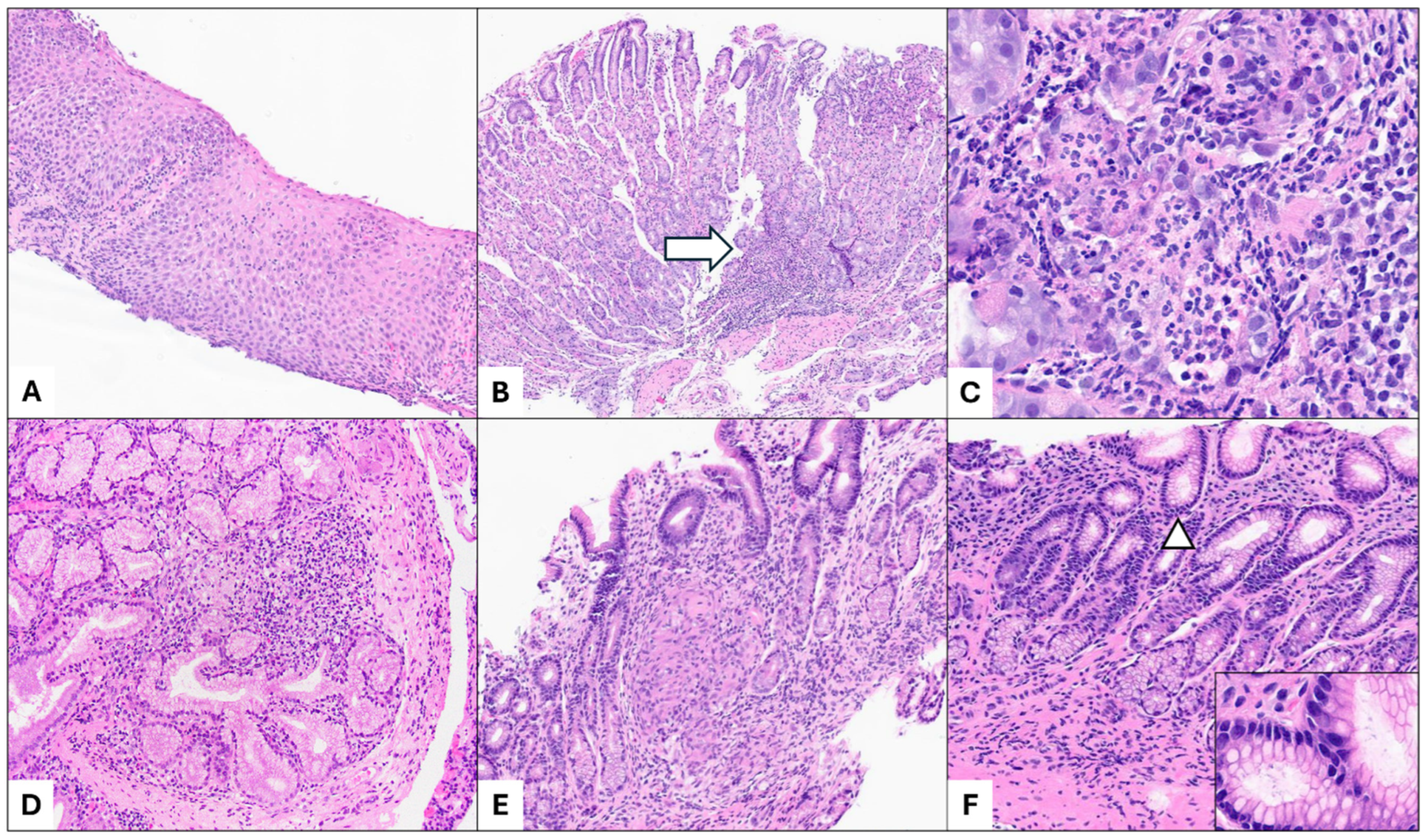
Figure 1. Upper gastrointestinal (GI) manifestations in pediatric inflammatory bowel disease (IBD) include (A) lymphocytic esophagitis, characterized by an increased number of intraepithelial lymphocytes with dilated intercellular spaces and parakeratosis (H&E, 4×). (B,C) Focally enhanced gastritis, marked by focal inflammation (white arrow) in the corpus mucosa, with neutrophilic infiltrates and associated epithelial injury (H&E, 4× and 20×). Notably, there is no significant background of chronic inflammation. (D) Brunner gland lobulitis in a patient with Crohn disease, showing focal acute inflammation involving mucosal Brunner glands (H&E, 10×). (E) Non-necrotizing granulomas, which most frequently affect the stomach in the upper GI tract, illustrated by antral mucosal involvement in a patient with Crohn disease (H&E, 10×). (F) Helicobacter infection should be ruled out in biopsies showing chronic active or inactive gastritis, as it may coexist with IBD (H&E, 10×). Microorganisms consistent with Helicobacter pylori are identified on H&E staining (white arrow and inset, H&E, 20×).
Diagnostics 15 00831 g001
Figure 2. Chronic granulomatous disease is characterized by (A) an epithelioid granuloma in the lamina propria of the colonic mucosa (H&E, 10×) and (B) pigmented macrophages (white arrows; H&E, 20×). There is no significant active inflammation or chronicity in this colonic mucosa.
Diagnostics 15 00831 g002
Table 1. Simplified items of PIBD classes proposed by Ledder et al. [11] (histologic criteria in bold italics). This table is reproduced from [11] with permission from Oxford University Press. It is not subject to the CC BY license of this article. For further use, please refer to the original publisher’s rights and permissions.
Class 1 (features incompatible with UC)
1. 
At least one well-formed granuloma anywhere in the GI tract (cryptolytic granulomas excluded);
2.
At least one of the following: deep ulcerations; cobblestoning; or stenosis anywhere in the small bowel or upper GI tract (stomach excluded);
3.
Fistulizing disease;
4.
Thickened jejunal or ileal bowel loops on imaging/significant small bowel inflammation on capsule endoscopy (backwash ileitis excluded);
5. 
Any ileal inflammation in the presence of normal cecum (incompatible with backwash ileitis);
Class 2 (features rarely found in UC)
6. 
Macro- and microscopically normal appearing skip lesions in untreated patients (rectal sparing and cecal patch excluded);
7. 
Complete rectal sparing (macroscopic and microscopic);
8. 
Macroscopically normal colon between the inflamed mucosa, but with microscopic inflammation (relative patchiness);
9.
Significant growth delay (height velocity < 2SD), not explained by other causes;
10. 
Transmural inflammation of the colon in the absence of severe colitis;
11.
Presence of any small and not deep ulcers in the small bowel and esophagus, or >5 small and not deep ulcers in the stomach or colon, with background normal mucosa, not explained by other causes;
12.
Positive ASCA in the presence of negative pANCA;
13. 
Reverse gradient of mucosal inflammation (proximal > distal; rectal sparing excluded);
14.
Deep ulcerations or severe cobblestoning of the stomach or scalloping of the duodenum, not explained by other causes;
Class 3 (features uncommon in UC)
15. 
Focal chronic duodenitis on histology;
16.
Focal active colitis on histology in more than one biopsy;
17.
Several (<5) aphthous ulcerations in the colon or in the stomach;
18.
Non-bloody diarrhea;
19. 
Focally enhanced gastritis on histology.
SD: standard deviation; ASCA: anti-Saccharomyces cerevisiae antibody; pANCA: perinuclear anti-neutrophil cytoplasmic antibody.
Table 2. Genotypes and phenotypes in monogenic forms of IBD.
Monogenic DisorderGenesIBD Type ReportedExtraintestinal Associations (in Order of Relative Frequency)
AgammaglobulinemiaBTK, PIK3CD, PIK3R1CD/CD-like, IBD, UC/UC-likeAutoimmunity, infection
Chronic enteropathy associated with SLCO2A1 gene SLCO2A1IBD, CD/CD-likeAutoimmunity
Chronic granulomatous disease CYBA, CYBB, NCF1, NCF2, NCF4Granulomatous colitis, IBD, CD/CD-like, UC/UC-like, IBD-U/ICAutoimmunity, infection, HLH, malignancy
CHAPLE syndromeCD55NDAutoimmunity, infection, malignancy
DOCK8 syndrome DOCK8UC/UC-like, CD/CD-like, IBDAutoimmunity, infection
Familial hemophagocytic lymphohistiocytosis 4 STXBP2NDAutoimmunity, infection, HLH
Glycogen storage disease type 1B SLC37A4IBD, CD/CD-likeInfection, hematolymphoid involvement, metabolic disease
Familial GUCY2C diarrhea syndrome GUCY2CCD/CD-like, IBD
G6PC3 deficiency G6PC3CD/CD-like, IBD Infection, dysmorphic features
Haploinsufficiency of A20TNFAIP3Intestinal BD, CD/CD-like, IBDAutoimmunity, infection
Hermansky-Pudlak syndrome HPS1, HPS4, HPS6CD/CD-like, IBD, UC/UC-likeAutoimmunity
Hoyeraal-Hreidarsson SyndromeDKC1, RTEL1UC/UC-like, CD/CD-likeDysmorphic features, infection
IL-10 and IL-10-receptor associated colitis IL-10, IL-10RA, IL-10RBIBD, CD/CD-like, IBD-U/IC Autoimmunity, infection, HLH
Immunodysregulation polyendocrinopathy enteropathy X-linked syndrome (IPEX)FOXP3IBD, CD/CD-like, UC/UC-like, IBD-U/ICAutoimmunity, infection
Immunodysregulation polyendocrinopathy enteropathy X-linked-like syndrome (IPEX-like)STAT1, STAT3, CTLA4, LRBA, IL21, IL2RA, IL2RB, MALT1IBD, CD/CD-like, UC/UC-like, IBD-UAutoimmunity, infection, malignancy, hematolymphoid involvement
Kindler syndrome FERMT1UC/UC-like, CD/CD-like
Leukocyte adhesion deficiency ITGB2NDInfection
Loeys-Dietz syndrome TGFBR1, TGFBR2UC/UC-like, CD/CD-like, IBDDysmorphic features, infection
Mevalonate kinase deficiency MVKNDAutoimmunity
Nuclear factor-kappa B Essential Modulator deficiency (NEMO)IKBKGIBD, CD/CD-like, intestinal BDDysmorphic features, infection
NLRC4-macrophage activating syndromeNLRC4NDAutoimmunity, infection, HLH
Niemann-Pick disease type C NPC1CD/CD-like, IBD-U/ICMetabolic disease
RIPK1 deficiencyRIPK1IBDAutoimmunity, infection
Severe combined immunodeficiency CD3G, DCLRE1C, IL2RG, LIG4, RAG1IBD, CD/CD-likeInfection
Trichohepatoenteric syndrome SKIV2L, TTC37IBD-U/IC, IBD, CD/CD-likeDysmorphic features, infection
TTC7A deficiencyTTC7AIBD, IBD-U/IC, CD/CD-likeAutoimmunity, infection, dysmorphic features, malignancy
TRIM22 deficiencyTRIM22NDAutoimmunity, infection
Wiskott-Aldrich syndrome/Wiskott-Aldrich-like syndrome (WAS/WAS-like)WAS, ARPC1BIBD, CD/CD-like, UC/UC-likeAutoimmunity, infection
XIAP deficiencyXIAPCD/CD-like, IBD, UC/UC-like, IBD-U/ICAutoimmunity, infection, HLH, hematolymphoid involvement, malignancy
CD: Crohn disease; IBD: inflammatory bowel disease; UC: ulcerative colitis; IBD-U: inflammatory bowel disease-unclassified; IC: indeterminate colitis; ND: not described; HLH: hemophagocytic lymphohistiocytosis.
Table 3. Histologic patterns and underlying causes of very-early-onset IBD.
PatternKey FindingsAssociated Monogenic DisordersOther Causes of Pattern
Chronic active enteritisActive inflammation (neutrophilic cryptitis and crypt abscesses)
Chronic mucosal changes (architectural changes and cellular metaplasia)
IL-10/R; CGD; XIAP; GUCY2C; familial Mediterranean fever; HPS types 1, 4, 6; GSD1; WAS; CVID; TTC7APolygenic IBD
Apoptosis/Epithelial injury patternEpithelial injury or apoptosis out of proportion to inflammatory activityDisorders resulting in B and T cell deficiency/dysregulation; SCID; Omenn syndrome; IPEX; TTC7A; dystrophic epidermolysis bullosa; mevalonate kinase deficiency; disorders of telomere maintenanceGvHD; antimetabolite immunosuppression, especially mycophenolate mofetil; acute GI transplant rejection; viral infection
Eosinophil-rich patternClusters of eosinophils in the lamina propria and eosinophilic infiltration of surface and crypt epitheliumOmenn syndromeNon-monogenic IBD; non-IBD etiologies for GI eosinophilia (e.g., allergy, parasitic infection, Langerhans cell histiocytosis)
Lymphocytic patternsLymphocytosis and nodular lymphoid hyperplasia
OR Lymphocyte or plasma cell depletion
AND Lack of active inflammation and chronic mucosal changes
Lymphocytosis: CVID; IgA deficiency; CTLA4-related disorders; T cell regulatory disorders; IPEX
Lymphocyte or plasma cell depletion: CVID; hypogammaglobulinemia; X-linked lymphoproliferative disorder type 1; SCID
Lymphocytosis: celiac disease; post-infectious enteritis (can be secondary to immunodeficiency), medication effect (especially NSAIDs); bacterial overgrowth
Lymphoid depletion: medical therapy (e.g., conditioning chemotherapy prior to bone marrow transplant); artifactual due to mucosal edema
Granulomatous patternNon-necrotizing granulomasCGD; IL-10/R; HPS types 1, 4, 6Misidentification of cryptolytic granuloma/germinal center; infectious granulomas (mycobacterial/fungal, usually necrotizing); or very rarely sarcoidosis
IL-10/R: IL-10 and IL-10-receptor associated colitis; CGD: chronic granulomatous disease; XIAP: X-linked inhibitor of apoptosis protein; GUCY2C: familial GUCY2C diarrhea syndrome; HPS: Hermansky-Pudlak syndrome; GSD1: glycogen storage disease type 1; WAS: Wiskott-Aldrich syndrome; CVID: common variable immune deficiency; SCID: severe combined immunodeficiency; IPEX: immunodysregulation polyendocrinopathy enteropathy X-linked syndrome; IBD: inflammatory bowel disease; GvHD: graft versus host disease; NSAIDs: non-steroidal anti-inflammatory drugs.
Disclaimer/Publisher’s Note: The statements, opinions and data contained in all publications are solely those of the individual author(s) and contributor(s) and not of MDPI and/or the editor(s). MDPI and/or the editor(s) disclaim responsibility for any injury to people or property resulting from any ideas, methods, instructions or products referred to in the content.