

Novel Automated Chemiluminescent Immunoassay for the Detection of Autoantibodies Against Aquaporin-4 in Neuromyelitis Optica Spectrum Disorders

Abstract
1. Introduction
2. Materials and Methods
2.1. Patients
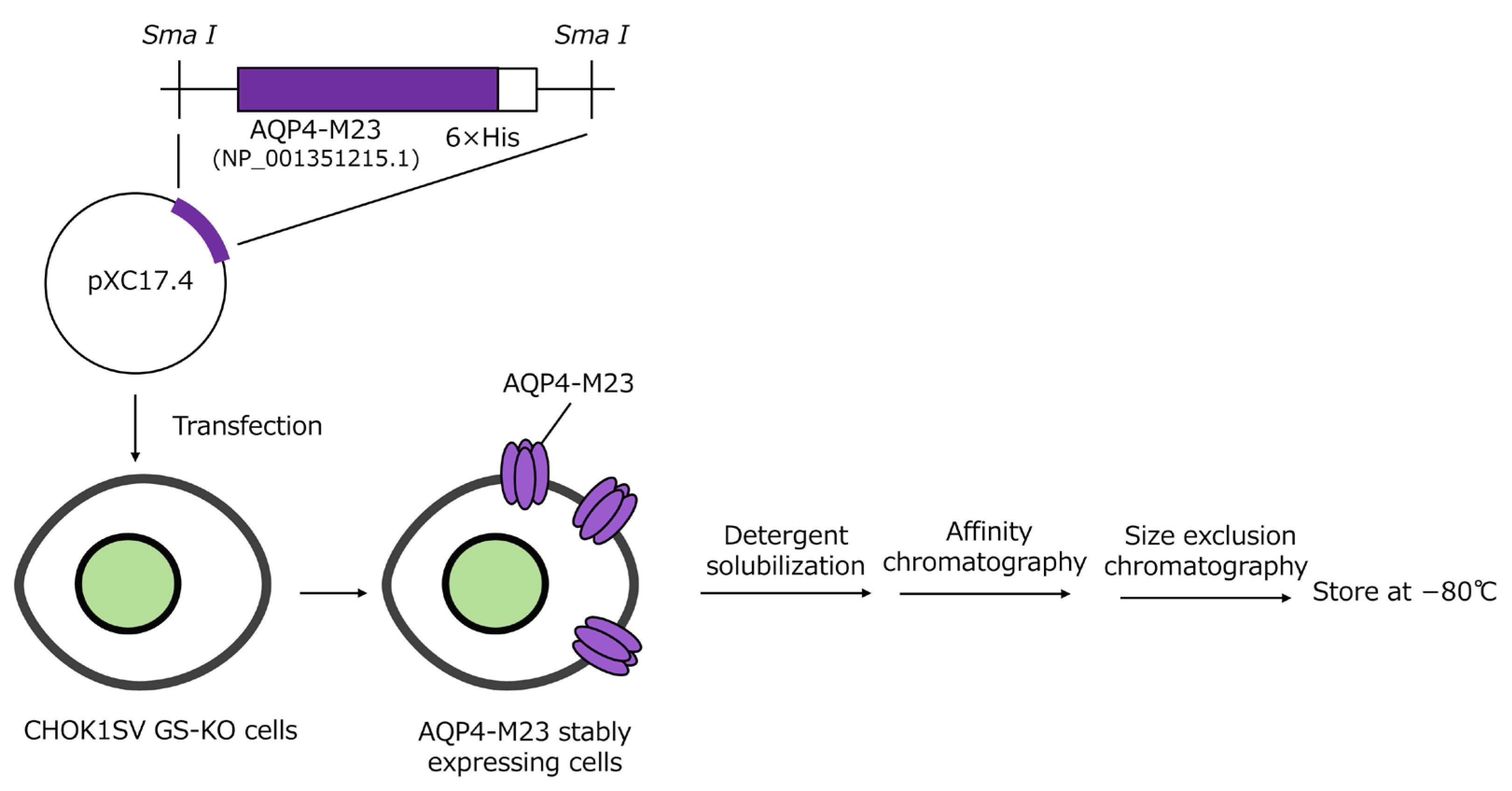
2.2. Generation of AQP4-M23 Stably Expressing Chinese Hamster Ovary Cell Line and Purification of AQP4-M23
2.3. Generation of Anti-AQP4 Monoclonal Antibody-Stable Expression Chinese Hamster Ovary Cell Line and Purification of Anti-AQP4 Monoclonal Antibody
2.4. AQP4-CLEIA
2.4.1. AQP4-CLEIA Assay Protocol
2.4.2. Calibrators for AQP4-CLEIA
2.5. Evaluation Methods for AQP4-CLEIA
2.5.1. Assay Precision
2.5.2. Limits of Quantification
2.5.3. Dilution Linearity
2.5.4. Hook Effect
2.5.5. Influence of Endogenous Substances
2.6. Statistical Analyses
3. Results
3.1. Analytical Performance of AQP4-CLEIA
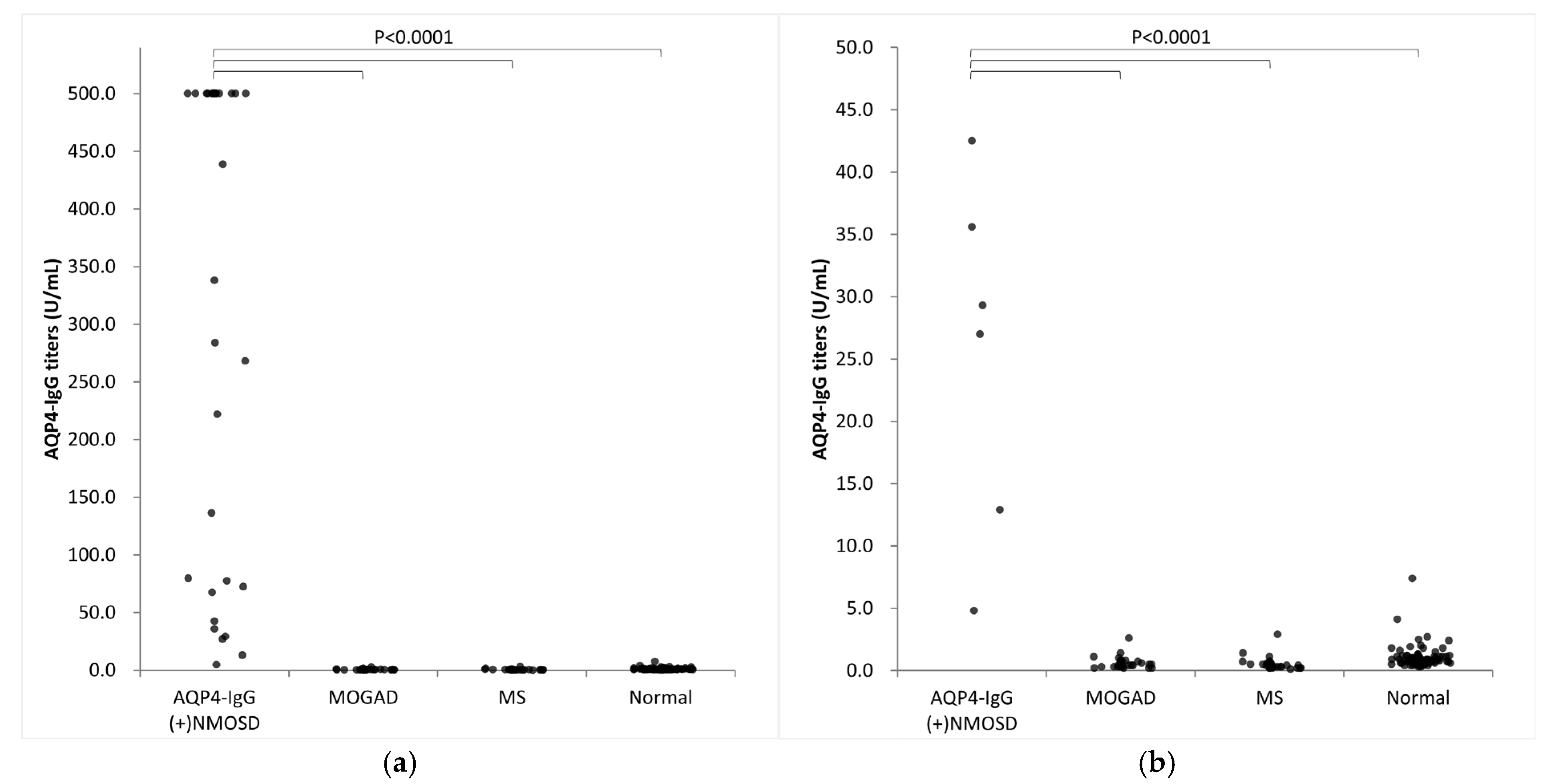
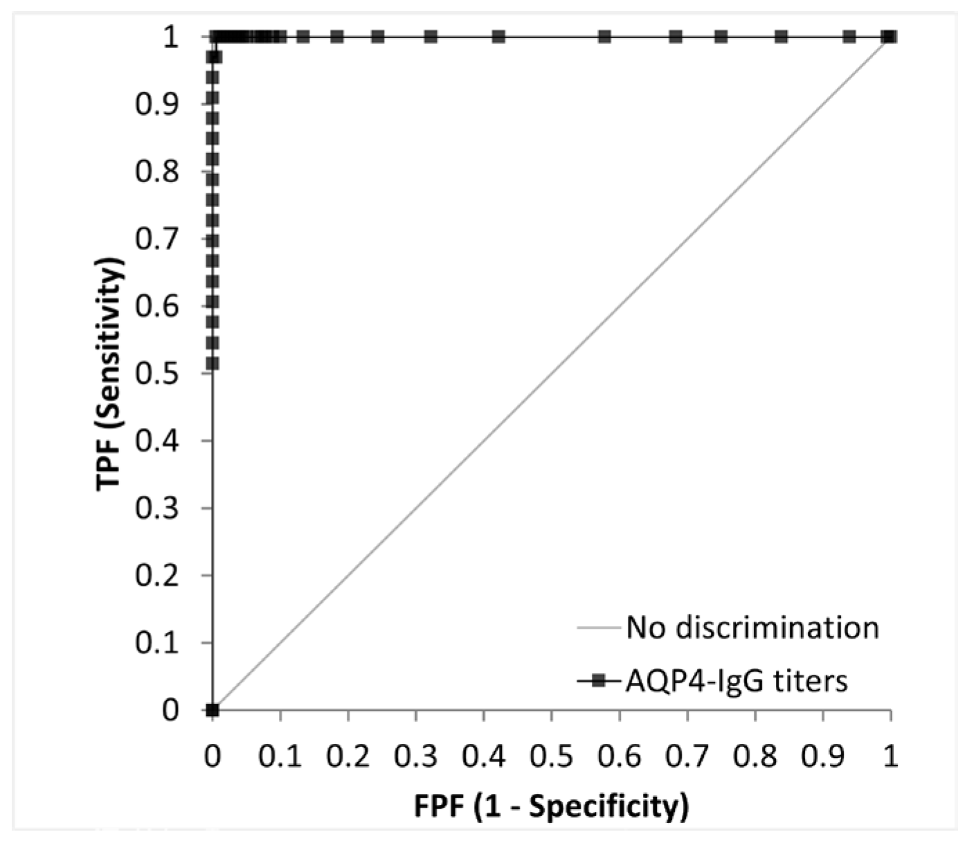
3.2. Clinical Validation in the Study Participants
4. Discussion
5. Conclusions
Author Contributions
Funding
Institutional Review Board Statement
Informed Consent Statement
Data Availability Statement
Acknowledgments
Conflicts of Interest
Abbreviations
| ALP | Alkaline phosphatase |
| AQP4-IgG | Aquaporin-4 immunoglobulin G antibodies |
| CBA | Cell-based assay |
| CI | Confidence interval |
| CLEIA | Chemiluminescent enzyme immunoassay |
| CLSI | Clinical and Laboratory Standards Institute |
| CNS | Central nervous system |
| ELISA | Enzyme-linked immunosorbent assay |
| LoQ | Limit of quantification |
| MOGAD | Myelin oligodendrocyte glycoprotein antibody-associated disease |
| MS | Multiple sclerosis |
| NMOSD | Neuromyelitis optica spectrum disorder |
| ROC | Receiver operating characteristic |
| SDS-PAGE | Sodium dodecyl sulfate–polyacrylamide electrophoresis |
| TE | Total error |
References
- Wingerchuk, D.M.; Banwell, B.; Bennett, J.L.; Cabre, P.; Carroll, W.; Chitnis, T.; de Seze, J.; Fujihara, K.; Greenberg, B.; Jacob, A.; et al. International Consensus Diagnostic Criteria for Neuromyelitis Optica Spectrum Disorders. Neurology 2015, 85, 177–189. [Google Scholar] [CrossRef] [PubMed]
- Papp, V.; Magyari, M.; Aktas, O.; Berger, T.; Broadley, S.A.; Cabre, P.; Jacob, A.; Kira, J.I.; Leite, M.I.; Marignier, R.; et al. Worldwide Incidence and Prevalence of Neuromyelitis Optica: A Systematic Review. Neurology 2021, 96, 59–77. [Google Scholar] [CrossRef] [PubMed]
- Hor, J.Y.; Asgari, N.; Nakashima, I.; Broadley, S.A.; Leite, M.I.; Kissani, N.; Jacob, A.; Marignier, R.; Weinshenker, B.G.; Paul, F.; et al. Epidemiology of Neuromyelitis Optica Spectrum Disorder and Its Prevalence and Incidence Worldwide. Front. Neurol. 2020, 11, 501. [Google Scholar] [CrossRef] [PubMed]
- Banwell, B.; Bennett, J.L.; Marignier, R.; Kim, H.J.; Brilot, F.; Flanagan, E.P.; Ramanathan, S.; Waters, P.; Tenembaum, S.; Graves, J.S.; et al. Diagnosis of Myelin Oligodendrocyte Glycoprotein Antibody-Associated Disease: International MOGAD Panel Proposed Criteria. Lancet Neurol. 2023, 22, 268–282. [Google Scholar] [CrossRef]
- Prain, K.; Woodhall, M.; Vincent, A.; Ramanathan, S.; Barnett, M.H.; Bundell, C.S.; Parratt, J.D.E.; Silvestrini, R.A.; Bukhari, W.; Australian and New Zealand NMO Collaboration; et al. AQP4 Antibody Assay Sensitivity Comparison in the Era of the 2015 Diagnostic Criteria for NMOSD. Front. Neurol. 2019, 10, 1028. [Google Scholar] [CrossRef]
- Kimbrough, D.J.; Fujihara, K.; Jacob, A.; Lana-Peixoto, M.A.; Leite, M.I.; Levy, M.; Marignier, R.; Nakashima, I.; Palace, J.; de Seze, J.; et al. Treatment of Neuromyelitis Optica: Review and Recommendations. Mult. Scler. Relat. Disord. 2012, 1, 180–187. [Google Scholar] [CrossRef]
- Shimizu, J.; Hatanaka, Y.; Hasegawa, M.; Iwata, A.; Sugimoto, I.; Date, H.; Goto, J.; Shimizu, T.; Takatsu, M.; Sakurai, Y.; et al. IFNβ-1b May Severely Exacerbate Japanese Optic-Spinal MS in Neuromyelitis Optica Spectrum. Neurology 2010, 75, 1423–1427. [Google Scholar] [CrossRef]
- Waters, P.J.; McKeon, A.; Leite, M.I.; Rajasekharan, S.; Lennon, V.A.; Villalobos, A.; Palace, J.; Mandrekar, J.N.; Vincent, A.; Bar-Or, A.; et al. Serologic Diagnosis of NMO: A Multicenter Comparison of aquaporin-4-IgG Assays. Neurology 2012, 78, 665–671. [Google Scholar] [CrossRef]
- Tampoia, M.; Abbracciavento, L.; Barberio, G.; Fabris, M.; Bizzaro, N.; Study Group on Autoimmune Diseases of the Italian Society of Clinical Pathology and Laboratory Medicine, Italy. A New M23-Based ELISA Assay for Anti-Aquaporin 4 Autoantibodies: Diagnostic Accuracy and Clinical Correlation. Auto Immun. Highlights 2019, 10, 5. [Google Scholar] [CrossRef]
- Cinquanta, L.; Fontana, D.E.; Bizzaro, N. Chemiluminescent Immunoassay Technology: What Does It Change in Autoantibody Detection? Auto Immun. Highlights 2017, 8, 9. [Google Scholar] [CrossRef]
- Thompson, A.J.; Banwell, B.L.; Barkhof, F.; Carroll, W.M.; Coetzee, T.; Comi, G.; Correale, J.; Fazekas, F.; Filippi, M.; Freedman, M.S.; et al. Diagnosis of Multiple Sclerosis: 2017 Revisions of the McDonald Criteria. Lancet Neurol. 2018, 17, 162–173. [Google Scholar] [CrossRef] [PubMed]
- Sato, D.K.; Nakashima, I.; Takahashi, T.; Misu, T.; Waters, P.; Kuroda, H.; Nishiyama, S.; Suzuki, C.; Takai, Y.; Fujihara, K.; et al. Aquaporin-4 Antibody-Positive Cases Beyond Current Diagnostic Criteria for NMO Spectrum Disorders. Neurology 2013, 80, 2210–2216. [Google Scholar] [CrossRef] [PubMed]
- Sato, D.K.; Callegaro, D.; Lana-Peixoto, M.A.; Waters, P.J.; de Haidar Jorge, F.M.; Takahashi, T.; Nakashima, I.; Apostolos-Pereira, S.L.; Talim, N.; Simm, R.F.; et al. Distinction Between MOG Antibody-Positive and AQP4 Antibody-Positive NMO Spectrum Disorders. Neurology 2014, 82, 474–481. [Google Scholar] [CrossRef] [PubMed]
- Werten, P.J.; Hasler, L.; Koenderink, J.B.; Klaassen, C.H.; de Grip, W.J.; Engel, A.; Deen, P.M. Large-Scale Purification of Functional Recombinant Human aquaporin-2. FEBS Lett. 2001, 504, 200–205. [Google Scholar] [CrossRef]
- Hiroaki, Y.; Tani, K.; Kamegawa, A.; Gyobu, N.; Nishikawa, K.; Suzuki, H.; Walz, T.; Sasaki, S.; Mitsuoka, K.; Kimura, K.; et al. Implications of the aquaporin-4 Structure on Array Formation and Cell Adhesion. J. Mol. Biol. 2006, 355, 628–639. [Google Scholar] [CrossRef]
- Miyazaki, K.; Abe, Y.; Iwanari, H.; Suzuki, Y.; Kikuchi, T.; Ito, T.; Kato, J.; Kusano-Arai, O.; Takahashi, T.; Nishiyama, S.; et al. Establishment of Monoclonal Antibodies Against the Extracellular Domain That Block Binding of NMO-IgG to AQP4. J. Neuroimmunol. 2013, 260, 107–116. [Google Scholar] [CrossRef]
- Westgard, J.O.; Carey, R.N.; Wold, S. Criteria for Judging Precision and Accuracy in Method Development and Evaluation. Clin. Chem. 1974, 20, 825–833. [Google Scholar] [CrossRef]
- Tanaka, R.; Takemura, M.; Sato, M.; Yamada, Y.; Nakagawa, T.; Horibe, T.; Hoshi, M.; Otaki, H.; Ito, H.; Seishima, M.; et al. Comparison of Chemiluminescence Enzyme Immunoassay (CLEIA) With ELISA for the Determination of Anti-cyclic Citrullinated Peptide Antibodies. Clin. Chim. Acta 2010, 411, 22–25. [Google Scholar] [CrossRef]
- Fujio, Y.; Kojima, K.; Hashiguchi, M.; Wakui, M.; Murata, M.; Amagai, M.; Yamagami, J. Validation of Chemiluminescent Enzyme Immunoassay in Detection of Autoantibodies in Pemphigus and Pemphigoid. J. Dermatol. Sci. 2017, 85, 208–215. [Google Scholar] [CrossRef]
- Waters, P.; Reindl, M.; Saiz, A.; Schanda, K.; Tuller, F.; Kral, V.; Nytrova, P.; Sobek, O.; Nielsen, H.H.; Barington, T.; et al. Multicentre Comparison of a Diagnostic Assay: Aquaporin-4 Antibodies in Neuromyelitis Optica. J. Neurol. Neurosurg. Psychiatry 2016, 87, 1005–1015. [Google Scholar] [CrossRef]
- Graus, F.; Titulaer, M.J.; Balu, R.; Benseler, S.; Bien, C.G.; Cellucci, T.; Cortese, I.; Dale, R.C.; Gelfand, J.M.; Geschwind, M.; et al. A Clinical Approach to Diagnosis of Autoimmune Encephalitis. Lancet Neurol. 2016, 15, 391–404. [Google Scholar] [CrossRef] [PubMed]
- Brooking, H.; Ananieva-Jordanova, R.; Arnold, C.; Amoroso, M.; Powell, M.; Betterle, C.; Zanchetta, R.; Furmaniak, J.; Smith, B.R. A Sensitive Non-isotopic Assay for GAD65 Autoantibodies. Clin. Chim. Acta 2003, 331, 55–59. [Google Scholar] [CrossRef] [PubMed]


| AQP4-IgG Titers of Samples | Precision | Intra-Assay Precision | Inter-Assay Precision | ||||
|---|---|---|---|---|---|---|---|
| Reagent Lot | Lot 1 | Lot 2 | Lot 3 | Lot 1 | Lot 2 | Lot 3 | |
| Low | Mean (U/mL) | 18.9 | 17.6 | 19.3 | 17.9 | 18.0 | 17.9 |
| SD (U/mL) | 0.74 | 0.94 | 0.93 | 0.97 | 0.98 | 1.04 | |
| CV | 3.9% | 5.3% | 4.8% | 5.4% | 5.4% | 5.8% | |
| Middle | Mean (U/mL) | 89.3 | 88.5 | 84.1 | 84.8 | 82.4 | 84.6 |
| SD (U/mL) | 5.50 | 2.62 | 2.10 | 6.24 | 5.70 | 6.48 | |
| CV | 6.2% | 3.0% | 2.5% | 7.4% | 6.9% | 7.7% | |
| High | Mean (U/mL) | 427.5 | 378.2 | 447.5 | 419.8 | 384.6 | 409.0 |
| SD (U/mL) | 11.73 | 20.27 | 12.04 | 22.90 | 18.96 | 30.39 | |
| CV | 2.7% | 5.4% | 2.7% | 5.5% | 4.9% | 7.4% | |
| Reagent Lot | Lot 1 | Lot 2 | |||||||||
|---|---|---|---|---|---|---|---|---|---|---|---|
| Sample No. | 1 | 2 | 3 | 4 | 5 | 1 | 2 | 3 | 4 | 5 | |
| Estimated value (U/mL) | 5.62 | 4.54 | 4.28 | 4.22 | 3.47 | 5.62 | 4.54 | 4.28 | 4.22 | 3.47 | |
| Day 1 | 1 | 5.73 | 4.77 | 4.92 | 4.30 | 3.73 | 5.28 | 4.92 | 4.82 | 4.32 | 3.58 |
| 2 | 6.06 | 4.69 | 4.82 | 4.28 | 3.41 | 6.12 | 5.06 | 4.82 | 4.45 | 3.22 | |
| 3 | 5.94 | 4.51 | 4.54 | 3.70 | 3.73 | 5.91 | 4.81 | 4.97 | 4.29 | 3.46 | |
| Day 2 | 1 | 6.26 | 4.50 | 4.72 | 4.35 | 2.98 | 5.16 | 4.82 | 5.20 | 4.95 | 4.01 |
| 2 | 6.15 | 4.12 | 4.76 | 4.06 | 3.67 | 5.04 | 4.69 | 5.08 | 4.08 | 3.49 | |
| 3 | 5.77 | 4.22 | 4.34 | 4.34 | 3.75 | 5.74 | 4.89 | 4.70 | 4.71 | 3.35 | |
| Day 3 | 1 | 4.75 | 3.94 | 4.42 | 3.75 | 3.14 | 6.02 | 4.55 | 4.48 | 4.20 | 3.38 |
| 2 | 5.67 | 4.10 | 4.56 | 4.15 | 3.18 | 5.98 | 4.86 | 4.64 | 3.58 | 3.14 | |
| 3 | 5.78 | 4.37 | 4.50 | 4.06 | 2.83 | 5.68 | 4.75 | 4.49 | 4.16 | 2.90 | |
| Mean (U/mL) | 5.79 | 4.36 | 4.62 | 4.11 | 3.38 | 5.66 | 4.82 | 4.80 | 4.30 | 3.39 | |
| SD (U/mL) | 0.44 | 0.28 | 0.19 | 0.25 | 0.36 | 0.40 | 0.15 | 0.25 | 0.39 | 0.31 | |
| Bias (U/mL) | 0.17 | −0.18 | 0.34 | −0.11 | −0.09 | 0.04 | 0.28 | 0.52 | 0.08 | −0.08 | |
| %TE | 18.68 | 16.48 | 17.01 | 14.22 | 23.23 | 15.00 | 12.49 | 23.88 | 20.43 | 20.12 | |
| %TE ≤ 20 | Pass | Pass | Pass | Pass | Fail | Pass | Pass | Fail | Fail | Fail | |
| Endogenous Substances | Concentration | % Difference from Control (95% CI) | ||
|---|---|---|---|---|
| AQP4-IgG Titers of Samples | ||||
| Low | Middle | High | ||
| Hemoglobin | 1089 mg/dL | −1.8% (−5.5, 1.8) | 3.9% (−1.6, 9.3) | −0.7% (−5.8, 4.4) |
| Free bilirubin | 46 mg/dL | −0.6% (−7.2, 6.0) | 0.9% (−2.8, 4.6) | −0.9% (−6.7, 4.9) |
| Conjugated bilirubin | 45 mg/dL | 4.1% (−0.9, 9.2) | 2.1% (−1.0, 5.3) | 5.6% (0.0, 11.3) |
| Chyle material | 1510 FTU | 1.9% (−4.1, 7.8) | −2.8% (−8.9, 3.3) | −0.7% (−5.8, 4.4) |
| Rheumatoid factor | 500 IU/mL | 3.6% (−1.4, 8.6) | 0.0% (−3.6, 3.6) | −0.5% (−3.7, 2.7) |
| Triglyceride | 2000 mg/dL | 0.0% (−4.8, 4.8) | 0.4% (−4.1, 4.8) | −1.0% (−4.1, 2.0) |
| Sample No. | AQP4-CBA | AQP4-CLEIA | Sample No. | AQP4-CBA | AQP4-CLEIA |
|---|---|---|---|---|---|
| (Titers) | (U/mL) | (Titers) | (U/mL) | ||
| 1 | 16 | 12.9 | 18 | 4096 | ≥500.0 |
| 2 | 64 | 4.8 | 19 | 8192 | ≥500.0 |
| 3 | 256 | 222.0 | 20 | 8192 | ≥500.0 |
| 4 | 256 | 136.3 | 21 | 8192 | ≥500.0 |
| 5 | 256 | 79.6 | 22 | 16,384 | ≥500.0 |
| 6 | 256 | 67.4 | 23 | 16,384 | ≥500.0 |
| 7 | 512 | 77.5 | 24 | 16,384 | ≥500.0 |
| 8 | 512 | 27.0 | 25 | 32,768 | ≥500.0 |
| 9 | 512 | 42.5 | 26 | 32,768 | ≥500.0 |
| 10 | 1024 | 35.6 | 27 | 32,768 | ≥500.0 |
| 11 | 1024 | 284.0 | 28 | 65,536 | ≥500.0 |
| 12 | 1024 | 72.3 | 29 | 65,536 | ≥500.0 |
| 13 | 1024 | 29.3 | 30 | 65,536 | ≥500.0 |
| 14 | 2048 | 438.6 | 31 | 65,536 | ≥500.0 |
| 15 | 4096 | 338.1 | 32 | 131,072 | ≥500.0 |
| 16 | 4096 | 268.1 | 33 | 131,072 | ≥500.0 |
| 17 | 4096 | ≥500.0 |
| Principle of Measurement | AQP4-CBA (Microscopic Live CBA) *1 | AQP4-CLEIA | AQP4-ELISA |
|---|---|---|---|
| Trade name | No commercial kit is available | Under consideration | RSR AQP4 Autoantibody ELISA Version 2 Kit |
| Manufacture | No commercial kit is available | Medical & Biological Laboratories Co., Ltd. | RSR Limited |
| Antigen | AQP4-M23 stably expressed HEK-293 cell lines *2 | Purified AQP4-M23 | Purified AQP4-M23 |
| Detection | Fluorochrome-labeled anti-human IgG pAb *2 | ALP-labeled anti-human IgG pAb | Biotinylated AQP4 and streptavidin peroxidase |
| Operation | Hand method | Fully automated | Hand method |
| Measurement time | Observer-dependent | 20 min | 3 h |
| Sample volume | Observer-dependent | 10 μL | 50 μL |
| Cutoff value | Observer-dependent | 10.0 U/mL | 3.0 U/mL |
| Measurement range | Observer-dependent | 4.6–500.0 U/mL | 1.5–80 U/mL, 1.5–40 U/mL *3 |
| Sensitivity | 92.0–100.0% [5,20] | 97.0% | 60.0–83.3% [5,9,20] |
| Specificity | 94.5–100.0% [5,20] | 100.0% | 96.9–100% [5,9,20] |
Disclaimer/Publisher’s Note: The statements, opinions and data contained in all publications are solely those of the individual author(s) and contributor(s) and not of MDPI and/or the editor(s). MDPI and/or the editor(s) disclaim responsibility for any injury to people or property resulting from any ideas, methods, instructions or products referred to in the content. |
© 2025 by the authors. Licensee MDPI, Basel, Switzerland. This article is an open access article distributed under the terms and conditions of the Creative Commons Attribution (CC BY) license (https://creativecommons.org/licenses/by/4.0/).
Share and Cite
Yamazaki, N.; Takahashi, T.; Misu, T.; Nishikawa, Y. Novel Automated Chemiluminescent Immunoassay for the Detection of Autoantibodies Against Aquaporin-4 in Neuromyelitis Optica Spectrum Disorders. Diagnostics 2025, 15, 298. https://doi.org/10.3390/diagnostics15030298
Yamazaki N, Takahashi T, Misu T, Nishikawa Y. Novel Automated Chemiluminescent Immunoassay for the Detection of Autoantibodies Against Aquaporin-4 in Neuromyelitis Optica Spectrum Disorders. Diagnostics. 2025; 15(3):298. https://doi.org/10.3390/diagnostics15030298
Chicago/Turabian StyleYamazaki, Nozomi, Toshiyuki Takahashi, Tatsuro Misu, and Yukihiro Nishikawa. 2025. "Novel Automated Chemiluminescent Immunoassay for the Detection of Autoantibodies Against Aquaporin-4 in Neuromyelitis Optica Spectrum Disorders" Diagnostics 15, no. 3: 298. https://doi.org/10.3390/diagnostics15030298
APA StyleYamazaki, N., Takahashi, T., Misu, T., & Nishikawa, Y. (2025). Novel Automated Chemiluminescent Immunoassay for the Detection of Autoantibodies Against Aquaporin-4 in Neuromyelitis Optica Spectrum Disorders. Diagnostics, 15(3), 298. https://doi.org/10.3390/diagnostics15030298