
Illuminating the Shadows: Innovation in Advanced Imaging Techniques for Myeloma Precursor Conditions

 , , , and
Abstract
1. Introduction
2. Role of Advanced Imaging in Myeloma Precursor Conditions
3. Whole-Body Low-Dose Computed Tomography for Myeloma Precursor Conditions
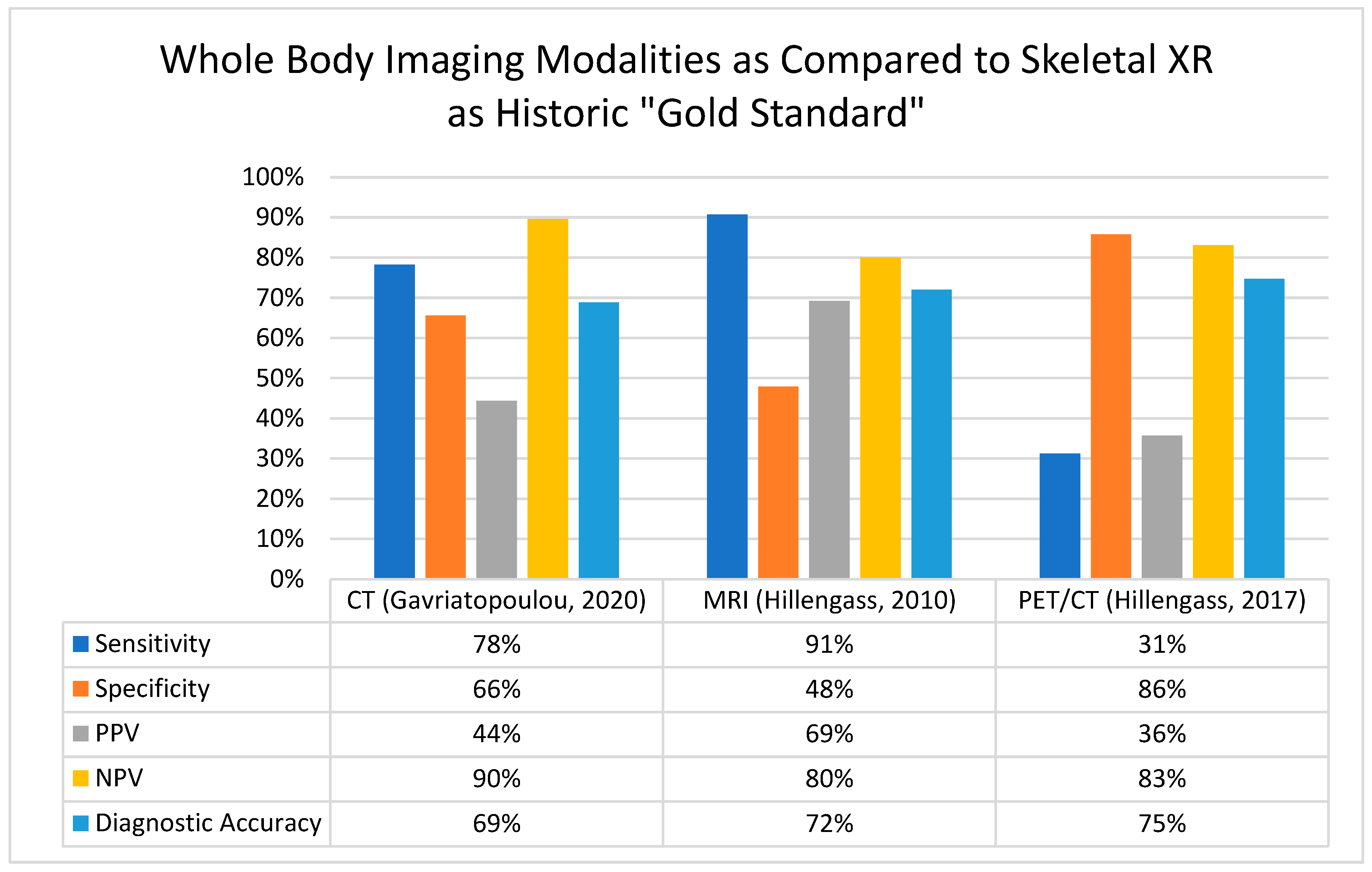
Summary—Whole-Body Low-Dose Computed Tomography for Myeloma Precursor Conditions
4. Whole-Body Magnetic Resonance Imaging for Myeloma Precursor Conditions
4.1. Focal Lesions on Whole-Body Magnetic Resonance Imaging
4.2. Focal Lesions: Volume, Kinetics, and Distribution
4.3. Diffuse Infiltrative Pattern on Whole-Body Magnetic Resonance Imaging
4.4. Summary—Whole-Body Magnetic Resonance Imaging for Myeloma Precursor Conditions
5. [18F]Fluorodeoxyglucose (FDG) Positron Emission Tomography–Computer Tomography Imaging for Myeloma Precursor Conditions
5.1. Patterns of Involvement on PET-CT: Diffuse vs. Focal, Without Osteolysis
5.2. The Role of Texture Analysis in FDG PET-CT Imaging of Smoldering Multiple Myeloma
5.3. Summary—PET-CT for Myeloma Precursor Conditions
6. Discussion
Author Contributions
Funding
Institutional Review Board Statement
Informed Consent Statement
Data Availability Statement
Conflicts of Interest
References
- Kristinsson, S.Y.; Rögnvaldsson, S.; Thorsteinsdottir, S.; Reed, E.R.; Oskarsson, J.T.T.; Petursdottir, I.; Sigurdardottir, G.A.; Vidarsson, B.; Onundarson, P.T.; Agnarsson, B.A.; et al. Screening for Monoclonal Gammopathy of Undetermined Significance: A Population-Based Randomized Clinical Trial. First Results from the Iceland Screens, Treats, or Prevents Multiple Myeloma (iStopMM) Study. Blood 2021, 138 (Suppl. S1), 156. [Google Scholar] [CrossRef]
- Thorsteinsdóttir, S.; Gíslason, G.K.; Aspelund, T.; Rögnvaldsson, S.; Óskarsson, J.Þ.; Sigurðardóttir, G.Á.; Þórðardóttir, Á.R.; Viðarsson, B.; Önundarson, P.T.; Agnarsson, B.A.; et al. Prevalence of smoldering multiple myeloma based on nationwide screening. Nat. Med. 2023, 29, 467–472. [Google Scholar] [CrossRef]
- Kyle, R.A.; Larson, D.R.; Therneau, T.M.; Dispenzieri, A.; Kumar, S.; Cerhan, J.R.; Rajkumar, S.V. Long-Term Follow-up of Monoclonal Gammopathy of Undetermined Significance. N. Engl. J. Med. 2018, 378, 241–249. [Google Scholar] [CrossRef]
- Kyle, R.A.; Remstein, E.D.; Therneau, T.M.; Dispenzieri, A.; Kurtin, P.J.; Hodnefield, J.M.; Larson, D.R.; Plevak, M.F.; Jelinek, D.F.; Fonseca, R.; et al. Clinical course and prognosis of smoldering (asymptomatic) multiple myeloma. N. Engl. J. Med. 2007, 356, 2582–2590. [Google Scholar] [CrossRef] [PubMed]
- Rajkumar, S.V.; Kyle, R.A.; Therneau, T.M.; Melton, L.J., 3rd; Bradwell, A.R.; Clark, R.J.; Larson, D.R.; Plevak, M.F.; Dispenzieri, A.; Katzmann, J.A. Serum free light chain ratio is an independent risk factor for progression in monoclonal gammopathy of undetermined significance. Blood 2005, 106, 812–817. [Google Scholar] [CrossRef]
- Lakshman, A.; Rajkumar, S.V.; Buadi, F.K.; Binder, M.; Gertz, M.A.; Lacy, M.Q.; Dispenzieri, A.; Dingli, D.; Fonder, A.L.; Hayman, S.R.; et al. Risk stratification of smoldering multiple myeloma incorporating revised IMWG diagnostic criteria. Blood Cancer J. 2018, 8, 59. [Google Scholar] [CrossRef] [PubMed]
- Mateos, M.V.; Kumar, S.; Dimopoulos, M.A.; González-Calle, V.; Kastritis, E.; Hajek, R.; De Larrea, C.F.; Morgan, G.J.; Merlini, G.; Goldschmidt, H.; et al. International Myeloma Working Group risk stratification model for smoldering multiple myeloma (SMM). Blood Cancer J. 2020, 10, 102. [Google Scholar] [CrossRef]
- Fernández de Larrea, C.; Isola, I.; Pereira, A.; Cibeira, M.T.; Magnano, L.; Tovar, N.; Rodríguez-Lobato, L.G.; Calvo, X.; Aróstegui, J.I.; Díaz, T.; et al. Evolving M-protein pattern in patients with smoldering multiple myeloma: Impact on early progression. Leukemia 2018, 32, 1427–1434. [Google Scholar] [CrossRef] [PubMed]
- Wu, V.; Moshier, E.; Leng, S.; Barlogie, B.; Cho, H.J.; Jagannath, S.; Madduri, D.; Mazumdar, M.; Parekh, S.; Chari, A. Risk stratification of smoldering multiple myeloma: Predictive value of free light chains and group-based trajectory modeling. Blood Adv. 2018, 2, 1470–1479. [Google Scholar] [CrossRef]
- Hill, E.; Mena, E.; Morrison, C.; Dew, A.; Choyke, P.; Lindenberg, L.; Kazandjian, D. Diagnostic performance of (18) F-FDG-PET/CT compared to standard skeletal survey for detecting bone destruction in smouldering multiple myeloma: Time to move forward. Br. J. Haematol. 2021, 193, 125–128. [Google Scholar] [CrossRef] [PubMed]
- Spinnato, P.; Filonzi, G.; Conficoni, A.; Facchini, G.; Ponti, F.; Sambri, A.; De Paolis, M.; Cavo, M.; Salizzoni, E.; Nanni, C. Skeletal Survey in Multiple Myeloma: Role of Imaging. Curr. Med. Imaging 2021, 17, 956–965. [Google Scholar] [CrossRef] [PubMed]
- Rajkumar, S.V.; Dimopoulos, M.A.; Palumbo, A.; Blade, J.; Merlini, G.; Mateos, M.V.; Kumar, S.; Hillengass, J.; Kastritis, E.; Richardson, P.; et al. International Myeloma Working Group updated criteria for the diagnosis of multiple myeloma. Lancet Oncol. 2014, 15, e538–e548. [Google Scholar] [CrossRef]
- Hillengass, J.; Moulopoulos, L.A.; Delorme, S.; Koutoulidis, V.; Mosebach, J.; Hielscher, T.; Drake, M.; Rajkumar, S.V.; Oestergaard, B.; Abildgaard, N.; et al. Whole-body computed tomography versus conventional skeletal survey in patients with multiple myeloma: A study of the International Myeloma Working Group. Blood Cancer J. 2017, 7, e599. [Google Scholar] [CrossRef]
- Simeone, F.J.; Harvey, J.P.; Yee, A.J.; O’Donnell, E.K.; Raje, N.S.; Torriani, M.; Bredella, M.A. Value of low-dose whole-body CT in the management of patients with multiple myeloma and precursor states. Skeletal Radiol. 2019, 48, 773–779. [Google Scholar] [CrossRef]
- Gavriatopoulou, M.; Βoultadaki, A.; Koutoulidis, V.; Ntanasis-Stathopoulos, I.; Bourgioti, C.; Malandrakis, P.; Fotiou, D.; Migkou, M.; Kanellias, N.; Eleutherakis-Papaiakovou, E.; et al. The Role of Low Dose Whole Body CT in the Detection of Progression of Patients with Smoldering Multiple Myeloma. Blood Cancer J. 2020, 10, 93. [Google Scholar] [CrossRef]
- Baffour, F.I.; Glazebrook, K.N.; Kumar, S.K.; Broski, S.M. Role of imaging in multiple myeloma. Am. J. Hematol. 2020, 95, 966–977. [Google Scholar] [CrossRef] [PubMed]
- Hillengass, J.; Usmani, S.; Rajkumar, S.V.; Durie, B.G.M.; Mateos, M.V.; Lonial, S.; Joao, C.; Anderson, K.C.; García-Sanz, R.; Riva, E.; et al. International myeloma working group consensus recommendations on imaging in monoclonal plasma cell disorders. Lancet Oncol. 2019, 20, e302–e312. [Google Scholar] [CrossRef]
- Moulopoulos, L.A.; Dimopoulos, M.A.; Smith, T.L.; Weber, D.M.; Delasalle, K.B.; Libshitz, H.I.; Alexanian, R. Prognostic significance of magnetic resonance imaging in patients with asymptomatic multiple myeloma. J. Clin. Oncol. 1995, 13, 251–256. [Google Scholar] [CrossRef] [PubMed]
- Florkow, M.C.; Willemsen, K.; Mascarenhas, V.V.; Oei, E.H.G.; van Stralen, M.; Seevinck, P.R. Magnetic Resonance Imaging Versus Computed Tomography for Three-Dimensional Bone Imaging of Musculoskeletal Pathologies: A Review. J. Magn. Reson. Imaging 2022, 56, 11–34. [Google Scholar] [CrossRef] [PubMed]
- Wang, J.; Zhang, B.; Zhang, R.; Zhang, L.; Jiang, W.; Jiang, Y. Role of whole-body diffusion-weighted imaging in evaluation of multiple myeloma. Medicine (Baltimore) 2021, 100, e27131. [Google Scholar] [CrossRef]
- Walker, R.; Barlogie, B.; Haessler, J.; Tricot, G.; Anaissie, E.; Shaughnessy, J.D., Jr.; Epstein, J.; van Hemert, R.; Erdem, E.; Hoering, A.; et al. Magnetic resonance imaging in multiple myeloma: Diagnostic and clinical implications. J. Clin. Oncol. 2007, 25, 1121–1128. [Google Scholar] [CrossRef]
- Hillengass, J.; Fechtner, K.; Weber, M.A.; Bäuerle, T.; Ayyaz, S.; Heiss, C.; Hielscher, T.; Moehler, T.M.; Egerer, G.; Neben, K.; et al. Prognostic significance of focal lesions in whole-body magnetic resonance imaging in patients with asymptomatic multiple myeloma. J. Clin. Oncol. 2010, 28, 1606–1610. [Google Scholar] [CrossRef] [PubMed]
- Dhodapkar, M.V.; Sexton, R.; Waheed, S.; Usmani, S.; Papanikolaou, X.; Nair, B.; Petty, N.; Shaughnessy, J.D., Jr.; Hoering, A.; Crowley, J.; et al. Clinical, genomic, and imaging predictors of myeloma progression from asymptomatic monoclonal gammopathies (SWOG S0120). Blood 2014, 123, 78–85. [Google Scholar] [CrossRef] [PubMed]
- Kastritis, E.; Moulopoulos, L.A.; Terpos, E.; Koutoulidis, V.; Dimopoulos, M.A. The prognostic importance of the presence of more than one focal lesion in spine MRI of patients with asymptomatic (smoldering) multiple myeloma. Leukemia 2014, 28, 2402–2403. [Google Scholar] [CrossRef] [PubMed]
- Wennmann, M.; Goldschmidt, H.; Mosebach, J.; Hielscher, T.; Bäuerle, T.; Komljenovic, D.; McCarthy, P.L.; Merz, M.; Schlemmer, H.P.; Raab, M.S.; et al. Whole-body magnetic resonance imaging plus serological follow-up for early identification of progression in smouldering myeloma patients to prevent development of end-organ damage. Br. J. Haematol. 2022, 199, 65–75. [Google Scholar] [CrossRef] [PubMed]
- Wennmann, M.; Kintzelé, L.; Piraud, M.; Menze, B.H.; Hielscher, T.; Hofmanninger, J.; Wagner, B.; Kauczor, H.U.; Merz, M.; Hillengass, J.; et al. Volumetry based biomarker speed of growth: Quantifying the change of total tumor volume in whole-body magnetic resonance imaging over time improves risk stratification of smoldering multiple myeloma patients. Oncotarget 2018, 9, 25254–25264. [Google Scholar] [CrossRef]
- Wennmann, M.; Hielscher, T.; Kintzelé, L.; Menze, B.H.; Langs, G.; Merz, M.; Sauer, S.; Kauczor, H.U.; Schlemmer, H.P.; Delorme, S.; et al. Analyzing Longitudinal wb-MRI Data and Clinical Course in a Cohort of Former Smoldering Multiple Myeloma Patients: Connections between MRI Findings and Clinical Progression Patterns. Cancers 2021, 13, 961. [Google Scholar] [CrossRef] [PubMed]
- Hildenbrand, N.; Klein, A.; Maier-Hein, K.; Wennmann, M.; Delorme, S.; Goldschmidt, H.; Hillengass, J. Identification of focal lesion characteristics in MRI which indicate presence of corresponding osteolytic lesion in CT in patients with multiple myeloma. Bone 2023, 175, 116857. [Google Scholar] [CrossRef] [PubMed]
- Wennmann, M.; Hielscher, T.; Kintzelé, L.; Menze, B.H.; Langs, G.; Merz, M.; Sauer, S.; Kauczor, H.U.; Schlemmer, H.P.; Delorme, S.; et al. Spatial Distribution of Focal Lesions in Whole-Body MRI and Influence of MRI Protocol on Staging in Patients with Smoldering Multiple Myeloma According to the New SLiM-CRAB-Criteria. Cancers 2020, 12, 2537. [Google Scholar] [CrossRef]
- Andrulis, M.; Bäuerle, T.; Goldschmidt, H.; Delorme, S.; Landgren, O.; Schirmacher, P.; Hillengass, J. Infiltration patterns in monoclonal plasma cell disorders: Correlation of magnetic resonance imaging with matched bone marrow histology. Eur. J. Radiol. 2014, 83, 970–974. [Google Scholar] [CrossRef]
- Hillengass, J.; Weber, M.A.; Kilk, K.; Listl, K.; Wagner-Gund, B.; Hillengass, M.; Hielscher, T.; Farid, A.; Neben, K.; Delorme, S.; et al. Prognostic significance of whole-body MRI in patients with monoclonal gammopathy of undetermined significance. Leukemia 2014, 28, 174–178. [Google Scholar] [CrossRef] [PubMed]
- Merz, M.; Hielscher, T.; Wagner, B.; Sauer, S.; Shah, S.; Raab, M.S.; Jauch, A.; Neben, K.; Hose, D.; Egerer, G.; et al. Predictive value of longitudinal whole-body magnetic resonance imaging in patients with smoldering multiple myeloma. Leukemia 2014, 28, 1902–1908. [Google Scholar] [CrossRef] [PubMed]
- Sun, M.; Cheng, J.; Ren, C.; Zhang, Y.; Li, Y.; Wang, L.; Zhang, S.; Lin, L. Evaluation of Diffuse Bone Marrow Infiltration Pattern in Monoclonal Plasma Cell Diseases by Quantitative Whole-body Magnetic Resonance Imaging. Acad. Radiol. 2022, 29, 490–500. [Google Scholar] [CrossRef]
- Siontis, B.; Kumar, S.; Dispenzieri, A.; Drake, M.T.; Lacy, M.Q.; Buadi, F.; Dingli, D.; Kapoor, P.; Gonsalves, W.; Gertz, M.A.; et al. Positron emission tomography-computed tomography in the diagnostic evaluation of smoldering multiple myeloma: Identification of patients needing therapy. Blood Cancer J. 2015, 5, e364. [Google Scholar] [CrossRef] [PubMed]
- Piñeiro-Fiel, M.; Moscoso, A.; Pubul, V.; Ruibal, Á.; Silva-Rodríguez, J.; Aguiar, P. A Systematic Review of PET Textural Analysis and Radiomics in Cancer. Diagnostics 2021, 11, 380. [Google Scholar] [CrossRef]
- Ghai, A.; Maji, D.; Cho, N.; Chanswangphuwana, C.; Rettig, M.; Shen, D.; DiPersio, J.; Akers, W.; Dehdashti, F.; Achilefu, S.; et al. Preclinical Development of CD38-Targeted [(89)Zr]Zr-DFO-Daratumumab for Imaging Multiple Myeloma. J. Nucl. Med. 2018, 59, 216–222. [Google Scholar] [CrossRef] [PubMed]
- Zamagni, E.; Nanni, C.; Gay, F.; Pezzi, A.; Patriarca, F.; Bellò, M.; Rambaldi, I.; Tacchetti, P.; Hillengass, J.; Gamberi, B.; et al. 18F-FDG PET/CT focal, but not osteolytic, lesions predict the progression of smoldering myeloma to active disease. Leukemia 2016, 30, 417–422. [Google Scholar] [CrossRef] [PubMed]
- Amini, B.; Nakache, Y.N.; Nardo, L.; Manasanch, E.E.; Sun, J.; Lenchik, L.; Boutin, R.D. Marrow uptake on FDG PET/CT is associated with progression from smoldering to symptomatic multiple myeloma. Skeletal Radiol. 2021, 50, 79–85. [Google Scholar] [CrossRef]
- Takahashi, M.E.S.; Mosci, C.; Souza, E.M.; Brunetto, S.Q.; Etchebehere, E.; Santos, A.O.; Camacho, M.R.; Miranda, E.; Lima, M.C.L.; Amorim, B.J.; et al. Proposal for a Quantitative (18)F-FDG PET/CT Metabolic Parameter to Assess the Intensity of Bone Involvement in Multiple Myeloma. Sci. Rep. 2019, 9, 16429. [Google Scholar] [CrossRef]
- Ripani, D.; Caldarella, C.; Za, T.; Rossi, E.; De Stefano, V.; Giordano, A. Progression to Symptomatic Multiple Myeloma Predicted by Texture Analysis-Derived Parameters in Patients Without Focal Disease at (18)F-FDG PET/CT. Clin. Lymphoma Myeloma Leuk. 2021, 21, 536–544. [Google Scholar] [CrossRef] [PubMed]
- Herrero Alvarez, N.; Michel, A.L.; Viray, T.D.; Mayerhoefer, M.E.; Lewis, J.S. (89)Zr-DFO-Isatuximab for CD38-Targeted ImmunoPET Imaging of Multiple Myeloma and Lymphomas. ACS Omega 2023, 8, 22486–22495. [Google Scholar] [CrossRef] [PubMed]
- Wei, W.; Zhang, Y.; Zhang, D.; Liu, Q.; An, S.; Chen, Y.; Huang, G.; Liu, J. Annotating BCMA Expression in Multiple Myelomas. Mol. Pharm. 2022, 19, 3492–3501. [Google Scholar] [CrossRef] [PubMed]

| Prognostic Indicator | Median TTP | 2-Year Progression Risk | Univariate HR (p-Value or 95%CI) | |
|---|---|---|---|---|
| MRI | Quantity of FL | |||
| >1 on whole body [21] | 1.1 years | 49.2% | 4.05 (<0.001) | |
| >1 on axial [23] | 1.3 years | |||
| >4 on whole body [28] | 80% | |||
| >3 on axial [28] | 80% | |||
| Total tumor volume of FL [25] | 1.48 (0.001) | |||
| TTV ≥ 7220 mm3 [24] | 80% | |||
| Speed of growth [25] | 1.99 (0.003) | |||
| Increase ≥ 114 mm3/mo [24] | 85.2% | |||
| Diffuse infiltration [21,25,34] | 1.77–3.14 (<0.001; 1.2–6.5) | |||
| PET/CT | Focal FDG uptake without osteolysis [34] | 1.1 years | 3.0 (1.6–5.7) | |
| SUV of L4 [35] | 1.698 (0.955–3.021) | |||
| Texture [36] | ||||
| Second order Femoral diaphysis | 1.1 years | |||
| Higher features L2–4 | 1.1 years |
| Low-risk MGUS | Imaging only as clinically indicated |
| High-risk MGUS | Low-dose whole-body CT for the baseline; thereafter, as clinically indicated or upon progression of myeloma labs |
| Low-risk SMM | Alternating DW MRI and PET/CT annually ×5 years from diagnosis or as clinically indicated; thereafter, as clinically indicated or upon progression of myeloma labs |
| High-risk SMM | Alternating DW MRI and PET/CT q6 mo ×5 years; thereafter, as clinically indicated or upon progression of myeloma labs or concern for evolving disease (e.g., declining hemoglobin, increasing M spike kinetics) [8,9] |
Disclaimer/Publisher’s Note: The statements, opinions and data contained in all publications are solely those of the individual author(s) and contributor(s) and not of MDPI and/or the editor(s). MDPI and/or the editor(s) disclaim responsibility for any injury to people or property resulting from any ideas, methods, instructions or products referred to in the content. |
© 2025 by the authors. Licensee MDPI, Basel, Switzerland. This article is an open access article distributed under the terms and conditions of the Creative Commons Attribution (CC BY) license (https://creativecommons.org/licenses/by/4.0/).
Share and Cite
Cicero, K.I.; Banerjee, R.; Kwok, M.; Dima, D.; Portuguese, A.J.; Chen, D.; Chalian, M.; Cowan, A.J. Illuminating the Shadows: Innovation in Advanced Imaging Techniques for Myeloma Precursor Conditions. Diagnostics 2025, 15, 215. https://doi.org/10.3390/diagnostics15020215
Cicero KI, Banerjee R, Kwok M, Dima D, Portuguese AJ, Chen D, Chalian M, Cowan AJ. Illuminating the Shadows: Innovation in Advanced Imaging Techniques for Myeloma Precursor Conditions. Diagnostics. 2025; 15(2):215. https://doi.org/10.3390/diagnostics15020215
Chicago/Turabian StyleCicero, Kara I., Rahul Banerjee, Mary Kwok, Danai Dima, Andrew J. Portuguese, Delphine Chen, Majid Chalian, and Andrew J. Cowan. 2025. "Illuminating the Shadows: Innovation in Advanced Imaging Techniques for Myeloma Precursor Conditions" Diagnostics 15, no. 2: 215. https://doi.org/10.3390/diagnostics15020215
APA StyleCicero, K. I., Banerjee, R., Kwok, M., Dima, D., Portuguese, A. J., Chen, D., Chalian, M., & Cowan, A. J. (2025). Illuminating the Shadows: Innovation in Advanced Imaging Techniques for Myeloma Precursor Conditions. Diagnostics, 15(2), 215. https://doi.org/10.3390/diagnostics15020215