The Predictive Value of Clinical Signs to Identify Shock in Critically Ill Patients

 , , and
Abstract
1. Introduction
2. Materials and Methods
2.1. Study Design and Ethical Approval
2.2. Study Setting and Database
2.3. Patient Population
2.4. Study Variables and Data Extraction
2.5. Definitions
2.6. Study Goals
2.7. Statistical Analysis
3. Results
3.1. Primary Study Goal
3.2. Secondary Study Goals
4. Discussion
4.1. Summary of Key Findings
4.2. Relationship with Previous Studies
4.3. Limitations
5. Conclusions
Supplementary Materials
Author Contributions
Funding
Institutional Review Board Statement
Informed Consent Statement
Data Availability Statement
Conflicts of Interest
Abbreviations
| CCRU | critical care resuscitation unit |
| ICU | intensive care unit |
| LR+ | positive likelihood ratio |
| LR− | negative likelihood ratio |
| SAPS II | Simplified Acute Physiology score II12 |
| SOFA | Sequential Organ Failure Assessment |
References
- Cecconi, M.; De Backer, D.; Antonelli, M.; Beale, R.; Bakker, J.; Hofer, C.; Jaeschke, R.; Mebazaa, A.; Pinsky, M.R.; Teboul, J.L.; et al. Consensus on circulatory shock and hemodynamic monitoring. Task force of the European Society of Intensive Care Medicine. Intensive Care Med. 2014, 40, 1795–1815. [Google Scholar] [CrossRef] [Scilit]
- Vincent, J.-L.; De Backer, D. Circulatory Shock. N. Engl. J. Med. 2013, 369, 1726–1734. [Google Scholar] [CrossRef] [Scilit]
- Singer, M.; Deutschman, C.S.; Seymour, C.W.; Shankar-Hari, M.; Annane, D.; Bauer, M.; Bellomo, R.; Bernard, G.R.; Chiche, J.-D.; Coopersmith, C.M.; et al. The Third International Consensus Definitions for Sepsis and Septic Shock (Sepsis-3). JAMA 2016, 315, 801. [Google Scholar] [CrossRef] [Scilit]
- Baran, D.A.; Grines, C.L.; Bailey, S.; Burkhoff, D.; Hall, S.A.; Henry, T.D.; Hollenberg, S.M.; Kapur, N.K.; O’Neill, W.; Ornato, J.P.; et al. SCAI Clinical Expert Consensus Statement on the Classification of Cardiogenic Shock: This Document Was Endorsed by the American College of Cardiology (ACC), the American Heart Association (AHA), the Society of Critical Care Medicine (SCCM), and the Society of Thoracic Surgeons (STS) in April 2019. Catheter. Cardiovasc. Interv. 2019, 94, 29–37. [Google Scholar] [CrossRef] [Scilit] [PubMed]
- Martin, J.T.; Alkhoury, F.; O’Connor, J.A.; Kyriakides, T.C.; Bonadies, J.A. ‘Normal’ vital signs belie occult hypoperfusion in geriatric trauma. Am. Surg. 2010, 76, 65–69. [Google Scholar] [CrossRef] [Scilit] [PubMed]
- Meregalli, A.; Oliveira, R.P.; Friedman, G. Occult hypoperfusion is associated with increased mortality in hemodynamically stable, high-risk, surgical patients. Crit. Care 2004, 8, R60. [Google Scholar] [CrossRef] [Scilit]
- Howell, M.D.; Donnino, M.; Clardy, P.; Talmor, D.; Shapiro, N.I. Occult hypoperfusion and mortality in patients with suspected infection. Intensive Care Med. 2007, 33, 1892–1899. [Google Scholar] [CrossRef] [Scilit]
- Lima, A.; Jansen, T.C.; van Bommel, J.; Ince, C.; Bakker, J. The prognostic value of the subjective assessment of peripheral perfusion in critically ill patients. Crit. Care Med. 2009, 37, 934–938. [Google Scholar] [CrossRef] [Scilit]
- Amson, H.; Vacheron, C.-H.; Thiolliere, F.; Piriou, V.; Magnin, M.; Allaouchiche, B. Core-to-skin temperature gradient measured by thermography predicts day-8 mortality in septic shock: A prospective observational study. J. Crit. Care 2020, 60, 294–299. [Google Scholar] [CrossRef] [Scilit] [PubMed]
- Mongkolpun, W.; Orbegozo, D.; Cordeiro, C.P.R.; Franco, C.J.C.S.; Vincent, J.-L.; Creteur, J. Alterations in Skin Blood Flow at the Fingertip Are Related to Mortality in Patients With Circulatory Shock. Crit. Care Med. 2020, 48, 443–450. [Google Scholar] [CrossRef] [Scilit]
- Walker, S.B.; Conlon, T.W.; Zhang, B.; Mensinger, J.L.; Fitzgerald, J.C.; Himebauch, A.S.; Glau, C.; Nishisaki, A.; Ranjit, S.; Nadkarni, V.; et al. Clinical Signs to Categorize Shock and Target Vasoactive Medications in Warm Versus Cold Pediatric Septic Shock. Pediatr. Crit. Care Med. 2020, 21, 1051–1058. [Google Scholar] [CrossRef] [Scilit]
- Kattan, E.; Hernández, G. The role of peripheral perfusion markers and lactate in septic shock resuscitation. J. Intensive Med. 2021, 2, 17–21. [Google Scholar] [CrossRef] [Scilit] [PubMed]
- Morin, A.; Missri, L.; Urbina, T.; Bonny, V.; Gasperment, M.; Bernier, J.; Baudel, J.L.; Kattan, E.; Maury, E.; Joffre, J.; et al. Relationship between skin microvascular blood flow and capillary refill time in critically ill patients. Crit. Care 2025, 29, 57. [Google Scholar] [CrossRef] [Scilit]
- Ait-Oufella, H.; Bige, N.; Boelle, P.Y.; Pichereau, C.; Alves, M.; Bertinchamp, R.; Baudel, J.L.; Galbois, A.; Maury, E.; Guidet, B. Capillary refill time exploration during septic shock. Intensive Care Med. 2014, 40, 958–964. [Google Scholar] [CrossRef] [Scilit]
- Hariri, G.; Joffre, J.; Leblanc, G.; Bonsey, M.; Lavillegrand, J.R.; Urbina, T.; Guidet, B.; Maury, E.; Bakker, J.; Ait-Oufella, H. Narrative review: Clinical assessment of peripheral tissue perfusion in septic shock. Ann. Intensive Care 2019, 9, 37. [Google Scholar] [CrossRef] [Scilit]
- Jacquet-Lagrèze, M.; Pernollet, A.; Kattan, E.; Ait-Oufella, H.; Chesnel, D.; Ruste, M.; Schweizer, R.; Allaouchiche, B.; Hernandez, G.; Fellahi, J.L. Prognostic value of capillary refill time in adult patients: A systematic review with meta-analysis. Crit. Care 2023, 27, 473. [Google Scholar] [CrossRef] [Scilit]
- Tschoellitsch, T.; Noitz, M.; Türk, M.; Meier, J.; Dünser, M.W. The value of clinical signs as indicators of shock. Intensive Care Med. 2023, 49, 1413–1415. [Google Scholar] [CrossRef] [Scilit]
- Hiemstra, B.; Eck, R.J.; Keus, F.; van der Horst, I.C.C. Clinical examination for diagnosing circulatory shock. Curr. Opin. Crit. Care 2017, 23, 293–301. [Google Scholar] [CrossRef] [Scilit]
- Hernández, G.; Ospina-Tascón, G.A.; Damiani, L.P.; Estenssoro, E.; Dubin, A.; Hurtado, J.; Friedman, G.; Castro, R.; Alegría, L.; Teboul, J.-L.; et al. Effect of a Resuscitation Strategy Targeting Peripheral Perfusion Status vs Serum Lactate Levels on 28-Day Mortality Among Patients With Septic Shock. JAMA 2019, 321, 654. [Google Scholar] [CrossRef] [Scilit] [PubMed]
- Ait-Oufella, H.; Lemoinne, S.; Boelle, P.Y.; Galbois, A.; Baudel, J.L.; Lemant, J.; Joffre, J.; Margetis, D.; Guidet, B.; Maury, E.; et al. Mottling score predicts survival in septic shock. Intensive Care Med. 2011, 37, 801–807. [Google Scholar] [CrossRef] [Scilit] [PubMed]
- Le Gall, J.R.; Lemeshow, S.; Saulnier, F. A new Simplified Acute Physiology Score (SAPS II) based on a European/North American multicenter study. JAMA 1993, 270, 2957–2963. [Google Scholar] [CrossRef] [Scilit]
- Moreno, R.; Vincent, J.-L.; Matos, R.; Mendonça, A.; Cantraine, F.; Thijs, L.; Takala, J.; Sprung, C.; Antonelli, M.; Bruining, H.; et al. The use of maximum SOFA score to quantify organ dysfunction/failure in intensive care. Results of a prospective, multicentre study. Intensive Care Med. 1999, 25, 686–696. [Google Scholar] [CrossRef] [Scilit] [PubMed]
- Koch, E.; Lovett, S.; Nghiem, T.; Riggs, R.A.; Rech, M.A. Shock index in the emergency department: Utility and limitations. Open Access Emerg. Med. 2019, 11, 179–199. [Google Scholar] [CrossRef] [Scilit] [PubMed]
- De Backer, D.; Biston, P.; Devriendt, J.; Madl, C.; Chochrad, D.; Aldecoa, C.; Brasseur, A.; Defrance, P.; Gottignies, P.; Vincent, J.-L. Comparison of dopamine and norepinephrine in the treatment of shock. N. Engl. J. Med. 2010, 362, 779–789. [Google Scholar] [CrossRef] [Scilit]
- Ait-Oufella, H.; Bourcier, S.; Alves, M.; Galbois, A.; Baudel, J.-L.; Margetis, D.; Bige, N.; Offenstadt, G.; Maury, E.; Guidet, B. Alteration of skin perfusion in mottling area during septic shock. Ann. Intensive Care 2013, 3, 31. [Google Scholar] [CrossRef] [Scilit]
- Brunauer, A.; Koköfer, A.; Bataar, O.; Gradwohl-Mathis, I.; Dankl, D.; Bakker, J.; Dünser, M.W. Changes in peripheral perfusion relate to visceral organ perfusion in early septic shock: A pilot study. J. Crit. Care 2016, 35, 105–109. [Google Scholar] [CrossRef] [Scilit]
- Filho, R.R.; de Freitas Chaves, R.C.; Assunção, M.S.C.; Neto, A.S.; De Freitas, F.M.; Romagnoli, M.L.; Silva, E.; Lattanzio, B.; Dubin, A.; Corrêa, T.D. Assessment of the peripheral microcirculation in patients with and without shock: A pilot study on different methods. J. Clin. Monit. Comput. 2020, 34, 1167–1176. [Google Scholar] [CrossRef] [Scilit]
- Vazquez, R.; Gheorghe, C.; Kaufman, D.; Manthous, C.A. Accuracy of bedside physical examination in distinguishing categories of shock: A pilot study. J. Hosp. Med. 2010, 5, 471–474. [Google Scholar] [CrossRef] [Scilit]
- Hernandez, G.; Pedreros, C.; Veas, E.; Bruhn, A.; Romero, C.; Rovegno, M.; Neira, R.; Bravo, S.; Castro, R.; Kattan, E.; et al. Evolution of peripheral vs metabolic perfusion parameters during septic shock resuscitation. A clinical-physiologic study. J. Crit. Care 2012, 27, 283–288. [Google Scholar] [CrossRef] [Scilit]
- Merdji, H.; Curtiaud, A.; Aheto, A.; Studer, A.; Harjola, V.P.; Monnier, A.; Duarte, K.; Girerd, N.; Kibler, M.; Ait-Oufella, H.; et al. Performance of Early Capillary Refill Time Measurement on Outcomes in Cardiogenic Shock: An Observational, Prospective Multicentric Study. Am. J. Respir. Crit. Care Med. 2022, 206, 1230–1238. [Google Scholar] [CrossRef] [Scilit] [PubMed]
- Huang, W.; Xiang, H.; Hu, C.; Wu, T.; Zhang, D.; Ma, S.; Hu, B.; Li, J. Association of Sublingual Microcirculation Parameters and Capillary Refill Time in the Early Phase of ICU Admission. Crit. Care Med. 2023, 51, 913–923. [Google Scholar] [CrossRef] [Scilit] [PubMed]
- Ferraris, A.; Bouisse, C.; Thiollière, F.; Piriou, V.; Allaouchiche, B. Mottling Incidence and Mottling Score According to Arterial Lactate Level in Septic Shock Patients. Indian J. Crit. Care Med. 2020, 24, 672–676. [Google Scholar] [CrossRef] [Scilit]
- Luo, J.C.; Luo, M.H.; Zhang, Y.J.; Liu, W.J.; Ma, G.G.; Hou, J.Y.; Su, Y.; Hao, G.W.; Tu, G.W.; Luo, Z. Skin mottling score assesses peripheral tissue hypoperfusion in critically ill patients following cardiac surgery. BMC Anesthesiol. 2024, 24, 130. [Google Scholar] [CrossRef] [Scilit]
- Merdji, H.; Bataille, V.; Curtiaud, A.; Bonello, L.; Roubille, F.; Levy, B.; Lim, P.; Schneider, F.; Khachab, H.; Dib, J.C.; et al. Mottling as a prognosis marker in cardiogenic shock. Ann. Intensive Care 2023, 13, 80. [Google Scholar] [CrossRef] [Scilit] [PubMed]
- Bourcier, S.; Pichereau, C.; Boelle, P.-Y.; Nemlaghi, S.; Dubée, V.; Lejour, G.; Baudel, J.-L.; Galbois, A.; Lavillegrand, J.-R.; Bigé, N.; et al. Toe-to-room temperature gradient correlates with tissue perfusion and predicts outcome in selected critically ill patients with severe infections. Ann. Intensive Care 2016, 6, 63. [Google Scholar] [CrossRef] [Scilit]
- Caputo, N.; Reilly, J.; Kanter, M.; West, J. A retrospective analysis of the respiratory adjusted shock index to determine the presence of occult shock in trauma patients. J. Trauma Acute Care Surg. 2018, 84, 674–678. [Google Scholar] [CrossRef] [Scilit] [PubMed]
- Jentzer, J.C.; van Diepen, S.; Barsness, G.W.; Henry, T.D.; Menon, V.; Rihal, C.S.; Naidu, S.S.; Baran, D.A. Cardiogenic Shock Classification to Predict Mortality in the Cardiac Intensive Care Unit. J. Am. Coll. Cardiol. 2019, 74, 2117–2128. [Google Scholar] [CrossRef] [Scilit]
- El-Menyar, A.; Al Habib, K.F.; Zubaid, M.; Alsheikh-Ali, A.A.; Sulaiman, K.; Almahmeed, W.; Amin, H.; AlMotarreb, A.; Ullah, A.; Suwaidi, J.A. Utility of shock index in 24,636 patients presenting with acute coronary syndrome. Eur. Heart J. Acute Cardiovasc. Care 2020, 9, 546–556. [Google Scholar] [CrossRef] [Scilit]
- Berger, T.; Green, J.; Horeczko, T.; Hagar, Y.; Garg, N.; Suarez, A.; Panacek, E.; Shapiro, N. Shock index and early recognition of sepsis in the emergency department: Pilot study. West J. Emerg. Med. 2013, 14, 168–174. [Google Scholar] [CrossRef] [Scilit]
- Baser, M.R.; Ganta, R.; Neeradi, C.; Varatharajan, S. Serum Lactate Levels and Their Correlation With Hospital Outcomes in ICU Patients With Shock: A Cross-Sectional Study at a Tertiary Care Center. Cureus 2025, 17, e86564. [Google Scholar] [CrossRef] [Scilit]
- Poloujadoff, M.-P.; Lapostolle, F.; Lockey, D.; Amathieu, R.; Merouani, M.; Galinski, M.; Adnet, F. Survival of severely shocked patients who present with absent radial pulse and unrecordable blood pressure in the pre-hospital phase. Resuscitation 2006, 69, 185–189. [Google Scholar] [CrossRef] [Scilit] [PubMed]



| All | Shock | No Shock | p-Value | ||
|---|---|---|---|---|---|
| N | 317 | 86 | 231 | ||
| Age | years | 68 (55–78) | 69 (63–81) | 68 (53–77) | 0.03 * |
| Male sex | n (%) | 193 (60.9%) | 56 (65.1%) | 137 (59.3%) | 0.42 |
| Body mass index | kg/m2 | 25.9 (22.5–31.2) | 25.3 (22.5–29.4) | 26.4 (22.7–32.1) | 0.23 |
| Comorbid conditions | |||||
| arterial hypertension | n (%) | 168 (53%) | 45 (52.3%) | 123 (53.2%) | 0.98 |
| diabetes mellitus | n (%) | 102 (32.2%) | 29 (33.7%) | 73 (31.6%) | 0.82 |
| chronic respiratory disease | n (%) | 93 (29.3%) | 14 (16.3%) | 79 (34.2%) | 0.003 * |
| coronary artery disease | n (%) | 80 (25.2%) | 26 (30.2%) | 54 (23.4%) | 0.27 |
| hypercholesterinaemia | n (%) | 78 (24.6%) | 27 (31.4%) | 51 (22.1%) | 0.12 |
| Source of admission | |||||
| emergency department | n (%) | 255 (80.4%) | 70 (81.4%) | 185 (80.1%) | 0.92 |
| hospital ward | n (%) | 62 (19.6%) | 16 (18.6%) | 46 (19.9%) | |
| Chief complaint | |||||
| respiratory failure | n (%) | 85 (26.8%) | 13 (15.1%) | 72 (31.2%) | 0.006 * |
| arterial hypotension | n (%) | 30 (9.5%) | 18 (20.9%) | 12 (5.2%) | <0.001 * |
| gastrointestinal bleeding | n (%) | 26 (8.2%) | 13 (15.1%) | 13 (5.6%) | 0.01 * |
| intoxication | n (%) | 24 (7.6%) | 0 (0%) | 24 (10.4%) | <0.001 * |
| sepsis/infection | n (%) | 24 (7.6%) | 12 (14.0%) | 14 (6.1%) | 0.04 * |
| neurological disorder | n (%) | 18 (5.7%) | 3 (3.5%) | 15 (6.5%) | 0.42 |
| heart failure/cardiogenic pulmonary edema | n (%) | 15 (4.7%) | 5 (5.8%) | 10 (4.3%) | 0.56 |
| arrhythmia | n (%) | 12 (3.8%) | 7 (8.1%) | 5 (2.2%) | 0.02 * |
| acute abdomen | n (%) | 10 (3.2%) | 7 (8.1%) | 3 (1.3%) | 0.005 * |
| diabetic ketoacidosis | n (%) | 8 (2.5%) | 2 (2.3%) | 6 (2.6%) | 1 |
| others | n (%) | 65 (20.5%) | 6 (7.0%) | 57 (24.7%) | <0.001 * |
| Shock criteria | |||||
| elevated lactate levels due to circulatory causes | n (%) | 82 (25.9%) | 82 (95.3%) | 0 (0%) | |
| need for vasopressor infusion | n (%) | 49 (15.5%) | 49 (57%) | 0 (0%) | |
| Type of shock | |||||
| hypovolaemic | n (%) | 44 (13.9%) | 44 (51.2%) | n.a. | |
| cardiogenic | n (%) | 26 (8.2%) | 26 (30.2%) | n.a. | |
| vasodilatory/distributive | n (%) | 11 (3.5%) | 11 (12.8%) | n.a. | |
| obstructive | n (%) | 5 (1.6%) | 5 (5.8%) | n.a. | |
| Critical care interventions | |||||
| vasopressor infusion | n (%) | 49 (15.5%) | 49 (57%) | 0 (0%) | <0.001 * |
| blood transfusion | n (%) | 25 (7.9%) | 13 (15.1%) | 12 (5.2%) | 0.007 * |
| non-invasive MV | n (%) | 90 (28.4%) | 16 (18.6%) | 74 (32%) | 0.03 * |
| invasive MV | n (%) | 42 (13.2%) | 20 (23.3%) | 22 (9.5%) | 0.003 * |
| SAPS II | points | 35 (31–39) | 36 (31–42) | 34 (30.8–38) | 0.26 |
| SOFA score | points | 4 (2–6) | 6 (3.3–9) | 3 (2–5) | <0.001 * |
| Need for emergency surgery | n (%) | 19 (6%) | 12 (14%) | 7 (3%) | <0.001 * |
| Disposition at CCRU discharge | |||||
| ICU | n (%) | 119 (37.5%) | 43 (50%) | 76 (32.9%) | 0.008 * |
| hospital ward | n (%) | 171 (53.9%) | 30 (34.9%) | 141 (61%) | <0.001 * |
| operating theatre | n (%) | 14 (4.4%) | 10 (11.6%) | 4 (1.7%) | <0.001 * |
| discharge | n (%) | 10 (3.2%) | 0 (0%) | 10 (4.3%) | 0.07 |
| died in CCRU | n (%) | 3 (0.9%) | 3 (3.5%) | 0 (0%) | 0.02 * |
| ICU length of stay (n = 119) | days | 5 (3–9) | 5 (3–9) | 5 (3–9) | 0.72 |
| Hospital length of stay | days | 10 (5–17) | 11 (5–18) | 9 (5–16) | 0.45 |
| 28-day mortality | n (%) | 48 (15.1%) | 24 (27.9%) | 24 (10.4%) | <0.001 * |
| All | Shock | No Shock | p-Value | ||
|---|---|---|---|---|---|
| n | 317 | 86 | 231 | ||
| Vital parameters | |||||
| heart rate | bpm | 102 (84–122) | 110 (86–138) | 100 (82–120) | 0.02 * |
| systolic arterial blood pressure | mmHg | 118 (90–150) | 90 (76–110) | 130 (104–160) | <0.001 * |
| mean arterial blood pressure | mmHg | 82 (67–102) | 63 (54–78) | 90 (73–107) | <0.001 * |
| plethysmographic SO2 | % | 94 (90–96) | 95 (91–97) | 94 (89–96) | 0.24 |
| body temperature | °C | 36.6 (36–37.2) | 36.5 (36–37.2) | 36.6 (36–37.2) | 0.4 |
| Laboratory results | |||||
| pH | 7.33 (7.22–7.4) | 7.3 (7.18–7.4) | 7.33 (7.25–7.4) | 0.26 | |
| base deficit | mmol/L | −2.4 (−7–0.8) | −6.5 (−11.3–−2.7) | −1.3 (−4.3–1.7) | <0.001 * |
| lactate | mmol/L | 2.1 (1.1–4) | 4.3 (2.8–6.5) | 1.3 (1–2.5) | <0.001 * |
| platelets | G/L | 245 (181–313) | 252 (179–312) | 242 (181–312) | 0.96 |
| creatinine | mg/dL | 1.13 (0.81–1.88) | 1.62 (1.07–2.38) | 1.04 (0.77–1.48) | <0.001 * |
| bilirubin | mg/dL | 0.5 (0.32–0.8) | 0.69 (0.42–1.09) | 0.43 (0.3–0.73) | <0.001 * |
| Clinical signs | |||||
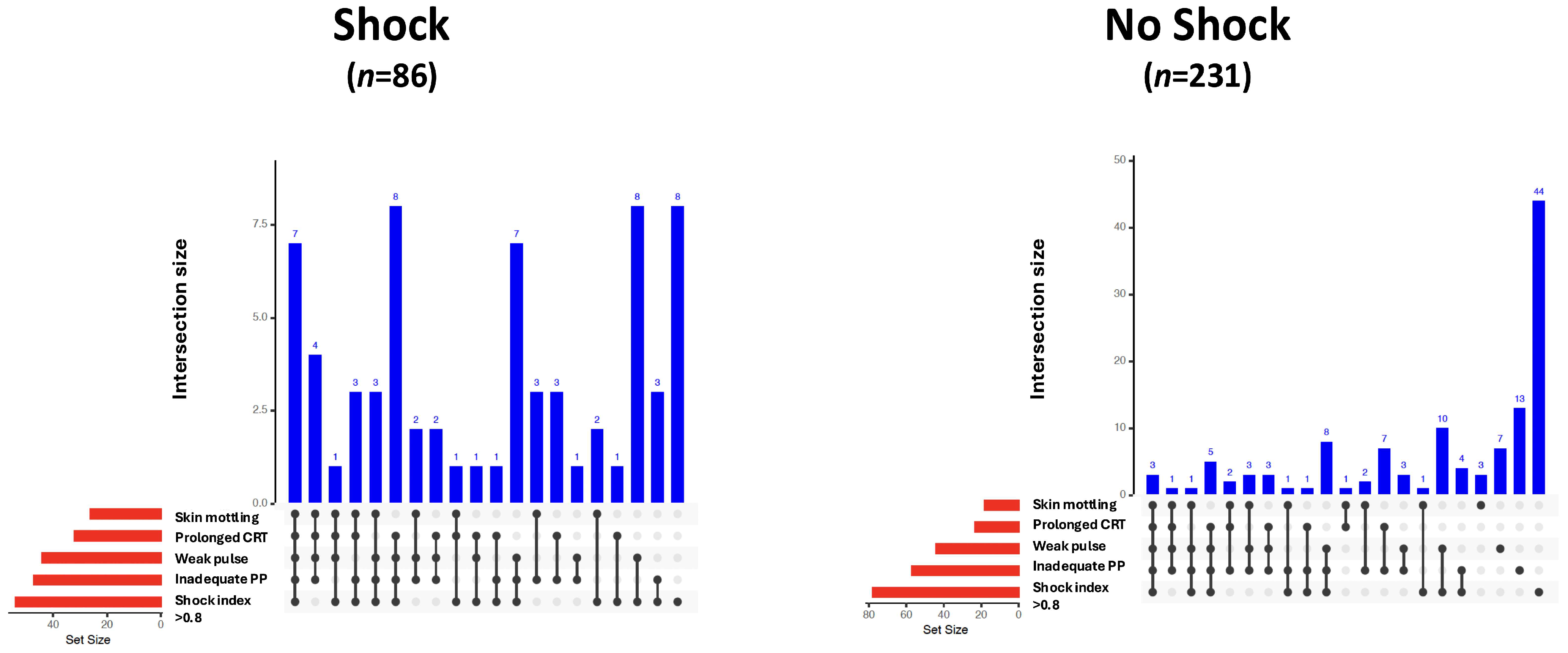
| tachycardia | n (%) | 155 (48.9%) | 49 (57%) | 106 (45.9%) | 0.1 |
| shock index > 0.8 | n (%) | 148 (46.7%) | 66 (76.7%) | 82 (35.5%) | <0.001 * |
| altered mental state | n (%) | 127 (41.2%) | 39 (47%) | 88 (39.1%) | 0.26 |
| inadequate peripheral perfusion | n (%) | 116 (38.3%) | 57 (67.9%) | 59 (26.9%) | <0.001 * |
| weak/thready radial pulse | n (%) | 95 (31.9%) | 48 (61.5%) | 47 (21.4%) | <0.001 * |
| prolonged capillary refill time | n (%) | 57 (19.4%) | 34 (44.2%) | 23 (10.6%) | <0.001 * |
| skin mottling | n (%) | 49 (16.2%) | 31 (37.8%) | 18 (8.2%) | <0.001 * |
| diaphoresis | n (%) | 29 (9.6%) | 10 (12.3%) | 19 (8.6%) | 0.46 |
| Predictive Value to Identify Shock | |||||||
| Sensitivity | Specificity | +LR | −LR | Accuracy | OR (95%CI) | p-Value | |
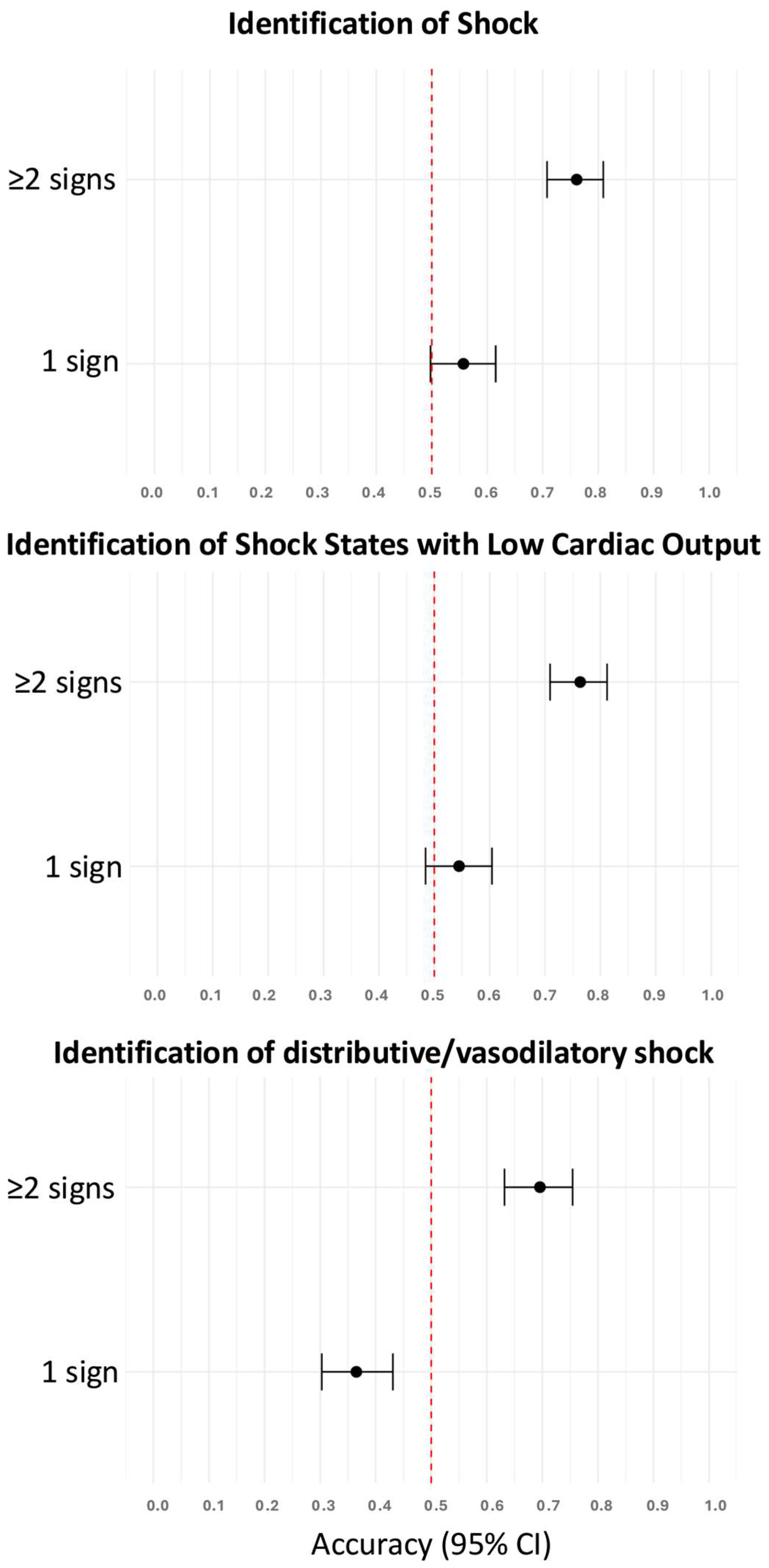
| Skin mottling | 0.38 | 0.92 | 4.62 | 0.68 | 0.77 | 6.82 (3.54–13.16) | <0.001 * |
| Prolonged capillary refill time | 0.44 | 0.89 | 4.17 | 0.62 | 0.78 | 6.67 (3.57–12.45) | <0.001 * |
| Shock index > 0.8 | 0.77 | 0.64 | 2.15 | 0.36 | 0.68 | 5.96 (3.37–10.52) | <0.001 * |
| Weak radial pulse | 0.62 | 0.79 | 2.88 | 0.49 | 0.74 | 5.89 (3.37–10.30) | <0.001 * |
| Inadequate peripheral perfusion | 0.68 | 0.73 | 2.52 | 0.44 | 0.72 | 5.73 (3.31–9.89) | <0.001 * |
| Tachycardia | 0.57 | 0.54 | 1.24 | 0.80 | 0.55 | 1.56 (0.95–2.57) | 0.10 |
| Diaphoresis | 0.12 | 0.91 | 1.43 | 0.96 | 0.70 | 1.49 (0.66–3.36) | 0.46 |
| Altered mental state | 0.47 | 0.61 | 1.20 | 0.87 | 0.57 | 1.38 (0.83–2.29) | 0.26 |
| Predictive Value to Identify Shock States with Low Cardiac Output | |||||||
| Sensitivity | Specificity | +LR | −LR | Accuracy | OR (95%CI) | p-Value | |
| Prolonged capillary refill time | 0.48 | 0.89 | 4.50 | 0.58 | 0.80 | 7.71 (4.04–14.71) | <0.001 * |
| Skin Mottling | 0.40 | 0.92 | 4.92 | 0.65 | 0.79 | 7.57 (3.86–14.85) | <0.001 * |
| Inadequate peripheral perfusion | 0.71 | 0.73 | 2.64 | 0.39 | 0.73 | 6.72 (3.73–12.09) | <0.001 * |
| Weak radial pulse | 0.63 | 0.79 | 2.93 | 0.47 | 0.75 | 6.18 (3.43–11.16) | <0.001 * |
| Shock index >0.8 | 0.76 | 0.64 | 2.13 | 0.37 | 0.67 | 5.72 (3.15–10.36) | <0.001 * |
| Altered mental state | 0.47 | 0.61 | 1.19 | 0.88 | 0.57 | 1.36 (0.8–2.31) | 0.32 |
| Tachycardia | 0.53 | 0.54 | 1.16 | 0.86 | 0.54 | 1.35 (0.8–2.27) | 0.32 |
| Diaphoresis | 0.11 | 0.91 | 1.30 | 0.97 | 0.72 | 1.34 (0.56–3.22) | 0.67 |
| Predictive Value to Identify Distributive/Vasodilatory Shock | |||||||
| Sensitivity | Specificity | +LR | −LR | Accuracy | OR (95%CI) | p-Value | |
| Shock index > 0.8 | 0.82 | 0.64 | 2.29 | 0.28 | 0.65 | 8.12 (1.71–38.49) | <0.01 * |
| Tachycardia | 0.82 | 0.54 | 1.78 | 0.34 | 0.55 | 5.31 (1.12–25.1) | 0.04 * |
| Weak radial pulse | 0.55 | 0.79 | 2.55 | 0.58 | 0.77 | 4.42 (1.29–15.11) | 0.03 * |
| Skin mottling | 0.20 | 0.92 | 2.44 | 0.87 | 0.89 | 2.81 (0.55–14.22) | 0.47 |
| Diaphoresis | 0.20 | 0.91 | 2.32 | 0.88 | 0.88 | 2.64 (0.52–13.36) | 0.51 |
| Inadequate peripheral perfusion | 0.45 | 0.73 | 1.69 | 0.75 | 0.72 | 2.26 (0.66–7.68) | 0.32 |
| Prolonged capillary refill time | 0.20 | 0.89 | 1.89 | 0.89 | 0.86 | 2.11 (0.42–10.54) | 0.68 |
| Altered mental state | 0.50 | 0.61 | 1.28 | 0.82 | 0.60 | 1.56 (0.44–5.53) | 0.72 |
Disclaimer/Publisher’s Note: The statements, opinions and data contained in all publications are solely those of the individual author(s) and contributor(s) and not of MDPI and/or the editor(s). MDPI and/or the editor(s) disclaim responsibility for any injury to people or property resulting from any ideas, methods, instructions or products referred to in the content. |
© 2025 by the authors. Licensee MDPI, Basel, Switzerland. This article is an open access article distributed under the terms and conditions of the Creative Commons Attribution (CC BY) license (https://creativecommons.org/licenses/by/4.0/).
Share and Cite
Noitz, M.; Preining, S.; Jenny, D.; Langthaler, S.; Erblich, R.; Tschoellitsch, T.; Meier, J.; Dünser, M.W. The Predictive Value of Clinical Signs to Identify Shock in Critically Ill Patients. Diagnostics 2025, 15, 2252. https://doi.org/10.3390/diagnostics15172252
Noitz M, Preining S, Jenny D, Langthaler S, Erblich R, Tschoellitsch T, Meier J, Dünser MW. The Predictive Value of Clinical Signs to Identify Shock in Critically Ill Patients. Diagnostics. 2025; 15(17):2252. https://doi.org/10.3390/diagnostics15172252
Chicago/Turabian StyleNoitz, Matthias, Sabine Preining, Dominik Jenny, Simon Langthaler, Romana Erblich, Thomas Tschoellitsch, Jens Meier, and Martin W. Dünser. 2025. "The Predictive Value of Clinical Signs to Identify Shock in Critically Ill Patients" Diagnostics 15, no. 17: 2252. https://doi.org/10.3390/diagnostics15172252
APA StyleNoitz, M., Preining, S., Jenny, D., Langthaler, S., Erblich, R., Tschoellitsch, T., Meier, J., & Dünser, M. W. (2025). The Predictive Value of Clinical Signs to Identify Shock in Critically Ill Patients. Diagnostics, 15(17), 2252. https://doi.org/10.3390/diagnostics15172252