Case Report

Beyond the Urogenital Tract, the Role of Ureaplasma parvum in Invasive Infection in Adults: A Case Series and Literature Review

Department of Infectious Disease, Peking University First Hospital, Beijing 100034, China
*
Author to whom correspondence should be addressed.
These authors contributed equally to this work.
Diagnostics 2025, 15(17), 2242; https://doi.org/10.3390/diagnostics15172242
Submission received: 31 July 2025 / Revised: 21 August 2025 / Accepted: 1 September 2025 / Published: 4 September 2025
(This article belongs to the Section Diagnostic Microbiology and Infectious Disease)

Abstract

Background/Objectives: Ureaplasma parvum (Up) is an opportunistic pathogen associated with urogenital tract infections, pregnancy complications, and reproductive system diseases. Advances in molecular diagnostics have expanded its pathogenic spectrum to include invasive conditions such as arthritis, meningitis, and pneumonia. However, the pathogenic significance of Up remains controversial. Methods: This study retrospectively analyzed nine adult cases of Up detected by metagenomic next-generation sequencing (mNGS) between 2023 and 2024. Results: Patients, aged 21 to 70 years, predominantly had underlying immunosuppressive conditions (66.7%). Infections involved the urinary system (44.4%), respiratory system (33.3%), and peritoneal cavity (22.2%). Symptomatic relief was achieved in five cases following treatment with tetracyclines, quinolones or tigecycline. Conclusions: These findings highlight Up as a potential causative agent of invasive infections, particularly in immunocompromised patients. Up has potential pathogenic significance, whether it is detected as a single pathogen or as a coexisting pathogen.

1. Introduction

Globally, Ureaplasma spp. colonizes 40–80% of asymptomatic American women, 8.9% of young Norwegians, and 20–47% of symptomatic patients in the United States, China, and Greece [1]. Ureaplasma parvum (Up), a member of the Family Mycoplasmataceae and the genus Ureaplasma spp., is characterized by its unique biological features: the absence of a cell wall, the dependence on host urease for metabolic activity, and the capacity for vertical transmission. Up typically functions as an opportunistic pathogen, frequently colonizing the mucosal surfaces of the urogenital tract [2]. Recent studies have established the significant pathogenicity of Up in pediatric infections and urogenital diseases in adults. In neonates, Up can cause pneumonia, meningitis, and bronchopulmonary dysplasia through vertical transmission from mother to child [3,4,5]. In women of childbearing age, Up colonization is closely associated with pregnancy complications such as spontaneous abortion, premature rupture of membranes, and preterm labor [6,7]. While in men, it has been related to urethritis and infertility [8,9].
With the widespread application of molecular diagnostic techniques in these years, such as metagenomic next-generation sequencing (mNGS), the detection of Up in adult infections has expanded beyond the urogenital system. Reports suggested that Up may be involved in invasive diseases such as septic arthritis, meningitis, pneumonia, and peritoneal infections [10,11,12,13]. However, most Up colonized individuals do not develop clinical disease. This has generated considerable debate regarding its pathogenic role in adult non-urogenital infections, specifically whether Up functions as a true pathogen or merely represents incidental detection of a commensal organism. Answering this question is crucial for precise clinical diagnosis and treatment. Based on this background, this study retrospectively analyzed adult infection cases detected Up by mNGS—from January 2023 to December 2024 at Peking University First Hospital, a national tertiary referral and academic medical center in Beijing, China. We systematically summarized the clinical characteristics, infection spectrum, and treatment outcomes of these cases. Relevant studies were also reviewed to explore the pathogenic significance of Up in adult non-genitourinary infections, as well as to reassess its pathogenic potential through molecular diagnostic techniques. By integrating multidimensional evidence, we aim to provide new insights into the clinical significance of Up.

2. Methods

This study employed a retrospective review of medical records to investigate the clinical characteristics, microbiological findings, and treatment outcomes of patients with Up detected by mNGS. This study was conducted in accordance with the Declaration of Helsinki and was approved by the Institutional Review Board (IRB) of Peking University First Hospital (Approval No. 2025-078-001), which waived the requirement for individual informed consent.

2.1. Patients Selection and Data Collection

This study included nine adult patients who tested positive for Up by mNGS between January 2023 and December 2024. The inclusion criteria were as follows: (1) positive mNGS results for Up; (2) age ≥ 18 years.
Demographic and clinical data were extracted from the electronic medical records, including age, gender, comorbidities, clinical symptoms, and infection site. Underlying conditions were categorized as immunosuppressive or non-immunosuppressive based on the presence of malignancies, diabetes mellitus, kidney injury, peritoneal dialysis, or obesity.

2.2. Specimen Collection and Next-Generation Sequencing

A total of 9 clinical specimens were analyzed using mNGS, including urine, blood, sputum, and bronchoalveolar lavage fluid (BALF) samples. Specimens were collected according to standard clinical protocols and processed for nucleic acid extraction. Library preparation was performed by using an NGS Automatic Library Preparation System (MatriDx Biotech Corp., Hangzhou, China). The quality of DNA was assessed using a BioAnalyzer 2100 (Agilent Technologies; Santa Clara, CA, USA) combined with quantitative PCR to measure the adapters before sequencing. Each sample generated a total of 0.1–1 million reads. A sequencing depth of 0.1–1 million reads per sample was adopted owing to host nucleic acid depletion, which reduces human-derived sequences by over two orders of magnitude. This enrichment of microbial reads improves pathogen detection, meets clinical diagnostic standards, and lowers sequencing demand, providing cost-effective benefits for routine application. Raw sequenced reads underwent quality control processing to eliminate short (length < 35 bp), low-quality, and low complexity reads, along with the sequencing adapters. Sequences from the host were excluded by aligning them to the human-specific database in NCBI (GRCh38.p13), utilizing Bowtie2 (version 2.3.5.1, https://sourceforge.net/projects/bowtie-bio/files/bowtie2/2.3.5.1/bowtie2-2.3.5.1-linux-x86_64.zip, accessed on 30 June 2025). Kraken2 (source code, https://github.com/DerrickWood/kraken2, accessed on 30 June 2025) was used for rapid classification, followed by secondary alignment with Bowtie2 to enhance classification accuracy. When inconsistencies arose between the results of Kraken2 and Bowtie2, the classification of reads was determined using BLAST (version 2.9.0, ftp://ftp.ncbi.nlm.nih.gov/blast/executables/blast+/2.9.0/ncbi-blast-2.9.0+-src.tar.gz, accessed on 30 June 2025). The numbers of reads per one million sequencing reads were calculated. Microbial reads from a library were reported if: (1) the sequencing data met specific quality control thresholds (library concentration > 1 pM, Q20 > 85%, Q30 > 80%); (2) the negative control in the same sequencing run either did not contain the species or the reads per one million sequencing reads ratio (sample to negative control) was ≥5, a cutoff established for differentiating true positives from background contaminates [14,15]. The positive control comprised a synthetic mixture of an RNA virus (influenza A), a DNA virus (Epstein–Barr virus), a bacterium (Klebsiella pneumoniae), and a fungus (Candida albicans), validating assay performance across pathogen classes. Reagent-derived contaminants were assessed with negative controls, and only taxa detected in samples but absent from controls were considered clinically relevant.
mNGS analyses were performed, with the aim of identifying microbial pathogens (bacteria, viruses, fungi, and other microorganisms) and quantifying their relative abundances and sequence number. The relative abundance and sequence number of Up was recorded for each positive sample, and co-existing microbial species were also documented.

2.3. Treatment and Outcome Assessment

Treatment strategies were determined based on the clinical judgment of the treating physicians, with some patients receiving targeted antibiotic therapy based on the mNGS results. The antibiotics used included tetracyclines and quinolones. Treatment outcomes were assessed based on the resolution of clinical symptoms and persistence of infections. Complete symptom resolution was defined as the absence of fever, pain, or other infection-related symptoms within 2 weeks of follow-up. Persistent infections were defined as unresolved symptoms or sustained positive microbiological findings following initial treatment.

2.4. Statistical Analysis

All data are presented individually in tabular form. The exact value for each patient is listed; values that required anonymization are provided as ranges. Figure was generated with the built-in charting tools in Microsoft Office.

3. Results

As shown in Figure 1A, among these cases, 8 patients were confirmed to have infections by clinicians, including 4 cases of urinary tract infection, 2 cases of pulmonary infection, 1 case of peritoneal dialysis-associated peritonitis, and 1 case of multisite infection involving both pulmonary and abdominal system. The remaining patient was diagnosed with fever of unknown origin. The patients ranged in age from 21 to 70 years, with 5 females (55.6%) and 4 males (44.4%). 6 patients (66.7%) had underlying immunosuppressive conditions, including malignancies, acute or chronic renal failure, diabetes mellitus, one undergoing peritoneal dialysis. A total of 9 clinical specimens were analyzed by mNGS, comprising 4 urine samples, 3 blood samples, 1 sputum sample, and 1 BALF sample (Figure 1B). The relative abundance of Up ranged from 0.03% to 31.56%. Co-existing microbial species and their relative abundances are detailed in Table 1.
Case 1 was a 70-year-old elderly woman hospitalized for “non-Hodgkin’s lymphoma”. During her hospital stay, she developed fever with a rash. Laboratory and imaging tests revealed no significant abnormalities. Up was detected in her blood sample by mNGS, with a relative abundance of 0.68%. Patient in case 2 was an overweight young man with a diagnosis of fever of unknown origin. His mNGS detected only Up, with a relative abundance of 0.81%. He did not revisit our hospital for fever within two weeks. Case 3 presented with dyspnea and chest pain. Laboratory results indicated a mild increase in white blood cells (WBC). The patient was also found to have mycobacterium tuberculosis complex, with a relative abundance of 2.48%, while the relative abundance of Up was 0.06%. Patient in case 5 was a young woman undergoing long-term peritoneal dialysis, with underlying diabetes and hypertension. She presented with abdominal pain, and laboratory tests showed a percentage of neutrophils (NE%) of 89.7% and C-reactive protein of 197.21 mg/L. Subsequently, mNGS was performed on her peripheral blood, which indicated an infection with Up and Cytomegalovirus (CMV), with a relative abundance of Up and CMV both at 7.14%. Case 8 involved a middle-aged man with a malignant retroperitoneal pheochromocytoma. His initial lab results showed significantly elevated WBC and NE%. mNGS detected Pseudomonas aeruginosa (PA, 77.04%) and Up (0.03%).
Regarding treatment and prognosis, 5 patients (Cases 4, 5, 6, 7, and 9) received antibiotics targeting Up (3 treated with minocycline, 1 with tigecycline and 1 with moxifloxacin), achieving complete symptom resolution. 1 patient (Case 2) tested positive for Up but did not receive treatment. Another patient (Case 3), who tested positive for mycobacterium tuberculosis and Up, received anti-tuberculosis treatment outside the hospital. The remaining 2 patients did not receive targeted anti-infective treatment. Among them, Case 1 had persistent fever for more than 2 weeks. Case 8 improved within 2 weeks after receiving treatment targeting PA.

4. Discussion

In this case series, Up was detected in all nine patients by mNGS, emphasizing the diverse clinical manifestations and infection sites that expand current understanding of its pathogenic characteristics. To differentiate true infection from colonization when Up was detected, infectious-disease specialists systematically weighed mNGS data against each patient’s clinical context and therapeutic response. There are four patients in this series presented with urinary tract infections. Notably, Up was also isolated from blood, sputum, and BALF samples in patients diagnosed with abdominal infections (22.2%, 2/9) and pneumonia (33.3%, 3/9). Although distinguishing colonization from true infection remains critical, these findings underscore Up’s potential to cause multisystem infections, a phenomenon supported by emerging evidence in the literature.
Up is well-established as a common colonizer of the urogenital tract, with its primary clinical association being urogenital infections. Noma et al. demonstrated that Up—when co-detected with high-risk human papillomavirus—significantly elevates the risk of low-grade squamous intraepithelial lesions in the cervix [16]. Furthermore, Up is an established pathogen of urethritis, which is associated with urethral inflammation severity and its capacity to disrupt sperm quality, contributing to impaired fertility [8,9,17]. Bacterial loads of Up contribute to the development of inflammatory responses in the male urethra. In our study, case 4, 5, 6, 7, and 9 were those who improved after targeted treatment. Among them, case 4, 6, 7, and 9 had urinary tract infections, and their symptoms and/or inflammatory indicators were alleviated within two weeks after targeted treatment for Up. Therefore, our study further affirms the pathogenicity of Up in the urogenital tract.
Beyond its urogenital tropism, a growing body of evidence has expanded the recognized pathogenic scope of Up. Previous studies have reported Up as a causative agent of purulent arthritis [10], central nervous system infections [11], and rare presentations such as hyperammonemia [18]. In our study, we identified four patients with non-urogenital tract infections. Among them, three patients were diagnosed with pulmonary infections. In case 1, pneumonia was suspected based on her clinical symptoms. However, no targeted anti-infective therapy was administered, and her fever persisted for 46 days before resolving. This raises the possibility that failure to target Up contributed to the prolonged febrile episode. In case 5, the patient was diagnosed with an abdominal infection and treated with tigecycline. Within two weeks, her C-reactive protein decreased from 197.21 to 47.07 mg/L, NE% dropped from 89.7% to 53.8%, and her abdominal pain was alleviated, suggesting that Up played a significant role in her peritonitis. Collectively, these observations support the hypothesis that Up may represent a pathogen of clinical significance.
However, at the same time, cases 2, 3, and 8 also remind us that not all patients with Up detected by mNGS have pathogenic significance. We need to consider its relative abundance and clinical manifestations. In case 3, the patient was diagnosed with a pulmonary infection, and mNGS detected both mycobacterium tuberculosis complex and Up. After two weeks of anti-tuberculosis treatment outside the hospital, the patient did not return for follow-up due to persistent fever. In case 8, mNGS detected PA and Up; clinically, his pneumonia and peritoneal infection were attributed to PA. He was treated with ceftazidime-avibactam, teicoplanin, and inhaled amikacin, resulting in a marked decrease in WBC count and NE% within two weeks. Based on these observations, we conclude that Up is unlikely to be pathogenically relevant in these two cases.
Although Up infections have been sporadically reported in immunocompetent individuals [19,20], immunocompromised populations (e.g., patients with malignancies or post-transplantation) exhibit heightened susceptibility [11,18], as evidenced by the 66.7% (6/9) prevalence of immunosuppression in this case series. More importantly, among the five patients in whom Up was detected and who received targeted treatment, three were immunocompromised. Notably, these individuals showed significant improvement in both symptoms and inflammatory markers following treatment. These findings underscore the potential of Up as a pathogen responsible for invasive infections, particularly in immunosuppressed populations. Clearly defining targeted therapeutic strategies for Up infection is therefore of critical importance in improving clinical outcomes for immunocompromised patients.
Previous reports have confirmed the pathogenicity of Up as a single pathogen [21], a finding corroborated by case 6 in our series, where symptomatic resolution followed moxifloxacin therapy targeting Up, the sole pathogen detected. Notably, Up was identified alongside Mycobacterium tuberculosis complex, CMV, and Pseudomonas aeruginosa in BALF, sputum, and peripheral blood samples in our study. This aligns with Rehman et al.’s report of Up coexisting with multidrug-resistant PA, Haemophilus influenzae, human coronaviruses, Mycoplasma pneumoniae, and CMV in respiratory samples [22]. Although the clinical dominance of Up in such polymicrobial infections remains unclear, the therapeutic response to Up-targeted antimicrobials in our cases 4, 5, 7, and 9 provides compelling evidence of its pathogenic contribution. Specifically, in cases 4, 5, and 9, the relative abundance of Up was found to be lower than or equal to that of the coexisting pathogens. This suggests that Up may still play a significant role in the infection, even in the presence of other coinfecting organisms.
The precise mechanisms driving Up infections remain incompletely elucidated but are hypothesized to involve dysregulated activation of innate immunity. Notably, Up membrane lipoproteins stimulate the NF-κB pathway through TLR2/6/9 receptors, triggering a pro-inflammatory cytokine storm characterized by elevated IL-6 and TNF-α [23]. This distinct immunostimulatory profile may explain Up’s tropism for TLR-rich tissues and its ability to invade the central nervous system post–blood–brain barrier compromise, though mechanisms in other systems warrant further exploration.
Diagnosing Up infections presents dual challenges: conventional culture frequently fails due to the organism’s fastidious growth requirements, while asymptomatic colonization complicates clinical differentiation. Advances in molecular diagnostics, particularly mNGS, have revolutionized the field by enabling rapid and highly sensitive detection of pathogens. In this series, mNGS identified Up in custom culture-negative specimens, underscoring the indispensable role of molecular methods. For sterile sites, detection of Up via mNGS strongly suggests pathogenicity. Conversely, in colonization-prone regions (e.g., respiratory tract), interpretation requires integration of bacterial load, host immunity, and clinical context.
Therapeutic management of Up infections is complicated by its lack of a cell wall and inability to synthesize folate, rendering β-lactams, glycopeptides, sulfonamides, and diaminopyrimidines ineffective. The treatment of choice comprises tetracyclines, macrolides, and fluoroquinolones; however, rising resistance rates to these classes are increasingly documented [24]. In this series, tetracyclines and fluoroquinolones were prioritized based on Up’s biological susceptibility profile, achieving satisfactory short-term clinical outcomes. Notably, in central nervous system infections, agents with blood–brain barrier penetration (e.g., doxycycline combined with moxifloxacin) are preferable, as evidenced by their use in resolving Up-associated brain abscess [25].
While this study is limited by its retrospective design, small cohort size, and absence of comprehensive antimicrobial susceptibility profiles, it offers novel insights into Up as an emerging multisystem pathogen and demonstrates the superior diagnostic sensitivity of mNGS. These findings provide actionable data to inform clinical decision-making, particularly in atypical infection contexts. To advance understanding, future multicenter studies should address three critical gaps: (1) establishing site-specific diagnostic thresholds to differentiate Up colonization from true infection; (2) clarifying mechanistic links between immunosuppression and invasive Up disease; and (3) formulating precision treatment guidelines informed by molecular susceptibility testing. In summary, the optimal management of Up hinges on the integration of patients’ clinical case records with personalized antimicrobial regimens, especially for immunocompromised hosts.

5. Conclusions

Drawing on nine well-documented cases of adult invasive infection and an exhaustive literature synthesis, we demonstrate that Up is no longer confined to its historical niche as a mere urogenital commensal. The organism can act alone or with co-pathogens to produce clinically significant disease—particularly in immunocompromised hosts. Yet fundamental questions remain; integrated studies are urgently required to reposition Up from an ambiguous colonizer to an unequivocally actionable invasive pathogen.

Author Contributions

Conceptualization: L.H. and X.L. (Xiangyan Li); data curation: L.H., D.L., J.Y. and X.L. (Xueying Li); formal analysis: L.H., D.L., J.Y. and X.L. (Xueying Li); supervision: L.H. and X.L. (Xiangyan Li); writing—original draft: L.H. and X.L. (Xiangyan Li); writing—review and editing: X.L. (Xiangyan Li) and Y.W. All authors have read and agreed to the published version of the manuscript.

Funding

This research received no external funding.

Institutional Review Board Statement

The study was conducted in accordance with the Declaration of Helsinki, and approved by the Institutional Review Board of Peking University First Hospital (protocol code 2025-078-001, approval date 27 February 2025).

Informed Consent Statement

Informed consent was waived because the study involved a retrospective review of de-identified clinical records that posed no more than minimal risk, and requiring participants to return solely to sign forms would have imposed an undue burden.

Data Availability Statement

The datasets used and/or analyzed during the current study are available from the corresponding author on reasonable request.

Acknowledgments

We are thankful to the patients for the support given in providing the data.

Conflicts of Interest

All authors declared no conflicts of interest with this work.

Abbreviations

UpUreaplasma parvum
NGSnext-generation sequencing
mNGSmetagenomic next-generation sequencing
IRBInstitutional Review Board
BALFbronchoalveolar lavage fluid
CMVCytomegalovirus
NEneutrophils
WBCwhite blood cells
PAPseudomonas aeruginosa

References

  1. Kotrotsiou, T.; Exindari, M.; Diza, E.; Gioula, G.; Melidou, A.; Kaplanis, K.; Malisiovas, N. Prevalence and antimicrobial susceptibility of Ureaplasma urealyticum in asymptomatic women in Northern Greece. Hippokratia 2013, 17, 319–321. [Google Scholar]
  2. Bartkeviciene, D.; Opolskiene, G.; Bartkeviciute, A.; Arlauskiene, A.; Lauzikiene, D.; Zakareviciene, J.; Ramasauskaite, D. The impact of Ureaplasma infections on pregnancy complications. Libyan J. Med. 2020, 15, 1812821. [Google Scholar] [CrossRef] [PubMed]
  3. Cassell, G.H.; Waites, K.B.; Watson, H.L.; Crouse, D.T.; Harasawa, R. Ureaplasma urealyticum intrauterine infection: Role in prematurity and disease in newborns. Clin. Microbiol. Rev. 1993, 6, 69–87. [Google Scholar] [CrossRef] [PubMed]
  4. Qin, L.; Li, Y.H.; Cao, X.J.; Wang, X.J.; Mao, R.P.; Yang, H.Y.; Li, L. Clinical metagenomic sequencing for rapid diagnosis of neonatal meningitis caused by Ureaplasma parvum: A case report. Medicine 2022, 101, e28662. [Google Scholar] [CrossRef] [PubMed]
  5. Sánchez, P.J.; Regan, J.A. Vertical transmission of Ureaplasma urealyticum in full term infants. Pediatr. Infect. Dis. J. 1987, 6, 825–828. [Google Scholar] [CrossRef] [PubMed]
  6. Gray, D.J.; Robinson, H.B.; Malone, J.; Thomson, R.B., Jr. Adverse outcome in pregnancy following amniotic fluid isolation of Ureaplasma urealyticum. Prenat. Diagn. 1992, 12, 111–117. [Google Scholar] [CrossRef]
  7. Perni, S.C.; Vardhana, S.; Korneeva, I.; Tuttle, S.L.; Paraskevas, L.R.; Chasen, S.T.; Kalish, R.B.; Witkin, S.S. Mycoplasma hominis and Ureaplasma urealyticum in midtrimester amniotic fluid: Association with amniotic fluid cytokine levels and pregnancy outcome. Am. J. Obstet. Gynecol. 2004, 191, 1382–1386. [Google Scholar] [CrossRef]
  8. Deguchi, T.; Shimada, Y.; Horie, K.; Mizutani, K.; Seike, K.; Tsuchiya, T.; Yokoi, S.; Yasuda, M.; Ito, S. Bacterial loads of Ureaplasma parvum contribute to the development of inflammatory responses in the male urethra. Int. J. STD AIDS 2015, 26, 1035–1039. [Google Scholar] [CrossRef]
  9. Ito, K.; Akai, K.; Nishiumi, F.; Nakura, Y.; Ning Wu, H.; Kurata, T.; Onodera, A.; Kawai, Y.; Kajiyama, S.; Yanagihara, I. Ability of Ureaplasma parvum to invade mouse sperm, fertilize eggs through infected sperm, and impair mouse sperm function and embryo development. F S Sci. 2021, 2, 13–23. [Google Scholar] [CrossRef]
  10. Pan, X.; Xu, J.; Pan, L.; Wang, C.; Qiu, J.; Huang, X.; Yan, C.; Mao, M. Hyperammonemia in a septic patient with Ureaplasma parvum arthritis: A case report. BMC Infect. Dis. 2022, 22, 958. [Google Scholar] [CrossRef]
  11. Pailhoriès, H.; Chenouard, R.; Eveillard, M.; Kempf, M.; Pereyre, S.; Bébéar, C.; Lemarié, C. A case of Ureaplasma parvum meningitis in an adult after transphenoidal ablation of craniopharyngioma. Int. J. Infect. Dis. 2019, 84, 5–7. [Google Scholar] [CrossRef]
  12. Tadera, K.; Kitagawa, H.; Kitano, H.; Hara, T.; Kashiyama, S.; Nomura, T.; Omori, K.; Shigemoto, N.; Yokozaki, M.; Ohge, H. Prevalence of Mycoplasma hominis, Ureaplasma urealyticum, and Ureaplasma parvum detection in urine and respiratory tract samples in Hiroshima, Japan. Heliyon 2023, 9, e14543. [Google Scholar] [CrossRef] [PubMed]
  13. Chen, L.; Nie, Z.; Zhao, Y.; Bao, B. Peritonitis-associated with peritoneal dialysis following Ureaplasma parvum infection: A case report and literature review. Indian J. Med. Microbiol. 2023, 45, 100410. [Google Scholar] [CrossRef]
  14. Liu, D.; Zhou, H.; Xu, T.; Yang, Q.; Mo, X.; Shi, D.; Ai, J.; Zhang, J.; Tao, Y.; Wen, D.; et al. Multicenter assessment of shotgun metagenomics for pathogen detection. eBioMedicine 2021, 74, 103649. [Google Scholar] [CrossRef] [PubMed]
  15. Du, J.; Zhang, J.; Zhang, D.; Zhou, Y.; Wu, P.; Ding, W.; Wang, J.; Ouyang, C.; Yang, Q. Background Filtering of Clinical Metagenomic Sequencing with a Library Concentration-Normalized Model. Microbiol. Spectr. 2022, 10, e0177922. [Google Scholar] [CrossRef] [PubMed]
  16. Noma, I.H.Y.; Shinobu-Mesquita, C.S.; Suehiro, T.T.; Morelli, F.; De Souza, M.V.F.; Damke, E.; Da Silva, V.R.S.; Consolaro, M.E.L. Association of Righ-Risk Human Papillomavirus and Ureaplasma parvum Co-Infections with Increased Risk of Low-Grade Squamous Intraepithelial Cervical Lesions. Asian Pac. J. Cancer Prev. 2021, 22, 1239–1246. [Google Scholar] [CrossRef]
  17. Galdiero, M.; Trotta, C.; Schettino, M.T.; Cirillo, L.; Sasso, F.P.; Petrillo, F.; Petrillo, A. Normospermic Patients Infected with Ureaplasma parvum: Role of Dysregulated miR-122-5p, miR-34c-5, and miR-141-3p. Pathog. Immun. 2023, 8, 16–36. [Google Scholar] [CrossRef]
  18. Wu, J.; Hu, Y. A late-onset hyperammonemia syndrome caused by Ureaplasma parvum infection after kidney transplantation. Heliyon 2024, 10, e32134. [Google Scholar] [CrossRef]
  19. Asif, A.A.; Roy, M.; Ahmad, S. Rare case of Ureaplasma parvum septic arthritis in an immunocompetent patient. BMJ Case Rep. 2020, 13, e236396. [Google Scholar] [CrossRef]
  20. Suknuntha, K.; Lepak, A.J.; Rehrauer, W.M.; Chen, D.J. Identification of Ureaplasma parvum as a Cause of Culture-Negative Septic Monoarthitis Using 16S rRNA Gene Sequencing. Infect. Dis. Clin. Pract. 2019, 27, e12–e14. [Google Scholar] [CrossRef]
  21. Siles-Guerrero, V.; Cardona-Benavides, I.; Liébana-Martos, C.; Vázquez-Alonso, F.; Expósito-Ruiz, M.; Navarro-Marí, J.M.; Gutiérrez-Fernández, J. Recent clinical relevance of mono-genital colonization/infection by Ureaplasma parvum. Eur. J. Clin. Microbiol. Infect. Dis. 2020, 39, 1899–1905. [Google Scholar] [CrossRef] [PubMed]
  22. Rehman, S.U.; Day, J.; Afshar, B.; Rowlands, R.S.; Billam, H.; Joseph, A.; Guiver, M.; Maddocks, S.E.; Chalker, V.J.; Beeton, M.L. Molecular exploration for Mycoplasma amphoriforme, Mycoplasma fermentans and Ureaplasma spp. in patient samples previously investigated for Mycoplasma pneumoniae infection. Clin. Microbiol. Infect. 2021, 27, e1691–e1697. [Google Scholar] [CrossRef] [PubMed]
  23. Triantafilou, M.; De Glanville, B.; Aboklaish, A.F.; Spiller, O.B.; Kotecha, S.; Triantafilou, K. Synergic activation of toll-like receptor (TLR) 2/6 and 9 in response to Ureaplasma parvum & urealyticum in human amniotic epithelial cells. PLoS ONE 2013, 8, e61199. [Google Scholar] [CrossRef]
  24. Biernat-Sudolska, M.; Rojek-Zakrzewska, D.; Drożdż, K.; Bilska-Wilkosz, A. Antimicrobial Activity of N,N-Diethyldithiocarbamate against Ureaplasma parvum and Ureaplasma urealyticum. Int. J. Mol. Sci. 2023, 25, 40. [Google Scholar] [CrossRef]
  25. Madlener, M.; Breuninger, M.; Meißner, A.; Stetefeld, H.; Telentschak, S.; Wille, T.; van Eimeren, T.; Jung, N. Brain abscess with Ureaplasma parvum in a patient with granulomatosis with polyangiitis. Infection 2023, 51, 779–782. [Google Scholar] [CrossRef] [PubMed]
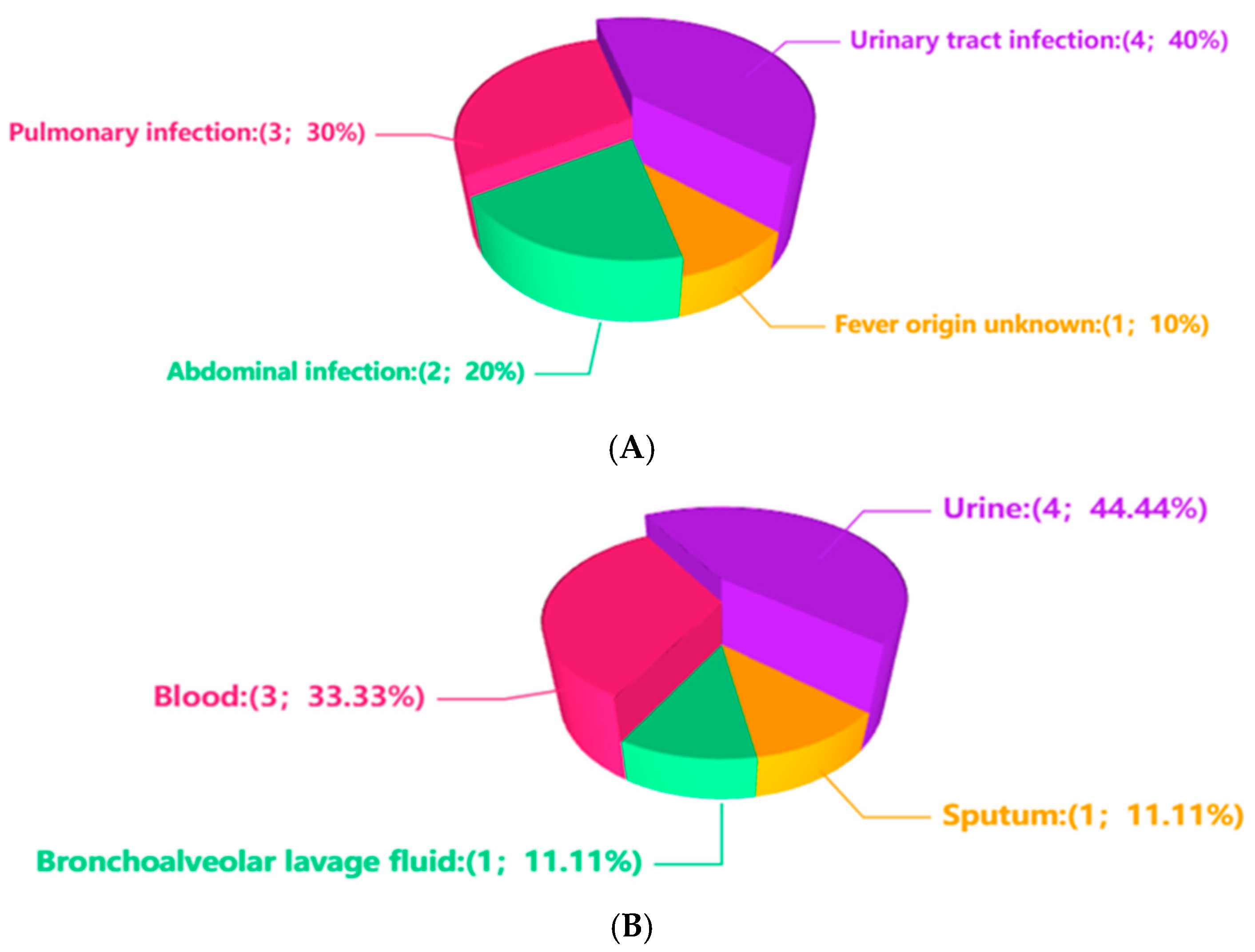
Figure 1. Infection sites and specimen types. (A) shows the infection site among the 9 cases. (B) shows the specimen type among the 9 samples.
Diagnostics 15 02242 g001
Table 1. Clinical characteristics, detected pathogens and prognosis in 9 cases.
CaseGendaAgeInfection SitesSpecimen TypesRelative Abundance of Up%Sequence Number of UpTarget TherapyPrognosis
1female70–80Pulmonary infectionblood<1≤1NoUnresolved infection
2male20–30Fever origin unknownblood<1≤1NoUnclear
3male40–50Pulmonary infectionBALF<11–100NoUnclear
4female40–50Urinary tract infectionurine1–10100–1000MinocyclineRelieved
5female30–40Abdominal infectionblood1–10≤1TigecyclineRelieved
6male20–30Urinary tract infectionurine>102000–3000MoxifloxacinRelieved
7female40–50Urinary tract infectionurine1–101000–2000MinocyclineRelieved
8male50–60Pulmonary infection, Abdominal infectionSputum<11–100NoRelieved
9female20–30Urinary tract infectionurine<1100–1000MinocyclineRelieved
CaseImmunosuppressed individualsOther microbial speciesRelative Abundance of other microbial species(%)Sequence number of other microbial species
1YesNoneNoneNone
2NoNoneNoneNone
3YesMycobacterium tuberculosis complex1–102000–3000
4NoLactobacillus crispatus>1070,000–80,000
5YesCytomegalovirus1–10≤1
6YesNoneNoneNone
7YesAcinetobacter junii<11–100
8YesPA>1090,000–100,000
9NoEnterococcusfaecalis1–101000–2000
Note: “20–30” indicates an age range of 20–30 years; “30–40” indicates an age range of 30–40 years; “40–50” indicates an age range of 40–50 years; “50–60” indicates an age range of 50–60 years; “70–80” indicates an age range of 70–80 years. “<1 (Relative abundance)” indicates a relative abundance < 1; “1–10 (Relative abundance)” indicates a relative abundance 1–10; “>10 (Relative abundance)” indicates a relative abundance > 10. “≤1 (Sequence number)” indicates a sequence number ≤ 1; “1–100 (Sequence number)” indicates a sequence number 1–100; “100–1000 (Sequence number)” indicates a sequence number 100–1000; “1000–2000 (Sequence number)” indicates a sequence number 1000–2000; “2000–3000 (Sequence number)” indicates a sequence number 2000–3000; “70,000–80,000 (Sequence number)” indicates a sequence number 70,000–80,000; “90,000–100,000 (Sequence number)” indicates a sequence number 90,000–100,000.
Disclaimer/Publisher’s Note: The statements, opinions and data contained in all publications are solely those of the individual author(s) and contributor(s) and not of MDPI and/or the editor(s). MDPI and/or the editor(s) disclaim responsibility for any injury to people or property resulting from any ideas, methods, instructions or products referred to in the content.