Article

Discovery of ETS1 as a New Gene Predisposing to Dilated Cardiomyopathy

1
Department of Geriatrics, Shanghai Fifth People′s Hospital, Fudan University, Shanghai 200240, China
2
Department of Cardiology, Shanghai Fifth People′s Hospital, Fudan University, Shanghai 200240, China
3
Department of Central Laboratory, Shanghai Fifth People′s Hospital, Fudan University, Shanghai 200240, China
4
Department of Cardiovascular Research Laboratory, Shanghai Fifth People′s Hospital, Fudan University, Shanghai 200240, China
*
Authors to whom correspondence should be addressed.
Diagnostics 2025, 15(16), 2031; https://doi.org/10.3390/diagnostics15162031 (registering DOI)
Submission received: 4 July 2025 / Revised: 2 August 2025 / Accepted: 12 August 2025 / Published: 13 August 2025
(This article belongs to the Special Issue Molecular Diagnosis and Medical Management of Cardiovascular Diseases)

Abstract

Background/Objectives: Dilated cardiomyopathy (DCM), defined as dilation and contractile dysfunction of the left or both cardiac ventricles, remains the most common category of primary myocardial disease worldwide. It is the most prevalent cause of chronic heart failure and the most common indication for cardiac transplantation in young subjects. Accumulating evidence increasingly highlights the substantial genetic defects underlying DCM. Nevertheless, the genetic ingredients accountable for DCM in a major percentage of patients remain indefinite. Methods: A multigenerational pedigree suffering from DCM and a total of 276 healthy volunteers employed as controls were recruited from the Chinese Han-ethnicity population. A whole-exome sequencing (WES) assay followed by a Sanger sequencing analysis of the genomic DNAs from the available family members was implemented. Functional characterization of the identified genetic variant was completed by dual-luciferase analysis. Results: A new heterozygous variation in the ETS1 (erythroblast transformation-specific 1) gene, NM_005238.4:c.447T>G;p.(Tyr149*), was identified by WES and validated by Sanger sequencing analysis to co-segregate with DCM in the whole DCM family. This nonsense ETS1 variant was not found in 276 control subjects. Functional examination elucidated that Tyr149*-mutant ETS1 lost the ability to transactivate its downstream target genes CLDN5 (claudin 5) and ALK1 (activin receptor-like kinase 1), two genes crucial for cardiovascular embryonic development and postnatal structural remodeling. Conclusions: The present investigation reveals ETS1 as a new gene predisposed to human DCM and indicates ETS1 haploinsufficiency as an alternative molecular pathogenesis underlying DCM, providing a potential molecular target for genetic counseling and early diagnosis as well as personalized prophylaxis of DCM.

1. Introduction

Dilated cardiomyopathy (DCM), which is clinically characterized by progressive enlargement and systolic dysfunction of the left or both cardiac ventricles without a known secondary cause such as valvular heart disease, coronary artery disease, metabolic derangement, essential hypertension, infiltrative disease, viral infection, nutrient deficiency, exposure of medications and toxins, autoimmune disease, or any others, represents the most prevalent category of primary myocardial disorder in humans worldwide, occurring in up to 0.45% of the general population [1,2,3]. There exists a slight woman preponderance in the prevalence of DCM, with an estimated male-to-female ratio of 3:4 [2]. DCM is associated with substantially enhanced risks for many detrimental clinical sequelae, encompassing ischemic stroke/thromboembolic complications [4,5,6], cardiac conduction block/abnormality [7,8,9], atrial fibrillation [10,11,12], chronic congestive heart failure [13,14,15,16,17,18], fatal/malignant ventricular dysrhythmias [19,20,21], and even cardiac demise [22,23,24]. In fact, in young individuals, DCM is the most prevalent cause of congestive heart failure and the most frequent indication for cardiac transplantation globally [25]. According to an observational study, approximately 26% of children with DCM experience either cardiac death or the necessity for a cardiac organ transplant within one year after initial diagnosis of DCM, with an additional increase of 1% per year thereafter [26]. Additionally, in adults aged 20–60 years, DCM is also the most common etiology of congestive/chronic heart failure and cardiac death, and the leading reason for heart transplant worldwide [25]. According to recent long-term studies of patients affected with nonischemic DCM, about 50% suffered from a composite of unplanned cardiovascular hospitalization, life-threatening/nonfatal arrhythmia, or cardiovascular demise over 12 years [27], and roughly 17% underwent cardiac death, heart transplant, or implantation of a ventricular assist device over 8 years [28]. In the United States alone, each year DCM causes approximately 46,000 hospitalizations and 10,000 deaths [29]. Consequently, DCM leads to substantial morbidity and mortality, and confers a tremendous socioeconomic burden on humans, underscoring the pressing need to unravel the causes underpinning DCM.
The etiology of DCM is extremely complex and highly diverse, and both exogenous/nonheritable risk factors and endogenous/genetic defectives may give rise to DCM [1,2,30,31]. Well-known acquired risk factors contributing to DCM include toxic chemicals (such as alcohol, clozapine, anabolic/adrenergic steroids, amphetamines, and anthracyclines), hemodynamic/mechanical factors (such as regurgitant/stenotic heart valve disease, anemia, athleticism/exercise, atrial tachycardias, premature ventricular beats, myocardial infarction/coronary artery disease, and pregnancy), inflammatory/infiltrative factors (such as immune myocarditis, pathogen myocarditis, and cardiac amyloidosis), endocrine/metabolic factors (such as diabetes mellitus, Cushing disease, acromegaly, pheochromocytoma, hypothyroidism, hyperlipidemia, and hyperthyroidism), hypertension, obesity, nutritional deficiency, obstructive sleep apnea, chronic kidney disease, hepatic cirrhosis, neuromuscular disease, severe sepsis, thoracic radiotherapy, and smoking [30,31]. However, there is a fast-growing body of data convincingly suggesting the pivotal effect of heritable determinants on the development of DCM, especially on the development of familial DCM, which is defined as the presence of ≥2 relatives with DCM or the presence of one relative with DCM along with sudden cardiac death before the age of 35 years [1,2]. It is reported that around 30% of DCM is familial [31], and in up to 50% of DCM patients, DCM is transmitted in an autosomal-dominant mode, though less common patterns of transmission, encompassing X-linked recessive inheritance and autosomal-recessive inheritance along with mitochondrial inheritance, have also been reported in some DCM patients [32]. At present, DCM-causing variations in >250 genes implicated in many distinct cellular processes have been identified, which include genes involved in force generation (sarcomere, Z-disk, sarcolemma, and intercalated disc proteins), cellular architecture (cytoskeleton, nuclear envelope, and desmosome proteins), gene expression (transcription factors and RNA-binding proteins), energy regulation (mitochondria), electrolyte homeostasis (ion channels and gap junction channels), and signaling transduction [1,2,33,34,35,36,37,38,39,40,41,42,43,44,45,46,47,48,49,50,51,52,53,54,55,56,57,58,59,60,61]. Notably, genetic defects responsible for DCM are discovered much more commonly in pediatric cases (54%) than in adult patients (27%) [2] Nevertheless, these known genetic determinants account for 25–50% of all DCM cases, strongly indicating that additional genetic components responsible for DCM remain to be identified [1,2,25].

2. Materials and Methods

2.1. Ethics

The current human research project was implemented in complete compliance with the tenets outlined in the Helsinki Declaration issued by the World Medical Association. The institutional Medical Ethics Committee of Shanghai Fifth People′s Hospital affiliated to Fudan University approved the research protocols (ethical approval no.: 2020-011; ethical approval date: 13 January 2021) that were applied to the current human research. Prior to commencing recruitment for the current research, all candidate research participants or their legal guardians signed informed consent forms to fulfill clinical and genetic analyses.

2.2. Recruitment and Basic Clinical Characterization of Study Participants

For the present study, a 42-member pedigree across five generations affected with DCM was discovered from the Han-race population in China (arbitrarily termed as Family DCM-101), from which available pedigree members were recruited. As control subjects, a cohort of 276 unrelated healthy volunteers with no family history of heart disease was enlisted from the same population in the same geographical area. All research participants underwent a comprehensive clinical assessment, including a thorough review of anamnestic personal and familial histories as well as medical history, an elaborate physical examination, transthoracic echocardiographic imaging, and standard 12-lead electrocardiography, along with routine laboratory tests. All DCM patients underwent a maximal exercise performance test. However, standard cardiac magnetic resonance imaging, coronary computed tomography angiography, and an endomyocardial biopsy were performed only when strongly indicated. As previously described [37], DCM was diagnosed by the existence of a left ventricular end-diastolic diameter >117% of the predicted value adjusted for age and body surface area and fractional shortening < 25% or ejection fraction <45% under the conditions of no obstructive coronary artery disease, sustained arterial hypertension, cardiac valve disorder, documented myocarditis, or systemic disorders. A total of 3 mL of peripheral venous blood samples was collected from each study individual and stored in a collecting tube containing an anticoagulant (acid-citrate dextrose) in a low-temperature (−80 °C) refrigerator. Extraction of genomic DNA from research participants’ whole blood leucocytes was conducted utilizing the QIAamp® DNA Blood Mini Kit (Cat. No. 51106; QIAGEN GmbH, Hilden, Germany).

2.3. Genetic Analysis of Research Participants

As described previously [62,63,64,65,66], a whole-exome sequencing (WES) assay was performed on genomic DNA samples from selected affected and unaffected members of Family DCM-101 suffering from DCM. Briefly, a sample of 5 μg of genomic DNA from a family member chosen for WES was randomly sheared via the Covaris® S2 Ultrasonicator (Covaris, Woburn, MA, USA) to generate various sizes of DNA fragments with a major length distribution of 100 to 500 bps, which were ligated with sequencing adapters and captured using the SureSelectXT Human All Exon V6 Kit (Cat. No. 5190-8864; Agilent Technologies, Santa Clara, CA, USA) to obtain a whole-exome library. The exomic DNA library was sequenced on the Illumina HiSeq® 4000 platform (Illumina, San Diego, CA, USA). Bioinformatic analyses of the sequence datasets produced by WES were implemented as narrated elsewhere [62,63,64,65,66]. In brief, alignment of read data to the referential human genome (build GRCh37/hg19) was implemented after removing low-quality reads by employing the Burrows–Wheeler Aligner (BWA; version 0.7.17) software package [67]. Thereafter, duplicated reads were ruled out, and the Genome Analysis Toolkit (GATK; version 3.8.1.0) software [68] was applied to call sequence variations (small insertions and deletions as well as single-nucleotide polymorphisms) at the targeted regions of an individual genome. All genetic variants were annotated by the ANNOVAR (version 2015) program [69]. Nonsynonymous variations, along with splicing donor/acceptor variations, were subject to further examination, encompassing a Sanger sequencing examination of a gene with a deleterious variation along with a segregation assessment among all the family members from Family DCM-101. Once a gene harboring a DCM-causing mutation was discovered by WES assay in Family DCM-101, a Sanger sequencing assay of the same gene (including the entire coding exons as well as all splicing junction sites) was performed in 276 unrelated control persons. Additionally, for each identified genetic variation, the databases of gnomAD (http://gnomad-sg.org/, accessed on 20 June 2025) and dbSNP (https://www.ncbi.nlm.nih.gov/, accessed on 20 June 2025) were consulted to verify its novelty.

2.4. Recombination of Gene Expression Plasmids

As depicted previously [70,71,72], total mRNAs were purified from human myocardial tissue specimens, from which cDNAs were yielded by routine reverse transcription of total mRNAs. A 1700-bp cDNA fragment containing the full-length ORF as well as partial 5′ and 3′ untranslated regions of the wild-type human erythroblast transformation-specific 1 (ETS1) gene (https://www.ncbi.nlm.nih.gov/nuccore/NM_005238.4, accessed on 16 July 2024) was amplified from purified human heart cDNA by polymerase chain reaction (PCR) using the PlatinumTM SuperFi II DNA Polymerase (Cat. No. 12361050; Invitrogen, Carlsbad, CA, USA) with the pair primers of 5′-GTCGCTAGCGAGATCGAGAGCGAACGAGG-3′ and 5′-GTCGCGGCCGCACCAACACGGCTGTCCTTGG-3′. The generated ETS1 cDNA was purified with the QIAEX II Gel Extraction Kit (Cat. No. 20051; QIAGEN GmbH, Hilden, Germany). The purified ETS1 cDNA and eukaryotic expression plasmid pCI-neo (Cat. No. E1841; Promega, Madison, WI, USA) were doubly cut with restriction endonucleases NheI (Cat. No. ER0971; Thermo Scientific, Waltham, MA, USA) and NotI (Cat. No. ER0595; Thermo Scientific, Waltham, MA, USA), purified employing the GeneJET™ Gel Extraction Kit (Cat. No. K0692; Thermo Scientific, Waltham, MA, USA), and ligated by T4 DNA ligase (Cat. No. 15224041; Invitrogen, Waltham, MA, USA) to create the wild-type ETS1-pCI plasmid ETS1-pCI. With the wild-type ETS1-pCI plasmid used as a PCR template, the Tyr149*-mutant ETS1-pCI expression plasmid was generated through site-targeted mutagenesis utilizing the GENEART® Site-Directed Mutagenesis System (Cat. No. K0692; A13282; Invitrogen, Waltham, MA, USA) with the complementary oligonucleotide primer pairs of 5′-AGTCAACCCAGCCTAGCCAGAATCCCGCTAT-3′ and 5′-ATAGCGGGATTCTGGCTAGGCTGGGTTGACT-3′ and was validated by Sanger sequencing assay. In addition, a 1101-bp promoter fragment of the human claudin 5 (CLDN5) gene (GenBank accession no.: NC_000011.10) was PCR-amplified from human genomic DNA with the DreamTaq™ DNA Polymerase (Cat. No. EP1702; Thermo Scientific, Waltham, MA, USA) and a CLDN5-specific pair of primers of 5′-CTAGGTACCGGAGGCCGCAGCAGTCATCC-3′ and 5′-CTAAAGCTTCTCAATCTTCACAGGGGCTG-3′. The produced CLDN5 promoter DNA as well as the reporter plasmid of pGL3-Basic (Cat. No. E1751; Promega, Madison, WI, USA) was doubly cut with the restriction enzymes KpnI (Cat. No. ER0521; Thermo Scientific, Waltham, MA, USA) and HindIII (Cat. No. ER0501; Thermo Scientific, Waltham, MA, USA), separated by agarose gel electrophoresis, extracted with the GenElute™ Gel Extraction Kit (Cat. No. NA1111; Sigma-Aldrich, St. Louis, MO, USA), and ligated with T4 DNA ligase (Cat. No. 15224041; Invitrogen, Waltham, MA, USA) to construct the CLDN5-luciferase (CLDN5-luc) reporter vector, which expresses firefly luciferase. Similarly, a 1690-bp promoter fragment (from −221 to −1910 upstream of the transcriptional initial site) of the human activin receptor-like kinase 1 (ALK1) gene (https://www.ncbi.nlm.nih.gov/nuccore/NC_000012.12?from=51906944&to=51923361&report=genbank, accessed on 16 July 2024) was PCR-amplified from human genomic DNA with the DreamTaq™ DNA Polymerase (Cat. No. EP1702; Thermo Scientific, Waltham, MA, USA) as well as an ALK1-specific pair of primers of 5′-GATGGTACCGACTCTCGCTTCTCAGGAC-3′ and 5′-GATAAGCTTCGCACCCGGGCCGGGCCTCC-3′. The yielded ALK1 promoter DNA as well as the reporter plasmid of pGL3-Basic (Cat. No. E1751; Promega, Madison, WI, USA) were digested with the restriction enzymes KpnI (Cat. No. ER0521; Thermo Scientific, Waltham, MA, USA) and HindIII (Cat. No. ER0501; Thermo Scientific, Waltham, MA, USA), separated by agarose gel electrophoresis, extracted with the GenElute™ Gel Extraction Kit (Cat. No. NA1111; Sigma-Aldrich, St. Louis, MO, USA), and ligated with T4 DNA ligase (Cat. No. 15224041; Invitrogen, Waltham, MA, USA) to construct the ALK1-luciferase (ALK1-luc) reporter vector, which expresses firefly luciferase. All the final recombinant expression plasmids were subject to confirmation by Sanger sequencing analysis.

2.5. Cell Transfection and Dual Reporter Gene Analysis

As narrated previously [37,73,74], HeLa cells were seeded into a Nunc™ 24-well microplate/multidish (Cat. No. 144530; Thermo Fisher Scientific, Rochester, NY, USA) at a density of 1 × 10⁵ cells/well, and routinely cultivated for 24 h before transient transfection with appropriate amounts of various expression plasmids using the Lipofectamine™ 3000 Transfection Reagent (Cat. No. L3000015; Invitrogen; Waltham, MA, USA). Specifically, HeLa cells were transiently transfected with the same amount (200 ng) of empty pCI, or wild-type ETS1-pCI, or Tyr149*-mutant ETS1-pCI, or 100 ng of empty pCI plus 100 ng of wild-type ETS1-pCI, or 100 ng of wild-type ETS1-pCI plus 100 ng of Tyr149*-mutant ETS1-pCI, together with 10 ng of pGL4.75 (Cat. No. E6931; Promega, Madison, WI, USA) and 1000 ng of CLDN5-luc or ALK1-luc. As an internal control, the pGL4.75 plasmid (Cat. No. E6931; Promega, Madison, WI, USA) expressing Renilla luciferase was co-transfected to balance transfection efficiency. HeLa cells were collected 36 h post-transfection, lysed, and the promoter-driven luciferase activities were quantitatively detected as described previously [37]. The promoter activity was measured as the ratio of firefly/Renilla luciferase luminescence intensity. Each recombinant expression plasmid, along with an internal control plasmid, was transiently transfected and detected three times in triplicate. Results are expressed as means of relative luciferase activity from three independent transfection experiments and standard deviations (SDs).

2.6. Statistical Analysis

Continuous data of promoter activities are presented as mean ± SD. All data were analyzed by using Student’s unpaired t-test for pairwise comparison or analysis of variance (ANOVA) followed by Tukey–Kramer HSD post hoc test for multi-group analysis. Differences were considered to be statistically significant at two-tailed p-values of <0.05.

3. Results

3.1. Demographic and Phenotypic Characteristics of the Recruited Family Affected with DCM

For the current investigation, as illustrated in Figure 1, a 42-member pedigree affected with DCM spanning five generations was identified as Family DCM-101 from the Chinese Han-ethnicity population. In this 42-member family with DCM, there were 37 living members, encompassing 19 male and 18 female family members. The proband of Family DCM-101 (IV-3) was first diagnosed with DCM at the age of 41 years. During the proband’s clinical follow-up, he experienced progressive fatigue and dyspnea on exertion, left ventricular dilation and systolic dysfunction on echocardiograms, and he received pharmacologic therapies (intravenous injections, infusions, and oral medications) for chronic cardiac failure, encompassing diuretics (furosemide and spironolactone), angiotensin-converting enzyme inhibitor (captopril), and a β-adrenergic receptor antagonist (metoprolol). His grandfather (II-1) and two other relatives (II-3 and I-1) had a past medical history of DCM and died of progressive congestive heart failure attributable to DCM. His grandmother (II-2) and another relative (I-2) had no medical history of DCM and died of a cerebral stroke.
Additionally, in Family DCM-101, all 10 affected members were diagnosed with DCM based on clinical symptoms, physical signs, and echocardiographic findings, while all unaffected members denied a medical history of DCM, with no echocardiographic abnormality. No individuals from Family DCM-101 had known environmental pathogenic factors predisposing to DCM, encompassing viral myocarditis, coronary heart disease, autoimmune disorders, rheumatic heart disease, and arterial hypertension. A genetic evaluation of the family (Family DCM-101) revealed that DCM was inherited as an autosomal-dominant trait in the whole pedigree, with complete penetrance. Notably, all 10 affected members also suffered mild facial dysmorphisms, including a high forehead, a broad nasal bridge with bulbous tip, and dysplastic little ears. Furthermore, in addition to DCM, three family members (III-8, III-11, and IV-12) also had atrial fibrillation, and one family member (IV-6) also had a minor membranous ventricular septal defect (VSD) that closed spontaneously within the first year of her life. The demographic and baseline phenotypic characteristics of the living family members with DCM from Family DCM-101 are outlined in Table 1.

3.2. Discovery of a New DCM-Causative Mutation in ETS1

A WES assay was completed in four DCM-affected members (III-2, III-8, IV-3, and IV-12 from Family DCM-101) and three non-DCM members (IV-2, V-2, and V-6 from Family DCM-101). A mean of 15,209 nonsynonymous genetic variations (ranging from 14,917 to 17,536) per family member passed filtering on the likely inheritance modes (the recessive genetic transmission mode was also encompassed during screening for potential causative genetic variations), of which 11 heterozygous missense and nonsense genetic variations passed filtering by ANNOVAR, with a minor allele frequency of <0.01%, and were carried by all the four DCM-affected members (III-2, III-8, IV-3, and IV-12 from Family DCM-101) who had experienced WES analysis, as given in Table 2.
Further Sanger sequencing assays of these genetic variations in all the family members available from Family DCM-101 revealed that only the heterozygous nonsense mutation of chr11:128,355,998T>G (GRCh37.p13/hg19: NC_000011.9), equivalent to chr11:128,560,220T>G (GRCh38.p14/hg38: NC_000011.10), or NM_005238.4:c.447T>G;p.(Tyr149*) in the erythroblast transformation-specific 1 (ETS1) gene, was verified to co-segregate with the DCM phenotype in the entire pedigree, i.e., present in all DCM members but absent in all unaffected members of Family DCM-101. However, Sanger sequencing assays of the other 10 genetic variations demonstrated that none was in co-segregation with the DCM phenotype in the whole family, suggesting that none of these 10 genetic variations is likely to underlie DCM in this family. In addition, a Sanger sequencing assay of the ETS1 gene in a total of 276 unrelated control people revealed no pathogenic mutations. The detected ETS1 mutation was absent in dbSNP (accessed on 20 June 2025) and gnomAD (accessed on 20 June 2025), suggesting a novel ETS1 mutation. The primers designed to specifically amplify the ETS1 gene (including the whole set of coding exons and splicing boundaries) by PCR are provided in Table 3.
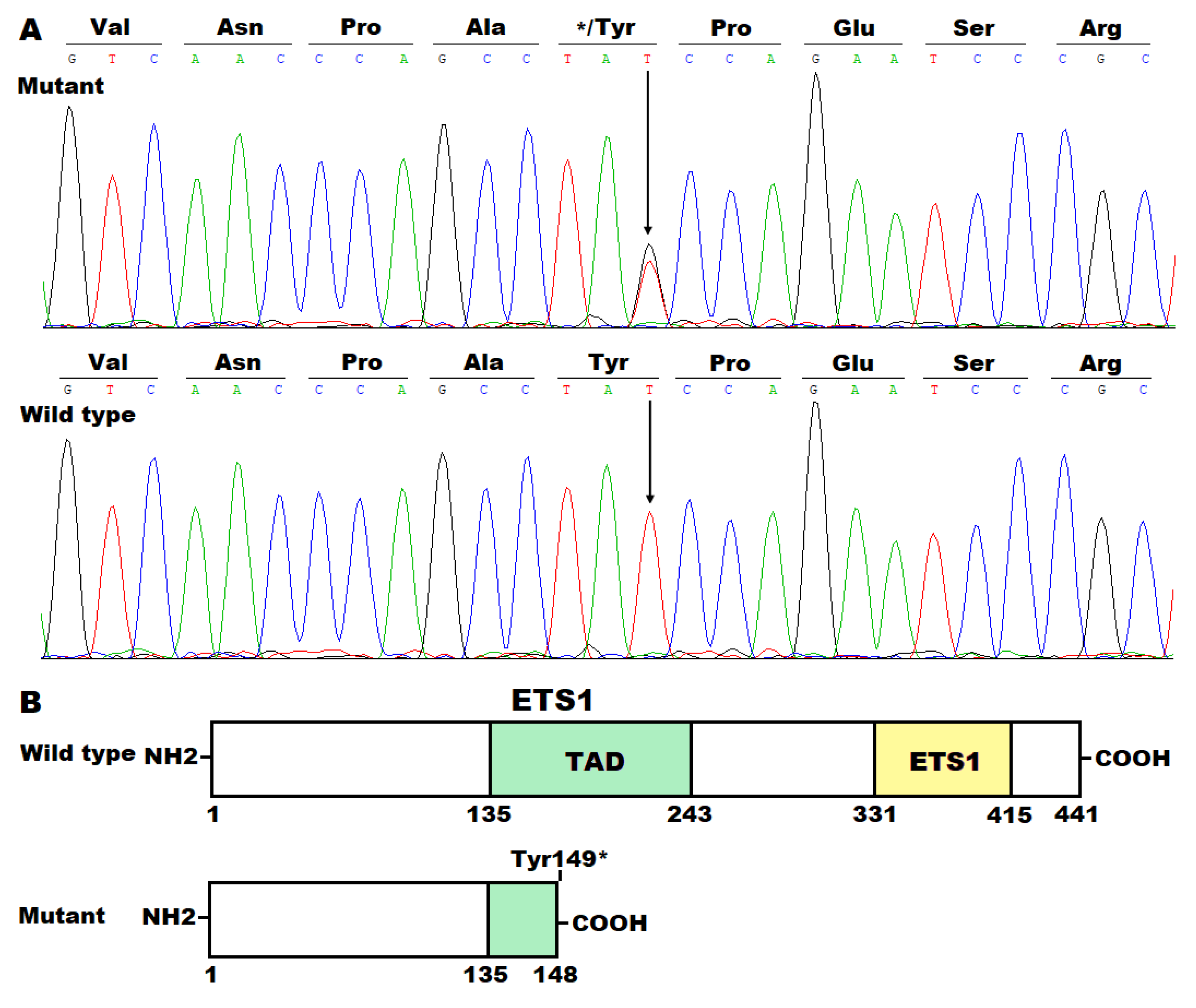
The DNA sequence chromatograms illustrating the heterozygous mutant ETS1 alleles (T/G) as well as wild-type ETS1 alleles (T/T) are provided in Figure 2A. The schematic diagrams delineating the functional motifs of the ETS1 proteins are given in Figure 2B.

3.3. Functional Loss of Tyr149*-Mutant ETS1 in Transactivating CLDN5

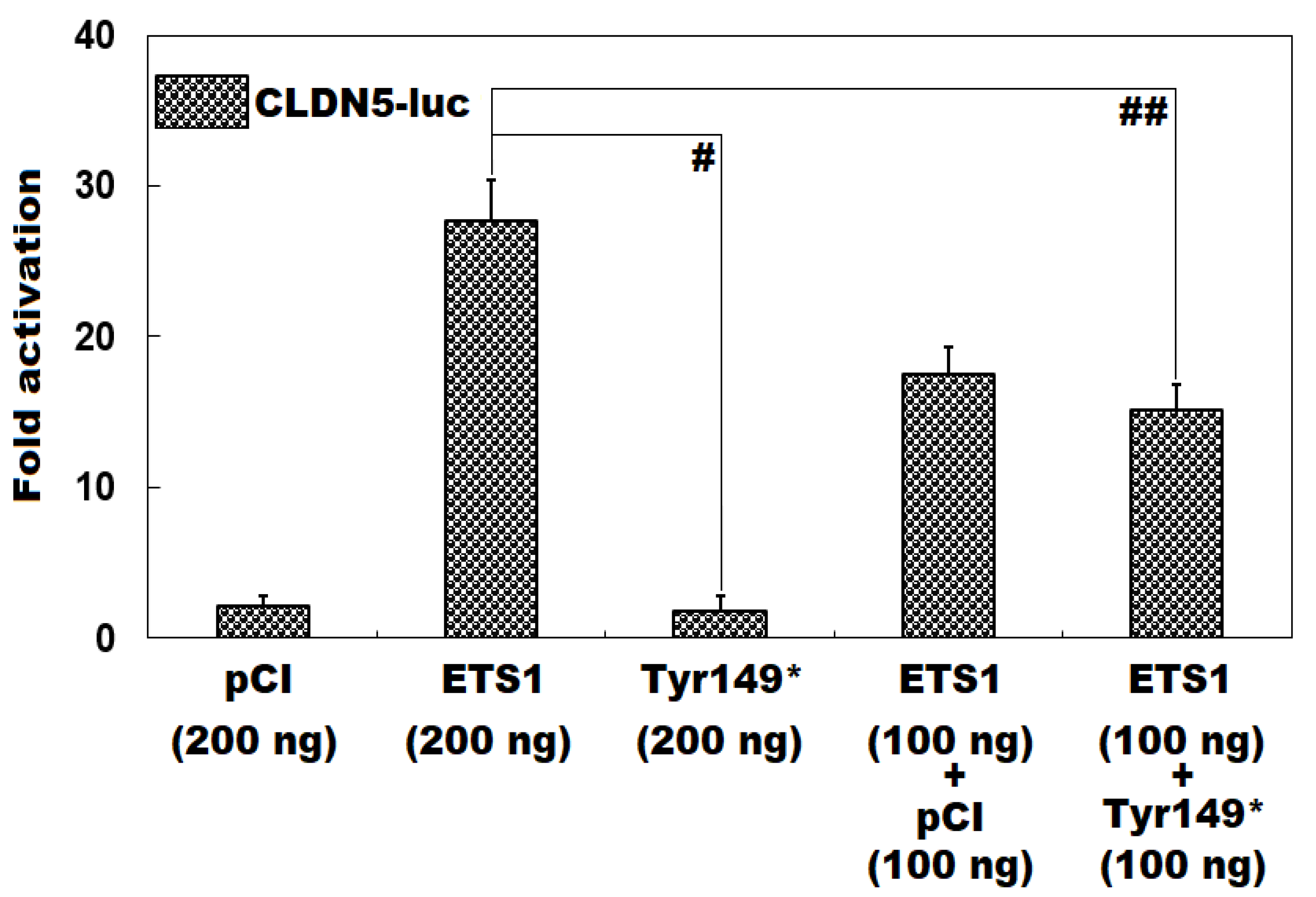
As exhibited in Figure 3, in HeLa cells routinely cultivated in vitro and transiently transfected with various eukaryotic expression plasmids, the wild-type ETS1-pCI-neo plasmid and Tyr149*-mutant ETS1-pCI-neo plasmid induced transcriptional activation of the CLDN5 promoter by ~28-fold and ~2-fold, respectively (Tyr149*-mutant ETS1-pCI-neo vs. wild-type ETS1-pCI-neo: t = 15.8273; p = 0.0001). When the wild-type ETS1-pCI-neo and Tyr149*-mutant ETS1-pCI-neo plasmids were co-transfected, the elicited transactivation activity was ~15-fold (wild-type ETS1-pCI-neo vs. wild-type ETS1-pCI-neo + Tyr149*-mutant ETS1-pCI-neo: t = 6.9335; p = 0.0023). When the comparisons among multiple groups were implemented, similar resultant data were yielded (F = 127.82, p = 1.534 × 10⁻⁸). Multiple-group comparisons were performed between 200 ng of empty pCI-neo plasmid and 200 ng of wild-type ETS1-pCI-neo plasmid (t = 25.59, p < 0.0001), between 200 ng of empty pCI-neo plasmid and 200 ng of Tyr149*-mutant ETS1-pCI-neo plasmid (t = 0.3533, p = 0.9989), between 200 ng of empty pCI-neo plasmid and 100 ng of empty pCI-neo plasmid + 100 ng of wild-type ETS1-pCI-neo plasmid (t = 15.4233, p < 0.0001), between 200 ng of empty pCI-neo plasmid and 100 ng of wild-type ETS1-pCI-neo plasmid + 100 ng of Tyr149*-mutant ETS1-pCI-neo plasmid (t = 13.04, p < 0.0001), between 200 ng of wild-type ETS1-pCI-neo plasmid and 200 ng of Tyr149*-mutant ETS1-pCI-neo plasmid (t = 25.9433, p < 0.0001), between 200 ng of wild-type ETS1-pCI-neo plasmid and 100 ng of empty pCI-neo plasmid + 100 ng of wild-type ETS1-pCI-neo plasmid (t = 10.1667, p = 0.0002), between 200 ng of wild-type ETS1-pCI-neo plasmid and 100 ng of wild-type ETS1-pCI-neo plasmid + 100 ng of Tyr149*-mutant ETS1-pCI-neo plasmid (t = 12.55, p < 0.0001), between 200 ng of Tyr149*-mutant ETS1-pCI-neo plasmid and 100 ng of empty pCI-neo plasmid + 100 ng of wild-type ETS1-pCI-neo plasmid (t = 15.7767, p < 0.0001), between 200 ng of Tyr149*-mutant ETS1-pCI-neo plasmid and 100 ng of wild-type ETS1-pCI-neo plasmid + 100 ng of Tyr149*-mutant ETS1-pCI-neo plasmid (t = 13.3933, p < 0.0001), and between 100 ng of empty pCI-neo plasmid + 100 ng of wild-type ETS1-pCI-neo plasmid and 100 ng of wild-type ETS1-pCI-neo plasmid + 100 ng of Tyr149*-mutant ETS1-pCI-neo plasmid (t = 2.3833, p = 0.4603).

3.4. Inability of Tyr149*-Mutant ETS1 to Transcriptionally Activate ALK1

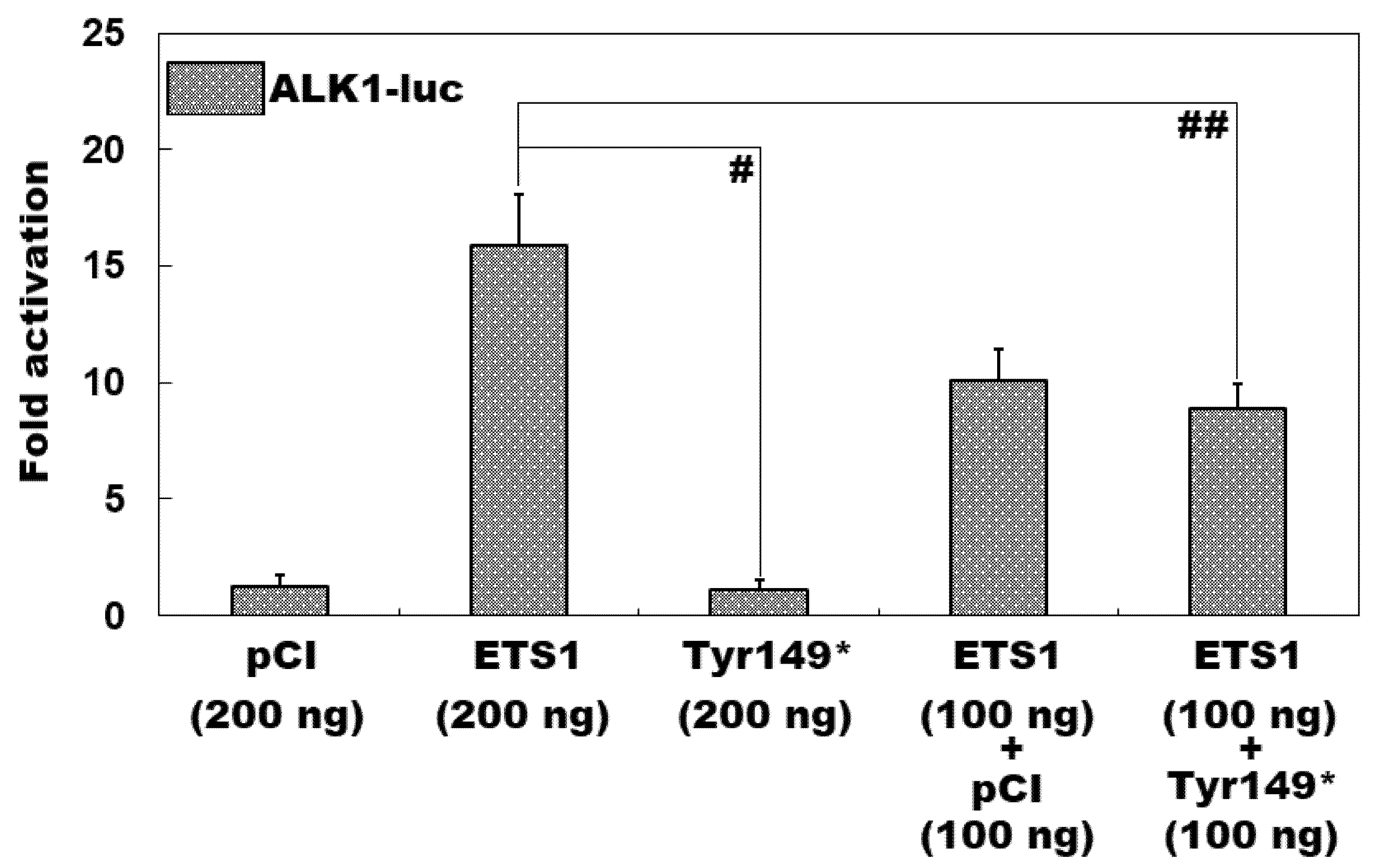
As displayed in Figure 4, in HeLa cells routinely cultured in vitro and transiently transfected with various eukaryotic expression plasmids, the wild-type ETS1-pCI-neo plasmid and Tyr149*-mutant ETS1-pCI-neo plasmid induced transcriptional activation of the ALK1 promoter by ~16-fold and ~1-fold, respectively (Tyr149*-mutant ETS1-pCI-neo vs. wild-type ETS1-pCI-neo: t = 11.3841; p = 0.0003). When the wild-type ETS1-pCI-neo and Tyr149*-mutant ETS1-pCI-neo plasmids were co-transfected, the elicited transactivation activity was ~9-fold (wild-type ETS1-pCI-neo vs. wild-type ETS1-pCI-neo + Tyr149*-mutant ETS1-pCI-neo: t = 4.9510; p = 0.0078). When the comparisons among multiple groups were implemented, similar statistical data were generated (F = 71.745, p = 2.524 × 10⁻⁷). Multiple-group comparisons were performed between 200 ng of empty pCI-neo plasmid and 200 ng of wild-type ETS1-pCI-neo plasmid (t = 14.6167, p < 0.0001), between 200 ng of empty pCI-neo plasmid and 200 ng of Tyr149*-mutant ETS1-pCI-neo plasmid (t = 0.1333, p = 0.9999), between 200 ng of empty pCI-neo plasmid and 100 ng of empty pCI-neo plasmid + 100 ng of wild-type ETS1-pCI-neo plasmid (t = 8.8067, p < 0.0001), between 200 ng of empty pCI-neo plasmid and 100 ng of wild-type ETS1-pCI-neo plasmid + 100 ng of Tyr149*-mutant ETS1-pCI-neo plasmid (t = 7.64, p = 0.0002), between 200 ng of wild-type ETS1-pCI-neo plasmid and 200 ng of Tyr149*-mutant ETS1-pCI-neo plasmid (t = 14.75, p < 0.0001), between 200 ng of wild-type ETS1-pCI-neo plasmid and 100 ng of empty pCI-neo plasmid + 100 ng of wild-type ETS1-pCI-neo plasmid (t = 5.81, p = 0.0018), between 200 ng of wild-type ETS1-pCI-neo plasmid and 100 ng of wild-type ETS1-pCI-neo plasmid + 100 ng of Tyr149*-mutant ETS1-pCI-neo plasmid (t = 6.9767, p = 0.0004), between 200 ng of Tyr149*-mutant ETS1-pCI-neo plasmid and 100 ng of empty pCI-neo plasmid + 100 ng of wild-type ETS1-pCI-neo plasmid (t = 8.94, p < 0.0001), between 200 ng of Tyr149*-mutant ETS1-pCI-neo plasmid and 100 ng of wild-type ETS1-pCI-neo plasmid + 100 ng of Tyr149*-mutant ETS1-pCI-neo plasmid (t = 7.7733, p = 0.0002), and between 100 ng of empty pCI-neo plasmid + 100 ng of wild-type ETS1-pCI-neo plasmid and 100 ng of wild-type ETS1-pCI-neo plasmid + 100 ng of Tyr149*-mutant ETS1-pCI-neo plasmid (t = 1.1667, p = 0.7976).

4. Discussion

In the present investigation, a five-generation pedigree of 42 members affected with DCM was enrolled. By means of a WES-based bioinformatics assay in the available family members, a heterozygous truncating ETS1 mutation, i.e., NM_005238.4:c.447T>G;p.(Tyr149*), was discovered and verified via a Sanger sequencing assay to co-segregate with DCM in the entire family. This ETS1 mutation was neither observed in the 552 reference human chromosomes from the Chinese Han population nor found in the population genetics databases of dbSNP and gnomAD. Quantitative biochemical measurement based on dual reporter genes demonstrated that Tyr149*-mutant ETS1 failed to transactivate the expression of CLDN5 (encoding claudin 5, an integral tight junction protein component) and ALK1 (coding for activin receptor-like kinase 1), two genes pivotal for proper cardiovascular embryonic development and postnatal structural remodeling [75,76,77,78,79,80,81,82,83,84,85]. These results cumulatively indicate that genetically compromised ETS1 gives rise to the development of DCM in humans.
In humans, ETS1 (also known as ETS-1, TPL-1, c-ETS-1, c-ETS, EWSR2, P54, or ETS) is mapped on chromosome 11q24.3; it contains 8 coding exons and encodes an integral tight junction protein with 441 amino acids, the founding member of the ETS-domain transcription factor family, which is defined by the existence of an evolutionarily conserved ETS DNA-binding motif that recognizes and binds to the core consensus DNA sequence of GGAA/T (a unique ETS-binding site) in the promoters/enhancers of target genes [86,87,88,89]. The ETS family of transcription factors currently comprises 28 members in humans and 27 members in mice, and they function as transcriptional regulators (activators or repressors) of numerous downstream target genes that are involved in an extensive assortment of important biological processes including cellular growth, proliferation, differentiation, migration, apoptosis, senescence, and death, hence playing crucial roles in embryonic organogenesis, tissue homeostasis, and the progression of diseases, including arteriosclerosis, autoimmune diseases, allergies, and tumors [86,87,88,89]. Among the ETS family, ETS1 is characterized by its broad expression, especially by its ample expression in the heart at all stages from fetuses to adult humans [90,91,92] and multiple species of animals [93,94,95,96,97,98]. Moreover, ETS1 has been validated to have a potent transcriptional regulation function, playing a key role in normal cardiovascular development and postnatal structural remodeling, and in mice, deletion of Ets1 leads to various cardiovascular developmental abnormalities, including VSD, double-outlet right ventricle (DORV), and ventricular noncompaction [93,94,95,96,97]. ETS1 contains two functionally critical structural domains, including a transcriptional activation domain (TAD) and an ETS domain responsible for recognizing and binding to the promoters/enhancers of target genes [86,87,88,89]. In the present research, we identified a novel heterozygous ETS1 mutation, NM_005238.4:c.447T>G;p.(Tyr149*), to co-segregate with DCM in the whole family. This mutation was anticipated to create a truncated ETS1 protein (Tyr149*), with the entire ETS domain along with a larger part of the TAD domain lost, hence, presumably, leading to a loss of transcriptional regulation function, which was confirmed by biochemical assays of its two representative target genes, CLDN5 and ALK1. These results support the hypothesis that the haploinsufficiency of ETS1 predisposes individuals to DCM.
It may be attributed to improper embryonic development and structural remodeling of the heart that ETS1 haploinsufficiency contributes to DCM. In mice (with a C57BL/6 genetic background), homozygous knockout of Ets1 led to almost complete perinatal lethality because of profound cardiovascular developmental defects, including a membranous VSD and an anomalous cartilage nodule in the heart [93]. Ye and colleagues [94] reported that in mice (with a C57/B6 background), ETS1 was highly expressed in the cardiac neural crest, endocardium, and vascular endothelium early during embryogenesis, and targeted disruption of Ets1 caused a large membranous VSD and a bifid cardiac apex, as well as a non-apex-forming left ventricle, a hallmark of hypoplastic left heart syndrome (HLHS). Lin et al. [95] reported that deletion of Ets1 in mice resulted in a cardiovascular developmental defect resembling a DORV in humans. Furthermore, endothelial-specific knockout of Ets1 in mice led to ventricular noncompaction and coronary vascular developmental anomaly, which reproduced the phenotype caused by global deletion of Ets1 [96]. Notably, Endothelial-specific ablation of Ets1 downregulated the expression levels of multiple target genes, including Alk1 and Cldn5 [96]. Recently, Nie and Bronner [98] demonstrated that in Xenopus, ETS1 was abundantly expressed in the cardiac neural crest and mesoderm during embryogenesis, and knockdown of Ets1 in the cardiac mesoderm gave rise to an HLHS-like ventricle, which was characteristic of an abundance of cardiomyocytes and a loss of endocardial cells, in line with a key role for ETS1 in mediating cellular fate determination between cardiac myocytes and endocardial cells [97,98]. Zhou and co-workers [99] reported that in the murine embryonic heart, the cardiomyocyte- and endothelial-restricted Gata4-occupied genomic regions were enriched for Ets1 and Nkx2-5 motifs, respectively, and both ETS1 and NKX2-5 transcription factors interacted with GATA4 to regulate heart development. Of note, both GATA4 and NKX2-5 have been causally linked to human DCM [100,101,102,103,104]. Additionally, in an experimental mouse model with autoimmune myocarditis, treatment with spironolactone (an aldosterone receptor antagonist) significantly alleviated myocardial hypertrophy, improved cardiac function, and diminished myocardial fibrosis via inhibition of ETS1 [105]. In rodent/rat and cellular models with myocardial ischemia/reperfusion (hypoxia/reoxygenation) injury, knockout of Kdm3a (coding for histone demethylase KDM3A) exacerbated myocardial injury and cardiac dysfunction, and deteriorated mitochondrial apoptosis and inflammation; conversely, overexpression of KDM3A could ameliorate such alterations by promoting ETS1 expression [106]. In chicken embryos, copious expression of ETS1 was observed in the heart and endothelial cells of blood vessels [107], and knockdown of Ets1 hampered the development of the myocardium and coronary system, resulting in a defect in myocardial perfusion [108]. In addition, in cultured ventricular myocytes from chick embryos, ETS1 was shown to transactivate the expression of inducible nitric oxide synthase, presumably via interaction with NF-kappaB [109]. In human embryonic stem cells, ETS1 was identified by single-cell reconstruction of differentiation trajectory to be a pivotal factor responsible for cardiac lineage commitment from a pluripotent status [110]. Taken together, these results highlight the crucial roles of ETS1 in embryonic cardiogenesis and postnatal cardiac remodeling, suggesting that an ETS1 loss-of-function mutation is a molecular defect underlying DCM in humans.
Previously, in humans, chromosome 11q terminal deletions (11q−) containing ETS1 and ETS1 variations have been implicated in the pathogenesis of Jacobsen syndrome (JS), a rare chromosomal/genetic disorder characterized by a wide spectrum of phenotypes with varying degrees of severity, including congenital heart defects, facial dysmorphisms, intellectual disability, autism and attention deficit, immunodeficiency, genitourinary tract deformities, growth restriction, and thrombocytopenia [111,112,113]. By comparative genomic hybridization and fluorescence in situ hybridization analyses in five patients from two families with JS, Conrad and partners [111] detected inherited interstitial deletions in the chromosomal region of 11q24.2-q24.3, the smallest ever reported at the JS locus. Specifically, in Family A, a 700 kb interstitial chromosomal deletion of 11q24.3 containing three genes, ETS1, FLI1, and SENCR, was observed; in Family B, a 1.5 Mb interstitial chromosomal deletion of 11q24.2-q24.3 containing eight genes, ARHGAP32, ETS1, TP53AIP1, FLI1, KCNJ1, C11orf45, SENCR, and KCNJ5, was observed [111]. Of the two JS families, all affected members had ETS1 haploinsufficiency and manifested such clinical characteristics of JS as developmental malformations (two patients from Family A had minor VSD), immune deficiency, and thrombocytopenia, but had normal cognitive development, consistent with an incomplete penetrance of JS [111]. Grossfeld et al. [112] conducted a prospective study of 110 JS patients with the terminal deletion of chromosome 11q diagnosed by karyotypic analysis, of whom 93 JS patients underwent a comprehensive cardiovascular assessment. As a result, 56% (52/93) of JS patients had severe cardiovascular developmental defects, of which the majority required surgical intervention [112]. In total, two-thirds of the JS patients had a variety of congenital heart defects, including membranous VSD, secundum atrial septal defects, aortic coarctation/stenosis, patent ductus arteriosus, atrioventricular canal defects, mitral valve stenosis, double-outlet right ventricle, bicuspid aortic valve, transposition of the great arteries, aberrant right subclavian artery, dextrocardia, a left-sided superior vena cava, and hypoplastic left heart syndrome [112]. By means of a genome-wide single-nucleotide polymorphism array and whole-exome sequencing assays in 538 trios with congenital heart defects, Glessner et al. [113] identified a de novo ETS1 frameshift variant (c.1044_1049delCAAGGAinsTT or p.Lys349SerfsX2) in one patient with congenital heart malformations (hypoplastic left heart syndrome). Additionally, the integrated data implied that ETS1 was the causal gene altered by chromosome 11q24.2-q25 deletions in JS [113]. In the current study, a novel ETS1 loss-of-function variant (c.447T>G or p.Tyr149*) was causally linked to familial DCM, hence expanding the phenotypic spectrum of ETS1 variations. Notably, there is an urgent need to investigate whether DCM occurs in small or large animals after knockout of Ets1 or knock-in of the DCM-causing Ets1 mutation, which is our future research direction. Additionally, the preparation and characterization of induced pluripotent stem cell-derived cardiomyocytes from patients and healthy individuals of the same family with DCM or patient-derived fibroblasts will help better understand the pathogenic effect of the mutated ETS1 gene on cardiac tissue [114,115,116,117].
To date, pathogenic variations in >250 genes have been implicated in the development of human DCM [1,2,33,34,35,36,37,38,39,40,41,42,43,44,45,46,47,48,49,50,51,52,53,54,55,56,57,58,59,60,61]. Nevertheless, there are still clear reasons why it is necessary to search for a new causative gene for DCM. First, due to substantial genetic heterogeneity, these known causative genes can merely explain 25–50% of all DCM cases, and the genetic determinants responsible for DCM in 50–75% of cases remain elusive, highlighting the need to identify new DCM-causative genes [1,2,25]. Second, the discovery of the DCM-causing mutation is of important clinical significance, since early interventions for DCM have been proven to be valuable in reducing morbidity and mortality [118]. Third, the identification of DCM-causing genes has a direct clinical impact on the families as well as the population involved, which immediately provides possibilities for antenatal/presymptomatic prevention of DCM through genetic screening of mutation carriers and prenatal testing, and ensures healthy family members reproduce healthy children [118]. Finally, the discovery of causative genes holds the potential to add more insight into the etiology and pathophysiology of diseases, and, therefore, potential improved therapy [118].

5. Conclusions

This investigation unveils ETS1 as a new gene accountable for DCM in humans, thereby offering novel insights into the pathogenesis of the disease and holding translational potential for future prophylactic and therapeutic strategies.

Author Contributions

Project administration, Z.-P.K. and Y.-Q.Y.; conceptualization, Z.-P.K. and Y.-Q.Y.; supervision, Z.-P.K. and Y.-Q.Y.; methodology, Z.-P.K., J.-N.G., Y.-J.X. and Y.-Q.Y.; formal analysis, Z.-P.K., J.-N.G., C.-X.Y., X.-L.L., S.Z., Y.-Z.B., Y.-J.X. and Y.-Q.Y.; validation, Z.-P.K. and Y.-Q.Y.; conduct of the experiment, Z.-P.K., J.-N.G., C.-X.Y., X.-L.L., S.Z., Y.-Z.B., Y.-J.X. and Y.-Q.Y.; visualization, Z.-P.K. and Y.-Q.Y.; collecting patients, Z.-P.K., J.-N.G., C.-X.Y., X.-L.L., Y.-J.X. and Y.-Q.Y.; data curation, Z.-P.K. and Y.-Q.Y.; funding acquisition, Z.-P.K.; writing—original draft preparation, Z.-P.K., J.-N.G., C.-X.Y., X.-L.L., S.Z., Y.-Z.B., Y.-J.X. and Y.-Q.Y.; writing—review and editing, Z.-P.K. and Y.-Q.Y. All authors have read and agreed to the published version of the manuscript.

Funding

This research was funded by the Natural Science Foundation of Minhang District, Shanghai, China (2024MHZ015).

Institutional Review Board Statement

This research was completed following the Declaration of Helsinki and approved by the Ethics Committee of Shanghai Fifth People’s Hospital, Fudan University (ethical approval no.: 2020-011; ethical approval date: 13 January 2021).

Informed Consent Statement

Informed consent was obtained from all subjects involved in the study. Written informed consent has been obtained from the patients to publish this paper.

Data Availability Statement

The data in favor of the discovery in this study are available upon reasonable request.

Acknowledgments

We cordially thank the research participants for participating in the current research.

Conflicts of Interest

The authors declare no conflict of interest.

References

  1. Newman, N.A.; Burke, M.A. Dilated Cardiomyopathy: A Genetic Journey from Past to Future. Int. J. Mol. Sci. 2024, 25, 11460. [Google Scholar] [CrossRef]
  2. Eldemire, R.; Mestroni, L.; Taylor, M.R.G. Genetics of Dilated Cardiomyopathy. Annu. Rev. Med. 2024, 75, 417–426. [Google Scholar] [CrossRef] [PubMed]
  3. McGurk, K.A.; Zhang, X.; Theotokis, P.; Thomson, K.; Harper, A.; Buchan, R.J.; Mazaika, E.; Ormondroyd, E.; Wright, W.T.; Macaya, D.; et al. The Penetrance of Rare Variants in Cardiomyopathy-Associated Genes: A Cross-sectional Approach to Estimating Penetrance for Secondary Findings. Am. J. Hum. Genet. 2023, 110, 1482–1495. [Google Scholar] [CrossRef] [PubMed]
  4. Rodríguez-González, E.; Martínez-Legazpi, P.; González-Mansilla, A.; Espinosa, M.Á.; Mombiela, T.; Guzmán De-Villoria, J.A.; Borja, M.G.; Díaz-Otero, F.; Gómez de Antonio, R.; Fernández-García, P.; et al. Cardiac stasis imaging, stroke, and silent brain infarcts in patients with nonischemic dilated cardiomyopathy. Am. J. Physiol. Heart Circ. Physiol. 2024, 327, H446–H453. [Google Scholar] [CrossRef] [PubMed]
  5. Ródenas-Alesina, E.; Lozano-Torres, J.; Tobías-Castillo, P.E.; Badia-Molins, C.; Calvo-Barceló, M.; Vila-Olives, R.; Casas-Masnou, G.; San Emeterio, A.O.; Soriano-Colomé, T.; Fernández-Galera, R.; et al. Risk of Stroke and Incident Atrial Fibrillation in Patients in Sinus Rhythm with Nonischemic Dilated Cardiomyopathy. Am. J. Cardiol. 2024, 233, 11–18. [Google Scholar] [CrossRef]
  6. Fan, Z.; Wu, C.; Wang, C.; Liu, C.; Fang, L.; Ma, L.; Zou, W.; Yuan, B.; Ji, Z.; Cai, B.; et al. Impact of Concurrent Ischaemic Stroke on Unfavourable Outcomes in Men and Women with Dilated Cardiomyopathy. Rev. Cardiovasc. Med. 2024, 25, 215. [Google Scholar] [CrossRef]
  7. Lakdawala, N.K.; Givertz, M.M. Dilated cardiomyopathy with conduction disease and arrhythmia. Circulation 2010, 122, 527–534. [Google Scholar] [CrossRef]
  8. Yuan, Y.; Yang, K.; Liu, Q.; Song, W.; Jin, D.; Zhao, S. Nonspecific intraventricular conduction delay predicts the prognosis of dilated cardiomyopathy. BMC Cardiovasc. Disord. 2023, 23, 409. [Google Scholar] [CrossRef]
  9. Jiménez-Blanco Bravo, M.; Alonso Salinas, G.L.; Parra Esteban, C.; Toquero Ramos, J.; Amores Luque, M.; Zamorano Gómez, J.L.; García-Izquierdo, E.; Álvarez-García, J.; Fernández Lozano, I.; Castro Urda, V. Right Bundle Branch Block Predicts Appropriate Implantable Cardioverter Defibrillator Therapies in Patients with Non-Ischemic Dilated Cardiomyopathy and a Prophylactic Implantable Cardioverter Defibrillator. Diagnostics 2024, 14, 1173. [Google Scholar] [CrossRef]
  10. Raafs, A.G.; Vos, J.L.; Henkens, M.T.H.M.; Verdonschot, J.A.J.; Sikking, M.; Stroeks, S.; Gerretsen, S.; Hazebroek, M.R.; Knackstedt, C.; Nijveldt, R.; et al. Left Atrial Strain Is an Independent Predictor of New-Onset Atrial Fibrillation in Dilated Cardiomyopathy. JACC Cardiovasc. Imaging 2023, 16, 991–992. [Google Scholar] [CrossRef]
  11. Iovănescu, M.L.; Hădăreanu, D.R.; Toader, D.M.; Florescu, C.; Istrătoaie, O.; Donoiu, I.; Militaru, C. The Impact of Atrial Fibrillation on All Heart Chambers Remodeling and Function in Patients with Dilated Cardiomyopathy-A Two- and Three-Dimensional Echocardiography Study. Life 2023, 13, 1421. [Google Scholar] [CrossRef]
  12. Siow, Y.K.; Lin, C.Y.; Chung, F.P.; Lin, Y.J.; Chang, S.L.; Lo, L.W.; Hu, Y.F.; Liao, J.N.; Chang, T.Y.; Tuan, T.C.; et al. Catheter ablation in patients with atrial fibrillation and dilated cardiomyopathy. Front. Cardiovasc. Med. 2024, 11, 1305485. [Google Scholar] [CrossRef] [PubMed]
  13. Zhou, L.; Peng, F.; Li, J.; Gong, H. Exploring novel biomarkers in dilated cardiomyopathy-induced heart failure by integrated analysis and in vitro experiments. Exp. Ther. Med. 2023, 26, 325. [Google Scholar] [CrossRef] [PubMed]
  14. Masè, M.; Rossi, M.; Setti, M.; Barbati, G.; Teso, M.V.; Ribichini, F.L.; Koni, M.; Stolfo, D.; Merlo, M.; Sinagra, G. Applicability and performance of heart failure prognostic scores in dilated cardiomyopathy: The real-world experience of an Italian referral center for cardiomyopathies. Int. J. Cardiol. 2024, 396, 131562. [Google Scholar] [CrossRef] [PubMed]
  15. Hammersley, D.J.; Mukhopadhyay, S.; Chen, X.; Jones, R.E.; Ragavan, A.; Javed, S.; Rajabali, H.; Androulakis, E.; Curran, L.; Mach, L.; et al. Precision prediction of heart failure events in patients with dilated cardiomyopathy and mildly reduced ejection fraction using multi-parametric cardiovascular magnetic resonance. Eur. J. Heart Fail. 2024, 26, 2553–2562. [Google Scholar] [CrossRef]
  16. Tsabedze, N.; du Plessis, A.; Mpanya, D.; Vorster, A.; Wells, Q.; Scholtz, L.; Manga, P. Cardiovascular Magnetic Resonance Imaging Findings in Africans with Idiopathic Dilated Cardiomyopathy. Diagnostics 2023, 13, 617. [Google Scholar] [CrossRef]
  17. Antoniou, N.; Kalaitzoglou, M.; Tsigkriki, L.; Baroutidou, A.; Tsaousidis, A.; Koulaouzidis, G.; Giannakoulas, G.; Charisopoulou, D. Speckle Tracking Echocardiography in Patients with Non-Ischemic Dilated Cardiomyopathy Who Undergo Cardiac Resynchronization Therapy: A Narrative Review. Diagnostics 2024, 14, 1178. [Google Scholar] [CrossRef]
  18. Winiarczyk, M.; Dziewięcka, E.; Wiśniowska-Śmiałek, S.; Stępień, A.; Graczyk, K.; Leśniak-Sobelga, A.; Hlawaty, M.; Woźniak, J.; Savitskaya, M.; Holcman, K.; et al. Left ventricular diastolic dysfunction worsens prognosis in patients with heart failure due to dilated cardiomyopathy. ESC Heart Fail. 2025, 12, 1183–1193. [Google Scholar] [CrossRef]
  19. Kimura, Y.; Beukers, H.K.C.; Rademaker, R.; Chen, H.S.; Ebert, M.; Jensen, T.; Piers, S.R.; Wijnmaalen, A.P.; de Riva, M.; Dekkers, O.M.; et al. Volume-Weighted Unipolar Voltage Predicts Heart Failure Mortality in Patients with Dilated Cardiomyopathy and Ventricular Arrhythmias. JACC Clin. Electrophysiol. 2023, 9, 965–975. [Google Scholar] [CrossRef]
  20. Ma, H.Y.; Xie, G.Y.; Tao, J.; Li, Z.Z.; Liu, P.; Zheng, X.J.; Wang, R.P. Identification of patients with nonischemic dilated cardiomyopathy at risk of malignant ventricular arrhythmias: Insights from cardiac magnetic resonance feature tracking. BMC Cardiovasc. Disord. 2024, 24, 29. [Google Scholar] [CrossRef]
  21. Amyar, A.; Al-Deiri, D.; Sroubek, J.; Kiang, A.; Ghanbari, F.; Nakamori, S.; Rodriguez, J.; Kramer, D.B.; Manning, W.J.; Kwon, D.; et al. Radiomic Cardiac MRI Signatures for Predicting Ventricular Arrhythmias in Patients with Nonischemic Dilated Cardiomyopathy. JACC Adv. 2025, 4, 101684. [Google Scholar] [CrossRef] [PubMed]
  22. Li, Y.; Xu, Y.; Li, W.; Guo, J.; Wan, K.; Wang, J.; Xu, Z.; Han, Y.; Sun, J.; Chen, Y. Cardiac MRI to Predict Sudden Cardiac Death Risk in Dilated Cardiomyopathy. Radiology 2023, 307, e222552. [Google Scholar] [CrossRef] [PubMed]
  23. Kantor, P.F.; Shi, L.; Colan, S.D.; Orav, E.J.; Wilkinson, J.D.; Hamza, T.H.; Webber, S.A.; Canter, C.E.; Towbin, J.A.; Everitt, M.D.; et al. Progressive Left Ventricular Remodeling for Predicting Mortality in Children with Dilated Cardiomyopathy: The Pediatric Cardiomyopathy Registry. J. Am. Heart Assoc. 2024, 13, e022557. [Google Scholar] [CrossRef] [PubMed]
  24. Street-de Palma, C.; Lim, Z.; Field, E.; Kaski, J.P.; Norrish, G. Imaging based risk factors for heart failure death in childhood dilated cardiomyopathy: A systematic review and meta-analysis. Front. Cardiovasc. Med. 2025, 12, 1568494. [Google Scholar] [CrossRef]
  25. Wang, S.; Zhang, Z.; He, J.; Liu, J.; Guo, X.; Chu, H.; Xu, H.; Wang, Y. Comprehensive review on gene mutations contributing to dilated cardiomyopathy. Front. Cardiovasc. Med. 2023, 10, 1296389. [Google Scholar] [CrossRef]
  26. Alexander, P.M.; Daubeney, P.E.; Nugent, A.W.; Lee, K.J.; Turner, C.; Colan, S.D.; Robertson, T.; Davis, A.M.; Ramsay, J.; Justo, R.; et al. Long-term outcomes of dilated cardiomyopathy diagnosed during childhood: Results from a national population-based study of childhood cardiomyopathy. Circulation 2013, 128, 2039–2046. [Google Scholar] [CrossRef]
  27. Hammersley, D.J.; Jones, R.E.; Owen, R.; Mach, L.; Lota, A.S.; Khalique, Z.; De Marvao, A.; Androulakis, E.; Hatipoglu, S.; Gulati, A.; et al. Phenotype, Outcomes and Natural History of Early-Stage Non-Ischaemic Cardiomyopathy. Eur. J. Heart Fail. 2023, 25, 2050–2059. [Google Scholar] [CrossRef]
  28. Merlo, M.; Cannatà, A.; Pio Loco, C.; Stolfo, D.; Barbati, G.; Artico, J.; Gentile, P.; De Paris, V.; Ramani, F.; Zecchin, M.; et al. Contemporary Survival Trends and Etiological Characterization in Non-Ischemic Dilated Cardiomyopathy. Eur. J. Heart Fail. 2020, 22, 1111–1121. [Google Scholar] [CrossRef]
  29. Kadhi, A.; Mohammed, F.; Nemer, G. The Genetic Pathways Underlying Immunotherapy in Dilated Cardiomyopathy. Front. Cardiovasc. Med. 2021, 8, 613295. [Google Scholar] [CrossRef]
  30. Lv, Y.; Gao, R.F.; Yang, C.X.; Xu, Y.J.; Yang, Y.Q. Increased gestational palmitic acid predisposes offspring to congenital heart disease. Cell Rep. Med. 2023, 4, 100984. [Google Scholar] [CrossRef]
  31. Peters, S.A.; Wright, L.; Yao, J.; McCall, L.; Thompson, T.; Thompson, B.; Johnson, R.; Huynh, Q.; Santiago, C.F.; Trainer, A.; et al. Environmental Risk Factors Are Associated with the Natural History of Familial Dilated Cardiomyopathy. J. Am. Heart Assoc. 2025, 14, e037311. [Google Scholar] [CrossRef]
  32. Tsabedze, N.; Ramsay, M.; Krause, A.; Wells, Q.; Mpanya, D.; Manga, P. The genetic basis for adult-onset idiopathic dilated cardiomyopathy in people of African descent. Heart Fail. Rev. 2023, 28, 879–892. [Google Scholar] [CrossRef]
  33. Chun, Y.W.; Miyamoto, M.; Williams, C.H.; Neitzel, L.R.; Silver-Isenstadt, M.; Cadar, A.G.; Fuller, D.T.; Fong, D.C.; Liu, H.; Lease, R.; et al. Impaired Reorganization of Centrosome Structure Underlies Human Infantile Dilated Cardiomyopathy. Circulation 2023, 147, 1291–1303. [Google Scholar] [CrossRef]
  34. Hofmeyer, M.; Haas, G.J.; Jordan, E.; Cao, J.; Kransdorf, E.; Ewald, G.A.; Morris, A.A.; Owens, A.; Lowes, B.; Stoller, D.; et al. Rare Variant Genetics and Dilated Cardiomyopathy Severity: The DCM Precision Medicine Study. Circulation 2023, 148, 872–881. [Google Scholar] [CrossRef]
  35. Stroeks, S.L.V.M.; Lunde, I.G.; Hellebrekers, D.M.E.I.; Claes, G.R.F.; Wakimoto, H.; Gorham, J.; Krapels, I.P.C.; Vanhoutte, E.K.; van den Wijngaard, A.; Henkens, M.T.H.M.; et al. Prevalence and Clinical Consequences of Multiple Pathogenic Variants in Dilated Cardiomyopathy. Circ. Genom. Precis. Med. 2023, 16, e003788. [Google Scholar] [CrossRef] [PubMed]
  36. Johnson, R.; Otway, R.; Chin, E.; Horvat, C.; Ohanian, M.; Wilcox, J.A.L.; Su, Z.; Prestes, P.; Smolnikov, A.; Soka, M.; et al. DMD-Associated Dilated Cardiomyopathy: Genotypes, Phenotypes, and Phenocopies. Circ. Genom. Precis. Med. 2023, 16, 421–430. [Google Scholar] [CrossRef] [PubMed]
  37. Gu, J.N.; Yang, C.X.; Ding, Y.Y.; Qiao, Q.; Di, R.M.; Sun, Y.M.; Wang, J.; Yang, L.; Xu, Y.J.; Yang, Y.Q. Identification of BMP10 as a Novel Gene Contributing to Dilated Cardiomyopathy. Diagnostics 2023, 13, 242. [Google Scholar] [CrossRef]
  38. Bertero, E.; Fracasso, G.; Eustachi, V.; Coviello, D.; Cecconi, M.; Giovinazzo, S.; Toma, M.; Merlo, M.; Sinagra, G.; Porto, I.; et al. Diagnostic yield and predictive value on left ventricular remodelling of genetic testing in dilated cardiomyopathy. ESC Heart Fail. 2023, 10, 2745–2750. [Google Scholar] [CrossRef]
  39. Sono, R.; Larrinaga, T.M.; Huang, A.; Makhlouf, F.; Kang, X.; Su, J.; Lau, R.; Arboleda, V.A.; Biniwale, R.; Fishbein, G.A.; et al. Whole-Exome Sequencing Identifies Homozygote Nonsense Variants in LMOD2 Gene Causing Infantile Dilated Cardiomyopathy. Cells 2023, 12, 1455. [Google Scholar] [CrossRef]
  40. Lian, H.; Song, S.; Chen, W.; Shi, A.; Jiang, H.; Hu, S. Genetic characterization of dilated cardiomyopathy patients undergoing heart transplantation in the Chinese population by whole-exome sequencing. J. Transl. Med. 2023, 21, 476. [Google Scholar] [CrossRef]
  41. Yu, T.; Yan, F.; Xu, Y.; Hunag, Y.; Gong, H.; Zhao, P.; Sun, D.; Zhang, Y.; Zhang, F.; He, X. Identification of a novel TNNI3 synonymous variant causing intron retention in autosomal recessive dilated cardiomyopathy. Gene 2023, 856, 147102. [Google Scholar] [CrossRef]
  42. Shi, H.Y.; Xie, M.S.; Guo, Y.H.; Yang, C.X.; Gu, J.N.; Qiao, Q.; Di, R.M.; Qiu, X.B.; Xu, Y.J.; Yang, Y.Q. VEZF1 loss-of-function mutation underlying familial dilated cardiomyopathy. Eur. J. Med. Genet. 2023, 66, 104705. [Google Scholar] [CrossRef] [PubMed]
  43. Majdalani, P.; Levitas, A.; Krymko, H.; Slanovic, L.; Braiman, A.; Hadad, U.; Dabsan, S.; Horev, A.; Zarivach, R.; Parvari, R. A Missense Variation in PHACTR2 Associates with Impaired Actin Dynamics, Dilated Cardiomyopathy, and Left Ventricular Non-Compaction in Humans. Int. J. Mol. Sci. 2023, 24, 1388. [Google Scholar] [CrossRef] [PubMed]
  44. Zheng, S.L.; Henry, A.; Cannie, D.; Lee, M.; Miller, D.; McGurk, K.A.; Bond, I.; Xu, X.; Issa, H.; Francis, C.; et al. Genome-wide association analysis provides insights into the molecular etiology of dilated cardiomyopathy. Nat. Genet. 2024, 56, 2646–2658. [Google Scholar] [CrossRef] [PubMed]
  45. Jurgens, S.J.; Rämö, J.T.; Kramarenko, D.R.; Wijdeveld, L.F.J.M.; Haas, J.; Chaffin, M.D.; Garnier, S.; Gaziano, L.; Weng, L.C.; Lipov, A.; et al. Genome-wide association study reveals mechanisms underlying dilated cardiomyopathy and myocardial resilience. Nat. Genet. 2024, 56, 2636–2645. [Google Scholar] [CrossRef]
  46. Li, Y.; Ma, K.; Dong, Z.; Gao, S.; Zhang, J.; Huang, S.; Yang, J.; Fang, G.; Li, Y.; Li, X.; et al. Frameshift variants in C10orf71 cause dilated cardiomyopathy in human, mouse, and organoid models. J. Clin. Investig. 2024, 134, e177172. [Google Scholar] [CrossRef]
  47. Amor-Salamanca, A.; Santana Rodríguez, A.; Rasoul, H.; Rodríguez-Palomares, J.F.; Moldovan, O.; Hey, T.M.; Delgado, M.G.; Cuenca, D.L.; de Castro Campos, D.; Basurte-Elorz, M.T.; et al. Role of TBX20 Truncating Variants in Dilated Cardiomyopathy and Left Ventricular Noncompaction. Circ. Genom. Precis. Med. 2024, 17, e004404. [Google Scholar] [CrossRef]
  48. Gao, X.; Pang, S.; Ding, L.; Yan, H.; Cui, Y.; Yan, B. Genetic and functional variants of the TBX20 gene promoter in dilated cardiomyopathy. Mol. Genet. Genom. Med. 2024, 12, e2355. [Google Scholar] [CrossRef]
  49. de Frutos, F.; Ochoa, J.P.; Webster, G.; Jansen, M.; Remior, P.; Rasmussen, T.B.; Sabater-Molina, M.; Barriales-Villa, R.; Girolami, F.; Cesar, S.; et al. Clinical Features and Outcomes of Pediatric MYH7-Related Dilated Cardiomyopathy. J. Am. Heart Assoc. 2024, 13, e036208. [Google Scholar] [CrossRef]
  50. Kraoua, L.; Louati, A.; Ahmed, S.B.; Abida, N.; Khemiri, M.; Menif, K.; Mrad, R.; Zaffran, S.; Jaouadi, H. Homozygous TNNI3 frameshift variant in a consanguineous family with lethal infantile dilated cardiomyopathy. Mol. Genet. Genom. Med. 2024, 12, e2486. [Google Scholar] [CrossRef]
  51. Ochoa, J.P.; Lalaguna, L.; Mirelis, J.G.; Dominguez, F.; Gonzalez-Lopez, E.; Salas, C.; Roustan, G.; McGurk, K.A.; Zheng, S.L.; Barton, P.J.R.; et al. Biallelic Loss of Function Variants in Myocardial Zonula Adherens Protein Gene (MYZAP) Cause a Severe Recessive Form of Dilated Cardiomyopathy. Circ. Heart Fail. 2024, 17, e011226. [Google Scholar] [CrossRef]
  52. Ha, C.; Kim, D.; Bak, M.; Park, J.H.; Kim, Y.G.; Jang, J.H.; Kim, J.W.; Choi, J.O.; Jang, M.A. CRYAB stop-loss variant causes rare syndromic dilated cardiomyopathy with congenital cataract: Expanding the phenotypic and mutational spectrum of alpha-B crystallinopathy. J. Hum. Genet. 2024, 69, 159–162. [Google Scholar] [CrossRef]
  53. Perret, C.; Proust, C.; Esslinger, U.; Ader, F.; Haas, J.; Pruny, J.F.; Isnard, R.; Richard, P.; Trégouët, D.A.; Charron, P.; et al. DNA-pools targeted-sequencing as a robust cost-effective method to detect rare variants: Application to dilated cardiomyopathy genetic diagnosis. Clin. Genet. 2024, 105, 185–189. [Google Scholar] [CrossRef]
  54. Tran, D.D.; Lien, N.T.K.; Tung, N.V.; Huu, N.C.; Nguyen, P.T.; Tien, D.A.; Thu, D.T.H.; Huy, B.Q.; Oanh, T.T.K.; Lien, N.T.P.; et al. Three Novel Pathogenic Variants in Unrelated Vietnamese Patients with Cardiomyopathy. Diagnostics 2024, 14, 2709. [Google Scholar] [CrossRef]
  55. Voinescu, O.R.; Ionescu, B.I.; Militaru, S.; Afana, A.S.; Sascau, R.; Vasiliu, L.; Onciul, S.; Dobrescu, M.A.; Cozlac, R.A.; Cozma, D.; et al. Genetic Characterization of Dilated Cardiomyopathy in Romanian Adult Patients. Int. J. Mol. Sci. 2024, 25, 2562. [Google Scholar] [CrossRef]
  56. d’Apolito, M.; Ranaldi, A.; Santoro, F.; Cannito, S.; Gravina, M.; Santacroce, R.; Ragnatela, I.; Margaglione, A.; D’Andrea, G.; Casavecchia, G.; et al. De Novo p.Asp3368Gly Variant of Dystrophin Gene Associated with X-Linked Dilated Cardiomyopathy and Skeletal Myopathy: Clinical Features and In Silico Analysis. Int. J. Mol. Sci. 2024, 25, 2787. [Google Scholar] [CrossRef]
  57. Mozafary Bazargany, M.; Esmaeili, S.; Hesami, M.; Houshmand, G.; Mahdavi, M.; Maleki, M.; Kalayinia, S. A novel likely pathogenic homozygous RBCK1 variant in dilated cardiomyopathy with muscle weakness. ESC Heart Fail. 2024, 11, 1472–1482. [Google Scholar] [CrossRef] [PubMed]
  58. Kubanek, M.; Binova, J.; Piherova, L.; Krebsova, A.; Kotrc, M.; Hartmannova, H.; Hodanova, K.; Musalkova, D.; Stranecky, V.; Palecek, T.; et al. Genotype is associated with left ventricular reverse remodelling and early events in recent-onset dilated cardiomyopathy. ESC Heart Fail. 2024, 11, 4127–4138. [Google Scholar] [CrossRef] [PubMed]
  59. León, P.; Franco, P.; Hinojosa, N.; Torres, K.; Moreano, A.; Romero, V.I. TTN novel splice variant in familial dilated cardiomyopathy and splice variants review: A case report. Front. Cardiovasc. Med. 2024, 11, 1387063. [Google Scholar] [CrossRef] [PubMed]
  60. Carigi, S.; Olivucci, G.; Cristalli, C.P.; Marzo, F.; Isidori, F.; Palmieri, S.; Schiavo, M.A.; Gualandi, F.; Amati, S.; Rocchetti, L.M.; et al. A single RBM20 missense variant is a potential contributor to dilated cardiomyopathy and/or isolated left ventricular dilatation in the Emilia Romagna region of Italy. Int. J. Cardiol. 2025, 423, 132999. [Google Scholar] [CrossRef]
  61. Jadidi, M.; Babaali, V.; InanlooRahatloo, K.; Salehi, N.; Mollazadeh, R. Identification of a rare variant in TNNT3 responsible for familial dilated cardiomyopathy through whole-exome sequencing and in silico analysis. Eur. J. Med. Res. 2025, 30, 424. [Google Scholar] [CrossRef]
  62. Jiang, W.F.; Sun, Y.M.; Qiu, X.B.; Wu, S.H.; Ding, Y.Y.; Li, N.; Yang, C.X.; Xu, Y.J.; Jiang, T.B.; Yang, Y.Q. Identification and Functional Investigation of SOX4 as a Novel Gene Underpinning Familial Atrial Fibrillation. Diagnostics 2024, 14, 2376. [Google Scholar] [CrossRef] [PubMed]
  63. Li, Y.J.; Wang, J.; Ye, W.G.; Liu, X.Y.; Li, L.; Qiu, X.B.; Chen, H.; Xu, Y.J.; Yang, Y.Q.; Bai, D.; et al. Discovery of GJC1 (Cx45) as a New Gene Underlying Congenital Heart Disease and Arrhythmias. Biology 2023, 12, 346. [Google Scholar] [CrossRef] [PubMed]
  64. Li, N.; Li, Y.J.; Guo, X.J.; Wu, S.H.; Jiang, W.F.; Zhang, D.L.; Wang, K.W.; Li, L.; Sun, Y.M.; Xu, Y.J.; et al. Discovery of TBX20 as a Novel Gene Underlying Atrial Fibrillation. Biology 2023, 12, 1186. [Google Scholar] [CrossRef]
  65. Cho, J.; Kim, J.; Song, J.S.; Uh, Y.; Lee, J.H.; Lee, H.S. Whole-Exome Sequencing and Analysis of the T Cell Receptor β and γ Repertoires in Rheumatoid Arthritis. Diagnostics 2024, 14, 529. [Google Scholar] [CrossRef]
  66. Fang, H.H.; Lee, C.L.; Chen, H.J.; Chuang, C.K.; Chiu, H.C.; Chang, Y.H.; Tu, Y.R.; Lo, Y.T.; Lin, H.Y.; Lin, S.P. Whole Exome Sequencing Facilitates Early Diagnosis of Lesch-Nyhan Syndrome: A Case Series. Diagnostics 2024, 14, 2809. [Google Scholar] [CrossRef]
  67. Li, H.; Durbin, R. Fast and accurate short read alignment with Burrows–Wheeler transform. Bioinformatics 2009, 25, 1754–1760. [Google Scholar] [CrossRef]
  68. McKenna, A.; Hanna, M.; Banks, E.; Sivachenko, A.; Cibulskis, K.; Kernytsky, A.; Garimella, K.; Altshuler, D.; Gabriel, S.; Daly, M. The Genome Analysis Toolkit: A MapReduce framework for analyzing next-generation DNA sequencing data. Genome Res. 2010, 20, 1297–1303. [Google Scholar] [CrossRef]
  69. Wang, K.; Li, M.; Hakonarson, H. ANNOVAR: Functional annotation of genetic variants from high-throughput sequencing data. Nucleic Acids Res. 2010, 38, e164. [Google Scholar] [CrossRef]
  70. Wang, Z.; Liu, X.Y.; Yang, C.X.; Zhou, H.M.; Li, Y.J.; Qiu, X.B.; Huang, R.T.; Cen, S.S.; Wang, Y.; Xu, Y.J.; et al. Discovery and functional investigation of BMP4 as a new causative gene for human congenital heart disease. Am. J. Transl. Res. 2024, 16, 2034–2048. [Google Scholar] [CrossRef]
  71. Abhinav, P.; Li, Y.J.; Huang, R.T.; Liu, X.Y.; Gu, J.N.; Yang, C.X.; Xu, Y.J.; Wang, J.; Yang, Y.Q. Somatic GATA4 mutation contributes to tetralogy of Fallot. Exp. Ther. Med. 2024, 27, 91. [Google Scholar] [CrossRef]
  72. Li, Y.J.; Zou, S.; Bian, Y.Z.; Liu, X.Y.; Yang, C.X.; Li, L.; Qiu, X.B.; Xu, Y.J.; Yang, Y.Q.; Huang, R.T. Chromosomal Location and Identification of TBX20 as a New Gene Responsible for Familial Bicuspid Aortic Valve. Diagnostics 2025, 15, 600. [Google Scholar] [CrossRef] [PubMed]
  73. Dong, B.B.; Li, Y.J.; Liu, X.Y.; Huang, R.T.; Yang, C.X.; Xu, Y.J.; Lv, H.T.; Yang, Y.Q. Discovery of BMP10 as a new gene underpinning congenital heart defects. Am. J. Transl. Res. 2024, 16, 109–125. [Google Scholar] [CrossRef] [PubMed]
  74. Yan, Z.; Dong, B.B.; Li, Y.J.; Yang, C.X.; Xu, Y.J.; Huang, R.T.; Liu, X.Y.; Yang, Y.Q. Identification and Functional Characterization of a Novel SOX4 Mutation Predisposing to Coffin-Siris Syndromic Congenital Heart Disease. Children 2025, 12, 608. [Google Scholar] [CrossRef] [PubMed]
  75. Jiang, S.; Liu, S.; Hou, Y.; Lu, C.; Yang, W.; Ji, T.; Yang, Y.; Yu, Z.; Jin, Z. Cardiac-specific overexpression of Claudin-5 exerts protection against myocardial ischemia and reperfusion injury. Biochim. Biophys. Acta Mol. Basis Dis. 2022, 1868, 166535. [Google Scholar] [CrossRef]
  76. Zhang, Y.; Chen, B.; Wang, M.; Liu, H.; Chen, M.; Zhu, J.; Zhang, Y.; Wang, X.; Wu, Y.; Liu, D.; et al. A novel function of claudin-5 in maintaining the structural integrity of the heart and its implications in cardiac pathology. Biochim. Biophys. Acta Mol. Basis Dis. 2024, 1870, 167274. [Google Scholar] [CrossRef] [PubMed]
  77. Burek, M.; Arias-Loza, P.A.; Roewer, N.; Förster, C.Y. Claudin-5 as a novel estrogen target in vascular endothelium. Arterioscler. Thromb. Vasc. Biol. 2010, 30, 298–304. [Google Scholar] [CrossRef]
  78. Sanford, J.L.; Edwards, J.D.; Mays, T.A.; Gong, B.; Merriam, A.P.; Rafael-Fortney, J.A. Claudin-5 localizes to the lateral membranes of cardiomyocytes and is altered in utrophin/dystrophin-deficient cardiomyopathic mice. J. Mol. Cell. Cardiol. 2005, 38, 323–332. [Google Scholar] [CrossRef]
  79. Milani-Nejad, N.; Schultz, E.J.; Slabaugh, J.L.; Janssen, P.M.; Rafael-Fortney, J.A. Myocardial Contractile Dysfunction Is Present without Histopathology in a Mouse Model of Limb-Girdle Muscular Dystrophy-2F and Is Prevented after Claudin-5 Virotherapy. Front. Physiol. 2016, 7, 539. [Google Scholar] [CrossRef]
  80. Delfín, D.A.; Xu, Y.; Schill, K.E.; Mays, T.A.; Canan, B.D.; Zang, K.E.; Barnum, J.A.; Janssen, P.M.; Rafael-Fortney, J.A. Sustaining cardiac claudin-5 levels prevents functional hallmarks of cardiomyopathy in a muscular dystrophy mouse model. Mol. Ther. 2012, 20, 1378–1383. [Google Scholar] [CrossRef]
  81. Luo, T.; Liu, H.; Chen, B.; Liu, H.; Abdel-Latif, A.; Kitakaze, M.; Wang, X.; Wu, Y.; Chou, D.; Kim, J.K. A Novel Role of Claudin-5 in Prevention of Mitochondrial Fission Against Ischemic/Hypoxic Stress in Cardiomyocytes. Can. J. Cardiol. 2021, 37, 1593–1606. [Google Scholar] [CrossRef]
  82. Mays, T.A.; Binkley, P.F.; Lesinski, A.; Doshi, A.A.; Quaile, M.P.; Margulies, K.B.; Janssen, P.M.; Rafael-Fortney, J.A. Claudin-5 levels are reduced in human end-stage cardiomyopathy. J. Mol. Cell. Cardiol. 2008, 45, 81–87. [Google Scholar] [CrossRef]
  83. Swager, S.A.; Delfín, D.A.; Rastogi, N.; Wang, H.; Canan, B.D.; Fedorov, V.V.; Mohler, P.J.; Kilic, A.; Higgins, R.S.D.; Ziolo, M.T.; et al. Claudin-5 levels are reduced from multiple cell types in human failing hearts and are associated with mislocalization of ephrin-B1. Cardiovasc. Pathol. 2015, 24, 160–167. [Google Scholar] [CrossRef]
  84. Capasso, T.L.; Li, B.; Volek, H.J.; Khalid, W.; Rochon, E.R.; Anbalagan, A.; Herdman, C.; Yost, H.J.; Villanueva, F.S.; Kim, K.; et al. BMP10-mediated ALK1 signaling is continuously required for vascular development and maintenance. Angiogenesis 2020, 23, 203–220. [Google Scholar] [CrossRef]
  85. Bhave, S.; Swain, L.; Qiao, X.; Martin, G.; Aryaputra, T.; Everett, K.; Kapur, N.K. ALK1 Deficiency Impairs the Wound-Healing Process and Increases Mortality in Murine Model of Myocardial Infarction. J. Cardiovasc. Transl. Res. 2024, 17, 496–504. [Google Scholar] [CrossRef] [PubMed]
  86. Garrett-Sinha, L.A. Review of Ets1 structure, function, and roles in immunity. Cell. Mol. Life Sci. 2013, 70, 3375–3390. [Google Scholar] [CrossRef] [PubMed]
  87. Shiu, Y.T.; Jaimes, E.A. Transcription Factor ETS-1 and Reactive Oxygen Species: Role in Vascular and Renal Injury. Antioxidants 2018, 7, 84. [Google Scholar] [CrossRef] [PubMed]
  88. Yang, Y.; Han, X.; Sun, L.; Shao, F.; Yin, Y.; Zhang, W. ETS Transcription Factors in Immune Cells and Immune-Related Diseases. Int. J. Mol. Sci. 2024, 25, 10004. [Google Scholar] [CrossRef]
  89. Wang, S.; Wan, L.; Zhang, X.; Fang, H.; Zhang, M.; Li, F.; Yan, D. ETS-1 in tumor immunology: Implications for novel anti-cancer strategies. Front. Immunol. 2025, 16, 1526368. [Google Scholar] [CrossRef]
  90. Szabo, L.; Morey, R.; Palpant, N.J.; Wang, P.L.; Afari, N.; Jiang, C.; Parast, M.M.; Murry, C.E.; Laurent, L.C.; Salzman, J. Statistically based splicing detection reveals neural enrichment and tissue-specific induction of circular RNA during human fetal development. Genome Biol. 2015, 16, 126. [Google Scholar] [CrossRef]
  91. Duff, M.O.; Olson, S.; Wei, X.; Garrett, S.C.; Osman, A.; Bolisetty, M.; Plocik, A.; Celniker, S.E.; Graveley, B.R. Genome-wide identification of zero nucleotide recursive splicing in Drosophila. Nature 2015, 521, 376–379. [Google Scholar] [CrossRef]
  92. Fagerberg, L.; Hallström, B.M.; Oksvold, P.; Kampf, C.; Djureinovic, D.; Odeberg, J.; Habuka, M.; Tahmasebpoor, S.; Danielsson, A.; Edlund, K.; et al. Analysis of the human tissue-specific expression by genome-wide integration of transcriptomics and antibody-based proteomics. Mol. Cell. Proteom. 2014, 13, 397–406. [Google Scholar] [CrossRef]
  93. Gao, Z.; Kim, G.H.; Mackinnon, A.C.; Flagg, A.E.; Bassett, B.; Earley, J.U.; Svensson, E.C. Ets1 is required for proper migration and differentiation of the cardiac neural crest. Development 2010, 137, 1543–1551. [Google Scholar] [CrossRef]
  94. Ye, M.; Coldren, C.; Benson, W.; Goldmuntz, E.; Ostrowski, M.; Watson, D.; Perryman, B.; Grossfeld, P. Deletion of ETS-1, a gene in the Jacobsen syndrome critical region, causes ventricular septal defects and abnormal ventricular morphology in mice. Hum. Mol. Genet. 2010, 19, 648–656. [Google Scholar] [CrossRef]
  95. Lin, L.; Pinto, A.; Wang, L.; Fukatsu, K.; Yin, Y.; Bamforth, S.D.; Bronner, M.E.; Evans, S.M.; Nie, S.; Anderson, R.H.; et al. ETS1 loss in mice impairs cardiac outflow tract septation via a cell migration defect autonomous to the neural crest. Hum. Mol. Genet. 2022, 31, 4217–4227. [Google Scholar] [CrossRef] [PubMed]
  96. Wang, L.; Lin, L.; Qi, H.; Chen, J.; Grossfeld, P. Endothelial Loss of ETS1 Impairs Coronary Vascular Development and Leads to Ventricular Non-Compaction. Circ. Res. 2022, 131, 371–387. [Google Scholar] [CrossRef] [PubMed]
  97. Grossfeld, P. ETS1 and HLHS: Implications for the Role of the Endocardium. J. Cardiovasc. Dev. Dis. 2022, 9, 219. [Google Scholar] [CrossRef] [PubMed]
  98. Nie, S.; Bronner, M.E. Dual developmental role of transcriptional regulator Ets1 in Xenopus cardiac neural crest vs. heart mesoderm. Cardiovasc. Res. 2015, 106, 67–75. [Google Scholar] [CrossRef]
  99. Zhou, P.; Zhang, Y.; Sethi, I.; Ye, L.; Trembley, M.A.; Cao, Y.; Akerberg, B.N.; Xiao, F.; Zhang, X.; Li, K.; et al. GATA4 Regulates Developing Endocardium Through Interaction with ETS1. Circ. Res. 2022, 131, e152–e168. [Google Scholar] [CrossRef]
  100. Zhao, L.; Xu, J.H.; Xu, W.J.; Yu, H.; Wang, Q.; Zheng, H.Z.; Jiang, W.F.; Jiang, J.F.; Yang, Y.Q. A novel GATA4 loss-of-function mutation responsible for familial dilated cardiomyopathy. Int. J. Mol. Med. 2014, 33, 654–660. [Google Scholar] [CrossRef]
  101. Li, J.; Liu, W.D.; Yang, Z.L.; Yuan, F.; Xu, L.; Li, R.G.; Yang, Y.Q. Prevalence and spectrum of GATA4 mutations associated with sporadic dilated cardiomyopathy. Gene 2014, 548, 174–181. [Google Scholar] [CrossRef] [PubMed]
  102. Costa, M.W.; Guo, G.; Wolstein, O.; Vale, M.; Castro, M.L.; Wang, L.; Otway, R.; Riek, P.; Cochrane, N.; Furtado, M.; et al. Functional characterization of a novel mutation in NKX2-5 associated with congenital heart disease and adult-onset cardiomyopathy. Circ. Cardiovasc. Genet. 2013, 6, 238–247. [Google Scholar] [CrossRef] [PubMed]
  103. Yuan, F.; Qiu, X.B.; Li, R.G.; Qu, X.K.; Wang, J.; Xu, Y.J.; Liu, X.; Fang, W.Y.; Yang, Y.Q.; Liao, D.N. A novel NKX2-5 loss-of-function mutation predisposes to familial dilated cardiomyopathy and arrhythmias. Int. J. Mol. Med. 2015, 35, 478–486. [Google Scholar] [CrossRef] [PubMed]
  104. Sveinbjornsson, G.; Olafsdottir, E.F.; Thorolfsdottir, R.B.; Davidsson, O.B.; Helgadottir, A.; Jonasdottir, A.; Jonasdottir, A.; Bjornsson, E.; Jensson, B.O.; Arnadottir, G.A.; et al. Variants in NKX2-5 and FLNC Cause Dilated Cardiomyopathy and Sudden Cardiac Death. Circ. Genom. Precis. Med. 2018, 11, e002151. [Google Scholar] [CrossRef]
  105. Wang, W.K.; Wang, B.; Cao, X.H.; Liu, Y.S. Spironolactone alleviates myocardial fibrosis via inhibition of Ets-1 in mice with experimental autoimmune myocarditis. Exp. Ther. Med. 2022, 23, 369. [Google Scholar] [CrossRef]
  106. Guo, X.; Zhang, B.F.; Zhang, J.; Liu, G.; Hu, Q.; Chen, J. The histone demthylase KDM3A protects the myocardium from ischemia/reperfusion injury via promotion of ETS1 expression. Commun. Biol. 2022, 5, 270. [Google Scholar] [CrossRef]
  107. Tahtakran, S.A.; Selleck, M.A. Ets-1 expression is associated with cranial neural crest migration and vasculogenesis in the chick embryo. Gene Expr. Patterns 2003, 3, 455–458. [Google Scholar] [CrossRef]
  108. Lie-Venema, H.; Gittenberger-de Groot, A.C.; van Empel, L.J.; Boot, M.J.; Kerkdijk, H.; de Kant, E.; DeRuiter, M.C. Ets-1 and Ets-2 transcription factors are essential for normal coronary and myocardial development in chicken embryos. Circ. Res. 2003, 92, 749–756. [Google Scholar] [CrossRef]
  109. Takahashi, T.; Sugishita, Y.; Kinugawa, K.; Shimizu, T.; Yao, A.; Harada, K.; Matsui, H.; Nagai, R. Ets-1 is involved in transcriptional regulation of the chick inducible nitric oxide synthase gene in embryonic ventricular myocytes. Mol. Cell. Biochem. 2001, 226, 57–65. [Google Scholar] [CrossRef]
  110. Ruan, H.; Liao, Y.; Ren, Z.; Mao, L.; Yao, F.; Yu, P.; Ye, Y.; Zhang, Z.; Li, S.; Xu, H.; et al. Single-cell reconstruction of differentiation trajectory reveals a critical role of ETS1 in human cardiac lineage commitment. BMC Biol. 2019, 17, 89. [Google Scholar] [CrossRef]
  111. Conrad, S.; Demurger, F.; Moradkhani, K.; Pichon, O.; Le Caignec, C.; Pascal, C.; Thomas, C.; Bayart, S.; Perlat, A.; Dubourg, C.; et al. 11q24.2q24.3 microdeletion in two families presenting features of Jacobsen syndrome, without intellectual disability: Role of FLI1, ETS1, and SENCR long noncoding RNA. Am. J. Med. Genet. A 2019, 179, 993–1000. [Google Scholar] [CrossRef]
  112. Grossfeld, P.D.; Mattina, T.; Lai, Z.; Favier, R.; Jones, K.L.; Cotter, F.; Jones, C. The 11q terminal deletion disorder: A prospective study of 110 cases. Am. J. Med. Genet. A 2004, 129A, 51–61. [Google Scholar] [CrossRef]
  113. Glessner, J.T.; Bick, A.G.; Ito, K.; Homsy, J.; Rodriguez-Murillo, L.; Fromer, M.; Mazaika, E.; Vardarajan, B.; Italia, M.; Leipzig, J.; et al. Increased frequency of de novo copy number variants in congenital heart disease by integrative analysis of single nucleotide polymorphism array and exome sequence data. Circ. Res. 2014, 115, 884–896. [Google Scholar] [CrossRef] [PubMed]
  114. Muhammad, E.; Levitas, A.; Singh, S.R.; Braiman, A.; Ofir, R.; Etzion, S.; Sheffield, V.C.; Etzion, Y.; Carrier, L.; Parvari, R. PLEKHM2 mutation leads to abnormal localization of lysosomes, impaired autophagy flux and associates with recessive dilated cardiomyopathy and left ventricular noncompaction. Hum. Mol. Genet. 2015, 24, 7227–7240. [Google Scholar] [CrossRef] [PubMed]
  115. Korover, N.; Etzion, S.; Cherniak, A.; Rabinski, T.; Levitas, A.; Etzion, Y.; Ofir, R.; Parvari, R.; Cohen, S. Functional defects in hiPSCs-derived cardiomyocytes from patients with a PLEKHM2-mutation associated with dilated cardiomyopathy and left ventricular non-compaction. Biol. Res. 2023, 56, 34. [Google Scholar] [CrossRef] [PubMed]
  116. Levitas, A.; Muhammad, E.; Zhang, Y.; Perea Gil, I.; Serrano, R.; Diaz, N.; Arafat, M.; Gavidia, A.A.; Kapiloff, M.S.; Mercola, M.; et al. A Novel Recessive Mutation in SPEG Causes Early Onset Dilated Cardiomyopathy. PLoS Genet. 2020, 16, e1009000. [Google Scholar] [CrossRef]
  117. Aspit, L.; Levitas, A.; Etzion, S.; Krymko, H.; Slanovic, L.; Zarivach, R.; Etzion, Y.; Parvari, R. CAP2 mutation leads to impaired actin dynamics and associates with supraventricular tachycardia and dilated cardiomyopathy. J. Med. Genet. 2019, 56, 228–235. [Google Scholar] [CrossRef]
  118. Parvari, R.; Levitas, A. The mutations associated with dilated cardiomyopathy. Biochem. Res. Int. 2012, 2012, 639250. [Google Scholar] [CrossRef]
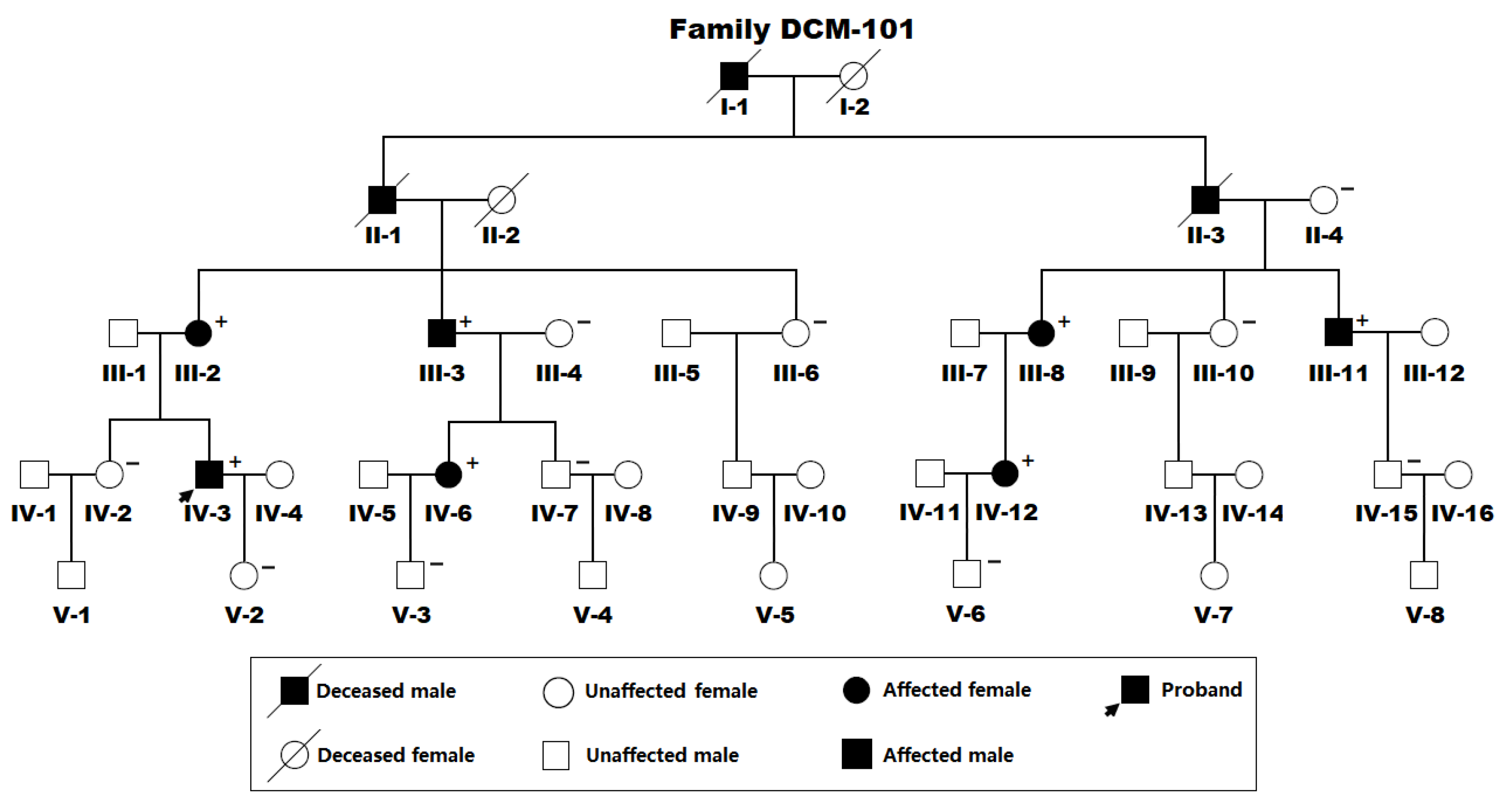
Figure 1. The pedigree of the studied family suffering from dilated cardiomyopathy. Family members are identified by generation and number (using a Roman and an Arabic numeral, respectively). “+” denotes a carrier of the detected ETS1 mutation in a heterogeneous status; “−” means a non-carrier.
Diagnostics 15 02031 g001
Figure 2. A novel ETS1 mutation identified to account for familial dilated cardiomyopathy. (A) Sequence chromatograms demonstrate the heterogeneous mutant ETS1 alleles (T/G) in the affected proband (mutant) and wild-type ETS1 alleles (T/T) in an unaffected individual (wild type). An arrow points to where the mutation occurs in the proband’s ETS1 allele. (B) The schematic diagrams signify the structural domains of the ETS1 protein. ETS1, erythroblast transformation-specific 1; TAD, transcription activation domain.
Diagnostics 15 02031 g002
Figure 3. Functional loss of Tyr149*-mutant ETS1 in transactivation of CLDN5. A dual reporter gene assay of the transactivation of the CLDN5 promoter-induced luciferase expression in cultivated HeLa cells by human wild-type ETS1-pCI-neo (ETS1) or Tyr149*-mutant ETS1-pCI-neo (Tyr149*), alone or together, showed that the Tyr149*-mutant ETS1 protein lost transactivation function. For each expression vector utilized, three independent cellular transfections were conducted and analyzed in triplicate. Herein, # and ## indicate p < 0.001 and p < 0.005.
Diagnostics 15 02031 g003
Figure 4. Inability of Tyr149*-mutant ETS1 to transactivate ALK1. A dual reporter gene assay of the transactivation of the ALK1 promoter-driven luciferase expression in HeLa cells cultured in vitro by human wild-type ETS1-pCI-neo (ETS1) or Tyr149*-mutant ETS1-pCI-neo (Tyr149*), singly or together, revealed that the Tyr149*-mutant ETS1 protein had no transcriptional function. For each plasmid utilized, three independent cellular transfections were performed and assayed in triplicate. Herein, # indicates p < 0.001 and ## indicates p < 0.01.
Diagnostics 15 02031 g004
Table 1. Demographic and baseline phenotypic characteristics of the affected pedigree members alive from Family DCM-101.
Individuals (Family DCM-101)SexesAges (Years)Cardiac PhenotypesLVEDD (mm)LVESD (mm)LVEF (%)LVFS (%)
III-2Female68DCM71623318
III-3Male65DCM76653014
III-8Female63DCM, AF70602713
III-11Male57DCM, AF69583216
IV-3Male45DCM58483618
IV-6Female40DCM, VSD64523819
IV-12Female38DCM, AF60504020
DCM, dilated cardiomyopathy; LVEF, left ventricular ejection fraction; AF, atrial fibrillation; LVESD, left ventricular end-systolic diameter; VSD, ventricular septal defect; LVEDD, left ventricular end-diastolic diameter; LVFS, left ventricular fractional shortening.
Table 2. Candidate genetic variations for dilated cardiomyopathy revealed by an exome-wide sequencing assay.
Chr.Position (hg19)Ref.Alt.GeneVariation
192,184,903GATGFBR3NM_003243.5: c.1532G>A; p.(Trp511*)
2141,777,525CGLRP1BNM_018557.3: c.1936C>G; p.(His646Asp)
371,096,210CGFOXP1NM_032682.6: c.547C>G; p.(Gln183Glu)
4157,693,868AGPDGFCNM_016205.3: c.673A>G; p.(Lys225Glu)
638,029,545GCZFAND3NM_021943.3: c.289G>C; p.(Glu97Gln)
7133,689,816CTEXOC4NM_021807.4: c.2500C>T; p.(Gln834*)
11128,355,998TGETS1NM_005238.4: c.447T>G; p.(Tyr149*)
1266,773,093GTGRIP1NM_021150.4: c.2432G>T; p.(Ser811Ile)
1586,266,500CGAKAP13NM_006738.6: c.6706C>G; p.(Leu2236Val)
1649,671,508TCZNF423NM_015069.5: c.1555T>C; p.(Cys519Arg)
1834,092,506GAFHOD3NM_025135.5: c.511G>A; p.(Ala171Thr)
Chr., chromosome; Ref., reference; Alt., alteration.
Table 3. Primers to specifically amplify the coding exons and splicing boundaries of the ETS1 gene.
Coding ExonsForward Primers (5′→3′)Reverse Primers (5′→3′)Amplicon Sizes (bp)
1AAAGTCGGATTTCCCCCGTCAGGGTTCTCGCTCATTTGGG515
2CTGGTGCTCTGGTGAGGATGCTGGTGCTCTGGTGAGGATG486
3TTGGCTGGCAGTAGTGACCTGGCACATTCCCACATACGTC571
4TCTTTAGACAAGGCCACAGTCATGGCCATCGTAGTGCACAGT568
5CATAGGGCCCTGTAGATGGTGGGACTTGGCTAAAACACCACG514
6CCCAAACGCTACTCCGACAGAAAGGGAGTCTCTCGTCGTC664
7GAAGAGGGATGTGGGAGTGCCACAGGAGGCAGCTCTATGG491
8TTGTGACTCCCATCCTTGCCGCTTTCCTTTCCCAACTGCG609
Disclaimer/Publisher’s Note: The statements, opinions and data contained in all publications are solely those of the individual author(s) and contributor(s) and not of MDPI and/or the editor(s). MDPI and/or the editor(s) disclaim responsibility for any injury to people or property resulting from any ideas, methods, instructions or products referred to in the content.