
Comparison of Tissue Factors in the Ontogenetic Aspects of Human Cholesteatoma


Abstract
1. Introduction
2. Materials and Methods
2.1. Tissue Samples
2.2. Immunohistochemical Analysis
2.3. Statistical Analysis
3. Results
3.1. Description of the Analyzed Tissue
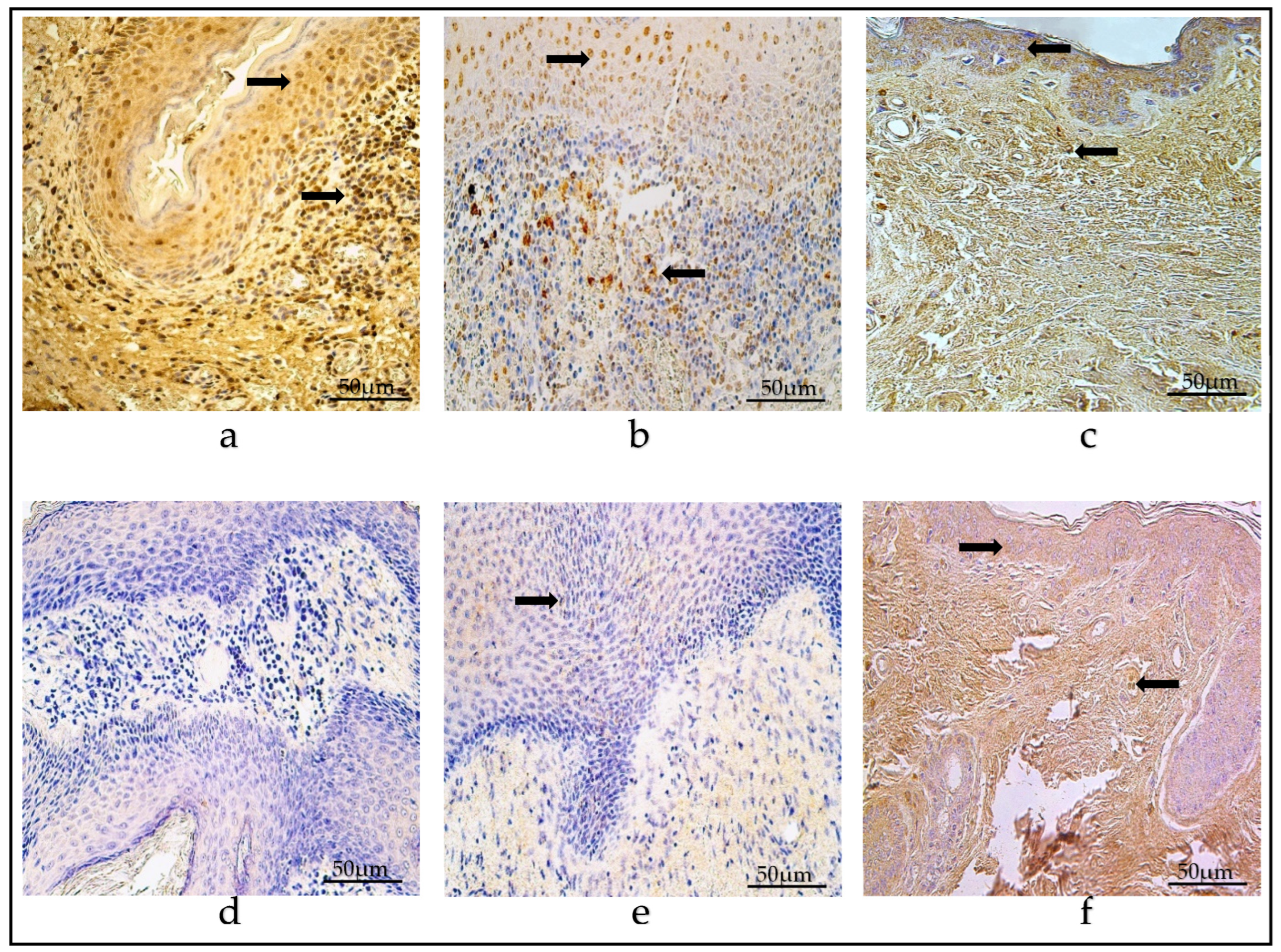
3.2. Immunohistochemistry of Defensins

| Groups | HβD-2 | HβD-4 | IL-1α | IL-10 | Ki-67 | NF-κβ | VEGF | SHH | MMP-2 | MMP-9 | TIMP-2 | TIMP-4 | ||||||||||||
|---|---|---|---|---|---|---|---|---|---|---|---|---|---|---|---|---|---|---|---|---|---|---|---|---|
| M | P | M | P | M | P | M | P | M | P | M | P | M | P | M | P | M | P | M | P | M | P | M | P | |
| Children | +/++ | + | 0/+-+ | 0/+-+ | +/++ | + | +/++ | + | 0/+ | 0/+-+ | ++ | + | ++ | 0/+-+ | ++ | +/++ | +/++ | + | 0/+-+ | 0/+ | + | 0/+ | ++/+++ | ++ |
| Adults | +/++ | + | + | 0/+ | +/++ | + | +/++ | + | 0/+ | 0/+ | ++ | + | +/++ | 0/+-+ | ++/+++ | +/++ | +/++ | + | 0/+ | 0/+ | + | 0/+ | ++/+++ | ++ |
| Control | E | CT | E | CT | E | CT | E | CT | E | CT | E | CT | E | CT | E | CT | E | CT | E | CT | E | CT | E | CT |
| + | 0-0/+ | + | 0/+ | 0/+-+ | 0/+-+ | +/++ | +/++ | 0-0/+ | 0-0/+ | 0/+ | 0/+-+ | ++/+++ | 0/+-+ | +/++ | 0/+-+ | + | + | + | 0/+-+ | +/++ | 0/+-+ | ++ | +/++ | |
3.3. Immunohistochemistry of Cytokines
3.4. Ki-67 Immunohistochemistry
3.5. NF-κβ Immunohistochemistry
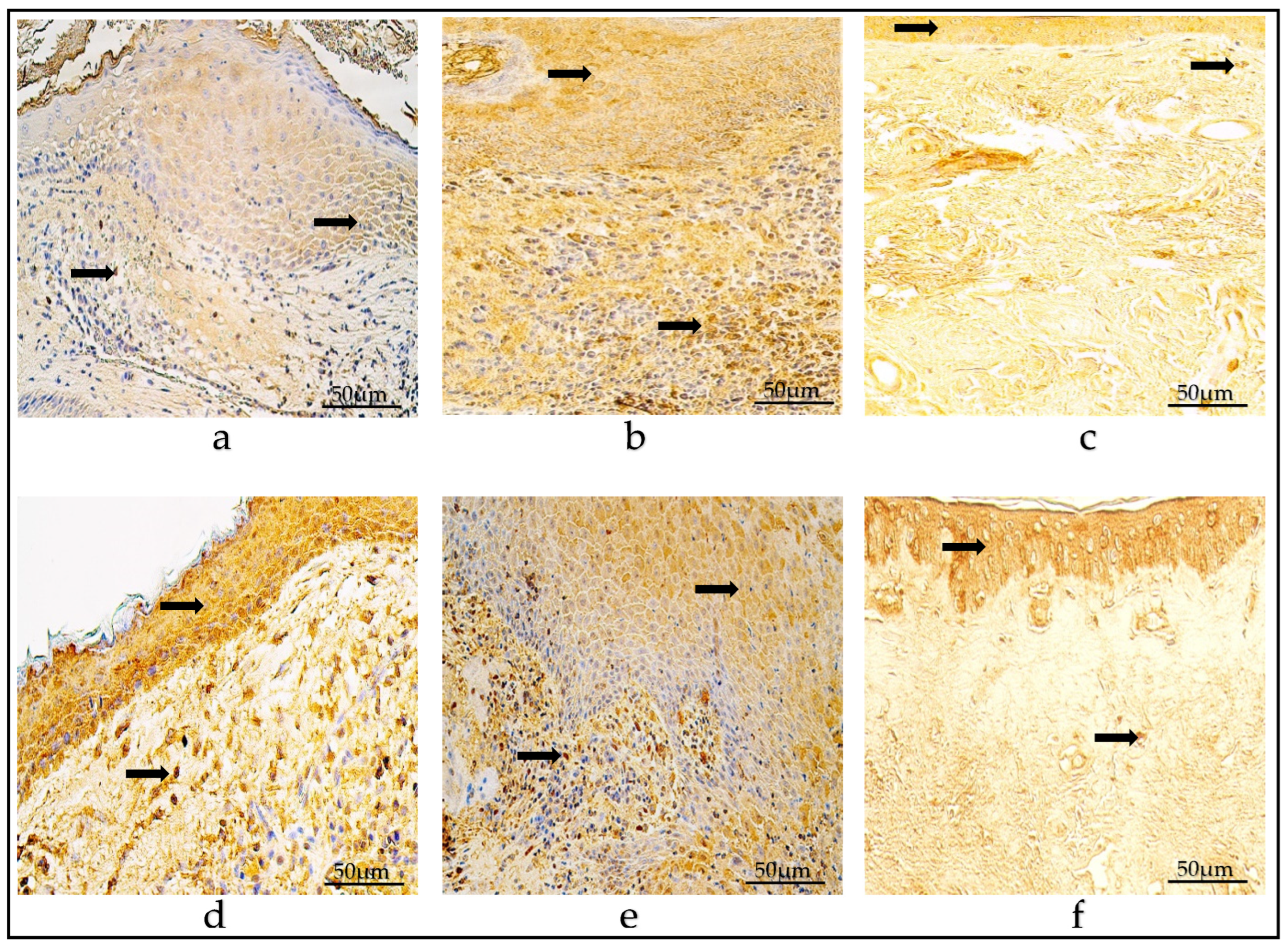
3.6. VEGF Immunohistochemistry
3.7. SHH Immunohistochemistry
3.8. Immunohistochemistry of Tissue-Remodeling Factors
3.9. Statistical Comparison between the Groups
| Detected Factor | Kruskal–Wallis Test | p-Value | |
|---|---|---|---|
| Children | Adults | ||
| HβD-2 matrix | HβD-2 matrix | 0.587 | >0.999 |
| HβD-2 perimatrix | HβD-2 perimatrix | 0.480 | >0.999 |
| HβD-4 matrix | HβD-4 matrix | 0.868 | >0.999 |
| HβD-4 perimatrix | HβD-4 perimatrix | −0.719 | >0.999 |
| IL-1α matrix | IL-1α matrix | −0.039 | >0.999 |
| IL-1α perimatrix | IL-1α perimatrix | 0.717 | >0.999 |
| IL-10 matrix | IL-10 matrix | 0.043 | >0.999 |
| IL-10 perimatrix | IL-10 perimatrix | 0.868 | >0.999 |
| Ki-67 matrix | Ki-67 matrix | 0.886 | >0.999 |
| Ki-67 perimatrix | Ki-67 perimatrix | 0.908 | >0.999 |
| NF-κβ matrix | NF-κβ matrix | −0.079 | >0.999 |
| NF-κβ perimatrix | NF-κβ perimatrix | 0.039 | >0.999 |
| VEGF matrix | VEGF matrix | −0.694 | >0.999 |
| VEGF perimatrix | VEGF perimatrix | 0.486 | >0.999 |
| SHH matrix | SHH matrix | 0.482 | >0.999 |
| SHH perimatrix | SHH perimatrix | 0.363 | >0.999 |
| MMP-2 matrix | MMP-2 matrix | 0.415 | >0.999 |
| MMP-2 perimatrix | MMP-2 perimatrix | 0.986 | 0.972 |
| MMP-9 matrix | MMP-9 matrix | −0.365 | >0.999 |
| MMP-9 perimatrix | MMP-9 perimatrix | 1.290 | 0.591 |
| TIMP-2 matrix | TIMP-2 matrix | 0.576 | >0.999 |
| TIMP-2 perimatrix | TIMP-2 perimatrix | 0.958 | >0.999 |
| TIMP-4 matrix | TIMP-4 matrix | −0.205 | >0.999 |
| TIMP-4 perimatrix | TIMP-4 perimatrix | 0.159 | >0.999 |
| Detected Factor | Kruskal–Wallis Test | p-Value |
|---|---|---|
| Adult HβD-2 P vs. Control HβD-2 CT | 2.815 | 0.015 |
| Children HβD-2 P vs. Control HβD-2 CT | 2.498 | 0.038 |
| Children HβD-4 M vs. Control HβD-4 E | −2.132 | 0.099 * |
| Adult Ki-67 M vs. Control Ki-67 E | 3.697 | 0.001 |
| Children Ki-67 M vs. Control Ki-67 E | 3.110 | 0.006 |
| Adult Ki-67 P vs. Control Ki-67 CT | 2.577 | 0.030 |
| Adult NF-κβ M vs. Control NF-κβ E | 2.864 | 0.013 |
| Children NF-κβ M vs. Control NF-κβ E | 2.915 | 0.011 |
| Adult VEGF M vs. Control VEGF E | −2.146 | 0.096 * |
| Adult SHH P vs. Control SHH CT | 3.146 | 0.005 |
| Children SHH P vs. Control SHH CT | 2.906 | 0.011 |
| Children TIMP-2 M vs. Control TIMP-2 E | -2.207 | 0.082 * |
3.10. Correlations between Tissue Factors in Patient Groups
| Factor 1 | Factor 2 | Spearman’s Correlation Coefficient; p Value | |
|---|---|---|---|
| MMP-2 matrix | MMP-2 perimatrix | Children r = 0.803; p = 0.000 | Adult r = 0.574; p = 0.003 |
| MMP-2 matrix | TIMP-2 matrix | Children r = 0.622; p = 0.001 | Adult r = 0.484; p = 0.014 |
| MMP-2 matrix | SHH matrix | Children r = 0.786; p = 0.000 | Adult r = 0.719; p = 0.000 |
| MMP-2 matrix | NF-κβ matrix | Children r = 0.677; p = 0.000 | Adult r = 0.399; p = 0.048 |
| MMP-2 perimatrix | SHH matrix | Children r = 0.786; p = 0.000 | Adult r = 0.453; p = 0.023 |
| MMP-2 perimatrix | SHH perimatrix | Children r = 0.653; p = 0.000 | Adult r = 0.460; p = 0.021 |
| MMP-9 matrix | IL-1α matrix | Children r = 0.549; p = 0.004 | Adult r = 0.426; p = 0.034 |
| MMP-9 matrix | IL-10 matrix | Children r = 0.418; p = 0.038 | Adult r = 0.458; p = 0.021 |
| MMP-9 perimatrix | TIMP-4 perimatrix | Children r = 0.490; p = 0.013 | Adult r = 0.664; p = 0.000 |
| MMP-9 perimatrix | IL-1α matrix | Children r = 0.642; p = 0.001 | Adult r = 0.435; p = 0.030 |
| MMP-9 perimatrix | IL-1α perimatrix | Children r = 0.714; p = 0.000 | Adult r = 0.608; p = 0.001 |
| MMP-9 perimatrix | IL-10 perimatrix | Children r = 0.468; p = 0.018 | Adult r = 0.601; p = 0.001 |
| MMP-9 perimatrix | NF-κβ perimatrix | Children r = 0.614; p = 0.001 | Adult r = 0.790; p = 0.000 |
| MMP-9 perimatrix | Ki-67 perimatrix | Children r = 0.624; p = 0.001 | Adult r = 0.677; p = 0.000 |
| MMP-9 perimatrix | HβD-2 matrix | Children r = 0.487; p = 0.014 | Adult r = 0.464; p = 0.019 |
| TIMP-2 matrix | TIMP-2 perimatrix | Children r = 0.685; p = 0.000 | Adult r = 0.676; p = 0.000 |
| TIMP-2 matrix | SHH matrix | Children r = 0.537; p = 0.006 | Adult r = 0.478; p = 0.016 |
| TIMP-2 matrix | NF-κβ matrix | Children r = 0.504; p = 0.010 | Adult r = 0.473; p = 0.017 |
| TIMP-2 matrix | HβD-2 matrix | Children r = 0.505; p = 0.010 | Adult r = 0.416; p = 0.038 |
| TIMP-2 perimatrix | IL-1α matrix | Children r = 0.457; p = 0.022 | Adult r = 0.505; p = 0.010 |
| TIMP-2 perimatrix | IL-10 matrix | Children r = 0.423; p = 0.035 | Adult r = 0.630; p = 0.001 |
| TIMP-4 matrix | TIMP-4 perimatrix | Children r = 0.841; p = 0.000 | Adult r = 0.431; p = 0.031 |
| TIMP-4 matrix | SHH matrix | Children r = 0.681; p = 0.000 | Adult r = 0.436; p = 0.029 |
| TIMP-4 matrix | NF-κβ matrix | Children r = 0.738; p = 0.000 | Adult r = 0.540; p = 0.005 |
| TIMP-4 perimatrix | IL-1α matrix | Children r = 0.641; p = 0.001 | Adult r = 0.457; p = 0.022 |
| TIMP-4 perimatrix | IL-1α perimatrix | Children r = 0.663; p = 0.000 | Adult r = 0.637; p = 0.001 |
| TIMP-4 perimatrix | IL-10 matrix | Children r = 0.638; p = 0.001 | Adult r = 0.425; p = 0.034 |
| TIMP-4 perimatrix | IL-10 perimatrix | Children r = 0.721; p = 0.000 | Adult r = 0.638; p = 0.001 |
| TIMP-4 perimatrix | NF-κβ matrix | Children r = 0.654; p = 0.000 | Adult r = 0.422; p = 0.036 |
| TIMP-4 perimatrix | NF-κβ perimatrix | Children r = 0.457; p = 0.022 | Adult r = 0.749; p = 0.000 |
| TIMP-4 perimatrix | Ki-67 matrix | Children r = 0.481; p = 0.015 | Adult r = 0.402; p = 0.046 |
| TIMP-4 perimatrix | Ki-67 perimatrix | Children r = 0.414; p = 0.040 | Adult r = 0.651; p = 0.000 |
| TIMP-4 perimatrix | HβD-2 matrix | Children r = 0.568; p = 0.003 | Adult r = 0.404; p = 0.045 |
| TIMP-4 perimatrix | HβD-2 perimatrix | Children r = 0.397; p = 0.049 | Adult r = 0.545; p = 0.005 |
| SHH matrix | NF-κβ matrix | Children r = 0.753; p = 0.000 | Adult r = 0.549; p = 0.005 |
| SHH perimatrix | Ki-67 matrix | Children r = 0.746; p = 0.000 | Adult r = 0.646; p = 0.000 |
| SHH perimatrix | HβD-2 perimatrix | Children r = 0.428; p = 0.033 | Adult r = 0.527; p = 0.007 |
| IL-1α matrix | IL-1α perimatrix | Children r = 0.716; p = 0.000 | Adult r = 0.557; p = 0.004 |
| IL-1α matrix | IL-10 matrix | Children r = 0.709; p = 0.000 | Adult r = 0.813; p = 0.000 |
| IL-1α matrix | IL-10 perimatrix | Children r = 0.720; p = 0.000 | Adult r = 0.762; p = 0.000 |
| IL-1α matrix | NF-κβ perimatrix | Children r = 0.406; p = 0.044 | Adult r = 0.519; p = 0.008 |
| IL-1α matrix | HβD-2 matrix | Children r = 0.700; p = 0.000 | Adult r = 0.827; p = 0.000 |
| IL-1α perimatrix | IL-10 perimatrix | Children r = 0.694; p = 0.000 | Adult r = 0.640; p = 0.001 |
| IL-1α perimatrix | NF-κβ perimatrix | Children r = 0.510; p = 0.009 | Adult r = 0.692; p = 0.000 |
| IL-1α perimatrix | Ki-67 perimatrix | Children r = 0.441; p = 0.027 | Adult r = 0.583; p = 0.002 |
| IL-1α perimatrix | HβD-2 matrix | Children r = 0.630; p = 0.001 | Adult r = 0.499; p = 0.011 |
| IL-10 matrix | IL-10 perimatrix | Children r = 0.668; p = 0.000 | Adult r = 0.801; p = 0.000 |
| IL-10 matrix | NF-κβ perimatrix | Children r = 0.536; p = 0.006 | Adult r = 0.554; p = 0.004 |
| IL-10 matrix | VEGF matrix | Children r = 0.559; p = 0.004 | Adult r = 0.611; p = 0.001 |
| IL-10 matrix | HβD-2 matrix | Children r = 0.828; p = 0.000 | Adult r = 0.841; p = 0.000 |
| IL-10 matrix | HβD-2 perimatrix | Children r = 0.677; p = 0.000 | Adult r = 0.462; p = 0.020 |
| IL-10 perimatrix | NF-κβ matrix | Children r = 0.602; p = 0.001 | Adult r = 0.396; p = 0.050 |
| IL-10 perimatrix | VEGF matrix | Children r = 0.696; p = 0.000 | Adult r = 0.687; p = 0.000 |
| IL-10 perimatrix | HβD-2 matrix | Children r = 0.592; p = 0.002 | Adult r = 0.686; p = 0.000 |
| IL-10 perimatrix | HβD-2 perimatrix | Children r = 0.516; p = 0.008 | Adult r = 0.687; p = 0.000 |
| NF-κβ matrix | VEGF matrix | Children r = 0.595; p = 0.002 | Adult r = 0.414; p = 0.039 |
| NF-κβ matrix | HβD-2 matrix | Children r = 0.750; p = 0.000 | Adult r = 0.418; p = 0.038 |
| NF-κβ perimatrix | Ki-67 perimatrix | Children r = 0.494; p = 0.012 | Adult r = 0.571; p = 0.003 |
| NF-κβ perimatrix | VEGF matrix | Children r = 0.637; p = 0.001 | Adult r = 0.474; p = 0.017 |
| NF-κβ perimatrix | HβD-2 matrix | Children r = 0.621; p = 0.001 | Adult r = 0.526; p = 0.007 |
| NF-κβ perimatrix | HβD-2 perimatrix | Children r = 0.635; p = 0.001 | Adult r = 0.748; p = 0.000 |
| Ki-67 matrix | HβD-2 perimatrix | Children r = 0.520; p = 0.008 | Adult r = 0.498; p = 0.011 |
| VEGF matrix | VEGF perimatrix | Children r = 0.745; p = 0.000 | Adult r = 0.429; p = 0.032 |
| VEGF matrix | HβD-2 perimatrix | Children r = 0.509; p = 0.009 | Adult r = 0.413; p = 0.040 |
| HβD-2 matrix | HβD-2 perimatrix | Children r = 0.748; p = 0.000 | Adult r = 0.477; p = 0.016 |
4. Discussion
4.1. Human Beta Defensins
4.2. Pro- and Anti-Inflammatory Cytokines
4.3. Proliferation Marker Ki-67
4.4. Transcription Factor NF-κβ
4.5. Angiogenetic Factor
4.6. Sonic Hedgehog
4.7. Remodeling Factors
5. Conclusions
Supplementary Materials
Author Contributions
Funding
Institutional Review Board Statement
Informed Consent Statement
Data Availability Statement
Acknowledgments
Conflicts of Interest
References
- Bhutta, M.F.; Williamson, I.G.; Sudhoff, H.H. Cholesteatoma. BMJ 2011, 342, d1088. [Google Scholar] [CrossRef]
- Kuo, C.L.; Shiao, A.S.; Yung, M.; Sakagami, M.; Sudhoff, H.; Wang, C.H.; Hsu, C.H.; Lien, C.F. Updates and knowledge gaps in cholesteatoma research. Biomed. Res. Int. 2015, 2015, 854024. [Google Scholar] [CrossRef]
- Britze, A.; Møller, M.L.; Ovesen, T. Incidence, 10-year recidivism rate and prognostic factors for cholesteatoma. J. Laryngol. Otol. 2017, 131, 319–328. [Google Scholar] [CrossRef]
- Olszewska, E.; Wagner, M.; Bernal-Sprekelsen, M.; Ebmeyer, J.; Dazert, S.; Hildmann, H.; Sudhoff, H. Etiopathogenesis of cholesteatoma. Eur. Arch. Otorhinolaryngol. 2004, 261, 6–24. [Google Scholar] [CrossRef] [PubMed]
- Harder, J.; Bartels, J.; Christophers, E.; Schröder, J.M. A peptide antibiotic from human skin. Nature 1997, 387, 861. [Google Scholar] [CrossRef] [PubMed]
- Schröder, J.M.; Harder, J. Human beta-defensin-2. Int. J. Biochem. Cell Biol. 1999, 31, 645–651. [Google Scholar] [CrossRef] [PubMed]
- Cieślik, M.; Bagińska, N.; Górski, A.; Jończyk-Matysiak, E. Human β-Defensin 2 and Its Postulated Role in Modulation of the Immune Response. Cells 2021, 10, 2991. [Google Scholar] [CrossRef] [PubMed]
- Nishimura, M.; Abiko, Y.; Kusano, K.; Yamazaki, M.; Saitoh, M.; Mizoguchi, I.; Jinbu, Y.; Noguchi, T.; Kaku, T. Localization of human beta-defensin 3 mRNA in normal oral epithelium, leukoplakia, and lichen planus: An in situ hybridization study. Med. Electron. Microsc. 2003, 36, 94–97. [Google Scholar] [CrossRef] [PubMed]
- Song, J.J.; Chae, S.W.; Woo, J.S.; Lee, H.M.; Jung, H.H.; Hwang, S.J. Differential expression of human beta defensin 2 and human beta defensin 3 in human middle ear cholesteatoma. Ann. Otol. Rhinol. Laryngol. 2007, 116, 235–240. [Google Scholar] [CrossRef] [PubMed]
- Park, K.; Moon, S.K.; Choung, Y.H.; Choi, H.S. Expression of beta-defensins in human middle ear cholesteatoma. Acta Otolaryngol. 2003, 123, 236–240. [Google Scholar] [CrossRef] [PubMed]
- García, J.R.; Krause, A.; Schulz, S.; Rodríguez-Jiménez, F.J.; Klüver, E.; Adermann, K.; Forssmann, U.; Frimpong-Boateng, A.; Bals, R.; Forssmann, W.G. Human beta-defensin 4: A novel inducible peptide with a specific salt-sensitive spectrum of antimicrobial activity. FASEB J. 2001, 15, 1819–1821. [Google Scholar] [CrossRef]
- Dambergs, K.; Sumeraga, G.; Pilmane, M. Complex Evaluation of Tissue Factors in Pediatric Cholesteatoma. Children 2021, 8, 926. [Google Scholar] [CrossRef]
- Dambergs, K.; Sumeraga, G.; Pilmane, M. Morphopathogenesis of Adult Acquired Cholesteatoma. Medicina 2023, 59, 306. [Google Scholar] [CrossRef]
- Schürmann, M.; Goon, P.; Sudhoff, H. Review of potential medical treatments for middle ear cholesteatoma. Cell Commun. Signal. 2022, 20, 148. [Google Scholar] [CrossRef]
- Dinarello, C.A. The interleukin-1 family: 10 years of discovery. FASEB J. 1994, 8, 1314–1325. [Google Scholar] [CrossRef]
- Bujía, J.; Kim, C.; Ostos, P.; Sudhoff, H.; Kastenbauer, E.; Hültner, L. Interleukin 1 (IL-1) and IL-1-receptor antagonist (IL-1-RA) in middle ear cholesteatoma: An analysis of protein production and biological activity. Eur. Arch. Otorhinolaryngol. 1996, 253, 252–255. [Google Scholar] [CrossRef]
- Lee, Y.M.; Fujikado, N.; Manaka, H.; Yasuda, H.; Iwakura, Y. IL-1 plays an important role in the bone metabolism under physiological conditions. Int. Immunol. 2010, 22, 805–816. [Google Scholar] [CrossRef]
- Mosser, D.M.; Zhang, X. Interleukin-10: New perspectives on an old cytokine. Immunol. Rev. 2008, 226, 205–218. [Google Scholar] [CrossRef] [PubMed]
- Sabat, R.; Grütz, G.; Warszawska, K.; Kirsch, S.; Witte, E.; Wolk, K.; Geginat, J. Biology of interleukin-10. Cytokine Growth Factor Rev. 2010, 21, 331–344. [Google Scholar] [CrossRef] [PubMed]
- Jung, M.; Sabat, R.; Krätzschmar, J.; Seidel, H.; Wolk, K.; Schönbein, C.; Schütt, S.; Friedrich, M.; Döcke, W.D.; Asadullah, K.; et al. Expression profiling of IL-10-regulated genes in human monocytes and peripheral blood mononuclear cells from psoriatic patients during IL-10 therapy. Eur. J. Immunol. 2004, 34, 481–493. [Google Scholar] [CrossRef] [PubMed]
- Kuczkowski, J.; Sakowicz-Burkiewicz, M.; Iżycka-Świeszewska, E.; Mikaszewski, B.; Pawełczyk, T. Expression of tumor necrosis factor-α, interleukin-1α, interleukin-6 and interleukin-10 in chronic otitis media with bone osteolysis. ORL J. Otorhinolaryngol. Relat. Spec. 2011, 73, 93–99. [Google Scholar] [CrossRef]
- Yeşilova, M.; Görür, K.; Ismi, O.; Özcan, C.; Büyükafşar, K. The Role of Rho/Rho-Kinase Pathway in the Pathogenesis of Cholesteatoma. Otol. Neurotol. 2017, 38, 516–520. [Google Scholar] [CrossRef]
- Araz Server, E.; Kalaycık Ertugay, Ç.; Baykal Koca, S.; Longur, E.S.; Yiğit, Ö.; Demirhan, H.; Çakır, Y. Predictive Role of Ki-67 and Proliferative-Cell Nuclear Antigen (PCNA) in Recurrent Cholesteatoma. J. Int. Adv. Otol. 2019, 15, 38–42. [Google Scholar] [CrossRef] [PubMed]
- Scholzen, T.; Gerdes, J. The Ki-67 protein: From the known and the unknown. J. Cell Physiol. 2000, 182, 311–322. [Google Scholar] [CrossRef]
- Hamed, M.A.; Nakata, S.; Shiogama, K.; Suzuki, K.; Sayed, R.H.; Nishimura, Y.; Iwata, N.; Sakurai, K.; Badawy, B.S.; Inada, K.I.; et al. Cytokeratin 13, Cytokeratin 17, and Ki-67 Expression in Human Acquired Cholesteatoma and Their Correlation with Its Destructive Capacity. Clin. Exp. Otorhinolaryngol. 2017, 10, 213–220. [Google Scholar] [CrossRef] [PubMed]
- Zhang, Q.; Lenardo, M.J.; Baltimore, D. 30 Years of NF-κB: A Blossoming of Relevance to Human Pathobiology. Cell 2017, 168, 37–57. [Google Scholar] [CrossRef] [PubMed]
- Giuliani, C.; Bucci, I.; Napolitano, G. The Role of the Transcription Factor Nuclear Factor-kappa B in Thyroid Autoimmunity and Cancer. Front. Endocrinol. 2018, 9, 471. [Google Scholar] [CrossRef]
- Byun, J.Y.; Yune, T.Y.; Lee, J.Y.; Yeo, S.G.; Park, M.S. Expression of CYLD and NF-kappaB in human cholesteatoma epithelium. Mediat. Inflamm. 2010, 2010, 796315. [Google Scholar] [CrossRef] [PubMed]
- Fukudome, S.; Wang, C.; Hamajima, Y.; Ye, S.; Zheng, Y.; Narita, N.; Sunaga, H.; Fujieda, S.; Hu, X.; Feng, L.; et al. Regulation of the angiogenesis of acquired middle ear cholesteatomas by inhibitor of DNA binding transcription factor. JAMA Otolaryngol. Head Neck Surg. 2013, 139, 273–278. [Google Scholar] [CrossRef] [PubMed]
- Duffy, A.M.; Bouchier-Hayes, D.J.; Harmey, J.H. Vascular Endothelial Growth Factor (VEGF) and Its Role in Non-Endothelial Cells: Autocrine Signalling by VEGF. In Madame Curie Bioscience Database; Landes Bioscience: Austin, TX, USA, 2013. [Google Scholar]
- Ankamreddy, H.; Bok, J.; Groves, A.K. Uncovering the secreted signals and transcription factors regulating the development of mammalian middle ear ossicles. Dev. Dyn. 2020, 249, 1410–1424. [Google Scholar] [CrossRef]
- Wright, C.G. Development of the human external ear. J. Am. Acad. Audiol. 1997, 8, 379–382. [Google Scholar]
- Brito, J.M.; Teillet, M.A.; Le Douarin, N.M. Induction of mirror-image supernumerary jaws in chicken mandibular mesenchyme by Sonic Hedgehog-producing cells. Development 2008, 135, 2311–2319. [Google Scholar] [CrossRef]
- Chiang, C.; Litingtung, Y.; Lee, E.; Young, K.E.; Corden, J.L.; Westphal, H.; Beachy, P.A. Cyclopia and defective axial patterning in mice lacking Sonic hedgehog gene function. Nature 1996, 383, 407–413. [Google Scholar] [CrossRef]
- Chole, R.A. The molecular biology of bone resorption due to chronic otitis media. Ann. N. Y. Acad. Sci. 1997, 830, 95–109. [Google Scholar] [CrossRef]
- Morales, D.S.; Penido Nde, O.; da Silva, I.D.; Stávale, J.N.; Guilherme, A.; Fukuda, Y. Matrix metalloproteinase 2: An important genetic marker for cholesteatomas. Braz. J. Otorhinolaryngol. 2007, 73, 51–57. [Google Scholar] [CrossRef] [PubMed]
- Juhász, A.; Sziklai, I.; Rákosy, Z.; Ecsedi, S.; Adány, R.; Balázs, M. Elevated level of tenascin and matrix metalloproteinase 9 correlates with the bone destruction capacity of cholesteatomas. Otol. Neurotol. 2009, 30, 559–565. [Google Scholar] [CrossRef] [PubMed]
- Galis, Z.S.; Khatri, J.J. Matrix metalloproteinases in vascular remodeling and atherogenesis: The good, the bad, and the ugly. Circ. Res. 2002, 90, 251–262. [Google Scholar] [CrossRef]
- Chakrabarti, S.; Patel, K.D. Matrix metalloproteinase-2 (MMP-2) and MMP-9 in pulmonary pathology. Exp. Lung Res. 2005, 31, 599–621. [Google Scholar] [CrossRef] [PubMed]
- Nikolov, A.; Popovski, N. Role of Gelatinases MMP-2 and MMP-9 in Healthy and Complicated Pregnancy and Their Future Potential as Preeclampsia Biomarkers. Diagnostics 2021, 11, 480. [Google Scholar] [CrossRef] [PubMed]
- Bergers, G.; Brekken, R.; McMahon, G.; Vu, T.H.; Itoh, T.; Tamaki, K.; Tanzawa, K.; Thorpe, P.; Itohara, S.; Werb, Z.; et al. Matrix metalloproteinase-9 triggers the angiogenic switch during carcinogenesis. Nat. Cell Biol. 2000, 2, 737–744. [Google Scholar] [CrossRef] [PubMed]
- Ezhilarasan, R.; Jadhav, U.; Mohanam, I.; Rao, J.S.; Gujrati, M.; Mohanam, S. The hemopexin domain of MMP-9 inhibits angiogenesis and retards the growth of intracranial glioblastoma xenograft in nude mice. Int. J. Cancer 2009, 124, 306–315. [Google Scholar] [CrossRef]
- Givvimani, S.; Tyagi, N.; Sen, U.; Mishra, P.K.; Qipshidze, N.; Munjal, C.; Vacek, J.C.; Abe, O.A.; Tyagi, S.C. MMP-2/TIMP-2/TIMP-4 versus MMP-9/TIMP-3 in transition from compensatory hypertrophy and angiogenesis to decompensatory heart failure. Arch. Physiol. Biochem. 2010, 116, 63–72. [Google Scholar] [CrossRef][Green Version]
- Schönermark, M.; Mester, B.; Kempf, H.G.; Bläser, J.; Tschesche, H.; Lenarz, T. Expression of matrix-metalloproteinases and their inhibitors in human cholesteatomas. Acta Oto-Laryngol. 1996, 116, 451–456. [Google Scholar] [CrossRef]
- Suchozebrska-Jesionek, D.; Szymański, M.; Kurzepa, J.; Gołabek, W.; Stryjecka-Zimmer, M. Gelatinolytic activity of matrix metalloproteinases 2 and 9 in middle ear cholesteatoma. J. Otolaryngol. Head Neck Surg. 2008, 37, 628–632. [Google Scholar]
- Pilmane, M.; Shine, J.; Iismaa, T.P. Distribution of galanin immunoreactivity in the bronchi of humans with tuberculosis. Ann. N. Y. Acad. Sci. 1998, 863, 445–449. [Google Scholar] [CrossRef]
- Moon, S.K.; Lee, H.Y.; Li, J.D.; Nagura, M.; Kang, S.H.; Chun, Y.M.; Linthicum, F.H.; Ganz, T.; Andalibi, A.; Lim, D.J. Activation of a Src-dependent Raf-MEK1/2-ERK signaling pathway is required for IL-1alpha-induced upregulation of beta-defensin 2 in human middle ear epithelial cells. Biochim. Biophys. Acta 2002, 1590, 41–51. [Google Scholar] [CrossRef]
- Wehkamp, K.; Schwichtenberg, L.; Schröder, J.M.; Harder, J. Pseudomonas aeruginosa- and IL-1beta-mediated induction of human beta-defensin-2 in keratinocytes is controlled by NF-kappaB and AP-1. J. Investig. Dermatol. 2006, 126, 121–127. [Google Scholar] [CrossRef]
- Kanda, N.; Kamata, M.; Tada, Y.; Ishikawa, T.; Sato, S.; Watanabe, S. Human β-defensin-2 enhances IFN-γ and IL-10 production and suppresses IL-17 production in T cells. J. Leukoc. Biol. 2011, 89, 935–944. [Google Scholar] [CrossRef] [PubMed]
- Yetiser, S.; Satar, B.; Aydin, N. Expression of epidermal growth factor, tumor necrosis factor-alpha, and interleukin-1alpha in chronic otitis media with or without cholesteatoma. Otol. Neurotol. 2002, 23, 647–652. [Google Scholar] [CrossRef] [PubMed]
- Artono; Surarto, B.; Purnami, N.; Hutahaen, F.; Mahardhika, M.R. The Association of IL-1 Alpha Level and TNF Alpha Expressions on Bone Destruction in Chronic Suppurative Otitis Media and Cholesteatoma. Indian. J. Otolaryngol. Head Neck Surg. 2020, 72, 1–7. [Google Scholar] [CrossRef] [PubMed]
- Kusano, K.; Miyaura, C.; Inada, M.; Tamura, T.; Ito, A.; Nagase, H.; Kamoi, K.; Suda, T. Regulation of matrix metalloproteinases (MMP-2, -3, -9, and -13) by interleukin-1 and interleukin-6 in mouse calvaria: Association of MMP induction with bone resorption. Endocrinology 1998, 139, 1338–1345. [Google Scholar] [CrossRef]
- Mertz, P.M.; DeWitt, D.L.; Stetler-Stevenson, W.G.; Wahl, L.M. Interleukin 10 suppression of monocyte prostaglandin H synthase-2. Mechanism of inhibition of prostaglandin-dependent matrix metalloproteinase production. J. Biol. Chem. 1994, 269, 21322–21329. [Google Scholar] [CrossRef]
- Lee, E.J.; Kim, H.S. The anti-inflammatory role of tissue inhibitor of metalloproteinase-2 in lipopolysaccharide-stimulated microglia. J. Neuroinflamm. 2014, 11, 116. [Google Scholar] [CrossRef]
- Sikka, K.; Sharma, S.C.; Thakar, A.; Dattagupta, S. Evaluation of epithelial proliferation in paediatric and adult cholesteatomas using the Ki-67 proliferation marker. J. Laryngol. Otol. 2012, 126, 460–463. [Google Scholar] [CrossRef]
- Bujía, J.; Holly, A.; Antolí-Candela, F.; Tapia, M.G.; Kastenbauer, E. Immunobiological peculiarities of cholesteatoma in children: Quantification of epithelial proliferation by MIB1. Laryngoscope 1996, 106, 865–868. [Google Scholar] [CrossRef]
- Chung, J.H.; Lee, S.H.; Park, C.W.; Kim, K.R.; Tae, K.; Kang, S.H.; Oh, Y.H.; Pyo, J.Y. Expression of Apoptotic vs Antiapoptotic Proteins in Middle Ear Cholesteatoma. Otolaryngol. Head Neck Surg. 2015, 153, 1024–1030. [Google Scholar] [CrossRef]
- Akdogan, V.; Yilmaz, I.; Canpolat, T.; Ozluoglu, L.N. Role of Langerhans cells, Ki-67 protein and apoptosis in acquired cholesteatoma: Prospective clinical study. J. Laryngol. Otol. 2013, 127, 252–259. [Google Scholar] [CrossRef]
- Hamajima, Y.; Komori, M.; Preciado, D.A.; Choo, D.I.; Moribe, K.; Murakami, S.; Ondrey, F.G.; Lin, J. The role of inhibitor of DNA-binding (Id1) in hyperproliferation of keratinocytes: The pathological basis for middle ear cholesteatoma from chronic otitis media. Cell Prolif. 2010, 43, 457–463. [Google Scholar] [CrossRef]
- Shuman Moss, L.A.; Jensen-Taubman, S.; Stetler-Stevenson, W.G. Matrix metalloproteinases: Changing roles in tumor progression and metastasis. Am. J. Pathol. 2012, 181, 1895–1899. [Google Scholar] [CrossRef]
- Zhu, J.; Zhang, X.; Ai, L.; Yuan, R.; Ye, J. Clinicohistopathological implications of MMP/VEGF expression in retinoblastoma: A combined meta-analysis and bioinformatics analysis. J. Transl. Med. 2019, 17, 226. [Google Scholar] [CrossRef] [PubMed]
- Olszewska, E.; Chodynicki, S.; Chyczewski, L. Znaczenie angiogenezy w patogenezie perlaka ucha środkowego u dorosłych [Role of angiogenesis in the pathogenesis of cholesteatoma in adults]. Otolaryngol. Pol. 2004, 58, 559–563. [Google Scholar]
- Viac, J.; Palacio, S.; Schmitt, D.; Claudy, A. Expression of vascular endothelial growth factor in normal epidermis, epithelial tumors and cultured keratinocytes. Arch. Dermatol. Res. 1997, 289, 158–163. [Google Scholar] [CrossRef]
- Ma, J.; Tian, L.; Cheng, J.; Chen, Z.; Xu, B.; Wang, L.; Li, C.; Huang, Q. Sonic hedgehog signaling pathway supports cancer cell growth during cancer radiotherapy. PLoS ONE 2013, 8, e65032. [Google Scholar] [CrossRef]
- de Carvalho Dornelles, C.; da Costa, S.S.; Meurer, L.; Rosito, L.P.; da Silva, A.R.; Alves, S.L. Comparison of acquired cholesteatoma between pediatric and adult patients. Eur. Arch. Otorhinolaryngol. 2009, 266, 1553–1561. [Google Scholar] [CrossRef]
- Banerjee, A.R.; James, R.; Narula, A.A. Matrix metalloproteinase-2 and matrix metalloproteinase-9 in cholesteatoma and deep meatal skin. Clin. Otolaryngol. Allied Sci. 1998, 23, 345–347. [Google Scholar] [CrossRef] [PubMed]
- Rezende, C.E.; Souto, R.P.; Rapoport, P.B.; Campos, L.D.; Generato, M.B. Cholesteatoma gene expression of matrix metalloproteinases and their inhibitors by RT-PCR. Braz. J. Otorhinolaryngol. 2012, 78, 116–121. [Google Scholar] [CrossRef]
- Kaya, İ.; Avcı, Ç.B.; Şahin, F.F.; Özateş, N.P.; Sezgin, B.; Kurt, C.Ç.; Bilgen, C.; Kirazlı, T. Evaluation of significant gene expression changes in congenital and acquired cholesteatoma. Mol. Biol. Rep. 2020, 47, 6127–6133. [Google Scholar] [CrossRef] [PubMed]
- Sun, J. Matrix metalloproteinases and tissue inhibitor of metalloproteinases are essential for the inflammatory response in cancer cells. J. Signal Transduct. 2010, 2010, 985132. [Google Scholar] [CrossRef]
- Preciado, D.A. Biology of cholesteatoma: Special considerations in pediatric patients. Int. J. Pediatr. Otorhinolaryngol. 2012, 76, 319–321. [Google Scholar] [CrossRef]


| Grading Scale | Explanation of Grading Scale | Percentage of Factor-Positive Cells in the Visual Field |
|---|---|---|
| 0 | No positive structures | 0% |
| 0/+ | Occasional positive structures | 12.5% |
| + | Few positive structures | 25% |
| +/++ | Few-to-moderate number of positive structures | 37.5% |
| ++ | Moderate number of positive structures | 50% |
| ++/+++ | Moderate-to-numerous positive structures | 62.5% |
| +++ | Numerous positive structures | 75% |
| +++/++++ | Numerous-to-abundant structures | 87.5% |
| ++++ | An abundance of positive structures in the visual field | 100% |
Disclaimer/Publisher’s Note: The statements, opinions and data contained in all publications are solely those of the individual author(s) and contributor(s) and not of MDPI and/or the editor(s). MDPI and/or the editor(s) disclaim responsibility for any injury to people or property resulting from any ideas, methods, instructions or products referred to in the content. |
© 2024 by the authors. Licensee MDPI, Basel, Switzerland. This article is an open access article distributed under the terms and conditions of the Creative Commons Attribution (CC BY) license (https://creativecommons.org/licenses/by/4.0/).
Share and Cite
Dambergs, K.; Sumeraga, G.; Pilmane, M. Comparison of Tissue Factors in the Ontogenetic Aspects of Human Cholesteatoma. Diagnostics 2024, 14, 662. https://doi.org/10.3390/diagnostics14060662
Dambergs K, Sumeraga G, Pilmane M. Comparison of Tissue Factors in the Ontogenetic Aspects of Human Cholesteatoma. Diagnostics. 2024; 14(6):662. https://doi.org/10.3390/diagnostics14060662
Chicago/Turabian StyleDambergs, Kristaps, Gunta Sumeraga, and Māra Pilmane. 2024. "Comparison of Tissue Factors in the Ontogenetic Aspects of Human Cholesteatoma" Diagnostics 14, no. 6: 662. https://doi.org/10.3390/diagnostics14060662
APA StyleDambergs, K., Sumeraga, G., & Pilmane, M. (2024). Comparison of Tissue Factors in the Ontogenetic Aspects of Human Cholesteatoma. Diagnostics, 14(6), 662. https://doi.org/10.3390/diagnostics14060662