A New Method of Predicting Final Mandibular Length Based on the Morphology of Cervical Vertebrae


Abstract
1. Introduction
2. Materials and Methods
2.1. Data Source
2.2. Cephalometric Measurements
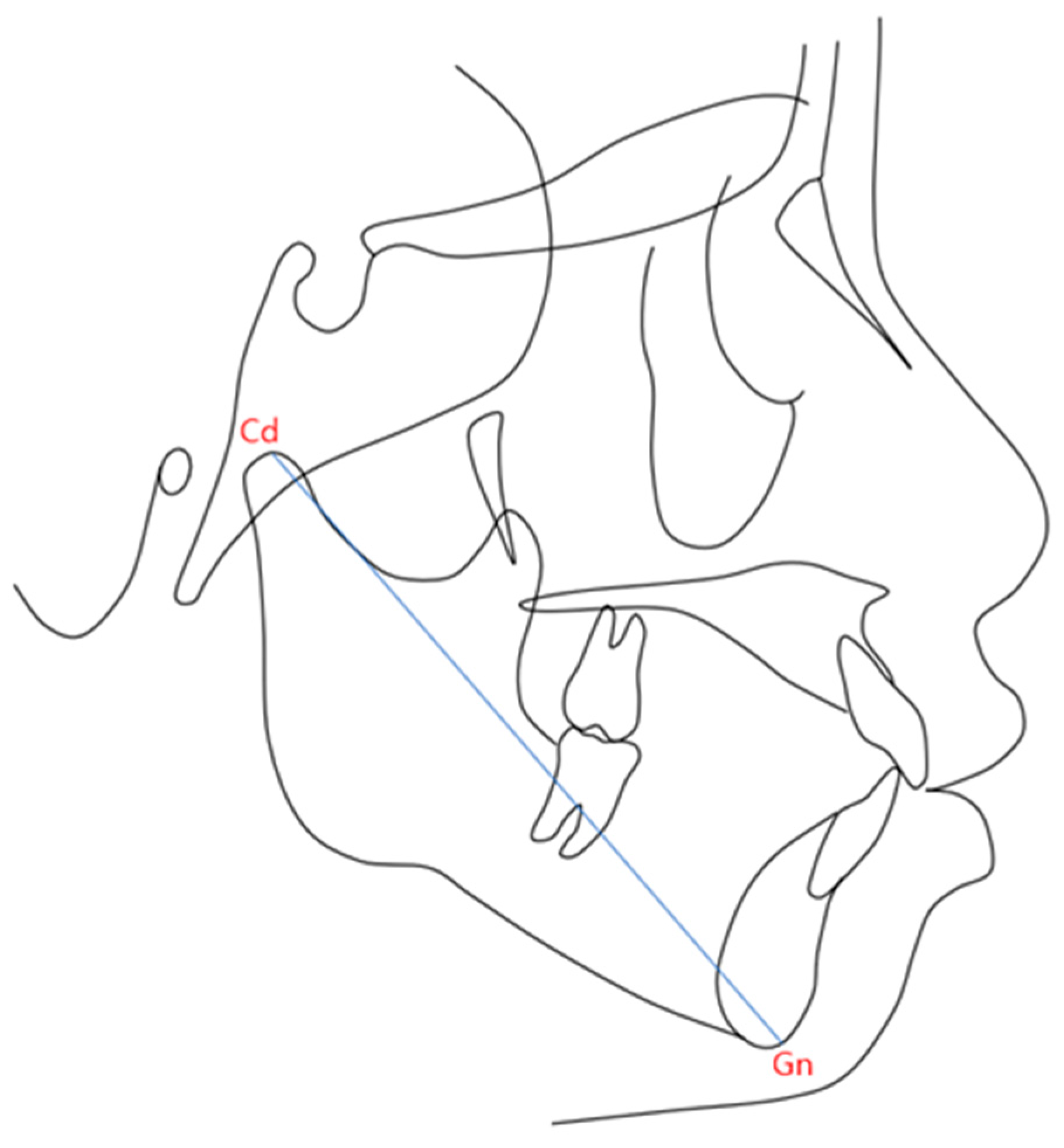
- Condyle (Cd): The most superior point on the mandibular condyle.
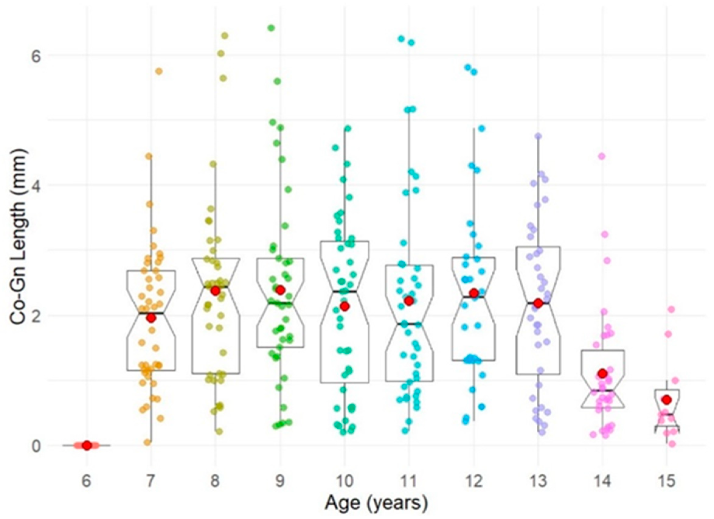
- Gnathion (Gn): The most anterior and inferior point of the mandibular contour, determined by bisecting the angle formed by the mandibular border (Menton-Gonion) and the facial plane (Nasion–Pogonion). The distance between Cd and Gn was defined as the mandibular length (Figure 1).
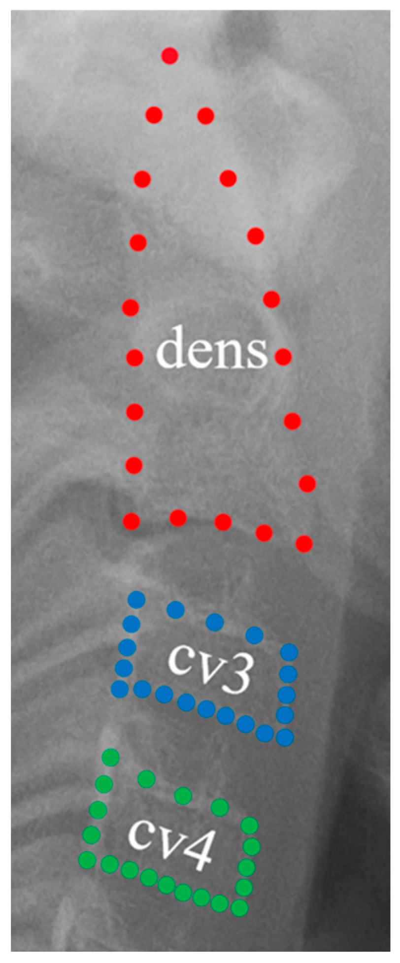
2.3. Cervical Vertebrae Analysis
2.4. Statistical Analysis
3. Results
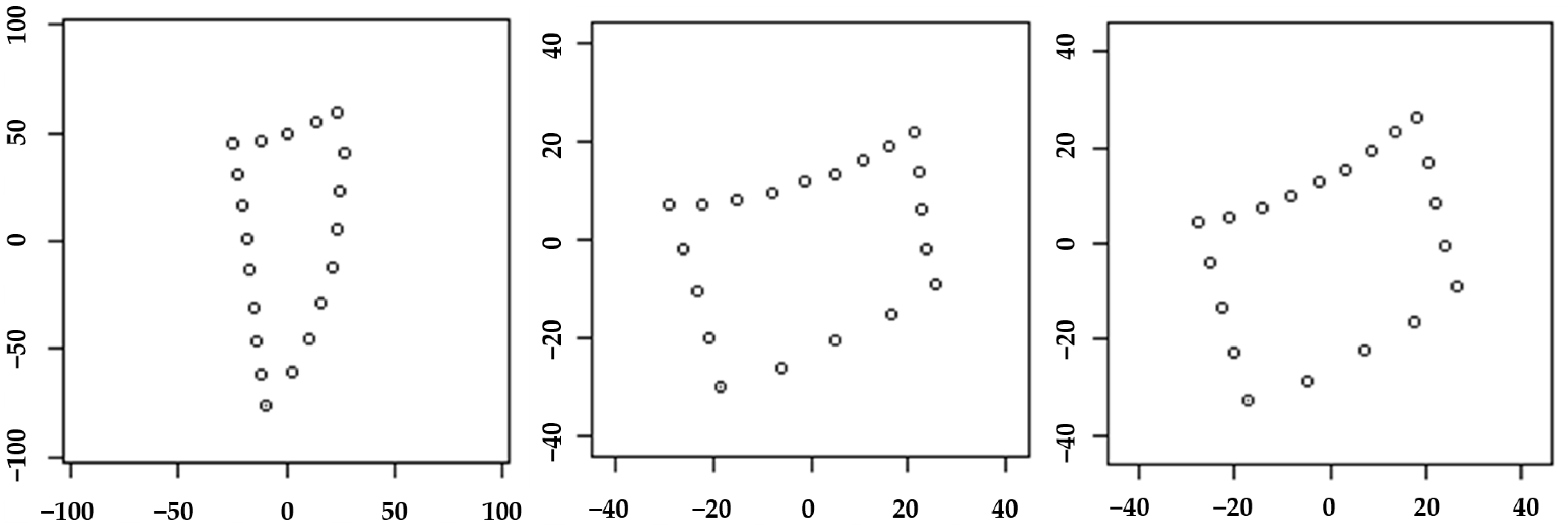
3.1. Statistical Shape Analysis
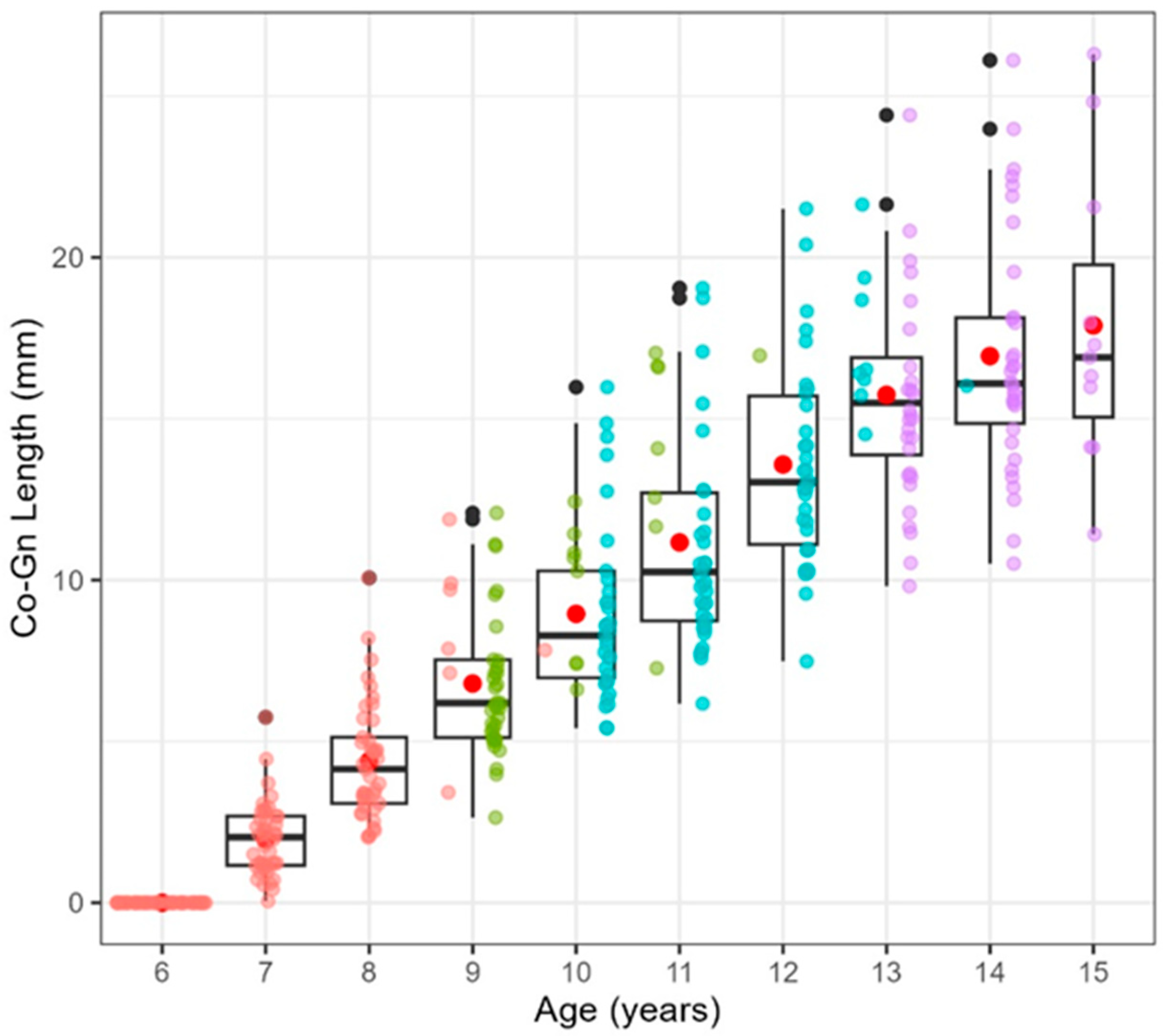
3.2. Linear Mixed Model for Mandibular Length Prediction
4. Discussion
5. Conclusions
Author Contributions
Funding
Institutional Review Board Statement
Informed Consent Statement
Data Availability Statement
Conflicts of Interest
References
- Moore, R.N.; Moyer, B.A.; DuBois, L.M. Skeletal maturation and craniofacial growth. Am. J. Orthod. Dentofac. Orthop. 1990, 98, 33–40. [Google Scholar] [CrossRef]
- Manabe, A.; Ishida, T.; Kanda, E.; Ono, T. Evaluation of maxillary and mandibular growth patterns with cephalometric analysis based on cervical vertebral maturation: A Japanese cross-sectional study. PLoS ONE 2022, 17, e0265272. [Google Scholar] [CrossRef]
- Mito, T.; Sato, K.; Mitani, H. Predicting mandibular growth potential with cervical vertebral bone age. Am. J. Orthod. Dentofac. Orthop. 2003, 124, 173–177. [Google Scholar] [CrossRef]
- Moon, J.H.; Kim, M.G.; Hwang, H.W.; Cho, S.J.; Donatelli, R.E.; Lee, S.J. Evaluation of an individualized facial growth prediction model based on the multivariate partial least squares method. Angle Orthod. 2022, 92, 705–713. [Google Scholar] [CrossRef]
- Rudolph, D.J.; White, S.E.; Sinclair, P.M. Multivariate prediction of skeletal Class II growth. Am. J. Orthod. Dentofac. Orthop. 1998, 114, 283–291. [Google Scholar] [CrossRef] [PubMed]
- Moon, J.H.; Shin, H.K.; Lee, J.M.; Cho, S.J.; Park, J.A.; Donatelli, R.E.; Lee, S.J. Comparison of individualized facial growth prediction models based on the partial least squares and artificial intelligence. Angle Orthod. 2024, 94, 207–215. [Google Scholar] [CrossRef] [PubMed]
- Sato, K.; Mito, T.; Mitani, H. An accurate method of predicting mandibular growth potential based on bone maturity. Am. J. Orthod. Dentofac. Orthop. 2001, 120, 286–293. [Google Scholar] [CrossRef]
- Larkin, A.; Kim, J.S.; Kim, N.; Baek, S.H.; Yamada, S.; Park, K.; Tai, K.; Yanagi, Y.; Park, J.H. Accuracy of artificial intelligence-assisted growth prediction in skeletal Class I preadolescent patients using serial lateral cephalograms for a 2-year growth interval. Orthod. Craniofacial Res. 2024, 27, 535–543. [Google Scholar] [CrossRef]
- Wood, T.; Anigbo, J.O.; Eckert, G.; Stewart, K.T.; Dundar, M.M.; Turkkahraman, H. Prediction of the post-pubertal mandibular length and Y axis of growth by using various machine learning techniques: A retrospective longitudinal study. Diagnostics 2023, 13, 1553. [Google Scholar] [CrossRef]
- Miller, C.A.; Hwang, S.J.; Cotter, M.M.; Vorperian, H.K. Cervical vertebral body growth and emergence of sexual dimorphism: A developmental study using computed tomography. J. Anat. 2019, 234, 764–777. [Google Scholar] [CrossRef] [PubMed]
- Brandt, S. Letter to the Editor: In defense of bonding. Am. J. Orthod. 1975, 68, 458. [Google Scholar] [CrossRef] [PubMed]
- Chen, F.; Terada, K.; Hanada, K. A new method of predicting mandibular length increment on the basis of cervical vertebrae. Angle Orthod. 2004, 74, 630–634. [Google Scholar] [CrossRef]
- Mito, T.; Sato, K.; Mitani, H. Cervical vertebral bone age in girls. Am. J. Orthod. Dentofac. Orthop. 2002, 122, 380–385. [Google Scholar] [CrossRef]
- Sousa-e-Silva, P.; Coelho-E-Silva, M.J.; Seabra, A.; Costa, D.C.; Martinho, D.V.; Duarte, J.P.; Oliveira, T.; Gonçalves-Santos, J.; Rodrigues, I.; Ribeiro, L.P.; et al. Skeletal age assessed by TW2 using 20-bone, carpal and RUS score systems: Intra-observer and inter-observer agreement among male pubertal soccer players. PLoS ONE 2022, 17, e0271386. [Google Scholar] [CrossRef]
- King, D.G.; Steventon, D.M.; O’Sullivan, M.P.; Cook, A.M.; Hornsby, V.P.; Jefferson, I.G.; King, P.R. Reproducibility of bone ages when performed by radiology registrars: An audit of Tanner and Whitehouse II versus Greulich and Pyle methods. Br. J. Radiol. 1994, 67, 848–851. [Google Scholar] [CrossRef]
- Franchi, L.; Nieri, M.; McNamara, J.A., Jr.; Giuntini, V. Predicting mandibular growth based on CVM stage and gender and with chronological age as a curvilinear variable. Orthod. Craniofacial Res. 2021, 24, 414–420. [Google Scholar] [CrossRef] [PubMed]
- Galvão, M.C.S.; Sato, J.R.; Coelho, E.C. Dahlberg formula: A novel approach for its evaluation. Dent. Press. J. Orthod. 2012, 17, 115–124. [Google Scholar] [CrossRef]
- Shin, S.M.; Kim, Y.I.; Choi, Y.S.; Yamaguchi, T.; Maki, K.; Cho, B.H.; Park, S.B. The skeletal maturation status estimated by statistical shape analysis: Axial images of Japanese cervical vertebra. Dentomaxillofac. Radiol. 2015, 44, 20140323. [Google Scholar] [CrossRef]
- Sandoval, C.; Díaz, A.; Manríquez, G. Assessing cervical spine and craniofacial morphology in Class II and Class III malocclusions: A geometric morphometric approach. Cranio 2024, 42, 450–460. [Google Scholar] [CrossRef]
- Al-Taai, N.; Persson, M.; Ransjö, M.; Levring Jäghagen, E.; Fors, R.; Westerlund, A. Craniofacial changes from 13 to 62 years of age. Eur. J. Orthod. 2022, 44, 556–565. [Google Scholar] [CrossRef]
- Baccetti, T.; Reyes, B.C.; McNamara, J.A., Jr. Craniofacial changes in Class III malocclusion as related to skeletal and dental maturation. Am. J. Orthod. Dentofac. Orthop. 2007, 132, 171.e1–171.e12. [Google Scholar] [CrossRef]
- O’Reilly, M.T.; Yanniello, G.J. Mandibular growth changes and maturation of cervical vertebrae--a longitudinal cephalometric study. Angle Orthod. 1988, 58, 179–184. [Google Scholar] [CrossRef] [PubMed]
- Baccetti, T.; Franchi, L.; McNamara, J.A., Jr. An improved version of the cervical vertebral maturation (CVM) method for the assessment of mandibular growth. Angle Orthod. 2002, 72, 316–323. [Google Scholar] [CrossRef] [PubMed]
- Chandrasekar, R.; Chandrasekhar, S.; Sundari, K.K.S.; Ravi, P. Development and validation of a formula for objective assessment of cervical vertebral bone age. Prog. Orthod. 2020, 21, 38. [Google Scholar] [CrossRef] [PubMed]
- Byun, B.R.; Kim, Y.I.; Yamaguchi, T.; Maki, K.; Son, W.S. Quantitative assessment of cervical vertebral maturation using cone beam computed tomography in Korean girls. Comput. Math. Methods Med. 2015, 2015, 405912. [Google Scholar] [CrossRef] [PubMed]
- Altan, M.; Nebioğlu Dalci, Ö.; İseri, H. Growth of the cervical vertebrae in girls from 8 to 17 years. A longitudinal study. Eur. J. Orthod. 2012, 34, 327–334. [Google Scholar] [CrossRef]
- Chen, L.L.; Xu, T.M.; Jiang, J.H.; Zhang, X.Z.; Lin, J.X. Quantitative cervical vertebral maturation assessment in adolescents with normal occlusion: A mixed longitudinal study. Am. J. Orthod. Dentofac. Orthop. 2008, 134, 720.e1–720.e7; discussion 720–721. [Google Scholar] [CrossRef] [PubMed]
- Cameriere, R.; Giuliodori, A.; Zampi, M.; Galić, I.; Cingolani, M.; Pagliara, F.; Ferrante, L. Age estimation in children and young adolescents for forensic purposes using fourth cervical vertebra (C4). Int. J. Leg. Med. 2015, 129, 347–355. [Google Scholar] [CrossRef]
- Tanner, J.M.; Whitehouse, R.H.; Marubini, E.; Resele, L.F. The adolescent growth spurt of boys and girls of the Harpenden growth study. Ann. Hum. Biol. 1976, 3, 109–126. [Google Scholar] [CrossRef]
- Grave, K.; Townsend, G. Cervical vertebral maturation as a predictor of the adolescent growth spurt. Aust. Orthod. J. 2003, 19, 25–32, Erratum in Aust. Orthod. J. 2003, 19, 44A. [Google Scholar] [CrossRef]

| Predictors (n = 370) | Estimates | Confidence Interval | p-Value |
|---|---|---|---|
| (Intercept) | 82.850 | 82.58–83.11 | <0.001 |
| age-6 | 2.250 | 2.12–2.37 | <0.001 |
| initial value adjust mean | 1.010 | 0.98–1.04 | <0.001 |
| (age-6) × initial value adj | 0.040 | 0.02–0.05 | <0.001 |
| cv4 PC1 | −0.320 | −0.47–−0.18 | <0.001 |
| Random Effects | |||
| σ2 | 1.2 | ||
| τ00 | 0.19id | ||
| τ11 | 0.14id.visit | ||
| ρ01 | 0.55id | ||
| N | 44id | ||
| Marginal R2/Conditional R2 | 0.957/0.990 | ||
| Deviance | 1263.838 | ||
| AIC | 1305.775 | ||
| AICc | 1306.275 | ||
| log-Likelihood | −643.887 | ||
| BIC | 1328.435 | ||
Disclaimer/Publisher’s Note: The statements, opinions and data contained in all publications are solely those of the individual author(s) and contributor(s) and not of MDPI and/or the editor(s). MDPI and/or the editor(s) disclaim responsibility for any injury to people or property resulting from any ideas, methods, instructions or products referred to in the content. |
© 2024 by the authors. Licensee MDPI, Basel, Switzerland. This article is an open access article distributed under the terms and conditions of the Creative Commons Attribution (CC BY) license (https://creativecommons.org/licenses/by/4.0/).
Share and Cite
Yamaguchi, M.; Kim, Y.-I.; Park, H.; Yamaguchi, T. A New Method of Predicting Final Mandibular Length Based on the Morphology of Cervical Vertebrae. Diagnostics 2024, 14, 2879. https://doi.org/10.3390/diagnostics14242879
Yamaguchi M, Kim Y-I, Park H, Yamaguchi T. A New Method of Predicting Final Mandibular Length Based on the Morphology of Cervical Vertebrae. Diagnostics. 2024; 14(24):2879. https://doi.org/10.3390/diagnostics14242879
Chicago/Turabian StyleYamaguchi, Manami, Yong-Il Kim, Heetae Park, and Tetsutaro Yamaguchi. 2024. "A New Method of Predicting Final Mandibular Length Based on the Morphology of Cervical Vertebrae" Diagnostics 14, no. 24: 2879. https://doi.org/10.3390/diagnostics14242879
APA StyleYamaguchi, M., Kim, Y.-I., Park, H., & Yamaguchi, T. (2024). A New Method of Predicting Final Mandibular Length Based on the Morphology of Cervical Vertebrae. Diagnostics, 14(24), 2879. https://doi.org/10.3390/diagnostics14242879