Case Report

Persistent Müllerian Duct Syndrome with Supernumerary Testicles Due to a Novel Homozygous Variant in the AMHR2 Gene and Literature Review

by
Luminita Nicoleta Cima
1,2,
Iustina Grosu
1,
Isabela Magdalena Draghici
2,3,*,
Augustina Cornelia Enculescu
4,
Adela Chirita-Emandi
5,6,
Nicoleta Andreescu
5,6,
Maria Puiu
5,6,
Carmen Gabriela Barbu
1,2 and
Simona Fica
1,2
1
Endocrinology Department, Elias Emergency University Hospital, 011461 Bucharest, Romania
2
Faculty of General Medicine, Carol Davila University of Medicine and Pharmacy, 050474 Bucharest, Romania
3
Pediatric Surgery Department, Maria Sklodowska Curie Emergency Hospital for Children, 077120 Bucharest, Romania
4
Pathology Department, Maria Sklodowska Curie Emergency Hospital for Children, 077120 Bucharest, Romania
5
Department of Microscopic Morphology, Genetics Discipline, Center of Genomic Medicine, University of Medicine and Pharmacy “Victor Babeș”, 400347 Timișoara, Romania
6
Regional Center of Medical Genetics Timiș, Clinical Emergency Hospital for Children “Louis Țurcanu”, 300011 Timișoara, Romania
*
Author to whom correspondence should be addressed.
Diagnostics 2024, 14(23), 2621; https://doi.org/10.3390/diagnostics14232621
Submission received: 26 September 2024 / Revised: 18 November 2024 / Accepted: 19 November 2024 / Published: 21 November 2024
(This article belongs to the Section Pathology and Molecular Diagnostics)

Abstract

:
Introduction: Persistent Müllerian duct syndrome (PMDS) is a rare disorder of sex development (DSD) caused by mutations in the genes coding anti-Müllerian hormone (AMH) or the AMH receptor, characterized by the persistence of Müllerian derivatives, the uterus and/or fallopian tubes, in otherwise normally virilized boys. Testicular regression syndrome is common in PMDS, yet the association with supernumerary testis has been reported in only two patients where genetic testing was not performed. Method: Thus, we report an individual with this particular association caused by a previously unreported homozygous variant in the AMHR2 gene to enable future genotype–phenotype correlations in this rare disorder. In addition, a search of PMDS associated with congenital anomalies reported in the literature was performed to provide a comprehensive overview of this pathology. Results: We present the case of a 13-year-old boy with a history of bilateral cryptorchidism. Two attempts of right orchidopexy were performed at the age of 4 and 5 years. At that time, exploratory laparoscopy identified an intra-abdominal left testicle. In addition, a fibrous structure extending from the left intra-abdominal testicle to the deep inguinal ring (Müllerian duct remnants) and a medially located abdominal mass, bilaterally fixated to the parietal peritoneum (uterine remnant), were detected. The left testicular biopsy revealed immature prepubertal testicular tissue. The uterine remnant was dissected and removed and the left orchidopexy was performed. The karyotype was 46, XY without other numerical or structural chromosomal abnormalities. Reinterventions on the left testicle were performed at the age of 9 and 12 years when a testicular remnant was identified in the left inguinal canal and removed. Three months after left orchidectomy, ultrasound followed by abdominopelvic MRI identified a structure resembling a testis in the left inguinal area. Another surgical exploration was performed, and a mass located outside (lateral) the inguinal canal was found. A biopsy from the suspected mass was performed. The histopathologic examination showed characteristics of immature prepubertal testis. The patient was later referred to our clinic with the suspicion of DSD. Serum AMH and inhibin B were normal. Therefore, the diagnosis of PMDS was suspected. Genetic testing was performed using next-generation sequencing in a gene panel that included AMH and AMHR2 genes. A homozygous variant classified as likely pathogenic in the AMHR2 gene was identified but remains unreported in the literature (NC_000012.11:g.53823315T>C in exon 8 of the AMHR2 gene). Conclusions: A high degree of suspicion and awareness is needed to diagnose this condition in order to avoid iterative surgery. The coexistence of two extremely rare conditions (PMDS and supernumerary testes) has been reported previously in only two patients, yet the association could have a common pathophysiologic background. Our case, reporting a novel AMHR2 variant, highlights the importance of genetic testing in these individuals in order to elucidate a possible genotype–phenotype correlation.

1. Introduction

Persistent Müllerian duct syndrome (PMDS) is a rare disorder of sex development (DSD) caused by mutations in the genes coding anti-Müllerian hormone (AMH) or the AMH receptor, characterized by the persistence of Müllerian derivatives (MDs), the uterus and/or fallopian tubes, in otherwise normally virilized boys [1]. At the time of writing, fewer than 300 cases have been reported in the literature. Testicular regression syndrome is common in PMDS, yet the association with supernumerary testis has been reported in only two individuals, in whom genetic testing was not performed [2,3]. Thus, we aim to report an individual with this particular association caused by a previously unreported homozygous variant in the AMHR2 gene to enable future genotype–phenotype correlations in this rare disorder. In addition, a search of PMDS associated with congenital anomalies reported in the literature was performed to provide a comprehensive overview of this pathology.

2. Case Report

We present the case of a patient diagnosed with PMDS during laparoscopic exploration for bilateral cryptorchidism, who was later diagnosed with polyorchidism in the context of redo interventions for recurrent undescended testes. This study was conducted in compliance with the Declaration of Helsinki (updated in 2013). Informed written approval was acquired from the patient (guardian of the patient) for the publication of this case report and the associated photos.

2.1. At Age 4 Years

A full-term male, without relevant family or prenatal history, was evaluated at 4 years of age for bilateral cryptorchidism and bilateral inguinal hernia. The patient had no history of parents’ consanguinity. On clinical examination, the external genital organs had a male phenotype, with a stretched penile length within age-appropriate limits and a normally located external urethral meatus. At this point, no hormonal tests were performed in order to assess gonadal function. Only TSH, FT4, and prolactin were collected with normal values. Pelvic MRI revealed an intrabdominal, hypoplastic right testis and another structure, situated in the left paramedian retrovesical area, resembling an atrophic testis. At that time (February 2013), the patient underwent bilateral inguinotomy under general anesthesia. Right orchidopexy with pelvic fixation, exploration of the left inguinal region, and bilateral herniorrhaphy were performed. During the exploration of the left inguinal region, the left testicle was not detected; thus, the left orchidopexy could not be performed at that time.

2.2. At Age 5 Years

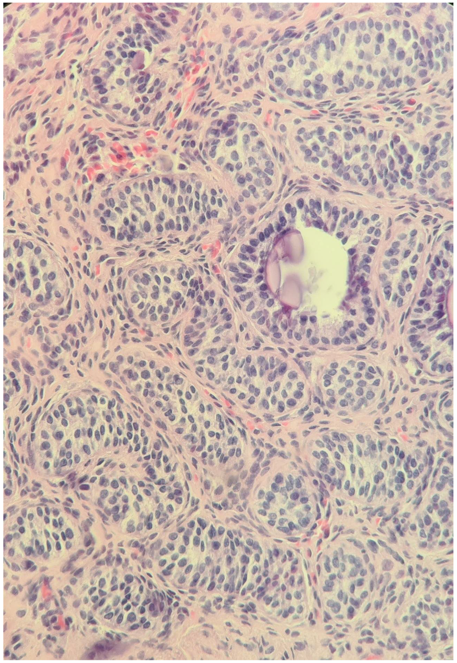
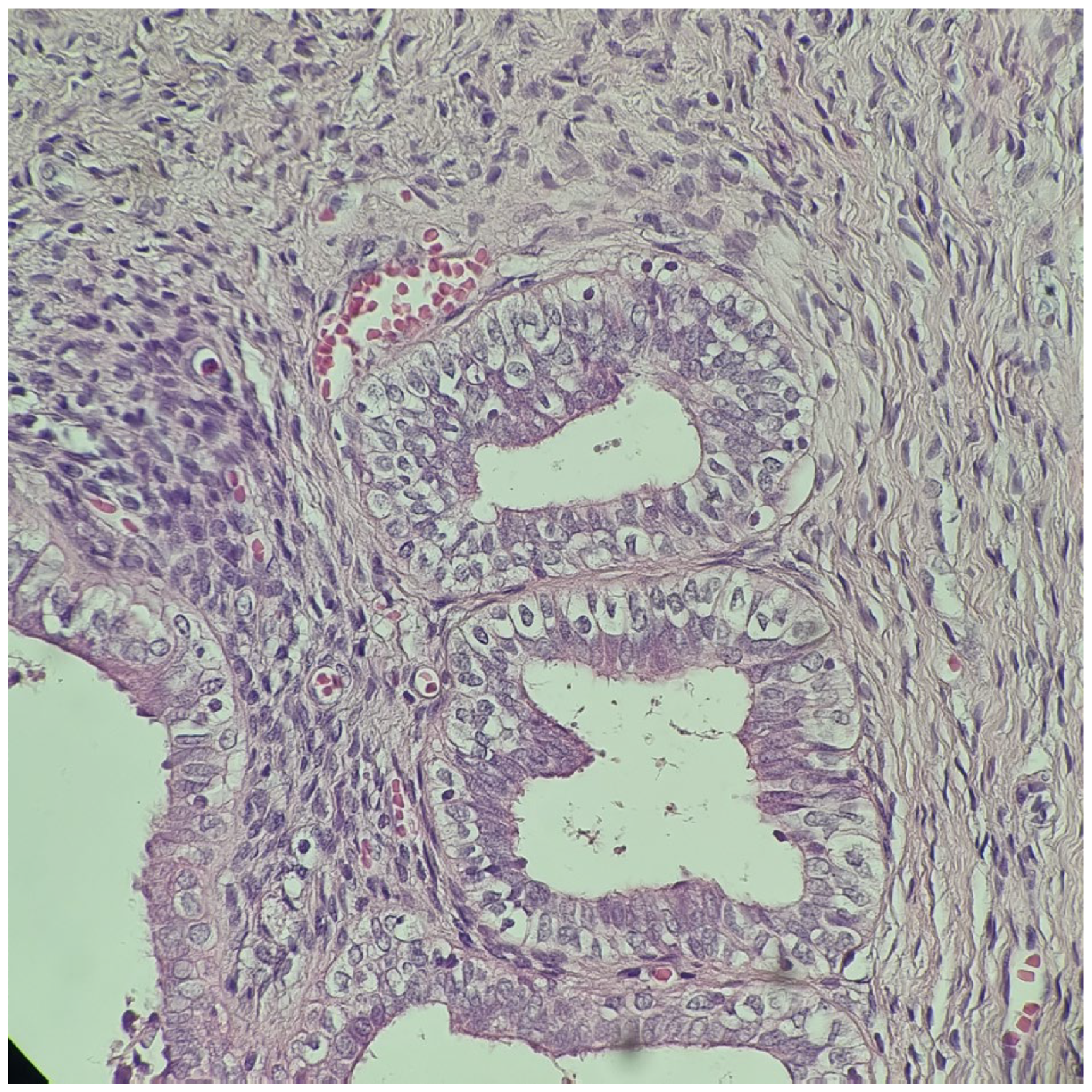
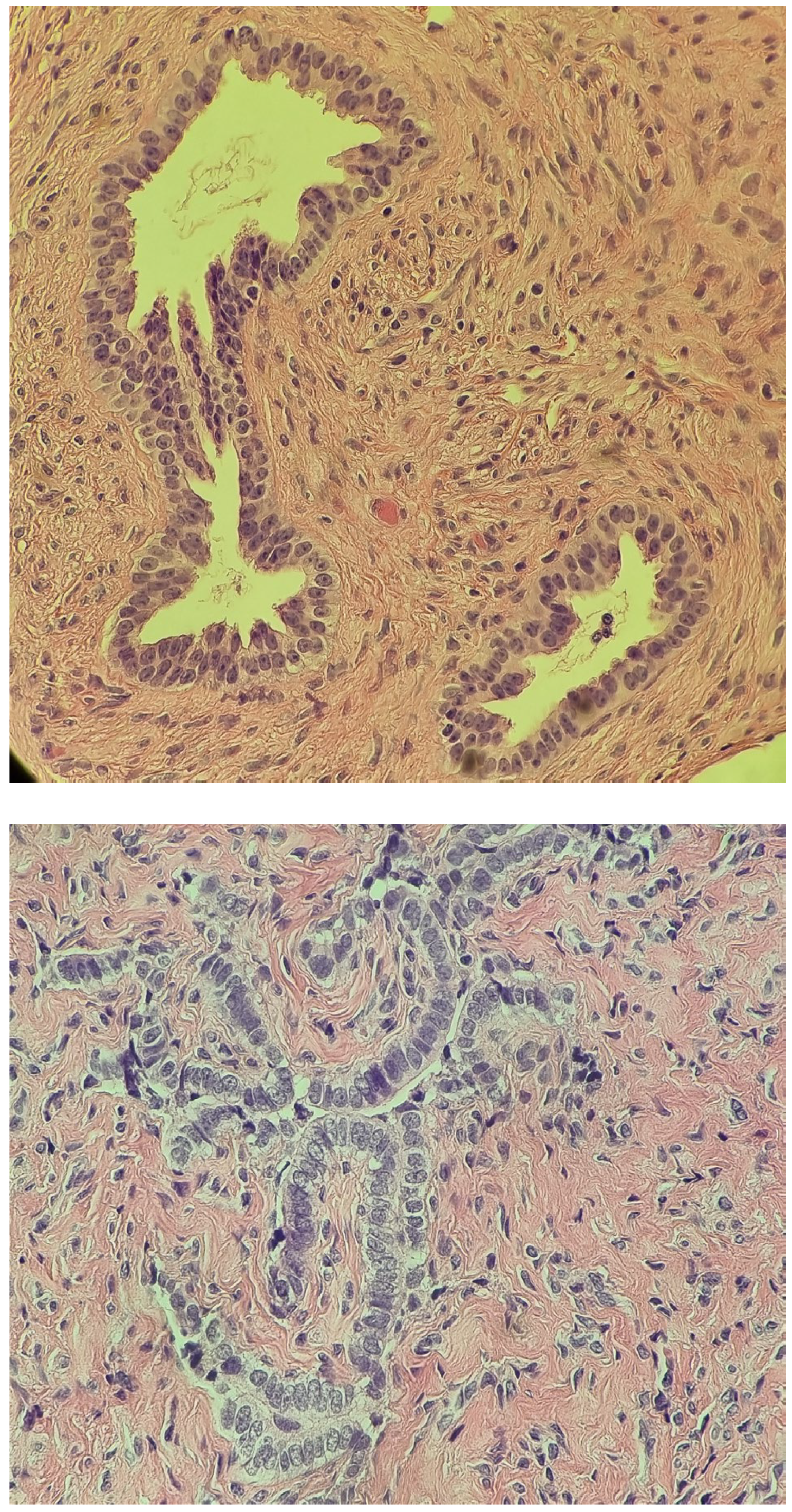
After orchidopexy, the right testis ascended again in the abdomen, and in May 2014, at the age of 5, the patient presented for the first time to our clinic (“Maria Sklodowska Curie” Emergency Hospital for Children, Bucharest, Romania). The preoperative investigation (MRI) revealed the presence of a left paramedian retrovesical lesion, possibly the left ectopic testicle, as well as a right testicle located in the upper 1/3 of the right inguinal canal. The Pregnyl stimulation test indicated viable testicular tissue. Under general anesthesia, exploratory laparoscopy was performed, which revealed a left intra-abdominal testicle with a modified appearance and an anatomical fibrous structure medially located that was bilaterally fixated to the parietal peritoneum, extended from the left testis to the right deep inguinal ring, resembling a rudimentary uterus. A biopsy of the left ectopic testicle was performed and the histopathologic exam showed characteristics of immature testicular tissue (Figure 1). At this point, an iterative right inguinotomy was also performed, which detected in the right inguinal canal a testicle fixed at this level (probably during the first surgical intervention, which was not performed in our clinic), and a right orchidopexy was carried out. Laparoscopic exploration was repeated in October 2014, during which the structure was dissected and removed and left orchidopexy was performed. Histopathology examination confirmed the Müllerian nature of the mass (Figure 2). The karyotype was 46, XY and the sex-determining region (SRY) on the Y chromosome was identified via fluorescence in situ hybridization (FISH).

2.3. At Age 9–12 Years

The follow-up of the patient consisted of annual scrotal ultrasound examinations. In April 2018, at the age of 9, we observed that the left testicle had an elevated position, with it being at the level of the external opening of the left inguinal canal, and surgery was performed again through an incision at the level of the left hemiscrotum and the testicle was descended. On this occasion, we observed that the left testicle was hypotrophic. The right testicle was located in the right hemiscrotum.
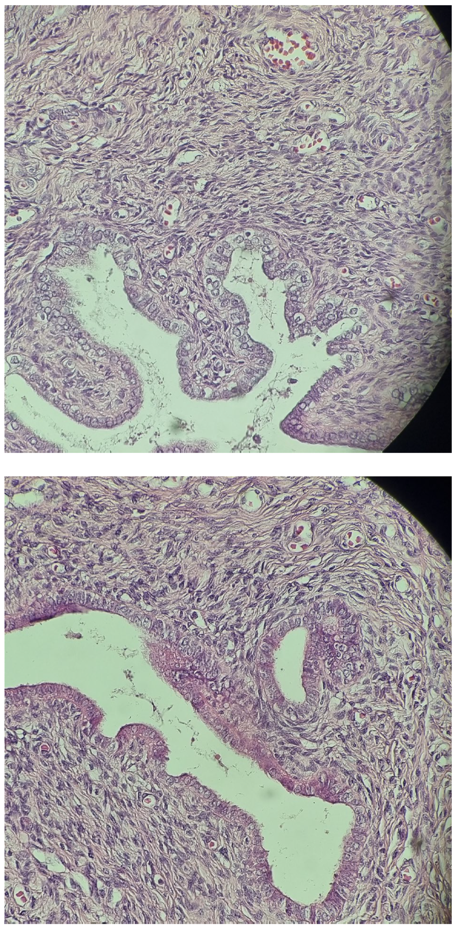
In September 2021, at the age of 12, surgery was repeated on the left hemiscrotum, and a small atrophic left testicle was discovered, for which an orchidectomy was performed, taking into account the risk of malignancy. The histopathologic examination was consistent with epididymis, ductus deferens, without specific features of masculine gonads, and seminiferous tubules, respectively (Figure 3), which were initially seen at the biopsy performed in May 2014 from the left undescended testis (Figure 1). The left testis that was initially intra-abdominal and was descended and fixed into the scrotum at the age of 5 decreased in size in evolution (as it was discovered at the age of 9 when an iterative orchidopexy was performed) and finally crumbled (at the age of 12, the tissue that was removed had no gonadal tissue). The right testicle was still in the right scrotal bursa and measured 13/9/6 mm.
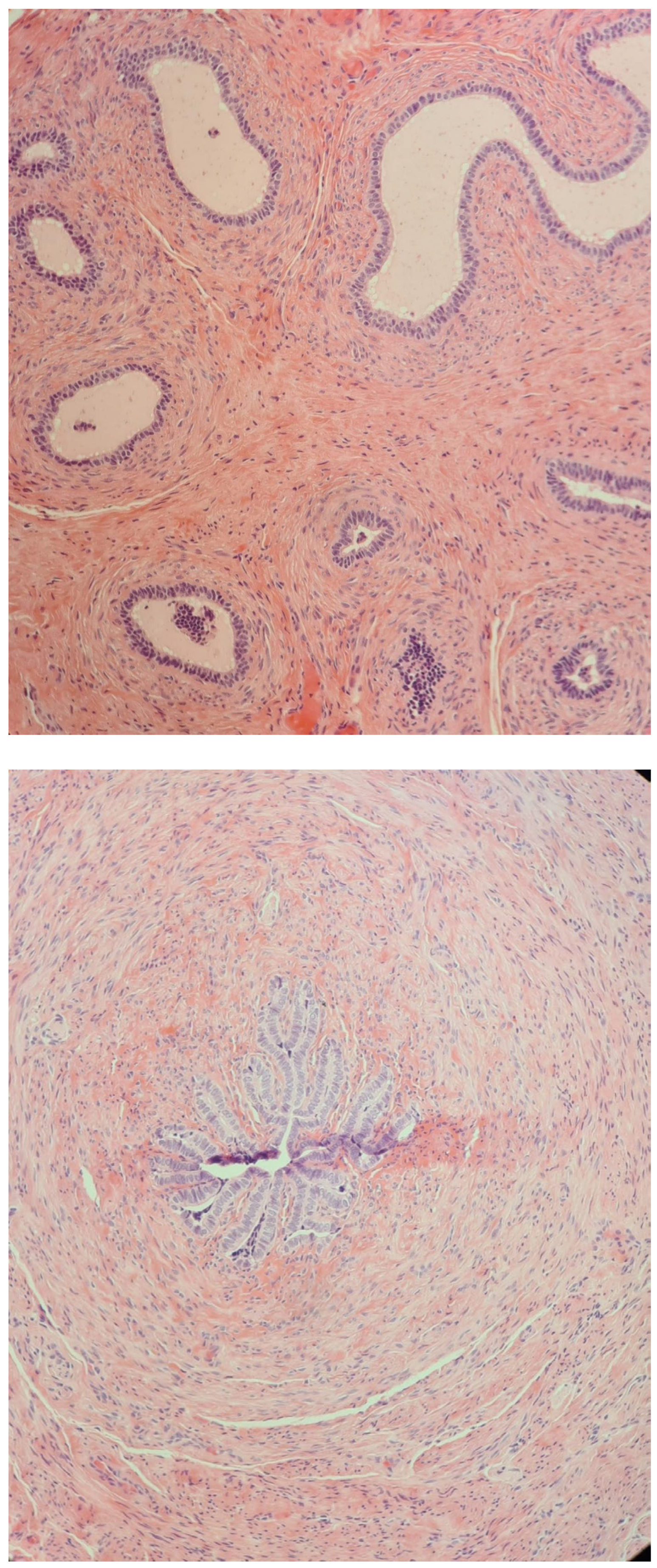
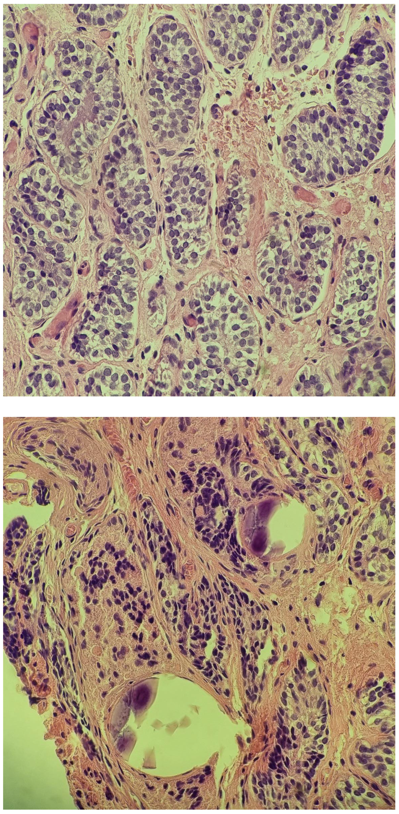
In February 2022, at the age of 13, following a routine ultrasound, a nodular image was discovered in the left inguinal region but outside the left inguinal canal, approximately 10–15 mm deep from the skin plane. The structure measured 15/10 mm and resembled a testis. The possibility of supernumerary gonads was raised. Another surgical intervention was performed and a biopsy from the suspected mass was retrieved. The histopathologic examination showed characteristics of immature prepubertal testis (Figure 4).

2.4. At Age 13 Years

At the age of 13 years, the patient was referred to our clinic with the suspicion of DSD. On physical examination, the patient’s height was 150.6 cm (−1.23 SD), their weight was 48.5 kg, their BMI = 21.4 kg/m2 (percentile 81), their Tanner stage was 2, and a small right testis was palpable in the scrotum, while the left one was identified in the inguinal area. Scrotal ultrasound was consistent with the clinical findings, showing a right hypoplastic right testis of 14/6.2 mm and a left testis of 16/8.9 mm in the inferior region of the inguinal area. Hormonal studies were performed to determine testicular function and revealed age-appropriate levels of luteinizing hormone (LH), follicle-stimulating hormone (FSH), and testosterone as follows: FSH = 3.03 mIU/mL (0.4–4.6), LH = 0.434 mIU/mL (0.1–7.8), testosterone = 3.12 ng/dL (Tanner 2: 2.5–432). Serum AMH and inhibin B were normal for age (serum AMH level 13.970 ng/mL; normal range: 2.079–30.656, serum inhibin B = 33.3 pg/mL; normal range: 16.610–278.870). Tumoral markers were within limits (LDH = 148 U/L, normal range: 105–233, AFP ≤ 0.908 ng/mL, normal range: <40, BETA-HCG ≤ 0.70 mIU/mL, normal range: <5).
Considering the male karyotype and the coexistence of testes, Wolffian duct structures, and Müllerian duct derivates, the diagnosis of PMDS was raised. Taking into account the normal levels of AMH, the AMHR2 gene was believed to be causative.

2.5. Genetic Testing

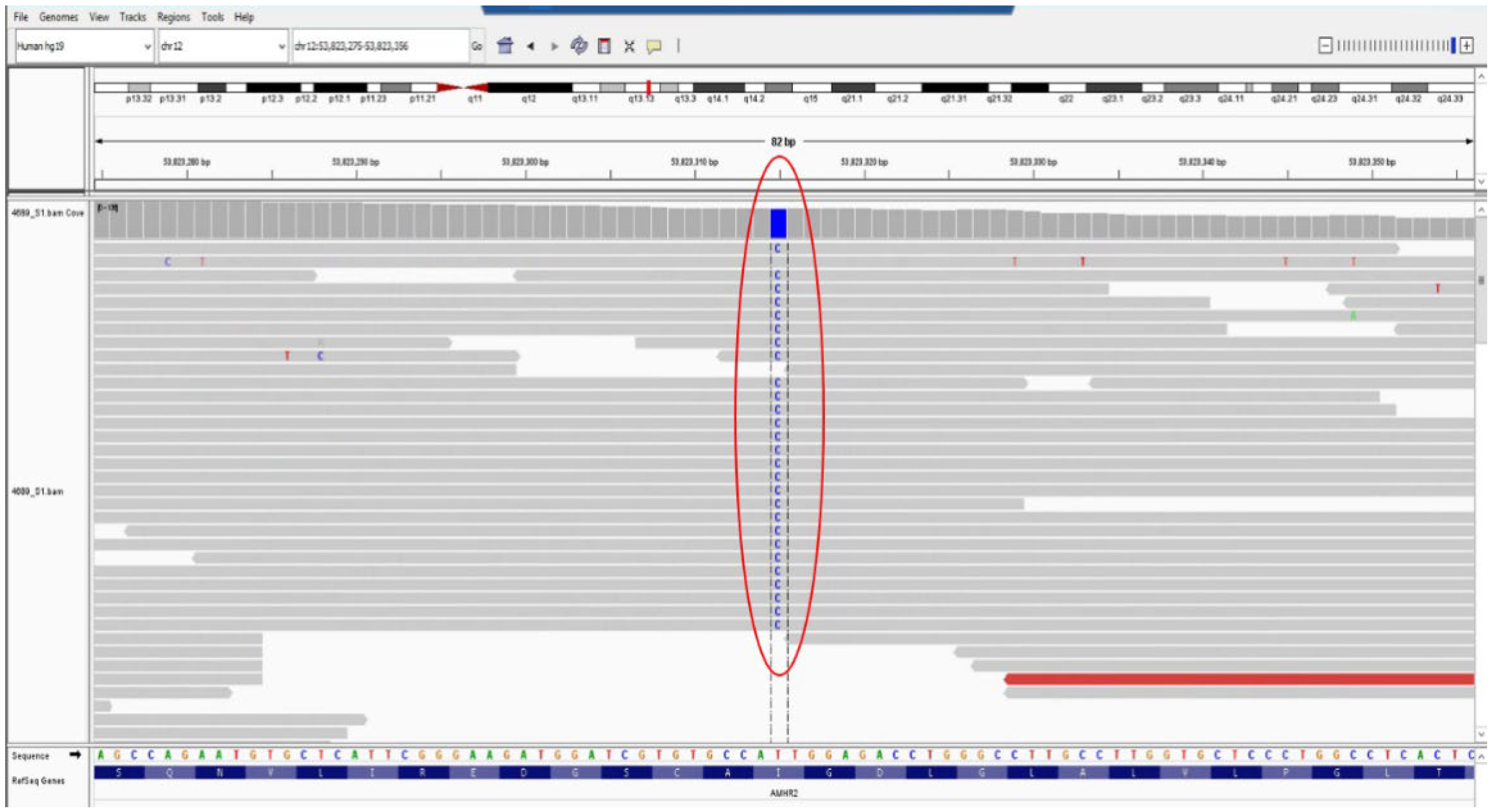
Genetic testing was performed using next-generation sequencing (NGS) with the TruSightOne panel (Illumina, San Diego, CA, USA) on a MiSeq (Illumina) Sequencer at the Timis Regional Centre of Medical Genetics, affiliated with “Louis Turcanu” Emergency Hospital for Children in collaboration with the Center for Genomic Medicine Victor Babes University of Medicine and Pharmacy Timisoara. Libraries were generated according to the manufacturer’s protocols using TruSightOne kits (Illumina Inc., San Diego, CA, USA). Targeted DNA sequencing was performed on TruSightOne library v1.0, targeting 4813 genes. The secondary analysis used the Illumina MiSeq Reporter 2.6.2.3 platform, incorporating FASTQ alignment (using Burrows–Wheeler Align) and variant extraction (using SAMtools and GATK). Sequences were mapped to GRCh37 (“hg19”), retaining reads with a median quality score genotype quality (GQ) greater than 30, variant frequency greater than 20%, variant depth greater than 20, and strand bias less than −10. The VFC annotation was performed using ANNOVAR. The variant frequency datasets were analyzed using gnomAD version 4.1. In silico prediction relied on Combined Annotation-Dependent Depletion (CADD) scores as a tool that integrates multiple annotations such as conversion metrics, functional genomic data, transcript information, and protein level scores and computes a score that indicates the variant effect. All DNA sequencing results were manually reviewed by two clinical geneticists to prioritize variants and subsequent reporting of consensus variants. The sequencing analysis, bioinformatics filtering strategy, data interpretation, and reporting are also presented elsewhere [4]. Variants were classified according to the American College of Medical Genetics and Genomics (ACMG) guidelines in relation to the patient’s phenotype [5]. Pathogenic, likely pathogenic, and variants of uncertain significance (VUS) related to the phenotype were reported for clinical use. The test showed a likely pathogenic variant in a homozygous state, in AMHR2 gene NM_020547.3:c.1046T>C, NP_065434.1:p.(Ile349Thr). This sequence change results in the replacement of isoleucine with threonine at position 349 of the AMHR2 protein (p.Ile349Thr) (Figure 5 showing the variant in Integrative Genomics Viewer (IGV)). Isoleucine 349 is located in a conserved region of the Protein Kinase functional domain, within AMHR2. UniProt protein AMHR2_HUMAN homo_sapiens proteome sequences have 65 missense/in-frame variants (22 pathogenic variants, 34 uncertain variants, and 9 benign variants), which qualifies as likely pathogenic. The detected variant is present in population databases with a very low frequency (gnomAD v2.1.1, ƒ = 0.00001591, no individual with homozygous status). To date, the variant has not been reported in other individuals with diseases associated with the AMHR2 gene. Most of the in silico prediction algorithms used suggest that the variant could have a deleterious impact on the protein. In addition, the patient’s phenotype is suggestive and specific for disorders associated with the AMHR2 gene. For these reasons, the variant was classified with likely pathogenic significance according to ACMG guidelines [5]. Unfortunately, carrier testing of the variants in the parents was not performed.

3. Literature Review

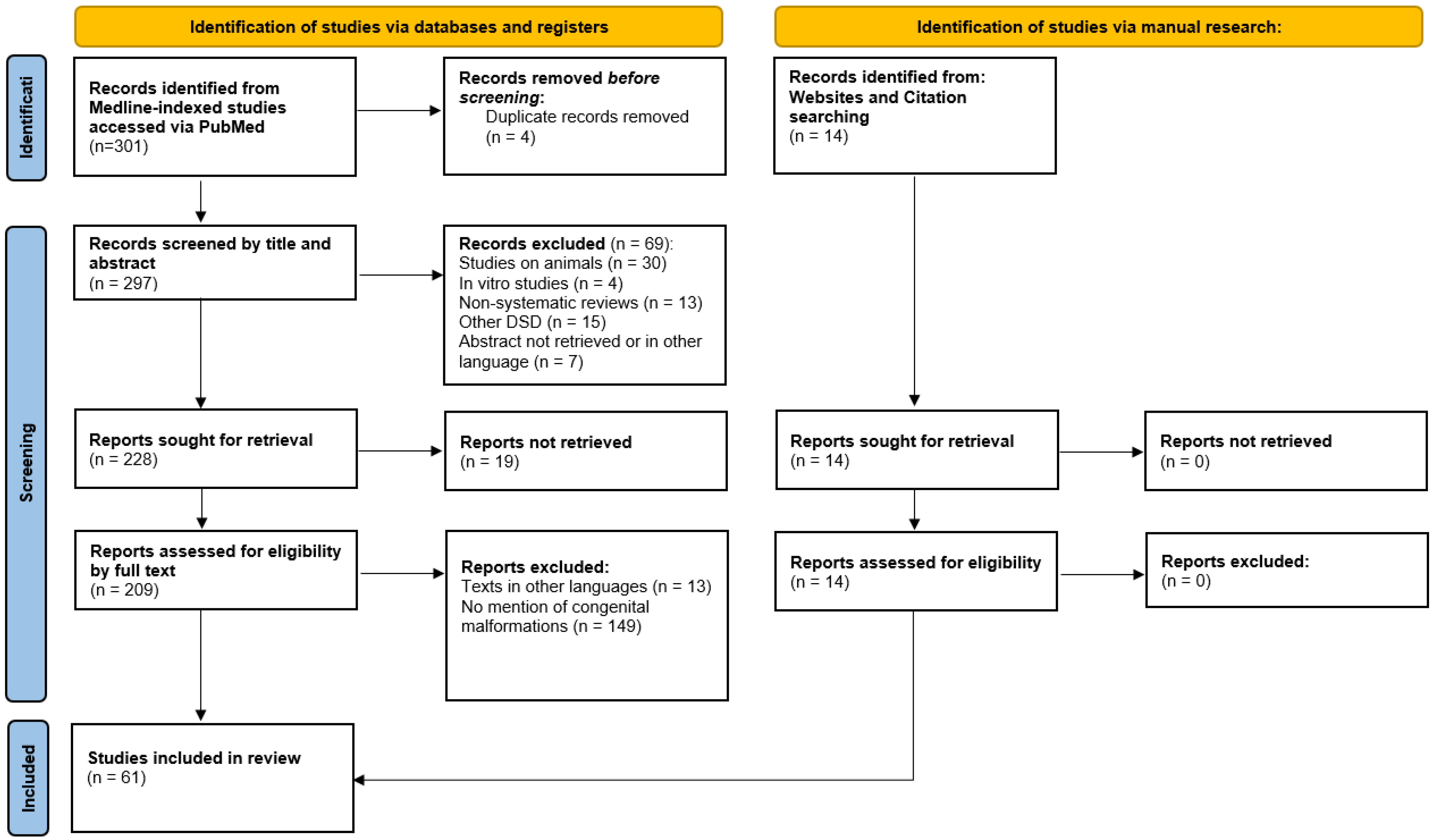
We conducted a comprehensive literature review on PMDS to provide an in-depth understanding of this rare pathology (Table 1). Our primary source of data was Medline-indexed studies accessed via PubMed. The aim was to include articles that reported cases of PMDS and highlighted clinical presentations, laboratory investigations, genetic and histopathological findings, and the outcomes of the disease, with a focus on the ones that reported the association with congenital anomalies. Our search included terms such as ‘Persistent Müllerian duct syndrome’ + ‘congenital anomalies’, ‘supernumerary testes’, ‘polyorchidism’, ‘genetic testing’, and ‘AMHR2 gene mutations’ (Figure 6).
Following this protocol, we initially identified 301 articles, spanning from the earliest available date to 1 February 2024. After title and abstract screening, 228 eligible studies were sought for retrieval. Records were excluded from the research if they involved non-human studies, non-systematic reviews, other DSDs apart from PMDS, or abstracts in other languages. The full text of 19 reports could not be retrieved. In total, 14 additional relevant articles were also identified through a manual review of reference lists. The full text of the studies was reassessed and, based on a detailed evaluation of content, 61 articles mentioning an association between PMDS and at least one congenital malformation were ultimately included in the final manuscript. The reports that referred to the association between PMDS and hypospadias were removed from the analysis considering the fact that undervirilization is an indicator of testicular dysgenesis rather than PMDS.

4. Discussion

PMDS is a rare form of 46, XY DSD that presents a wide range of clinical manifestations, diagnostic findings, and treatment approaches. This discussion aims to compare the findings from a recent case with those from a comprehensive review of the cases published in the literature.
PMDS is a 46, XY DSD, characterized by an ineffective AMH, described by Nilson in 1939 [58]. In the absence of AMH action, Müllerian ducts persist and differentiate into rudimentary forms of the fallopian tubes, the uterus, and the upper third of the vagina (due to the absence of estrogen). Caudally, these structures open into the posterior aspect of the urethra, near the verumontanum [59]. Leydig cells are functional, so virilization is complete, and Wolffian duct derivates are preserved [1]. The vas deferens pass nearby or are encapsulated within the uterine walls and open into the upper vagina, the female equivalent of the prostatic utricle. Excretory duct anomalies are nevertheless frequent in PMDS [60].
Our recent case refers to a patient without relevant family or prenatal history who was first evaluated at the age of 4 years for bilateral cryptorchidism and bilateral inguinal hernia. The clinical presentation of our patient is consistent with the female form of PMDS which occurs in 80% of cases [61]. The female type presents with bilateral impalpable testis (the testes remain intra-abdominal in a high ovarian position) and is usually diagnosed during laparoscopic exploration [62]. In a minority of cases, testis may prolapse, due to the long, flexible gubernaculum, into the processus vaginalis, dragging along the MD (male form). Classical male presentations are hernia uteri inguinalis (a hernia sac accommodating the testis and the MD derivates) or transverse testicular ectopia (an inguinal hernia containing both testes, the uterus, and the fallopian tubes) [60,63]. Usually, the male types (20%) are discovered during herniorrhaphy.
AMH is involved in the swelling reaction of the genito-inguinal ligament. In PMDS, this process is altered and the gubernaculum remains elongated, similar to the round ligament. Consequently, testes may reascend after orchidopexy, and this explains the necessity of recurrent bilateral orchidopexies in our patient.
The coexistence of Müllerian and Wolffian duct remnants can be encountered in other forms of DSD apart from PMDS. Such examples are partial testicular dysgenesis or ovotesticular DSD [64,65,66,67]. In contrast with PMDS, these conditions generally present with ambiguous genitalia and asymmetrically formed internal genital organs [1]. If there is any uncertainty, gonadal biopsies and a karyotype should be performed [63]. In our case, complete external virilization pointed toward PMDS. Nevertheless, considering that 10% of patients with ovotesticular DSDs present with a normal or almost normal penis [68] and taking into account the atypical clinical presentation (abnormal appearance and supernumerary gonads), further investigations were conducted. The 46, XY karyotype and the presence of normal testicular tissue support the working diagnosis.
Hormonal studies revealed that normal testicular function and serum AMH and inhibin B were normal for age in the present case. Testosterone production is usually unaffected in PMDS patients. Seric AMH levels can suggest the pathophysiology of the condition. Type 1 is routinely associated with low AMH values, while in type 2, AMH levels are normal or high [1,69].
PMDS shows an autosomal recessive inheritance [70], but de novo mutations are also possible [60,63,71,72]. Homozygous or compound heterozygous alterations in AMH associated with loss of function (type 1) or AMHR2 determining receptor resistance (type 2) [73] have been identified in approximately 88% of PMDS cases [60,74]. In a minority of cases, no specific variant has yet to be identified (idiopathic PMDS). The detection of PPP1R12A truncation mutations coding myosin phosphatase in five cases of PMDS suggests that myosin phosphatase is involved in Müllerian regression, independently of the AMH signaling cascade [49]. It seems that myosin phosphatase is required for cell mobility, which plays a major role in Müllerian regression; alternatively, PPP1R12A mutations could affect the AMH transduction pathway [49]. In our patient, a homozygous variant classified as likely pathogenic in the AMHR2 gene was identified through NGS, yet it remains unreported in the literature.
It has been reported that the AMHR2 gene is located on the long arm of chromosome 12 and that it contains eleven exons: three exons form the extracellular domain, responsible for binding AMH specifically, the fourth encodes the transmembrane region, and the remaining seven account for the intracellular domain with serine/threonine kinase function [70,73]. Natural mutations of the AMH type II receptor found in PMDS affect ligand binding, signal transduction, and cellular transport [75,76]. The missense variant described in our patient determines the replacement of the 349th amino acid, a highly conserved region in exon 8, from isoleucine to threonine (p.Ile349Thr), altering protein kinase’s function. However, functional studies have not been conducted; therefore, further work is needed to investigate the harmfulness of this variant.
Up to now, the most common genetic test performed to detect mutations of AMH or AMHR2 in relation to PMDS is Sanger sequencing with a few exceptions [8,74,77]. However, as suggested by Tosca et al., in the near future, NGS targeted to AMH and AMHR2 may become the method of choice for diagnosing PMDS, with the capacity for detecting both SNVs and CNVs in a single step [8]. In our patient, the variant was identified through NGS and, to the best of our knowledge, it has not been reported before in the literature.
Of particular interest in our case is the association between PMDS and supernumerary testes in the presence of a genetic diagnosis of this rare disease. Usually, congenital malformations, notably of intestinal and renal nature, are described in PMDS patients. In most cases, no significant variants in AMH or AMHR2 genes have been identified (idiopathic PMDS) [24,49,60]. Testicular abnormalities have also been reported in PMDS patients, mainly testicular regression syndrome. Testicular torsion is most probably the cause, due to the increased mobility of gonads and the long gubernaculum [78]. At the other end of the spectrum, the association with polyorchidism that we discovered in our patient is less often encountered. Polyorchidism is defined by the presence of more than two testes. At the present time, around 200 cases have been reported. The cause is still not established, but one explanation may be the division of the undifferentiated gonadal ridge by congenital peritoneal bands [79]. Apart from the presented case, only two other cases of PMDS with supernumerary testes have been documented. The first one presents a 46, XY patient with left hernia uteri inguinalis, left intra-abdominal testis, and two right scrotal testes [2]. The second case describes the occurrence of malignant degeneration of a supernumerary testis in a 46, XY patient with a female form of PMDS [3]. In both cases, no further genetic testing was performed. However, we would like to acknowledge the limitations regarding the evidence of a supernumerary testis in our case. At the age of 12 years, the tissue that was removed from the left side did not have gonadal tissue, although seminiferous tubules were initially seen at the biopsy performed in May 2014 (at the age of 5) from the left undescended testis. We excluded a testicular–epididymal dissociation on the left side because, in this case, the testicle remained fixed in the scrotum, while the vas deferens and the epididymis ascended into the inguinal canal. In our case, the epididymis was found in the scrotum (at the age of 12) and the testicle was found outside (anterolateral to) the inguinal canal (at the age of 13). Apart from this, the left testicle (which was initially intra-abdominal and was descended and fixed into the scrotum at the age of 5) was smaller than the right testicle, was of only a few mm, and decreased in size progressively (observed at the age of 9), until it crumbled (at the age of 12). The testicle found outside the left inguinal canal at the age of 13 was bigger than 1.5 cm; thus, it cannot be the same structure descended and fixed into the left scrotum. Therefore, we consider this to be strong evidence that the lesion identified at the age of 13 is a supernumerary testis.
As PMDS patients are at risk of malignant testicular degeneration, a differential diagnosis between a supernumerary testis and a lymph node metastasis from a testicular or MD remnant cancer was considered. Tumoral markers were within limits in our patient and the biopsy showed characteristics of immature testis. It has been shown that testicular malignant degeneration occurs in 33% of adults with PMDS [80]. A recent search on Medline/PubMed retrieved 44 articles (49 patients) on testicular tumors associated with PMDS, with the majority (59%) presenting with a large abdominal mass [81], but only five cases (10%) had a preceding history of appropriately managed cryptorchidism. The most commonly reported tumors in the literature were seminomas [82,83,84,85,86,87], but other types of tumors such as mixed germ cell tumors [88,89,90,91,92], embryonal carcinoma, yolk sac tumors, and teratoma were also encountered [63,93,94,95]. Some studies cite an incidence similar to that of cryptorchidism (5–18%) [63], while others suggest that the real incidence may be as high as 33% [60]. The American Urologic Association recommends early orchidopexy in patients with undescended testis by 6 months to decrease the risk of malignancy and emphasizes the importance of testicular self-examination after puberty for early detection [96]. At the time of writing, 12 cases of uterine neoplasma have been reported, affecting patients as young as 4 years [63]. Adenocarcinoma, adenosarcoma, and squamous cell carcinoma are the histopathological types found, and some case reports describe highly aggressive tumors [38,43,97,98]. Removal of Müllerian structures eliminates the risk. If resection is impossible out of fear of damaging the gonads or the excretory ducts; close surveillance consisting of regular pelvic ultrasounds or MRI should be envisioned [23,99]. Occasionally, other types of cancers have been described in patients with PMDS such as prostatic adenocarcinoma [100,101].
Even though clear guidelines for the management of PMDS have not yet been established, the main objectives that experts agree upon are the preservation of fertility and the prevention of malignant degeneration [1,60,63,102]. Unfortunately, despite a few reports of PMDS patients being able to father children, either spontaneously [83,84,103] or through assisted reproduction treatments [19,60,104], most affected males are infertile [1,105]. The mechanisms involved are cryptorchidism and late orchidopexy, male excretory duct anomalies, and iatrogenic lesions. In order to minimize this risk, early correction of cryptorchidism should be performed. In our case, the orchidopexies were performed after the age of 4 when the patient first approached the medical system. Müllerian structures pose a mechanical constraint to testicular descent and are therefore usually resected. During dissection, great attention should be paid to the conservation of the vas deferens and blood vessels, as these structures are in close proximity or even enclosed within the uterine walls [60]. As the vas deferens and vascular structures may be injured during orchiopexy, leading to subsequent infertility, patients should always be referred to experienced clinics [60]. If spermatogenesis is present, testicular sperm extraction followed by ICSI may be used to facilitate conception in cases of congenital or iatrogenic lesions of the male excretory ducts [8].
In our case, a dissection of the hypoplastic uterus was performed before the diagnosis of PMDS. Treatment of the remnants of MD remains controversial [102,106]. Previous studies have demonstrated that children with PMDS require the removal of MD remnants as they are also prone to malignant transformation [25,107] and predispose to urinary tract infections, periodic hematuria, stones, and urination disorders because they are connected with the seminal vesicle [108,109]. As PMDS patients have been reported to develop malignancy of Müllerian remnants originating from the mucosa, some authors suggest that the preferred surgical procedure should split the uterus in the middle, destroy the mucosal lining, and leave an intact pedicle of the myometrium. This procedure releases the testes to the ideal position and also protects the integrity and vascularity of the vas deferens and reduces the chance of malignancy [6,51,110].

5. Conclusions

A high degree of suspicion and awareness is needed to diagnose this condition in order to avoid iterative surgery. The coexistence of two extremely rare conditions (PMDS and supernumerary testes) has previously been reported in only two patients, yet the association could have a common pathophysiologic background. Our case, reporting a novel AMHR2 variant, highlights the importance of genetic testing in these individuals in order to elucidate a possible genotype–phenotype correlation. Further genetic studies on these individuals are needed in order to investigate this unusual association.

Author Contributions

Conceptualization, L.N.C., I.G., I.M.D. and S.F.; methodology, L.N.C., S.F., I.G. and I.M.D.; histology, A.C.E.; genetics, A.C.-E., N.A. and M.P.; writing—original draft preparation, L.N.C. and I.G.; writing—review and editing, A.C.-E., C.G.B. and S.F.; supervision, S.F. All authors have read and agreed to the published version of the manuscript.

Funding

This research received no external funding.

Institutional Review Board Statement

Not applicable.

Informed Consent Statement

Informed consent was obtained from the mother of the child involved in the study. Written informed consent was obtained from the child’s mother to publish this paper.

Data Availability Statement

The data presented in this study are available from the corresponding author upon request. The data are not publicly available due to the confidentiality of personal data.

Acknowledgments

The Regional Center of Medical Genetics Timiș, Clinical Emergency Hospital for Children “Louis Țurcanu” Timișoara, Romania, is part of ERN ITHACA, a European reference network for rare malformation syndromes and rare intellectual and neurodevelopmental disorders.

Conflicts of Interest

The authors declare no conflicts of interest.

References

  1. Josso, N.; Rey, R.A. What Does AMH Tell Us in Pediatric Disorders of Sex Development? Front. Endocrinol. 2020, 11, 619. [Google Scholar] [CrossRef] [PubMed]
  2. Renton, C.J.C. A Case of Polyorchidism with Intersex. J. Urol. 1975, 113, 720–724. [Google Scholar] [CrossRef] [PubMed]
  3. Kulkarni, J.N.; Bhansali, M.S.; Tongaonkar, H.B.; Kamat, M.R.; Borges, A.M. Carcinoma in the Third Testis in a Case of Polyorchidism and Persistent Müllerian Structure Syndrome. Eur. Urol. 1992, 22, 174–176. [Google Scholar] [CrossRef] [PubMed]
  4. Chirita-Emandi, A.; Andreescu, N.; Zimbru, C.G.; Tutac, P.; Arghirescu, S.; Serban, M.; Puiu, M. Challenges in Reporting Pathogenic/Potentially Pathogenic Variants in 94 Cancer Predisposing Genes—In Pediatric Patients Screened with NGS Panels. Sci. Rep. 2020, 10, 223. [Google Scholar] [CrossRef] [PubMed]
  5. Richards, S.; Aziz, N.; Bale, S.; Bick, D.; Das, S.; Gastier-Foster, J.; Grody, W.W.; Hegde, M.; Lyon, E.; Spector, E.; et al. Standards and Guidelines for the Interpretation of Sequence Variants: A Joint Consensus Recommendation of the American College of Medical Genetics and Genomics and the Association for Molecular Pathology. Genet. Med. 2015, 17, 405–424. [Google Scholar] [CrossRef]
  6. Tian, H.-J.; Wu, D.-H.; Ru, W.; Wu, D.-W.; Tao, C.; Chen, G.-J.; Yuan, J.-N.; Fu, J.-F.; Tang, D.-X. Surgical Management and Molecular Diagnosis of Persistent Müllerian Duct Syndrome in Chinese Patients. Asian J. Androl. 2022, 24, 78. [Google Scholar] [CrossRef]
  7. Silveri, M.; Zaccara, A.; Cappa, M. A Simplified Management of Transverse Testicular Ectopia in Patients with Persistent Mullerian Duct Syndrome. Urol. J. 2020, 18, 237–239. [Google Scholar] [CrossRef]
  8. Tosca, L.; Giltay, J.C.; Bouvattier, C.; Klijn, A.J.; Bouligand, J.; Lambert, A.S.; Lecerf, L.; Josso, N.; Tachdjian, G.; Picard, J.Y. Persistent Müllerian Duct Syndrome Due to Anti-Müllerian Hormone Receptor 2 Microdeletions: A Diagnostic Challenge. Hum. Reprod. 2020, 35, 999–1003. [Google Scholar] [CrossRef]
  9. Chung, H.S.; Kim, S.-O.; Yu, H.S.; Kim, S.-S.; Kwon, D.D. Transverse Testicular Ectopia Associated with Persistent Müllerian Duct Syndrome Treated by Transseptal Orchiopexy. Medicine 2018, 97, e13305. [Google Scholar] [CrossRef]
  10. Saleem, M.; Ather, U.; Mirza, B.; Iqbal, S.; Sheikh, A.; Shaukat, M.; Sheikh, M.T.; Ahmad, F.; Rehan, T. Persistent Mullerian Duct Syndrome: A 24-Year Experience. J. Pediatr. Surg. 2016, 51, 1721–1724. [Google Scholar] [CrossRef]
  11. Bowen, D.K.; Matulewicz, R.S.; Gong, E.M. Preservation of Müllerian Structures with Laparoscopic Management of Intra-Abdominal Testes in Persistent Müllerian Duct Syndrome. J. Pediatr. Urol. 2016, 12, 65–66. [Google Scholar] [CrossRef] [PubMed]
  12. Morikawa, S.; Moriya, K.; Ishizu, K.; Tajima, T. Two Heterozygous Mutations of the AMH Gene in a Japanese Patient with Persistent Müllerian Duct Syndrome. J. Pediatr. Endocrinol. Metab. 2014, 27, 1223–1226. [Google Scholar] [CrossRef] [PubMed]
  13. Ju, X.; Li, Z.; Zhang, C.; Qin, C.; Shao, P.; Li, J.; Li, P.; Cao, Q.; Zhang, W.; Wang, Z.; et al. Clinical Aspects and Molecular Genetics of Persistent Müllerian Duct Syndrome Associated with Transverse Testicular Ectopia: Report of Three Cases. Urol. Int. 2013, 90, 83–86. [Google Scholar] [CrossRef] [PubMed]
  14. Zhapa, E.; Castagnetti, M.; Alaggio, R.; Talenti, E.; Rigamonti, W. Testicular Fusion in a Patient with Transverse Testicular Ectopia and Persistent Mullerian Duct Syndrome. Urology 2010, 76, 62–64. [Google Scholar] [CrossRef] [PubMed]
  15. Wuerstle, M.; Lesser, T.; Hurwitz, R.; Applebaum, H.; Lee, S.L. Persistent Mullerian Duct Syndrome and Transverse Testicular Ectopia: Embryology, Presentation, and Management. J. Pediatr. Surg. 2007, 42, 2116–2119. [Google Scholar] [CrossRef]
  16. Marjanović, Z.O.; Perović, S.V.; Slavković, A.; Živanović, D.; Đorđević, I. Transverse Testicular Ectopia with and without Persistent Müllerian Duct Syndrome. Int. Urol. Nephrol. 2007, 39, 1167–1171. [Google Scholar] [CrossRef]
  17. Mouli, K.; Mccarthy, P.; Ray, P.; Ray, V.; Rosenthal, I.M. Persistent Müllerian Duct Syndrome in a Man with Transverse Testicular Ectopia. J. Urol. 1988, 139, 373–375. [Google Scholar] [CrossRef]
  18. Sloan, W.R.; Walsh, P.C. Familial Persistent Müllerian Duct Syndrome. J. Urol. 1976, 115, 459–461. [Google Scholar] [CrossRef]
  19. Fang, J.; Gao, G.; Liu, J.; Cai, L.; Cui, Y.; Yang, X. A Novel Mutation of AMHR2 in Two Brothers with Persistent Müllerian Duct Syndrome and Their Intracytoplasmic Sperm Injection Outcome. Mol. Genet. Genom. Med. 2021, 9, e1801. [Google Scholar] [CrossRef]
  20. Lima, M.; Morabito, A.; Libri, M.; Bertozzi, M.; Dòmini, M.; Lauro, V.; Strano, C.; Messina, P.; Tani, G. Laparoscopic Removal of a Persistent Müllerian Duct in a Male: Case Report. Eur. J. Pediatr. Surg. 2000, 10, 265–269. [Google Scholar] [CrossRef]
  21. Zhou, W.; Li, S.; Wang, H.; Yin, J.; Liu, X.; Jiang, J.; Zhou, G.; Wen, J. Diagnostic Value of Ultrasound in Children with Transverse Testicular Ectopia. Front. Pediatr. 2022, 10, 914139. [Google Scholar] [CrossRef] [PubMed]
  22. Dhua, A.; Varshney, A.; Bhatnagar, V. Transverse Testicular Ectopia with a Blind Ending Vas Deferens. Indian J. Urol. 2016, 32, 317. [Google Scholar] [CrossRef] [PubMed]
  23. Sancar, S.; Ozcakir, E.; Kaya, M. Management of the Patients with Persistent Müllerian Duct Syndrome: Is the Ultimate Goal Testicular Descent? Türk. Urol. Derg./Turk. J. Urol. 2018, 44, 166–171. [Google Scholar] [CrossRef] [PubMed]
  24. Sharma, U.K.; Thapa, D.K.; Pokhrel, D.; Shah, A.K. Persistent Mullerian Duct Syndrome with Polysplenia and Short Pancreas: A Case Report. J. Nepal Med. Assoc. 2019, 57, 123. [Google Scholar] [CrossRef] [PubMed]
  25. Farikullah, J.; Ehtisham, S.; Nappo, S.; Patel, L.; Hennayake, S. Persistent Müllerian Duct Syndrome: Lessons Learned from Managing a Series of Eight Patients over a 10-year Period and Review of Literature Regarding Malignant Risk from the Müllerian Remnants. BJU Int. 2012, 110, E1084–E1089. [Google Scholar] [CrossRef]
  26. Imbeaud, S.; Rey, R.; Berta, P.; Chaussain, J.-L.; Wit, J.-M.; Lustig, R.H.; Picard, J.-Y.; Josso, N. Testicular Degeneration in Three Patients with the Persistent Müllerian Duct Syndrome. Eur. J. Pediatr. 1995, 154, 187–190. [Google Scholar] [CrossRef]
  27. Scott, J.E.S. The Hutson Hypothesis A Clinical Study. Br. J. Urol. 1987, 60, 74–76. [Google Scholar] [CrossRef]
  28. Shah, D.S.; Shah, U.S.; Kumaresan, N. Persistent Mullerian Duct Syndrome: Rare Presentation in an Elderly Man. BMJ Case Rep. 2020, 13, e234890. [Google Scholar] [CrossRef]
  29. Aw, L.D.; Zain, M.M.; Esteves, S.C.; Humaidan, P. Persistent Mullerian Duct Syndrome: A Rare Entity with a Rare Presentation in Need of Multidisciplinary Management. Int. Braz. J. Urol. 2016, 42, 1237–1243. [Google Scholar] [CrossRef]
  30. Østergren, P.; Juul, A.; Azawi, N.H. Disorders of Sex Development Presenting as Unilateral Cryptorchidism. Scand. J. Urol. 2013, 47, 433–436. [Google Scholar] [CrossRef]
  31. Demir, O.; Kizer, O.; Sen, V.; Esen, A.A. Persistent Mullerian Duct Syndrome in Adult Men Diagnosed Using Laparoscopy. Urology 2011, 78, 566. [Google Scholar] [CrossRef] [PubMed]
  32. Beyribey, S.; Çetinkaya, M.; Adsan, Ö.; Memis, A.; Öztürk, B. Persistent Müllerian Duct Syndrome. Scand. J. Urol. Nephrol. 1993, 27, 563–565. [Google Scholar] [CrossRef] [PubMed]
  33. Wu, H.-C.; Chen, J.-H.; Lu, H.-F.; Shen, W.-C. Persistent Müllerian Duct Syndrome with Seminoma. Am. J. Roentgenol. 2000, 174, 102–104. [Google Scholar] [CrossRef] [PubMed]
  34. Imbeaud, S. A 27 Base-Pair Deletion of the Anti-Mullerian Type II Receptor Gene Is the Most Common Cause of the Persistent Mullerian Duct Syndrome. Hum. Mol. Genet. 1996, 5, 1269–1277. [Google Scholar] [CrossRef]
  35. Adamsbaum, C.; Rolland, Y.; Josso, N.; Kalifa, G. Radiological Findings in Three Cases of Persistent Müllerian Duct Syndrome. Pediatr. Radiol. 1993, 23, 55–56. [Google Scholar] [CrossRef]
  36. Sheehan, S.J.; Tobbia, I.N.; Ismail, M.A.; Kelly, D.G.; Duff, F.A. Persistent Müllerian Duct Syndrome: Review and Report of 3 Cases. Br. J. Urol. 1985, 57, 548–551. [Google Scholar] [CrossRef]
  37. Barad, A.K. Persistent Mullerian Duct Syndrome with Embryonal Cell Carcinoma along with Ectopic Cross Fused Kidney. J. Clin. Diagn. Res. 2016, 10, PD07. [Google Scholar] [CrossRef]
  38. Thiel, D.D.; Erhard, M.J. Uterine Adenosarcoma in a Boy with Persistent Müllerian Duct Syndrome: First Reported Case. J. Pediatr. Surg. 2005, 40, e29–e31. [Google Scholar] [CrossRef]
  39. Cansaran, S. Management of Transverse Testicular Ectopia with Persistent Mullerian Duct Syndrome. North Clin. Istanb. 2018, 5, 357–360. [Google Scholar] [CrossRef]
  40. Urioste, M.; Rodríguez, J.I.; Barcia, J.M.; Martín, M.; Escribá, R.; Pardo, M.; Camino, J.; Frías, M.L.M. Persistence of Müllerian Derivatives, Lymphangiectasis, Hepatic Failure, Postaxial Polydactyly, Renal and Craniofacial Anomalies. Am. J. Med. Genet. 1993, 47, 494–503. [Google Scholar] [CrossRef]
  41. Pappis, C.; Constantinides, C.; Chiotis, D.; Dacou-Voutetakis, C. Persistent Müllerian Duct Structures in Cryptorchid Male Infants: Surgical Dilemmas. J. Pediatr. Surg. 1979, 14, 128–131. [Google Scholar] [CrossRef] [PubMed]
  42. Barriola, M.; Jimenez, J.; Gutierrez-Hoyos, A.; Tovar, J. Cryptorchidism and Persistence of Müllerian Remnants in a Normal Male. Eur. J. Pediatr. Surg. 1981, 33, 369–373. [Google Scholar] [CrossRef] [PubMed]
  43. Dimasis, N.; Koukourikis, P.; Klampatsas, A.; Xirou, P.; Sountoulides, P. A Unique Case of Aggressive Uterine Cancer in a 45-Year-Old Man with Persistent Müllerian Duct Syndrome. Arch. Esp. Urol. 2019, 72, 435–438. [Google Scholar] [PubMed]
  44. Hossaini, D.; Wahdat, M.M.; Aklaqi, A.; Haidary, M. A Rare Case Report of Orchiopexy and Hysterectomy in an Afghan Boy with Persistent Müllerian Duct Syndrome. Int. J. Surg. Case Rep. 2024, 115, 109235. [Google Scholar] [CrossRef] [PubMed]
  45. van Haelst, M.M.; Hoogeboom, J.; Galjaard, R.H.; Kleijer, W.J.; den Hollander, N.S.; de Krijger, R.R.; Hennekam, R.C.M.; Niermeijer, M.F. Lymphangiectasia with Persistent Müllerian Derivatives: Confirmation of Autosomal Recessive Urioste Syndrome. Am. J. Med. Genet. 2001, 104, 65–68. [Google Scholar] [CrossRef]
  46. Bellini, C.; Bonioli, E.; Josso, N.; Belville, C.; Mazzella, M.; Costabel, S.; Sementa, A.R.; Marino, C.E.; Tomà, P.; Hennekam, R.C.M.; et al. Persistence of Müllerian Derivatives and Intestinal Lymphangiectasis in Two Newborn Brothers: Confirmation of the Urioste Syndrome. Am. J. Med. Genet. 2001, 104, 69–74. [Google Scholar] [CrossRef]
  47. Cass, D.T.; Hutson, J. Association of Hirschsprung’s Disease and Müllerian Inhibiting Substance Deficiency. J. Pediatr. Surg. 1992, 27, 1596–1599. [Google Scholar] [CrossRef]
  48. Cass, D. Aganglionosis: Associated Anomalies. J. Paediatr. Child Health 1990, 26, 351–354. [Google Scholar] [CrossRef]
  49. Picard, J.-Y.; Morin, G.; Devouassoux-Shisheboran, M.; Van der Smagt, J.; Klosowski, S.; Pienkowski, C.; Pierre-Renoult, P.; Masson, C.; Bole, C.; Josso, N. Persistent Müllerian Duct Syndrome Associated with Genetic Defects in the Regulatory Subunit of Myosin Phosphatase. Hum. Reprod. 2022, 37, 2952–2959. [Google Scholar] [CrossRef]
  50. Klosowski, S.; Abriak, A.; Morisot, C.; Bayart, H.; Belville, C.; Thelliez, P.; Gottrand, F.; Croquette, M.; Deroubaix, P. Atrésie Jejunale et Syndrome de Persistance Des Dérivés Müllériens. Arch. Pédiatr. 1997, 4, 1264–1265. [Google Scholar] [CrossRef]
  51. Bugrul, F.; Abali, Z.Y.; Kirkgoz, T.; Cerit, K.K.; Canmemis, A.; Turan, S.; Tugtepe, H.; Picard, J.-Y.; Bereket, A.; Guran, T. Persistent Müllerian Duct Syndrome: A Rare But Important Etiology of Inguinal Hernia and Cryptorchidism. Sex. Dev. 2019, 13, 264–270. [Google Scholar] [CrossRef]
  52. Josso, N.; Fekete, C.; Cachin, O.; Nezelof, C.; Rappaport, R. Persistence of müllerian ducts in male pseudohermaphroditism, and its relationship to cryptorchidism. Clin. Endocrinol. 1983, 19, 247–258. [Google Scholar] [CrossRef]
  53. Dadheech, D. A Rare Case Report of Inguinal Hernia with Persistent Mullerian Duct and Klinefelter Syndrome. J. Clin. Diagn. Res. 2016, 10, PD28. [Google Scholar] [CrossRef]
  54. Rehman, A.; Hasan, Z.; Amanat, S.; Shaukat, T.; Saeed, A.; Jamil, K.; Zaidi, A.; Akram, M. Combined Persistent Mullerian Duct Syndrome, Transverse Testicular Ectopia and Mosaic Klinefelter’s Syndrome. J. Coll. Physicians Surg. Pak. 2008, 18, 375–377. [Google Scholar]
  55. Delaney, D.P.; Kolon, T.F.; Zderic, S.A. Persistent Mullerian Duct Syndrome Associated with 47,XXY Genotype. J. Urol. 2004, 171, 852–853. [Google Scholar] [CrossRef]
  56. Panesar, N.S.; Yeung, V.T.F.; Chan, J.C.N.; Shek, C.C.; Nicholls, M.G.; Cockram, C.S. 17α-Hydroxylase Deficiency with Persistence of Müllerian Ducts in a Genotypic Male and Paradoxical Aldosterone Secretion. Postgrad. Med. J. 1993, 69, 159–162. [Google Scholar] [CrossRef]
  57. Van Maldergem, L.; Bachy, A.; Feldman, D.; Bouillon, R.; Maassen, J.; Dreyer, M.; Rey, R.; Holm, C.; Gillerot, Y. Syndrome of Lipoatrophic Diabetes, Vitamin D Resistant Rickets, and Persistent Müllerian Ducts in a Turkish Boy Born to Consanguineous Parents. Am. J. Med. Genet. 1996, 64, 506–513. [Google Scholar] [CrossRef]
  58. Nilson, O. Hernia Uteri Inguinalis Beim Manne. Acta Chir. Scand. 1939, 83, 231–249. [Google Scholar]
  59. Hutson, J.M.; Baker, M.L. A Hypothesis to Explain Abnormal Gonadal Descent in Persistent Mllerian Duct Syndrome. Pediatr. Surg. Int. 1994, 9, 542–543. [Google Scholar] [CrossRef]
  60. Picard, J.-Y.; Cate, R.L.; Racine, C.; Josso, N. The Persistent Müllerian Duct Syndrome: An Update Based Upon a Personal Experience of 157 Cases. Sex. Dev. 2017, 11, 109–125. [Google Scholar] [CrossRef] [PubMed]
  61. Hutson, J.M.; Grover, S.R.; O’connell, M.A.; Bouty, A.; Hanna, C.A. An Integrated Approach to Management Disorders/Differences of Sex Development, 2nd ed.; Springer: Singapore, 2020. [Google Scholar]
  62. Shenoy, K.; Dama, S.; Makam, R. Persistent Müllerian Duct Syndrome: A Surgical Surprise and Management during Laparoscopic Transabdominal Pre-Peritoneal Repair. J. Minim. Access Surg. 2023, 19, 155. [Google Scholar] [CrossRef] [PubMed]
  63. Chua, I.; Samnakay, N. Persistent Müllerian Duct Syndrome: Understanding the Challenges. Case Rep. Urol. 2022, 2022, 2643833. [Google Scholar] [CrossRef] [PubMed]
  64. Cools, M.; Nordenström, A.; Robeva, R.; Hall, J.; Westerveld, P.; Flück, C.; Köhler, B.; Berra, M.; Springer, A.; Schweizer, K.; et al. Caring for Individuals with a Difference of Sex Development (DSD): A Consensus Statement. Nat. Rev. Endocrinol. 2018, 14, 415–429. [Google Scholar] [CrossRef] [PubMed]
  65. León, N.Y.; Reyes, A.P.; Harley, V.R. A Clinical Algorithm to Diagnose Differences of Sex Development. Lancet Diabetes Endocrinol. 2019, 7, 560–574. [Google Scholar] [CrossRef]
  66. Ahmed, S.F.; Achermann, J.; Alderson, J.; Crouch, N.S.; Elford, S.; Hughes, I.A.; Krone, N.; McGowan, R.; Mushtaq, T.; O’Toole, S.; et al. Society for Endocrinology UK Guidance on the Initial Evaluation of a Suspected Difference or Disorder of Sex Development (Revised 2021). Clin. Endocrinol. 2021, 95, 818–840. [Google Scholar] [CrossRef]
  67. Wherrett, D.K. Approach to the Infant with a Suspected Disorder of Sex Development. Pediatr. Clin. N. Am. 2015, 62, 983–999. [Google Scholar] [CrossRef]
  68. Özdemir, M.; Kavak, R.P.; Yalcinkaya, I.; Guresci, K. Ovotesticular Disorder of Sex Development: An Unusual Presentation. J. Clin. Imaging Sci. 2019, 9, 34. [Google Scholar] [CrossRef]
  69. Kanakatti Shankar, R.; Dowlut-McElroy, T.; Dauber, A.; Gomez-Lobo, V. Clinical Utility of Anti-Mullerian Hormone in Pediatrics. J. Clin. Endocrinol. Metab. 2022, 107, 309–323. [Google Scholar] [CrossRef]
  70. Unal, E.; Haspolat, Y.K.; Yıldırım, R.; Tekin, S.; Demir, V.; Onay, H. A Novel Mutation of AMHR2 in Two Siblings with Persistent Müllerian Duct Syndrome. J. Clin. Res. Pediatr. Endocrinol. 2018, 10, 387. [Google Scholar] [CrossRef]
  71. Brunello, F.G.; Rey, R.A. AMH and AMHR2 Involvement in Congenital Disorders of Sex Development. Sex. Dev. 2022, 16, 138–146. [Google Scholar] [CrossRef]
  72. Chen, H.; Lin, P.; Yuan, X.; Chen, R. Two Novel AMHR2 Gene Variants in Monozygotic Twins with Persistent Müllerian Duct Syndrome: A Case Report and Functional Study. Mol. Genet. Genom. Med. 2022, 10, e1999. [Google Scholar] [CrossRef]
  73. Imbeaud, S.; Faure, E.; Lamarre, I.; Mattéi, M.-G.; di Clemente, N.; Tizard, R.; Carré-Eusèbe, D.; Belville, C.; Tragethon, L.; Tonkin, C.; et al. Insensitivity to Anti–Müllerian Hormone Due to a Mutation in the Human Anti–Müllerian Hormone Receptor. Nat. Genet. 1995, 11, 382–388. [Google Scholar] [CrossRef]
  74. Fernández-Cancio, M.; Viswanath, N.; Puzhankara, R.; Valiyaprambil Pavithran, P.; Mora-Palma, C.; Camats, N.; Audí, L.; Benito-Sanz, S. A Novel Homozygous AMRH2 Gene Mutation in a Patient with Persistent Müllerian Duct Syndrome. Sex. Dev. 2019, 13, 87–91. [Google Scholar] [CrossRef]
  75. Belville, C.; Marechal, J.-D.; Pennetier, S.; Carmillo, P.; Masgrau, L.; Messika-Zeitoun, L.; Galey, J.; Machado, G.; Treton, D.; Gonzales, J.; et al. Natural Mutations of the Anti-Mullerian Hormone Type II Receptor Found in Persistent Mullerian Duct Syndrome Affect Ligand Binding, Signal Transduction and Cellular Transport. Hum. Mol. Genet. 2009, 18, 3002–3013. [Google Scholar] [CrossRef]
  76. Howard, J.A.; Hart, K.N.; Thompson, T.B. Molecular Mechanisms of AMH Signaling. Front. Endocrinol. 2022, 13, 927824. [Google Scholar] [CrossRef]
  77. Hughes, L.A.; McKay-Bounford, K.; Webb, E.A.; Dasani, P.; Clokie, S.; Chandran, H.; McCarthy, L.; Mohamed, Z.; Kirk, J.M.W.; Krone, N.P.; et al. Next Generation Sequencing (NGS) to Improve the Diagnosis and Management of Patients with Disorders of Sex Development (DSD). Endocr. Connect. 2019, 8, 100–110. [Google Scholar] [CrossRef]
  78. Carachi, R.; Helmi, S.; Doss, E. Clinical Embryology 123; Springer International Publishing AG: Cham, Switzerland, 2019. [Google Scholar]
  79. Pliszka, A.; Wawrzyniak, A.; Walocha, J.; Musiał, A.; Bonczar, M.; Ostrowski, T.; Polguj, M.; Wysiadecki, G.; Clarke, E.; Tubbs, R.S.; et al. Embryological Basis of Polyorchidism Including Classification, Reproductive Potential, and Risk of Malignancy: A Review. Clin. Anat. 2023, 37, 405–412. [Google Scholar] [CrossRef]
  80. Picard, J.-Y.; Josso, N. Persistent Müllerian Duct Syndrome: An Update. Reprod. Fertil. Dev. 2019, 31, 1240. [Google Scholar] [CrossRef]
  81. Philips, M.R.; Menon, A.R.; Kumar, G.R.; Malik, K.; Chandrasekaran, S.; Ramaswamy, T.; Narayanaswamy, K.; Raja, A. Testicular Malignancy in Persistent Mullerian Duct Syndrome: Experience from an Apex Cancer Center with Review of Literature. Urol. Oncol. Semin. Orig. Investig. 2023, 41, 258.e1–258.e6. [Google Scholar] [CrossRef] [PubMed]
  82. Alanazi, A.B.; Aldhowayan, A.; Almuhanna, M.M.; Alghamdi, A.M. Persistent Mullerian Duct Syndrome (PMDS): Case Report and Review of Literature. Urol. Case Rep. 2022, 42, 102031. [Google Scholar] [CrossRef] [PubMed]
  83. Mansour, M.; Fattal, A.; Ouerdane, Y.; Alsuliman, T.; Kanjawi, O. A 35-Year-Old Father with Persistent Mullerian Duct Syndrome and Seminoma of the Right Undescended Testis: A Rare Case Report. Surg. Case Rep. 2021, 7, 271. [Google Scholar] [CrossRef] [PubMed]
  84. Modi, J.; Modi, D.; Bachani, L. Acute Urinary Retention Caused by Seminoma in a Case of Persistent Mullerian Duct Syndrome. Indian J. Pathol. Microbiol. 2015, 58, 83. [Google Scholar] [CrossRef] [PubMed]
  85. Rane, S.; Dangmali, D.; Vishwasrao, S.; Puranik, S. Persistent Mullerian Duct Syndrome with Testicular Seminoma in Transverse Testicular Ectopia. J. Hum. Reprod. Sci. 2018, 11, 300. [Google Scholar] [CrossRef] [PubMed]
  86. Bonnet, E.; Tartas, S.; Lazareth, A.; Jaouen, A.; Tamarelle, B.; You, B.; Maillet, D. Pure Seminoma and Concurrent Aggressive Lymphoma: Case Report of a Patient With Persistent Müllerian Duct Syndrome. Clin. Genitourin. Cancer 2019, 17, e369–e371. [Google Scholar] [CrossRef] [PubMed]
  87. Chiang, C.-Y.; Tsai, J.-W.; Wang, H.-P.; Sung, Y.-Z.; Chang, L.-C. Hernia Uterine Inguinale and Seminoma in Persistent Müllerian Duct Syndrome. Int. J. Surg. Pathol. 2010, 18, 440–442. [Google Scholar] [CrossRef]
  88. Feki, J.; Ennouri, S.; Frikha, R.; Keskes, L.; Boudawara, T.; Kammoun, H.; Rebai, T.; Slimen, M.H.; Khanfir, A. Germ Cell Tumors Revealing a Familial Persistent Müllerian Duct Syndrome. Gulf J. Oncol. 2022, 1, 71–73. [Google Scholar]
  89. Gul, U.J.; Hussain Zaidi, S.A.; Medhat, N.; Ahmad, D.; Khawaja, F.G. Persistent Mullerian Duct Syndrome. J. Ayub Med. Coll. Abbottabad 2021, 33 (Suppl. S1), S818–S822. [Google Scholar]
  90. Mohapatra, M.; Subramanya, Y. Persistent Müllerian Duct Syndrome of Mixed Anatomical Variant (Combined Male and Female Type) with Mixed Germ Cell Tumor of Left Intra-Abdominal Testis. Indian J. Pathol. Microbiol. 2016, 59, 212. [Google Scholar] [CrossRef]
  91. Al Harbi, T.Z.; Azzam, K.A.; Azzam, A.; Amin, T.; Bakshi, N. Incidentally Discovered Persistent Müllerian Duct Syndrome in a 45-Year-Old Male Presenting with Germ Cell Tumor and Bilateral Cryptorchidism: A Rare Case Report and Review of the Literature. Int. J. Surg. Case Rep. 2018, 43, 41–44. [Google Scholar] [CrossRef]
  92. Manassero, F.; Cuttano, M.G.; Morelli, G.; Salinitri, G.; Spurio, M.; Selli, C. Mixed Germ Cell Tumor after Bilateral Orchidopexy in Persistent Müllerian Duct Syndrome with Transverse Testicular Ectopia. Urol. Int. 2004, 73, 81–83. [Google Scholar] [CrossRef]
  93. Acero, M.G.; Moreno, O.; Gutiérrez, A.; Sánchez, C.; Cataño, J.G.; Suárez-Obando, F.; Rojas, A. Novel Homozygous Mutation in a Colombian Patient with Persistent Müllerian Duct Syndrome: Expanded Phenotype. Int. Braz. J. Urol. 2019, 45, 1064–1070. [Google Scholar] [CrossRef] [PubMed]
  94. Yazar, S.; Eren, H.; Acehan, T.; Bedir, R.; Gündoğdu, H.; Yüksel, A.O. A Rare Form of Persistent Mullerian Duct Syndrome: Transverse Testicular Ectopia with Germ Cell Testis Cancer and Hernia Uteri Inguinalis. Andrologia 2021, 53, e14229. [Google Scholar] [CrossRef]
  95. Samet, A.; Mseddi, M.A.; Bahloul, R.; Rebai, N.; Bahloul, A.; Slimène, M.H. Cancer on Cryptorchid Testis Revealing a Persistent Müllerian Duct Syndrome: A Rare Case. Urol. Case Rep. 2019, 26, 100977. [Google Scholar] [CrossRef]
  96. Kolon, T.F.; Herndon, C.D.A.; Baker, L.A.; Baskin, L.S.; Baxter, C.G.; Cheng, E.Y.; Diaz, M.; Lee, P.A.; Seashore, C.J.; Tasian, G.E.; et al. Evaluation and Treatment of Cryptorchidism: AUA Guideline. J. Urol. 2014, 192, 337–345. [Google Scholar] [CrossRef]
  97. Shinmura, Y.; Yokoi, T.; Tsutsui, Y. A Case of Clear Cell Adenocarcinoma of the Müllerian Duct in Persistent Müllerian Duct Syndrome. Am. J. Surg. Pathol. 2002, 26, 1231–1234. [Google Scholar] [CrossRef]
  98. Kovachev, S.M.; Nikolov, S.D.; Mihova, A.P. Uterine Leiomyoma in a Man with Persistent Müllerian Duct Syndrome and Seminoma. Isr. Med. Assoc. J. 2014, 16, 735–737. [Google Scholar]
  99. Gagliardi, F.; Lauro, A.; De Anna, L.; Tripodi, D.; Esposito, A.; Forte, F.; Pironi, D.; Lori, E.; Gentile, P.A.; Marino, I.R.; et al. The Risk of Malignant Degeneration of Müllerian Derivatives in PMDS: A Review of the Literature. J. Clin. Med. 2023, 12, 3115. [Google Scholar] [CrossRef]
  100. Umair, M.; Khan, A.U.; Arruda, J.B.; Lakhani, D.A.; Adelanwa, A.; Hadi, Y.B.; Markovich, B.; Salkini, M.W. Prostatic Adenocarcinoma in a Patient with Persistent Müllerian Duct Syndrome. Urol. Ann. 2022, 14, 398–402. [Google Scholar] [CrossRef]
  101. McCroskey, Z.; Koen, T.M.; Lim, D.J.; Divatia, M.K.; Shen, S.S.; Ayala, A.G.; Ro, J.Y. Prostatic Adenocarcinoma in the Setting of Persistent Müllerian Duct Syndrome: A Case Report. Hum. Pathol. 2018, 75, 125–131. [Google Scholar] [CrossRef]
  102. Ren, X.; Wu, D.; Gong, C. Persistent Mullerian Duct Syndrome: A Case Report and Review. Exp. Ther. Med. 2017, 14, 5779–5784. [Google Scholar] [CrossRef]
  103. Agrawal, A.S.; Kataria, R. Persistent Müllerian Duct Syndrome (PMDS): A Rare Anomaly the General Surgeon Must Know About. Indian J. Surg. 2015, 77, 217–221. [Google Scholar] [CrossRef]
  104. Du, Q.; Qiu, C.; Zhang, L.; Wang, Q.-Y.; Hong, K.; Liu, X.-L. Persistent Müllerian Duct Syndrome in an Assisted Reproductive Patient: A Novel Variant Impairs the Biosynthesis and Secretion of Anti-Müllerian Hormone (AMH). Asian J. Androl. 2023, 25, 534–536. [Google Scholar] [CrossRef]
  105. Liu, Y.; Wang, S.; Lan, R.; Yang, J. Identification of AMH and AMHR2 Variants Led to the Diagnosis of Persistent Müllerian Duct Syndrome in Three Cases. Genes 2022, 13, 159. [Google Scholar] [CrossRef]
  106. Natarajan, S.; Periasamy, M.; Rangasamy, S.; Mohan, S.; Sundararajan, P. Persistent Mullerian Duct Syndrome: A Single-Center Experience. J. Indian Assoc. Pediatr. Surg. 2018, 23, 203. [Google Scholar] [CrossRef]
  107. Niazi, S.A.K.; Mukhtar, M.U.; Hassan, R.; Mehmood, Q. Lessons Learned from Five Patients of Persistent Mullerian Duct Syndrome: A Case Series. Int. J. Surg. Case Rep. 2022, 97, 107459. [Google Scholar] [CrossRef]
  108. Manjunath, B.G.; Shenoy, V.G.; Raj, P. Persistent Müllerian Duct Syndrome: How to Deal with the Müllerian Duct Remnants—A Review. Indian J. Surg. 2010, 72, 16–19. [Google Scholar] [CrossRef]
  109. Patil, V.; Muktinaini, S.; Patil, R.; Verma, A. Persistent Müllerian Duct Syndrome: A Case Report. Indian J. Surg. 2013, 75, 460–462. [Google Scholar] [CrossRef]
  110. Mattone, M.C.; Lobo de la Vega, M.V.; Redondo, E.J.; D’Alessandro, P.; Perez Garrido, N.; Galluzzo, M.L.; Costanzo, M.; Zaidman, V.; Lazzati, J.M.; Berensztein, E.; et al. A Surgical and Clinical Approach to Persistent Müllerian Duct Syndrome: Laparoscopic, Histological, and Molecular Findings. Sex. Dev. 2023, 17, 1–7. [Google Scholar] [CrossRef] [PubMed]
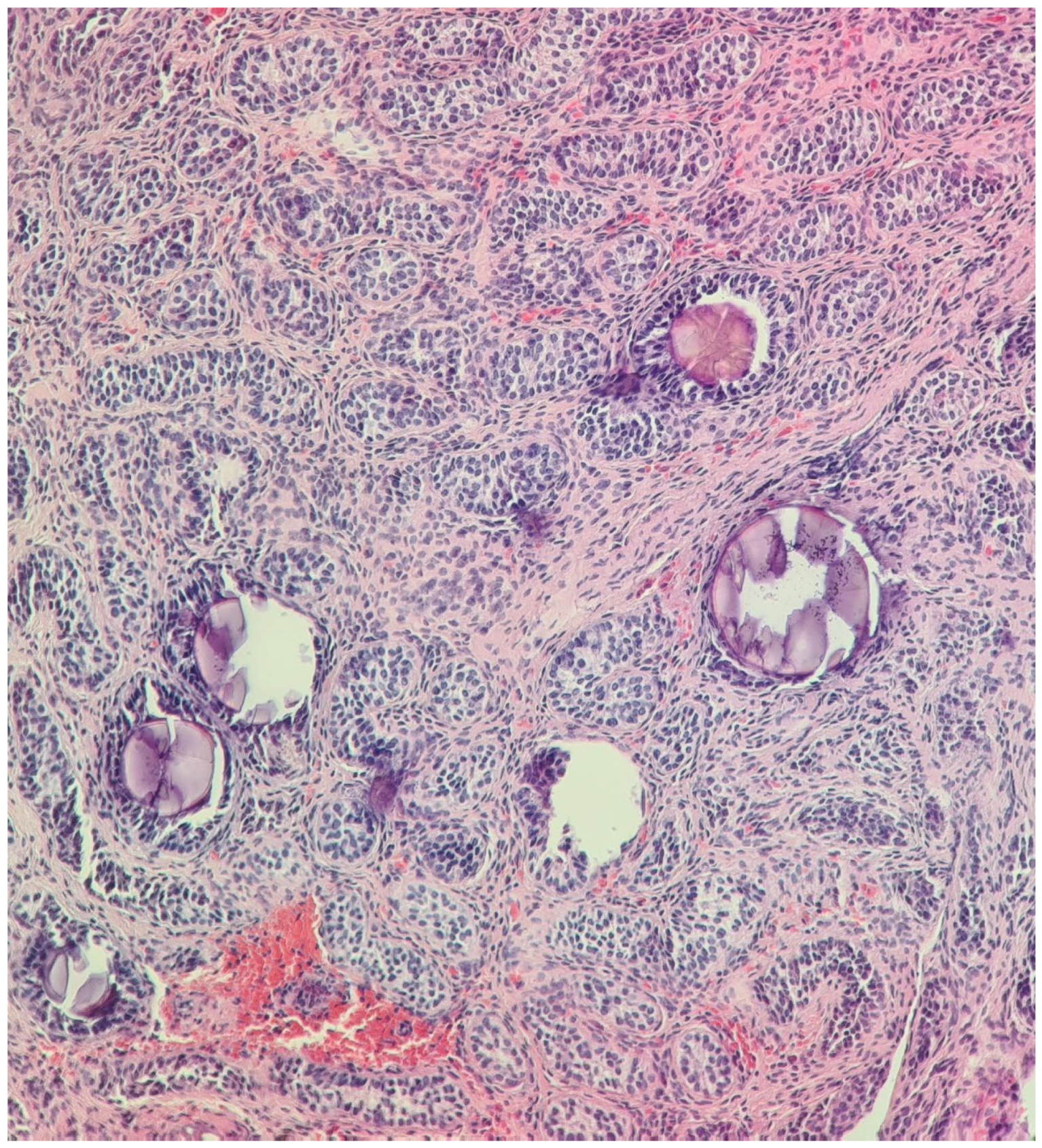
Figure 1. Undescended left testis biopsy: immature testis consisting of small seminiferous tubules without lumen and composed of immature Sertoli cells, with no evidence of spermatogenesis (May 2014). H&E stain, 200× and 400×.
Diagnostics 14 02621 g001aDiagnostics 14 02621 g001b
Figure 2. Rudimentary uterus with hypoplastic endometrial and myometrial layers. H&E stain, 400×.
Diagnostics 14 02621 g002aDiagnostics 14 02621 g002b
Figure 3. Structure of the hypoplastic vas deferens and epididymis with dilated tubules (2021), H&E stain, 200× and 400×.
Diagnostics 14 02621 g003aDiagnostics 14 02621 g003b
Figure 4. Tumor in the inguinal region: Immature testis consisting of small seminiferous tubules composed of mature and immature Sertoli cells, with no evidence of spermatogenesis; scattered microliths. H&E stain, 100× and 200×.
Diagnostics 14 02621 g004
Figure 5. Integrative Genomics Viewer (IGV) showing the homozygous variant NC_000012.11:g.53823315T>C in exon 8 of the AMHR2 gene in the patient.
Diagnostics 14 02621 g005
Figure 6. Flowchart depicting the search strategy employed to find the studies included in the review (following 2020 PRISMA guidelines).
Diagnostics 14 02621 g006
Table 1. Malformations associated with PMDS reported in the literature (from PubMed-indexed articles, accessed on 1 February 2024).
Malformations AffectingCasesGenetic Testing (If Available)Other Associated MalformationsObservationsReferences
Male excretory ducts
Vas deferens/epididymis dissociation, agenesia, fusion, duplication, etc.29 Not specific to PMDS, often associated with cryptorchidism[6,7,8,9,10,11,12,13,14,15,16,17,18,19,20,21,22]
Testes
Fused testes2 There is also a case of demonstrated fusion of the testes after birth[14,23]
Testicular regression syndrome (TRS)9 Late regression without hypospadiasWe excluded cases with a history of testicular torsion, inguinal hernia incarceration, or surgery in the inguinal–scrotal region[20,24,25,26,27,28,29,30,31,32,33,34,35,36]
Polyorchidism3AMHR2 mutation in the index case. No genetic testing in other cases. [2,3]
Urinary system
Crossed fused renal ectopia1 [37]
Multicystic kidney1 [38]
Kidney ectasia1 [39]
Kidney hypoplasia1 Intestinal lymphangiectasia, prenatal growth deficiency, hypertrophied alveolar ridges, redundant nuchal skin, and hepatomegaly [40]
Hydronephrosis3 Intestinal lymphangiectasia (2), pulmonary lymphangiectasia (1), prenatal growth deficiency (2), hypertrophied alveolar ridges (2), redundant nuchal skin (1), postaxial polydactyly (1), and hepatomegaly (2) [40,41]
Vesico-uretral reflux1 [42]
Ureteral duplication1 [43]
Gastrointestinal tract
Intestinal lymphangiectasia/lymphangiomyxoma 8 Prenatal growth deficiency (3), hypertrophied alveolar ridges (3), redundant nuchal skin (4), postaxial polydactyly (2), renal anomalies (2—hydronephrosis and 1—renal hypoplasia), and hepatomegaly (4) [40,44,45,46]
Pulmonary lymphangiectasia (2)
Hirschsprung disease2 [47,48]
Esophageal atresia1Idiopathic PMDS (PPP1R12A) [49]
Jejunal atresia1 [50]
Ileal atresia3Idiopathic PMDS (PPP1R12A) [49]
Spleen
Polysplenia1 Short pancreas (no body or tail) and unilateral TRS [24]
Vascular system
Pulmonary artery stenosis1 Facial dysmorphism [51]
Mesenteric vein abnormality1Trisomy 7Glaucoma [52]
Nervous system
Cerebellar ataxia and optic nerve atrophy1 [52]
Mental retardation1Monosomy 11Glaucoma and aniridia [52]
Sex chromosome abnormalities
Klinefelter syndrome347XXY (2), 46XY/47XXY (1) [53,54,55]
Adrenal glands
Congenital adrenal hyperplasia2 17 alpha-Hydroxylase deficiency (1) and 21 alpha-Hydroxylase deficiency (1) [25,56]
Metabolic abnormalities
Lipoatrophic diabetes and vitamin D-resistant rickets1Normal AMH gene [57]
Disclaimer/Publisher’s Note: The statements, opinions and data contained in all publications are solely those of the individual author(s) and contributor(s) and not of MDPI and/or the editor(s). MDPI and/or the editor(s) disclaim responsibility for any injury to people or property resulting from any ideas, methods, instructions or products referred to in the content.