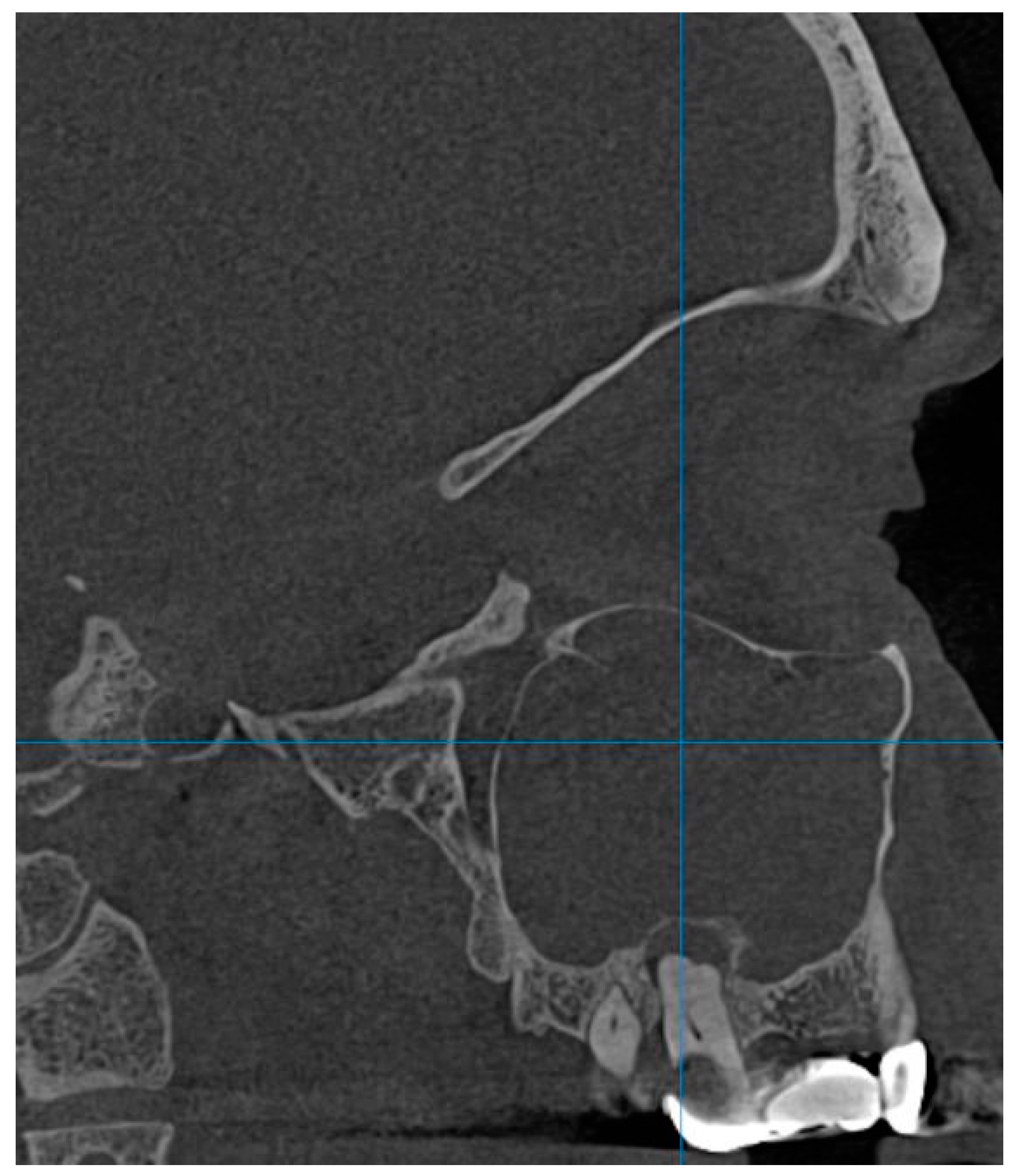
Preoperative Cone Beam Computed Topography Assessment of Maxillary Sinus Variations in Dental Implant Patients
Abstract
1. Introduction
2. Materials and Methods
2.1. Study Design
2.2. Image Analysis
2.3. Statistical Analysis
3. Results
4. Discussion
5. Conclusions
Author Contributions
Funding
Institutional Review Board Statement
Informed Consent Statement
Data Availability Statement
Conflicts of Interest
References
- Pelinsari Lana, J.; Moura Rodrigues Carneiro, P.; de Carvalho Machado, V.; Alencar, E.; de Souza, P.; Ricardo Manzi, F.; Campolina Rebello Horta, M. Anatomic variations and lesions of the maxillary sinus detected in cone beam computed tomography for dental implants. Clin. Oral Implants Res. 2012, 23, 1398–1403. [Google Scholar] [CrossRef] [PubMed]
- Pauwels, R.; Jacobs, R.; Singer, S.R.; Mupparapu, M. CBCT-based bone quality assessment: Are Hounsfield units applicable? Dentomaxillofacial Radiol. 2015, 44, 20140238. [Google Scholar] [CrossRef] [PubMed]
- Watanabe, H.; Honda, E.; Tetsumura, A.; Kurabayashi, T. A comparative study for spatial resolution and subjective image characteristics of a multi-slice CT and a cone-beam CT for dental use. Eur. J. Radiol. 2011, 77, 397–402. [Google Scholar] [CrossRef]
- Terlemez, A.; Tassoker, M.; Kizilcakaya, M.; Gulec, M. Comparison of cone-beam computed tomography and panoramic radiography in the evaluation of maxillary sinus pathology related to maxillary posterior teeth: Do apical lesions increase the risk of maxillary sinus pathology? Imaging Sci. Dent. 2019, 49, 115–122. [Google Scholar] [CrossRef]
- Krmpotić-Nemanić, J.; Draf, W.; Helms, J. Surgical Anatomy of Head and Neck; Springer: Berlin/Heidelberg, Germany, 1988. [Google Scholar] [CrossRef]
- Shankar, L.; Evans, K.; Marotta, T.R.; Yu, E.; Hawke, M.; Stammberger, H. An Atlas of Imaging of the Paranasal Sinuses; CRC Press: London, UK, 2021. [Google Scholar] [CrossRef]
- Şimşek Kaya, G.; Daltaban, Ö.; Kaya, M.; Kocabalkan, B.; Sindel, A.; Akdağ, M. The potential clinical relevance of anatomical structures and variations of the maxillary sinus for planned sinus floor elevation procedures: A retrospective cone beam computed tomography study. Clin. Implant. Dent. Relat. Res. 2019, 21, 114–121. [Google Scholar] [CrossRef]
- Traxler, H.; Windisch, A.; Geyerhofer, U.; Surd, R.; Solar, P.; Firbas, W. Arterial blood supply of the maxillary sinus. Clin. Anat. 1999, 12, 417–421. [Google Scholar] [CrossRef]
- Velasco-Torres, M.; Padial-Molina, M.; Alarcón, J.A.; Ovalle, F.; Catena, A.; Galindo-Moreno, P. Maxillary sinus dimensions with respect to the posterior superior alveolar artery decrease with tooth loss. Implant. Dent. 2016, 25, 464–470. [Google Scholar] [CrossRef]
- Gładysz, M.T.; Kruczała, Z.; Bąk, F.; Ochwat, K. The Role of the Alveolar Antral Artery in Oral and Maxillofacial Surgery: A Comprehensive Review. Transl. Res. Anat. 2024, 36, 100309. [Google Scholar] [CrossRef]
- Iwanaga, J.; Wilson, C.; Lachkar, S.; Tomaszewski, K.A.; Walocha, J.A.; Tubbs, R.S. Clinical anatomy of the maxillary sinus: Application to sinus floor augmentation. Anat. Cell Biol. 2019, 52, 17–24. [Google Scholar] [CrossRef]
- Rostetter, C.; Hungerbühler, A.; Blumer, M.; Rücker, M.; Wagner, M.; Stadlinger, B.; Lübbers, H.T. Cone beam computed tomography evaluation of the artery in the lateral wall of the maxillary sinus: Retrospective analysis of 602 sinuses. Implant. Dent. 2018, 27, 434–438. [Google Scholar] [CrossRef]
- Mardinger, O.; Abba, M.; Hirshberg, A.; Schwartz-Arad, D. Prevalence, diameter and course of the maxillary intraosseous vascular canal with relation to sinus augmentation procedure: A radiographic study. Int. J. Oral Maxillofac. Surg. 2007, 36, 735–738. [Google Scholar] [CrossRef] [PubMed]
- Varela-Centelles, P.; Loira-Gago, M.; Seoane-Romero, J.M.; Takkouche, B.; Monteiro, L.; Seoane, J. Detection of the posterior superior alveolar artery in the lateral sinus wall using computed tomography/cone beam computed tomography: A prevalence meta-analysis study and systematic review. Int. J. Oral Maxillofac. Surg. 2015, 44, 1405–1410. [Google Scholar] [CrossRef]
- Landis, R.; Koch, G. The Measurement of Observer Agreement for Categorical Data. Biometrics 1977, 33, 159–174. [Google Scholar] [CrossRef] [PubMed]
- Potter, R. Oral Radiology: Principles and Interpretation by Stuart White and Michael Pharoah. Oral Surg. Oral Med. Oral Pathol. Oral Radiol. Endodontology 2005, 99, 253. [Google Scholar] [CrossRef]
- Blake, F.A.S.; Blessmann, M.; Pohlenz, P.; Heiland, M. A new imaging modality for intraoperative evaluation of sinus floor augmentation. Int. J. Oral Maxillofac. Surg. 2008, 37, 183–185. [Google Scholar] [CrossRef]
- Amine, K.; Slaoui, S.; Kanice, F.Z.; Kissa, J. Evaluation of maxillary sinus anatomical variations and lesions: A retrospective analysis using cone beam computed tomography. J. Stomatol. Oral Maxillofac. Surg. 2020, 121, 484–489. [Google Scholar] [CrossRef]
- Danesh-Sani, S.A.; Movahed, A.; ElChaar, E.S.; Chong Chan, K.; Amintavakoli, N. Radiographic Evaluation of Maxillary Sinus Lateral Wall and Posterior Superior Alveolar Artery Anatomy: A Cone-Beam Computed Tomographic Study. Clin. Implant. Dent. Relat. Res. 2017, 19, 151–160. [Google Scholar] [CrossRef] [PubMed]
- Ilgüy, D.; Ilgüy, M.; Dolekoglu, S.; Fisekcioglu, E. Evaluation of the posterior superior alveolar artery and the maxillary sinus with CBCT. Oral Radiology Braz. Oral Res. 2013, 27, 431–437. [Google Scholar] [CrossRef] [PubMed]
- Shiki, K.; Tanaka, T.; Kito, S.; Wakasugi-Sato, N.; Matsumoto-Takeda, S.; Oda, M.; Nishimura, S.; Morimoto, Y. The significance of cone beam computed tomography for the visualization of anatomical variations and lesions in the maxillary sinus for patients hoping to have dental implant-supported maxillary restorations in a private dental office in Japan. Head. Face Med. 2014, 10, 20. [Google Scholar] [CrossRef]
- Naitoh, M.; Suenaga, Y.; Kondo, S.; Gotoh, K.; Ariji, E. Assessment of Maxillary Sinus Septa Using Cone-Beam Computed Tomography: Etiological Consideration. Clin. Implant. Dent. Relat. Res. 2009, 11, e52–e58. [Google Scholar] [CrossRef] [PubMed]
- Van Den Bergh, J.P.A.; Ten Bruggenkate, C.M.; Disch, F.J.M.; Tuinzing, D.B. Anatomical aspects of sinus floor elevations. Clin. Oral Implants Res. 2000, 11, 256–265. [Google Scholar] [CrossRef]
- Aimetti, M.; Romagnoli, R.; Ricci, G.; Massei, G. Maxillary sinus elevation: The effect of macrolacerations and microlacerations of the sinus membrane as determined by endoscopy. Int. J. Periodontics Restor. Dent. 2001, 21, 581–589. [Google Scholar]
- Güncü, G.N.; Yildirim, Y.D.; Wang, H.L.; Tözüm, T.F. Location of posterior superior alveolar artery and evaluation of maxillary sinus anatomy with computerized tomography: A clinical study. Clin. Oral Implant. Res. 2011, 22, 1164–1167. [Google Scholar] [CrossRef] [PubMed]
- Ritter, L.; Lutz, J.; Neugebauer, J.; Scheer, M.; Dreiseidler, T.; Zinser, M.J.; Mischkowski, R.A. Prevalence of pathologic findings in the maxillary sinus in cone-beam computerized tomography. Oral Surg. Oral Med. Oral Pathol. Oral Radiol. Endodontology 2011, 111, 634–640. [Google Scholar] [CrossRef]
- Bong-Hae, C.; Yun-Hoa, J. Prevalence of incidental paranasal sinus opacification in an adult dental population. Koreean J. Oral Maxilofac. Radiol. 2009, 39, 191–194. [Google Scholar]
- Whyte, A.; Boeddinghaus, R. The maxillary sinus: Physiology, development and imaging anatomy. Dentomaxillofacial Radiol. 2019, 48, 20190205. [Google Scholar] [CrossRef]
- Ta Velli, L.; Borgonovo, A.E.; Re, D.; Maiorana, C. Sinus presurgical evaluation: A literature review and a new classification proposal. Minerva Stomatologica. Ed. Minerva Medica 2017, 66, 115–131. [Google Scholar] [CrossRef]
- Mardinger, O.; Manor, I.; Mijiritsky, E.; Hirshberg, A. Maxillary sinus augmentation in the presence of antral pseudocyst: A clinical approach. Oral Surg. Oral Med. Oral Pathol. Oral Radiol. Endodontology 2007, 103, 180–184. [Google Scholar] [CrossRef] [PubMed]
- Yaman, H.; Yilmaz, S.; Karali, E.; Guclu, E.; Ozturk, O. Evaluation and Management of Antrochoanal Polyps. Clin. Exp. Otorhinolaryngol. 2010, 3, 110. [Google Scholar] [CrossRef] [PubMed]
- Basma, H.S.; Abou-Arraj, R.V. Management of a Large Artery During Maxillary Sinus Bone Grafting: A Case Report. Clin. Adv. Periodontics 2021, 11, 22–26. [Google Scholar] [CrossRef]
- Al-Ghurabi, Z.H.; Abdulrazaq, S.S. Vascular Precautions before Sinus Lift Procedure. J. Craniofacial Surg. 2018, 29, e116–e118. [Google Scholar] [CrossRef] [PubMed]
- Wang, L.; Gun, R.; Youssef, A.; Carrau, R.L.; Prevedello, D.M.; Otto, B.A.; Ditzel, L. Anatomical study of critical features on the posterior wall of the maxillary sinus: Clinical implications. Laryngoscope 2014, 124, 2451–2455. [Google Scholar] [CrossRef] [PubMed]
- Valente, N.A. Anatomical Considerations on the Alveolar Antral Artery as Related to the Sinus Augmentation Surgical Procedure. Clin. Implant. Dent. Relat. Res. 2016, 18, 1042–1050. [Google Scholar] [CrossRef] [PubMed]


| Anatomic Variation | Frequency | Gender Distribution | p Value 1 | Age Group Distribution | p Value 1 | |
|---|---|---|---|---|---|---|
| Pneumatization | 163 (81.5%) -alveolar 32.5% -multiple sites 67.5% | M: 97 (59.5%) F: 66 (40.5%) | 3.246 1 0.53 2 | A: 55 (33.7%) B: 91 (55.8%) C: 17 (10.4%) | 4.304 1 0.116 2 | |
| Antral septa | 97 (48.5%) | M: 52 (53.6%) F: 45 (46.4%) | 5.641 1 0.255 2 | A:35 (36.1%) B: 52 (53.6%) C: 10 (10.3%) | 1.462 1 0.481 2 | |
| Hypoplasia | 13 (6.5%) | M: 7 (53.8%) F: 6 (46.2%) | 0.141 1 0.842 2 | A: 7 (53.8%) B: 5 (38.5%) C: 1 (7.7%) | 2.498 1 0.287 2 | |
| Exostosis | 3 (1.5%) | M: 2 (66.6%) F: 1 (33.3%) | 0.128 1 0.597 2 | A:1 (33.3%) B:2 (66.6%) C:0 | 0.304 1 0.859 2 | |
| Pathological factors | ||||||
| Mucosal thickening | >3 mm 81 (40.5%) | M: 40 (49.4%) F:41 (50.6%) | 2.8061 0.063 2 | A: 1 B: 71 C: 9 | 65.605 1 <0.001 2 | |
| ≤3 mm 119 (59.5%) | M:73 (61.3%) F:46 (38.7%) | A: 67 B: 44 C: 8 | ||||
| Polypoid lesion | 35 (17.5%) | M: 23 (65.7%) F: 12 (34.3%) | 1.466 1 0.153 2 | A: 20 (57.1%) B: 15 (42.9%) C: 0 | 11.871 1 <0.001 2 | |
| Bone thickening | 7 (3.5%) | M: 1 (14.3%) F: 6 (85.7%) | 5.260 1 0.022 2 | A: 2 (28.6%) B: 4 (57.1%) C: 1 14.3%) | 0.349 1 0.840 2 | |
| Antrolith | 4 (2%) | M: 3 (75%) F: 1 (25%) | 0.568 1 0.414 2 | A: 1 (25% B: 2 (50%) C: 1 (25%) | 1.444 1 0.486 2 | |
| Sinusal floor discontinuity | 43 (21.5%) | Bone graft 36 (83.7%) | M: 20 (55.5%) F: 16 (44.4%) | 2.679 1 0.444 2 | A: 18 (50%) B: 11 (30.6%) C: 7 (19.4%) | 16.515 1 0.011 2 |
| Extraction 5 (11.6%) | M: 3 (60%) F: 2 (40%) | A: 2 (40%) B: 3 (60%) C: 0 | ||||
| Unclear reason 2 (4.7%) | M: 0 F: 2 (100%) | A: 0 B: 2 (100% C: 0 | ||||
| Foreign body | 3 (1.5%) | M: 1 (33.3%) F: 2 (66.6%) | 0.665 1 0.403 2 | A: 0 B: 2 (66.6%) C: 1 (33.3%) | 3.290 1 0.193 2 | |
| Opacifiation | 16 (8%) | M: 7 (43.8%) F: 9 (56.2%) | 1.150 1 0.208 2 | A: 6 (37.5%) B: 9 (56.3%) C: 1 (6.2%) | 0.171 1 0.918 2 | |
Disclaimer/Publisher’s Note: The statements, opinions and data contained in all publications are solely those of the individual author(s) and contributor(s) and not of MDPI and/or the editor(s). MDPI and/or the editor(s) disclaim responsibility for any injury to people or property resulting from any ideas, methods, instructions or products referred to in the content. |
© 2024 by the authors. Licensee MDPI, Basel, Switzerland. This article is an open access article distributed under the terms and conditions of the Creative Commons Attribution (CC BY) license (https://creativecommons.org/licenses/by/4.0/).
Share and Cite
Misăiloaie, A.; Tărăboanță, I.; Budacu, C.C.; Sava, A. Preoperative Cone Beam Computed Topography Assessment of Maxillary Sinus Variations in Dental Implant Patients. Diagnostics 2024, 14, 1929. https://doi.org/10.3390/diagnostics14171929
Misăiloaie A, Tărăboanță I, Budacu CC, Sava A. Preoperative Cone Beam Computed Topography Assessment of Maxillary Sinus Variations in Dental Implant Patients. Diagnostics. 2024; 14(17):1929. https://doi.org/10.3390/diagnostics14171929
Chicago/Turabian StyleMisăiloaie, Alexandru, Ionuț Tărăboanță, Cristian Constantin Budacu, and Anca Sava. 2024. "Preoperative Cone Beam Computed Topography Assessment of Maxillary Sinus Variations in Dental Implant Patients" Diagnostics 14, no. 17: 1929. https://doi.org/10.3390/diagnostics14171929
APA StyleMisăiloaie, A., Tărăboanță, I., Budacu, C. C., & Sava, A. (2024). Preoperative Cone Beam Computed Topography Assessment of Maxillary Sinus Variations in Dental Implant Patients. Diagnostics, 14(17), 1929. https://doi.org/10.3390/diagnostics14171929


