
Machine Learning on Ultrasound Texture Analysis Data for Characterizing of Salivary Glandular Tumors: A Feasibility Study

Abstract
1. Introduction
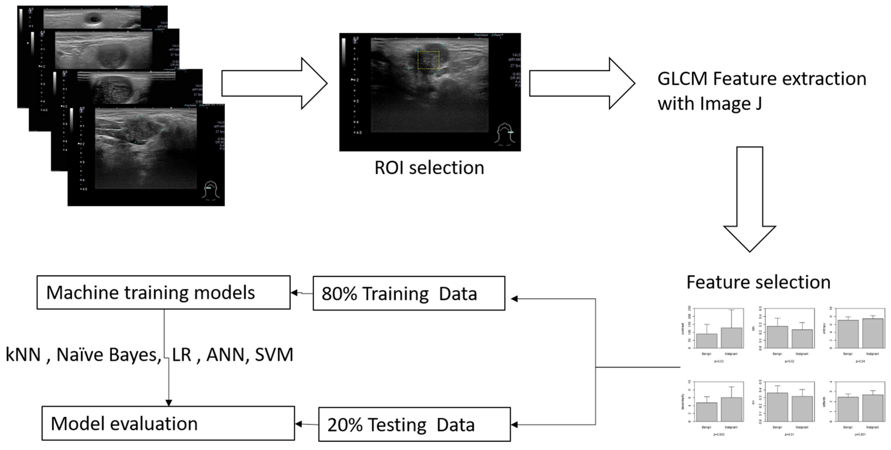
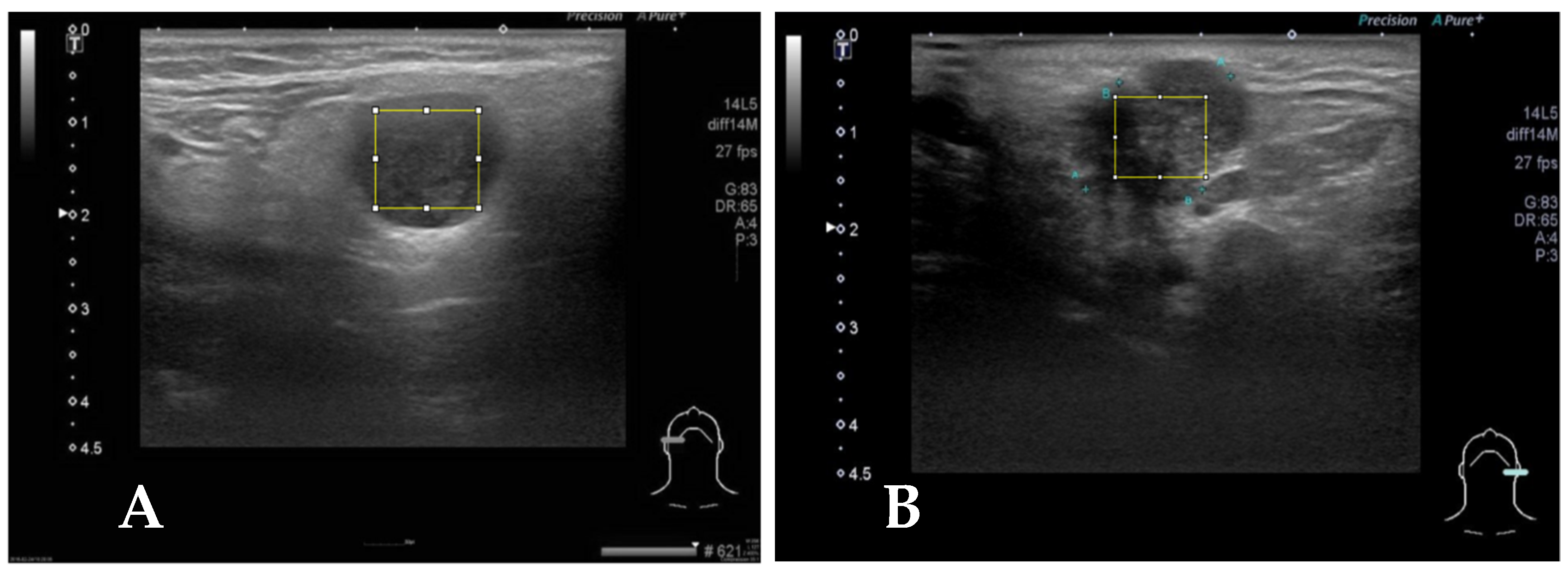
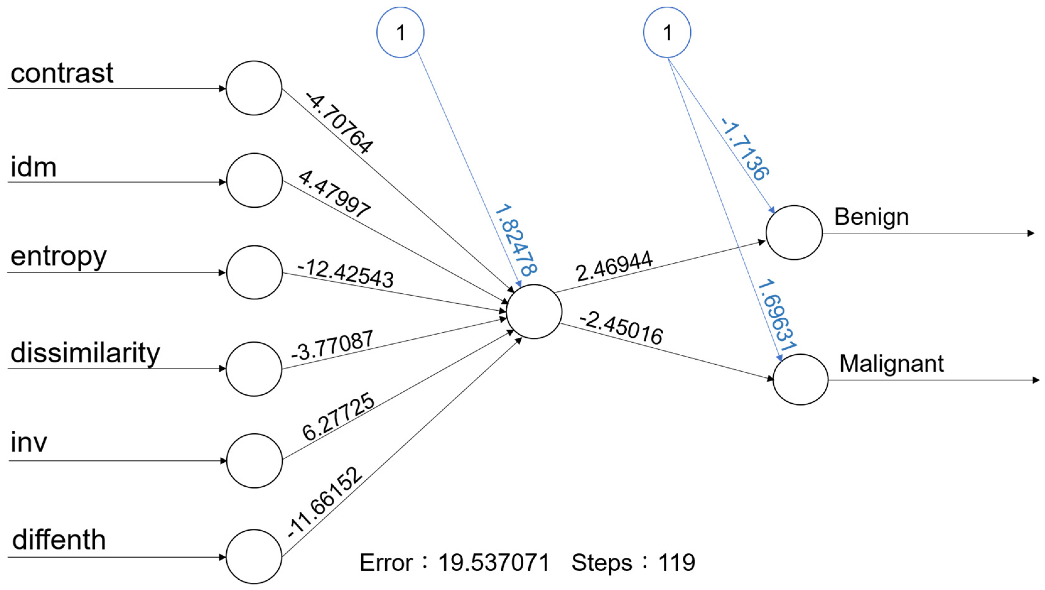
2. Materials and Methods
3. Results
4. Discussion
5. Conclusions
Author Contributions
Funding
Institutional Review Board Statement
Informed Consent Statement
Data Availability Statement
Conflicts of Interest
References
- Kuan, E.C.; Clair, J.M.S.; John, M.A.S. Evaluation of Parotid Lesions. Otolaryngol. Clin. N. Am. 2016, 49, 313–325. [Google Scholar] [CrossRef] [PubMed]
- Bozzato, A.; Zenk, J.; Greess, H.; Hornung, J.; Gottwald, F.; Rabe, C.; Iro, H. Potential of ultrasound diagnosis for parotid tumors: Analysis of qualitative and quantitative parameters. Otolaryngol. Head Neck Surg. 2007, 137, 642–646. [Google Scholar] [CrossRef] [PubMed]
- Haidar, Y.M.; Moshtaghi, O.; Mahmoodi, A.; Helmy, M.; Goddard, J.A.; Armstrong, W.B. The Utility of In-Office Ultrasound in the Diagnosis of Parotid Lesions. Otolaryngol. Head Neck Surg. 2017, 156, 511–517. [Google Scholar] [CrossRef]
- Liao, L.J.; Wen, M.H.; Yang, T.L. Point-of-care ultrasound in otolaryngology and head and neck surgery: A prospective survey study. J. Formos. Med. Assoc. 2021, 120, 1547–1553. [Google Scholar] [CrossRef]
- Kovacević, D.O.; Fabijanić, I. Sonographic diagnosis of parotid gland lesions: Correlation with the results of sonographically guided fine-needle aspiration biopsy. J. Clin. Ultrasound 2010, 38, 294–298. [Google Scholar] [CrossRef] [PubMed]
- Song, I.H.; Song, J.S.; Sung, C.O.; Roh, J.L.; Choi, S.H.; Nam, S.Y.; Kim, S.Y.; Lee, J.H.; Baek, J.H.; Cho, K.J. Accuracy of Core Needle Biopsy Versus Fine Needle Aspiration Cytology for Diagnosing Salivary Gland Tumors. J. Pathol. Transl. Med. 2015, 49, 136–143. [Google Scholar] [CrossRef] [PubMed]
- Cheng, P.C.; Chang, C.M.; Huang, C.C.; Lo, W.C.; Huang, T.W.; Cheng, P.W.; Liao, L.J. The diagnostic performance of ultrasonography and computerized tomography in differentiating superficial from deep lobe parotid tumours. Clin. Otolaryngol. 2019, 44, 286–292. [Google Scholar] [CrossRef]
- Lo, W.A.-O.; Chang, C.M.; Wang, C.T.; Cheng, P.W.; Liao, L.A.-O. A Novel Sonographic Scoring Model in the Prediction of Major Salivary Gland Tumors. Laryngoscope 2021, 131, E157–E162. [Google Scholar] [CrossRef] [PubMed]
- Haralick, R.M.; Shanmugam, K.; Dinstein, I.H. Textural features for image classification. IEEE Trans. Syst. Man Cybern. 1973, 6, 610–621. [Google Scholar] [CrossRef]
- Ghorayeb, S.R.; Bracero, L.A.; Blitz, M.J.; Rahman, Z.; Lesser, M.L. Quantitative Ultrasound Texture Analysis for Differentiating Preterm From Term Fetal Lungs. J. Ultrasound Med. 2017, 36, 1437–1443. [Google Scholar] [CrossRef] [PubMed]
- Bhatia, K.S.; Lam, A.C.; Pang, S.W.; Wang, D.; Ahuja, A.T. Feasibility Study of Texture Analysis Using Ultrasound Shear Wave Elastography to Predict Malignancy in Thyroid Nodules. Ultrasound Med. Biol. 2016, 42, 1671–1680. [Google Scholar] [CrossRef] [PubMed]
- Yang, X.; Tridandapani, S.; Beitler, J.J.; Yu, D.S.; Yoshida, E.J.; Curran, W.J.; Liu, T. Ultrasound GLCM texture analysis of radiation-induced parotid-gland injury in head-and-neck cancer radiotherapy: An in vivo study of late toxicity. Med. Phys. 2012, 39, 5732–5739. [Google Scholar] [CrossRef] [PubMed]
- Brattain, L.J.; Telfer, B.A.; Dhyani, M.; Grajo, J.R.; Samir, A.E. Machine learning for medical ultrasound: Status, methods, and future opportunities. Abdom. Radiol. 2018, 43, 786–799. [Google Scholar] [CrossRef] [PubMed]
- Yonetsu, K.; Ohki, M.; Fau-Kumazawa, S.; Kumazawa, S.; Fau-Eida, S.; Eida, S.; Fau-Sumi, M.; Sumi, M.; Fau-Nakamura, T.; Nakamura, T. Parotid tumors: Differentiation of benign and malignant tumors with quantitative sonographic analyses. Ultrasound Med. Biol. 2004, 30, 567–574. [Google Scholar] [CrossRef]
- R Core Team. R: A Language and Environment for Statistical Computing; R Foundation for Statistical Computing: Vienna, Austria, 2013. [Google Scholar]
- Lantz, B. Machine Learning with R: Expert Techniques for Predictive Modeling; Packt Publishing Ltd.: Birmingham, UK, 2019. [Google Scholar]
- Schneider, C.A.; Rasband Ws Fau-Eliceiri, K.W.; Eliceiri, K.W. NIH Image to ImageJ: 25 years of image analysis. Nat. Methods 2012, 9, 671–675. [Google Scholar] [CrossRef] [PubMed]
- Chi, J.; Walia, E.; Babyn, P.; Wang, J.; Groot, G.; Eramian, M. Thyroid Nodule Classification in Ultrasound Images by Fine-Tuning Deep Convolutional Neural Network. J. Digit. Imaging 2017, 30, 477–486. [Google Scholar] [CrossRef] [PubMed]
- Chai, Y.J.; Song, J.; Shaear, M.; Yi, K.H. Artificial intelligence for thyroid nodule ultrasound image analysis. Ann. Thyroid 2020, 5, 8. [Google Scholar] [CrossRef]
- Nwanganga, F.; Chapple, M. Practical Machine Learning in R; John Wiley & Sons: Hoboken, NJ, USA, 2020. [Google Scholar]
- Nanni, L.; Lumini, A.; Fau-Brahnam, S.; Brahnam, S. Local binary patterns variants as texture descriptors for medical image analysis. Artif. Intell. Med. 2010, 49, 117–125. [Google Scholar] [CrossRef] [PubMed]
- Materka, A.; Strzelecki, M. Texture Analysis Methods—A Review; COST B11 Report; Technical University of Lodz: Lodz, Poland; Institute of Electronics: Brussels, Belgium, 1998; Volume 10, p. 4968. [Google Scholar]
- Uddin, S.A.-O.; Khan, A.; Hossain, M.E.; Moni, M.A. Comparing different supervised machine learning algorithms for disease prediction. BMC Med. Inform. Decis. Mak. 2019, 19, 281. [Google Scholar] [CrossRef] [PubMed]

| Characteristics | Benign | Malignant | p-Value |
|---|---|---|---|
| Age | 50.5 ± 12.8 | 56.1 ± 17.8 | 0.06 |
| Gender (F/M) | 30/41 | 19/32 | 0.71 |
| Size-short axis | 1.58 ± 0.59 | 1.79 ± 0.60 | 0.06 |
| Size-long axis | 2.35 ± 0.95 | 2.51 ± 0.91 | 0.35 |
| Contrast | 90.2 ± 58.0 | 129.2 ± 115.4 | 0.03 |
| IDM | 0.28 ± 0.10 | 0.23 ± 0.09 | 0.02 |
| Entropy | 7.01 ± 0.87 | 7.39 ± 0.86 | 0.04 |
| Dissimilarity | 4.70 ± 1.53 | 6.08 ± 2.72 | 0.002 |
| INV | 0.36 ± 0.09 | 0.32 ± 0.09 | 0.01 |
| Diffenth | 2.47 ± 0.31 | 2.7 ± 0.41 | 0.0006 |
| Final diagnosis | Pleomorphic adenoma (29) | Metastatic carcinoma (26) | |
| Warthin’s tumor (24) | Invasive carcinoma (6) | ||
| Chronic sialadenitis (5) | Mucoepidermoid carcinoma (3) | ||
| Basal cell adenoma (4) | Acinic cell carcinoma (3) | ||
| Lymphoepithelial cyst (2) | Lymphoepithelial carcinoma (2) | ||
| Nodular fasciitis (2) | Adenoid cystic carcinoma (2) | ||
| Benign cyst (2) | Carcinoma ex-pleomorphic adenoma (2) | ||
| Epidermal cyst (1) | Adenocarcinoma (1) | ||
| Lipoma (1) | Diffuse large B cell lymphoma (1) | ||
| Reactive hyperplasia LN (1) | High-grade B cell lymphoma (1) | ||
| Blue round cell tumor (1) | |||
| Lymphoblastic lymphoma (1) | |||
| Squamous cell carcinoma (1) | |||
| Salivary ductal carcinoma (1) |
| Sensitivity | Specificity | Overall Accuracy | |
|---|---|---|---|
| kNN (k = 5) | 62.5 (38.8–86.2)% | 84.2 (67.8–100)% | 74.3 (59.8–88.8)% |
| Naïve Bay | 88.2 (72.9–100)% | 100% | 94.3 (86.6–100)% |
| Logistic regression | 75.0 (32.6–100)% | 71.4 (52.1–90.8)% | 72.0 (54.4–89.6)% |
| ANN | 60.0 (29.6–90.4)% | 100% | 84.0 (69.5–97.3)% |
| SVM | 87.5 (64.6–100)% | 69.2 (51.5–87.0)% | 73.5 (58.7–88.4)% |
Disclaimer/Publisher’s Note: The statements, opinions and data contained in all publications are solely those of the individual author(s) and contributor(s) and not of MDPI and/or the editor(s). MDPI and/or the editor(s) disclaim responsibility for any injury to people or property resulting from any ideas, methods, instructions or products referred to in the content. |
© 2024 by the authors. Licensee MDPI, Basel, Switzerland. This article is an open access article distributed under the terms and conditions of the Creative Commons Attribution (CC BY) license (https://creativecommons.org/licenses/by/4.0/).
Share and Cite
Liao, L.-J.; Cheng, P.-C.; Chan, F.-T. Machine Learning on Ultrasound Texture Analysis Data for Characterizing of Salivary Glandular Tumors: A Feasibility Study. Diagnostics 2024, 14, 1761. https://doi.org/10.3390/diagnostics14161761
Liao L-J, Cheng P-C, Chan F-T. Machine Learning on Ultrasound Texture Analysis Data for Characterizing of Salivary Glandular Tumors: A Feasibility Study. Diagnostics. 2024; 14(16):1761. https://doi.org/10.3390/diagnostics14161761
Chicago/Turabian StyleLiao, Li-Jen, Ping-Chia Cheng, and Feng-Tsan Chan. 2024. "Machine Learning on Ultrasound Texture Analysis Data for Characterizing of Salivary Glandular Tumors: A Feasibility Study" Diagnostics 14, no. 16: 1761. https://doi.org/10.3390/diagnostics14161761
APA StyleLiao, L.-J., Cheng, P.-C., & Chan, F.-T. (2024). Machine Learning on Ultrasound Texture Analysis Data for Characterizing of Salivary Glandular Tumors: A Feasibility Study. Diagnostics, 14(16), 1761. https://doi.org/10.3390/diagnostics14161761