
Pre- and Post-Operative Cone Beam Computed Tomography Assessment of the Temporomandibular Joint in Patients with Orthognathic Surgery

 ,
,  ,
, 
Abstract
1. Introduction
2. Materials and Methods
2.1. Study Design and Population
2.2. Operative and Post-Operative Data Evaluation
2.3. Cone-Beam CT (CBCT)
2.4. Image Evaluation
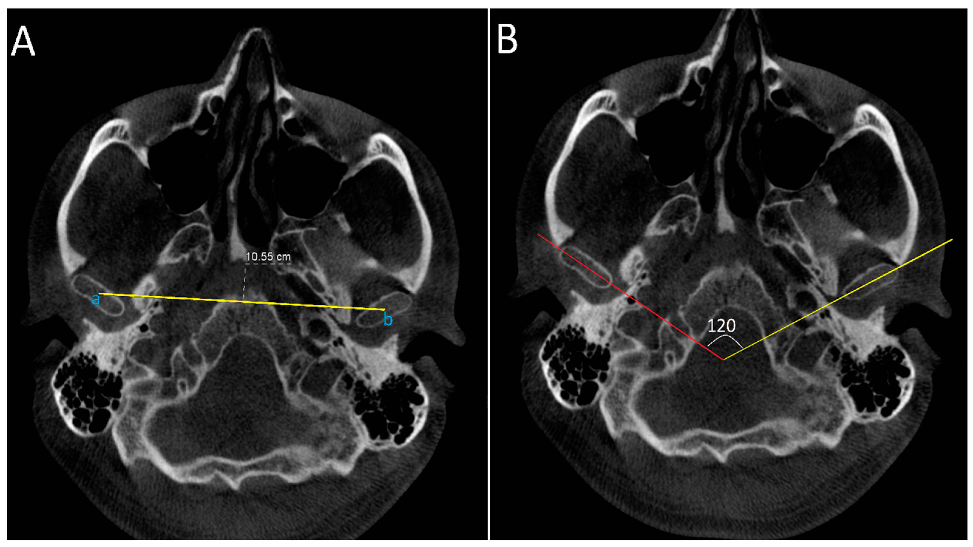
2.4.1. Axial Projection: Measurements Were Conducted on the Slice with the Largest Condylar Dimensions in the Axial Plane (Figure 2)
- Intercondylar distance: defined as the distance between midpoint of right and left condylar heads.
- Intercondylar angle: defined as the angle formed between lines connecting medial and lateral poles of the condylar heads on both sides.
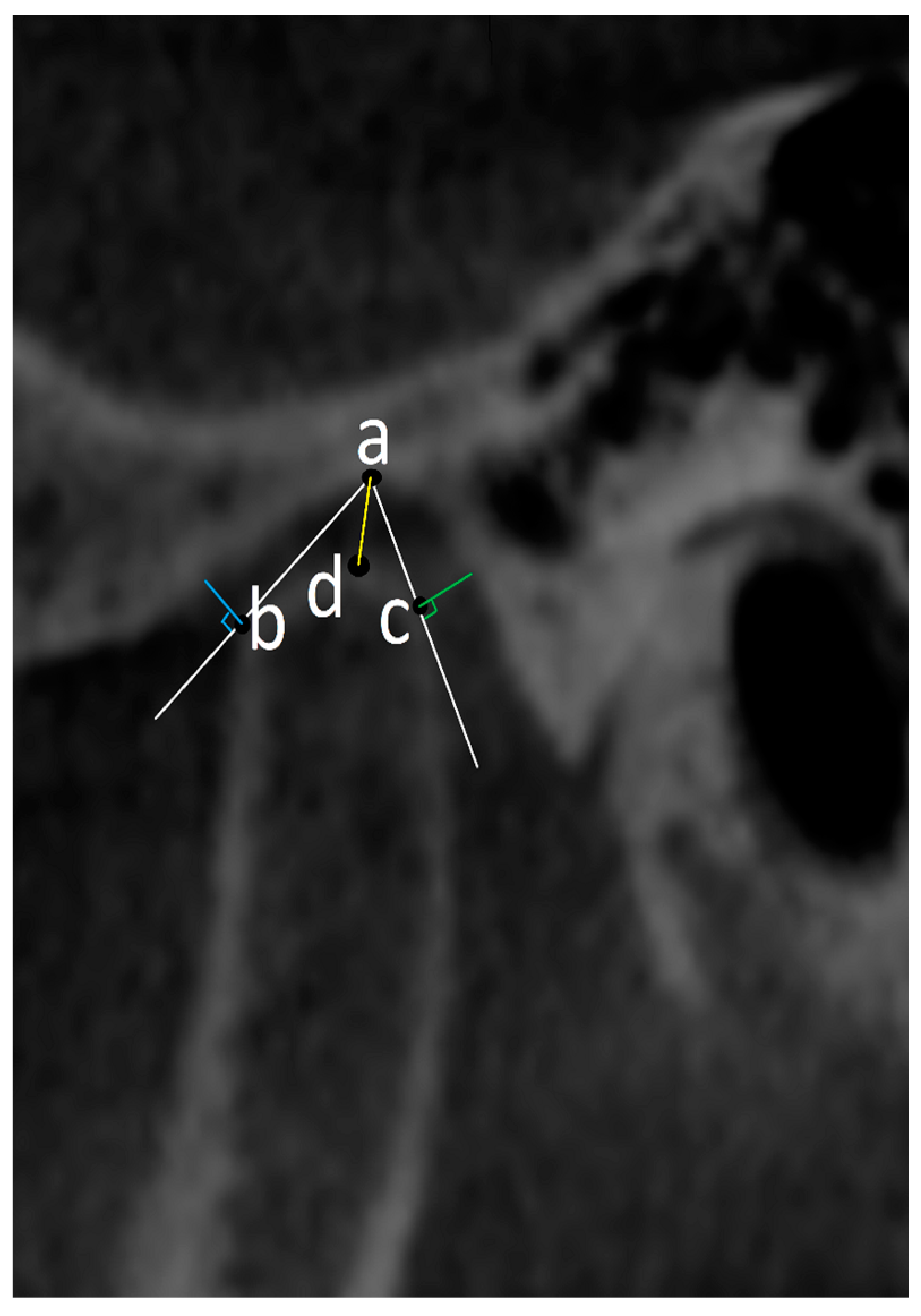
2.4.2. Oblique Sagittal Projection (Figure 3): Measurements Were Carried out on the Image in the Sagittal Plane Corresponding to the Middle Point of the Condyle in the Axial Plane and Coronal Plane on the Slices with the Largest Condylar Dimensions
- The uppermost point of the mandibular fossa is set as point (a) from which two tangent lines are drawn to join the most anterior and most posterior margins of the condylar head. By drawing perpendicular lines to these tangents, anterior (AJS) and posterior (PJS) joint spaces were measured. Another line joining point (a) and the most superior part of the condyle was used to measure the superior joint space (SJS).
- Condylar position was assessed after that using the formula according to the method of Pullinger and Hollender [16]:Condylar position ratio = ln (PJS/AJS)Ratio = ±0.25, condyle in concentric position.Ratio < −0.25, condyle in posterior position.Ratio > 0.25, condyle in anterior position.
2.4.3. Coronal Projection (Figure 4 and Figure 5): Measurements Were Carried out on the Image in the Coronal Plane Corresponding to the Middle Point of the Condyle on the Axial and Sagittal Planes on the Slices with the Largest Condylar Dimensions
- Medial joint space (MJS): distance between the medial pole of the mandibular condyle and medial wall of glenoid fossa.
- Lateral joint space (LJS): distance between the lateral pole of the mandibular condyle and lateral wall of glenoid fossa.
- Intercondylar distance: defined as distance between the midpoint of the right and left condylar heads.
- Intercondylar angle: defined as the angle formed between lines connecting the medial and lateral poles of the condylar heads on both sides.


2.5. Statistical Analysis
3. Results
Radiological Assessment of TMJ Joint Spaces and Condylar Position Analysis
4. Discussion
5. Conclusions
Author Contributions
Funding
Institutional Review Board Statement
Informed Consent Statement
Data Availability Statement
Conflicts of Interest
References
- Miotto, A.V.; Bonotto, D.V.; Silva, J.S.C.; De Souza, J.F.; Sebastiani, A.M.; Scariot, R. Temporomandibular Disorders at the Preoperative Time of Orthognathic Surgery. Diagnostics 2023, 13, 2922. [Google Scholar] [CrossRef] [PubMed]
- Ayyıldız, E.; Orhan, M.; Bahşi, İ.; Yalçin, E.D. Morphometric evaluation of the temporomandibular joint on cone-beam computed tomography. Surg. Radiol. Anat. 2021, 43, 975–996. [Google Scholar] [CrossRef] [PubMed]
- Monson, L.A. Bilateral Sagittal Split Osteotomy. Semin. Plast. Surg. 2013, 27, 145–148. [Google Scholar] [CrossRef]
- Holzinger, D.; Willinger, K.; Millesi, G.; Schicho, K.; Breuss, E.; Wagner, F.; Seemann, R. Changes of temporomandibular joint position after surgery first orthognathic treatment concept. Sci. Rep. 2019, 9, 2206. [Google Scholar] [CrossRef]
- Bahşi, I.; Orhan, M.; Kervancıoğlu, P.; Yalçın, E.D.; Aktan, A.M. Anatomical evaluation of nasopalatine canal on cone beam computed tomography images. Folia Morphol. 2019, 78, 153–162. [Google Scholar] [CrossRef] [PubMed]
- Dalili, Z.; Khaki, N.; Kia, S.; Salamat, F. Assessing joint space and condylar position in the people with normal function of temporomandibular joint with cone-beam computed tomography. Dent. Res. J. 2012, 9, 607–612. [Google Scholar] [CrossRef]
- Krishnamoorthy, B.; Mamatha, N.; Kumar, V.A. TMJ imaging by CBCT: Current scenario. Ann. Maxillofac. Surg. 2013, 3, 80–83. [Google Scholar] [CrossRef]
- Zhang, Z.-L.; Cheng, J.-G.; Li, G.; Zhang, J.-Z.; Zhang, Z.-Y.; Ma, X.-C. Measurement accuracy of temporomandibular joint space in Promax 3-dimensional cone-beam computerized tomography images. Oral Surg. Oral Med. Oral Pathol. Oral Radiol. 2012, 114, 112–117. [Google Scholar] [CrossRef]
- Chenin, D.L. 3D cephalometrics: The new norm. Alpha Omegan 2010, 103, 51–56. [Google Scholar] [CrossRef]
- Wen, J.; Liu, S.; Ye, X.; Xie, X.; Li, J.; Li, H.; Mei, L. Comparative study of cephalometric measurements using 3 imaging modalities. J. Am. Dent. Assoc. 2017, 148, 913–921. [Google Scholar] [CrossRef]
- Goncalves, J.R.; Wolford, L.M.; Cassano, D.S.; da Porciuncula, G.; Paniagua, B.; Cevidanes, L.H. Temporomandibular Joint Condylar Changes Following Maxillomandibular Advancement and Articular Disc Repositioning. J. Oral Maxillofac. Surg. 2013, 71, 1759.e1–1759.e15. [Google Scholar] [CrossRef] [PubMed]
- Harris, M.D.; Van Sickels, J.R.; Alder, M. Factors influencing condylar position after the bilateral sagittal split osteotomy fixed with bicortical screws. J. Oral Maxillofac. Surg. 1999, 57, 650–654. [Google Scholar] [CrossRef] [PubMed]
- Wang, L.C.; Lee, Y.H.; Tsai, C.Y.; Wu, T.J.; Teng, Y.Y.; Lai, J.P.; Lin, S.S.; Chang, Y.J. Postsurgical Stability of Temporomandibular Joint of Skeletal Class III Patients Treated with 2-Jaw Orthognathic Surgery via Computer-Aided Three-Dimensional Simulation and Navigation in Orthognathic Surgery (CASNOS). BioMed Res. Int. 2021, 6, 1563551. [Google Scholar] [CrossRef] [PubMed]
- Draenert, F.G.; Erbe, C.; Zenglein, V.; Kämmerer, P.W.; Wriedt, S.; Al Nawas, B. 3D Analysis of Condylar Position after Sagittal Split Osteotomy of the Mandible in Mono- and Bimaxillary Orthognathic Surgery—A Methodology Study in 18 Patients. J. Orofac. Orthop. Der Kieferorthopadie 2010, 71, 421–429. [Google Scholar] [CrossRef] [PubMed]
- Chen, S.; Lei, J.; Wang, X.; Fu, K.-Y.; Farzad, P.; Yi, B. Short- and Long-Term Changes of Condylar Position after Bilateral Sagittal Split Ramus Osteotomy for Mandibular Advancement in Combination with Le Fort I Osteotomy Evaluated by Cone-Beam Computed Tomography. J. Oral Maxillofac. Surg. 2013, 71, 1956–1966. [Google Scholar] [CrossRef] [PubMed]
- Pullinger, A.; Hollender, L. Variation in condyle-fossa relationships according to different methods of evaluation in tomograms. Oral Surg. Oral Med. Oral Pathol. 1986, 62, 719–727. [Google Scholar] [CrossRef] [PubMed]
- Pachnicz, D.; Ramos, A. Mandibular condyle displacements after orthognathic surgery-an overview of quantitative studies. Quant. Imaging Med. Surg. 2021, 11, 1628–1650. [Google Scholar] [CrossRef]
- An, S.-B.; Park, S.-B.; Kim, Y.-I.; Son, W.-S. Effect of post-orthognathic surgery condylar axis changes on condylar morphology as determined by 3-dimensional surface reconstruction. Angle Orthod. 2014, 84, 316–321. [Google Scholar] [CrossRef]
- Han, J.J.; Hwang, S.J. Three-dimensional analysis of postoperative returning movement of perioperative condylar displacement after bilateral sagittal split ramus osteotomy for mandibular setback with different fixation methods. J. Cranio-Maxillofacial Surg. 2015, 43, 1918–1925. [Google Scholar] [CrossRef]
- Ma, R.-H.; Li, G.; Yin, S.; Sun, Y.; Li, Z.-L.; Ma, X.-C. Quantitative assessment of condyle positional changes before and after orthognathic surgery based on fused 3D images from cone beam computed tomography. Clin. Oral Investig. 2020, 24, 2663–2672. [Google Scholar] [CrossRef]
- Park, S.-B.; Yang, Y.-M.; Kim, Y.-I.; Cho, B.-H.; Jung, Y.-H.; Hwang, D.-S. Effect of Bimaxillary Surgery on Adaptive Condylar Head Remodeling: Metric Analysis and Image Interpretation Using Cone-Beam Computed Tomography Volume Superimposition. J. Oral Maxillofac. Surg. 2012, 70, 1951–1959. [Google Scholar] [CrossRef] [PubMed]
- Kim, Y.-I.; Cho, B.-H.; Jung, Y.-H.; Son, W.-S.; Park, S.-B. Cone-beam computerized tomography evaluation of condylar changes and stability following two-jaw surgery: Le Fort I osteotomy and mandibular setback surgery with rigid fixation. Oral Surg. Oral Med. Oral Pathol. Oral Radiol. Endodontol. 2011, 111, 681–687. [Google Scholar] [CrossRef] [PubMed]

| Class II DFD n = 29 (36.7%) | Class III DFD n = 50 (63.3%) | Total n = 79 | ||
|---|---|---|---|---|
| Age (Years) | Mean ± SD | 28.3 ± 8.2 | 25.7 ± 10.2 | 26.62 ± 9.5 |
| Range | 16–52 | 16–58 | 16–58 | |
| Median (IQR) | 28 (10) | 20.5 (12) | 24 (13) | |
| Gender | Male | 13 (44.8%) | 25 (50%) | 38 (48.1%) |
| Female | 16 (55.2%) | 25 (50%) | 41 (51.9%) | |
| Surgical Approach | BSSO | 8 (27.6%) | 11 (22%) | 19 (24.05%) |
| BBSO + Le Fort I | 21 (72.4%) | 39 (78%) | 60 (75.95%) | |
| Pre-Operative | Post-Operative | p Value | |||
|---|---|---|---|---|---|
| Oblique Sagittal View | AJS (right) | Median (IQR) | 2.1 (1.1) | 2.2 (0.9) | 0.13 ‡ |
| AJS (left) | Median (IQR) | 2.1 (1.2) | 2.1 (1) | 0.26 ‡ | |
| SJS (right) | Median (IQR) | 2.5 (1.5) | 2.3 (1.1) | 0.005 ‡ | |
| SJS (left) | Mean ± SD | 2.8 ± 1.1 | 2.6 ± 1.2 | 0.008 * | |
| PJS (right) | Median (IQR) | 2.2 (1.4) | 2 (1) | 0.01 ‡ | |
| PJS (left) | Median (IQR) | 2.3 (1.2) | 1.9 (1.3) | 0.002 ‡ | |
| Coronal View | LJS (right) | Median (IQR) | 2.1 (1.2) | 1.9 (1.2) | 0.02 ‡ |
| LJS (left) | Mean ± SD | 2.3 ± 1.01 | 2.04 ± 0.95 | 0.006 * | |
| MJS (right) | Median (IQR) | 2.1 (1) | 1.9 (1.5) | 0.001 ‡ | |
| MJS (left) | Mean ± SD | 2.1 ± 0.6 | 1.9 ± 0.85 | 0.008 * | |
| Pre-Operative | Post-Operative | p Value | ||||
|---|---|---|---|---|---|---|
| Class II | Oblique Sagittal View | AJS (right) | Mean ± SD | 2.3 ± 1.1 | 2.4 ± 1 | 0.36 * |
| AJS (left) | 2.3 ± 1 | 2.3 ± 1.1 | 0.4 * | |||
| SJS (right) | 2.7 ± 1 | 2.5 ± 1 | 0.01 * | |||
| SJS (left) | 2.9 ± 1.1 | 2.7 ± 1.1 | 0.01 * | |||
| PJS (right) | 2.4 ± 1 | 2.1 ± 0.95 | 0.02 * | |||
| PJS (left) | Median (IQR) | 2.3 (1.2) | 2 (1.2) | 0.003 ‡ | ||
| Coronal View | LJS (right) | Mean ± SD | 2.3 ± 1 | 2.1 ± 1.1 | 0.04 * | |
| LJS (left) | 2.3 ± 1 | 2.1 ± 0.9 | 0.008 * | |||
| MJS (right) | 2.3 ± 0.8 | 2 ± 0.9 | 0.003 * | |||
| MJS (left) | 2.2 ± 0.68 | 1.9 ± 0.8 | 0.002 * | |||
| Class III | Oblique Sagittal View | AJS (right) | Median (IQR) | 1.9 (1) | 2 (0.7) | 0.58 ‡ |
| AJS (left) | 2 (1.2) | 1.9 (1.1) | 0.31 ‡ | |||
| SJS (right) | 2.4 (1.1) | 2.2 (1) | 0.004 ‡ | |||
| SJS (left) | Mean ± SD | 2.6 ± 1.1 | 2.4 ± 1.1 | 0.02 * | ||
| PJS (right) | 2.1 ± 0.81 | 1.98 ± 0.82 | 0.3 * | |||
| PJS (left) | Median (IQR) | 2 (1) | 1.9 (1.2) | 0.296 ‡ | ||
| Coronal View | LJS (right) | Median (IQR) | 1.9 (1.1) | 1.7 (0.7) | 0.04 ‡ | |
| LJS (left) | 1.95 (1.3) | 1.8 (1.2) | 0.02 ‡ | |||
| MJS (right) | 1.9 (0.7) | 1.7 (0.9) | 0.005 ‡ | |||
| MJS (left) | Mean ± SD | 1.99 ± 0.59 | 1.76 ± 0.66 | <0.001 * | ||
| Right Side | Left Side | ||||||||
|---|---|---|---|---|---|---|---|---|---|
| Pre-Operative | Post-Operative | p Value | Pre-Operative | Post-Operative | p Value | ||||
| Study population | Median (IQR) | 0.078 (0.79) | −0.0445 (0.69) | 0.01 ‡ | Median (IQR) | 0.07 (0.64) | 0 (0.71) | 0.004 ‡ | |
| Condylar position | Concentric | 25 (31.6%) | 30 (38.0%) | 0.82 § | Concentric | 33 (41.8%) | 31 (39.2%) | 0.22 § | |
| Anterior | 29 (36.7%) | 21 (26.6%) | Anterior | 26 (32.9%) | 19 (24.1%) | ||||
| Posterior | 25 (31.6%) | 28 (35.4%) | Posterior | 20 (25.3%) | 29 (36.7%) | ||||
| Class II DFD | Median (IQR) | 0.13 (0.81) | −0.19 (0.78) | 0.002 ‡ | Mean ± SD | 0.18 ± 0.5 | −0.24 ± 0.52 | <0.001 * | |
| Condylar position | Concentric | 9 (31%) | 8 (27.6%) | 0.26 § | Concentric | 8 (27.6%) | 9 (31%) | 0.32 § | |
| Anterior | 12 (41.4%) | 8 (27.6%) | Anterior | 14 (48.3%) | 6 (20.7%) | ||||
| Posterior | 8 (27.6%) | 13 (44.8%) | Posterior | 7 (24.1%) | 14 (48.3%) | ||||
| Class III DFD | Mean ± SD | −0.028 ± 0.56 | −0.061 ± 0.54 | 0.63 * | Median (IQR) | 0.04 (0.53) | 0.036 (0.57) | 0.66 ‡ | |
| Condylar position | Concentric | 16 (32%) | 22 (44%) | 0.27 § | Concentric | 25 (50%) | 22 (44%) | 0.46 § | |
| Anterior | 17 (34%) | 13 (26%) | Anterior | 12 (24%) | 13 (26%) | ||||
| Posterior | 17 (34%) | 15 (30%) | Posterior | 13 (26%) | 15 (30%) | ||||
| Pre-Operative | Post-Operative | p Value | ||||
|---|---|---|---|---|---|---|
| Whole Study population | Axial | Intercondylar angle | Mean ± SD | 139.5 ± 19.6 | 136.6 ± 19.9 | 0.001 * |
| Intercondylar distance | Median (IQR) | 10.03 (0.83) | 9.997 (0.83) | 0.62 ‡ | ||
| Coronal | Intercondylar angle | 150 (19) | 151 (23) | 0.71 ‡ | ||
| Intercondylar distance | 9.86 (0.67) | 9.84 (0.76) | 0.08 ‡ | |||
| Class II DFD | Axial | Intercondylar angle | Mean ± SD | 139.61 ± 19.27 | 136.55 ± 19.53 | <0.001 * |
| Intercondylar distance | 10.06 ± 0.65 | 10.02 ± 0.61 | 0.66 * | |||
| Coronal | Intercondylar angle | 149.72 ± 14.05 | 149.83 ± 15.06 | 0.88 * | ||
| Intercondylar distance | 9.93 ± 0.61 | 9.92 ± 0.58 | 0.29 * | |||
| Class III DFD | Axial | Intercondylar angle | Mean ± SD | 148.16 ± 14 | 144.7 ± 15.8 | <0.001 * |
| Intercondylar distance | Median (IQR) | 10.04 (0.79) | 9.95 (0.91) | 0.37 ‡ | ||
| Coronal | Intercondylar angle | Mean ± SD | 150.96 ± 13.4 | 150.6 ± 14.3 | 0.61 * | |
| Intercondylar distance | Median (IQR) | 9.81 (0.71) | 9.83 (0.7) | 0.21 ‡ | ||
Disclaimer/Publisher’s Note: The statements, opinions and data contained in all publications are solely those of the individual author(s) and contributor(s) and not of MDPI and/or the editor(s). MDPI and/or the editor(s) disclaim responsibility for any injury to people or property resulting from any ideas, methods, instructions or products referred to in the content. |
© 2024 by the authors. Licensee MDPI, Basel, Switzerland. This article is an open access article distributed under the terms and conditions of the Creative Commons Attribution (CC BY) license (https://creativecommons.org/licenses/by/4.0/).
Share and Cite
Vogl, T.J.; Zyada, W.; Helal, R.; Naguib, N.N.; Lingwal, N.; Nour-Eldin, N.-E.A. Pre- and Post-Operative Cone Beam Computed Tomography Assessment of the Temporomandibular Joint in Patients with Orthognathic Surgery. Diagnostics 2024, 14, 1389. https://doi.org/10.3390/diagnostics14131389
Vogl TJ, Zyada W, Helal R, Naguib NN, Lingwal N, Nour-Eldin N-EA. Pre- and Post-Operative Cone Beam Computed Tomography Assessment of the Temporomandibular Joint in Patients with Orthognathic Surgery. Diagnostics. 2024; 14(13):1389. https://doi.org/10.3390/diagnostics14131389
Chicago/Turabian StyleVogl, Thomas J., Wael Zyada, Rania Helal, Nagy N. Naguib, Neelam Lingwal, and Nour-Eldin A. Nour-Eldin. 2024. "Pre- and Post-Operative Cone Beam Computed Tomography Assessment of the Temporomandibular Joint in Patients with Orthognathic Surgery" Diagnostics 14, no. 13: 1389. https://doi.org/10.3390/diagnostics14131389
APA StyleVogl, T. J., Zyada, W., Helal, R., Naguib, N. N., Lingwal, N., & Nour-Eldin, N.-E. A. (2024). Pre- and Post-Operative Cone Beam Computed Tomography Assessment of the Temporomandibular Joint in Patients with Orthognathic Surgery. Diagnostics, 14(13), 1389. https://doi.org/10.3390/diagnostics14131389