Article

Low-Risk Women with Suspicious Microcalcifications in Mammography—Can an Additional Breast MRI Reduce the Biopsy Rate?

1
Department of Gynecology and Obstetrics, University Hospital Erlangen, Universitätsstraße 21-23, 91054 Erlangen, Germany
2
Radiologie München, Burgstraße 7, 80331 München, Germany
3
Medizinische Fakultät, Friedrich-Alexander-Universität Erlangen-Nürnberg (FAU), 91054 Erlangen, Germany
4
Department of Radiology, University Hospital Erlangen, Maximiliansplatz 3, 91054 Erlangen, Germany
*
Author to whom correspondence should be addressed.
Diagnostics 2023, 13(6), 1197; https://doi.org/10.3390/diagnostics13061197
Submission received: 23 February 2023 / Revised: 14 March 2023 / Accepted: 20 March 2023 / Published: 22 March 2023
(This article belongs to the Special Issue Advances in Diagnostic Medical Imaging in 2023)

Abstract

:
Background: In the German Mammography Screening Program, 62% of ductal carcinoma in situ (DCIS) and 38% of invasive breast cancers are associated with microcalcifications (MCs). Vacuum-assisted stereotactic breast biopsies are necessary to distinguish precancerous lesions from benign calcifications because mammographic discrimination is not possible. The aim of this study was to investigate if breast magnetic resonance imaging (MRM) could assist the evaluation of MCs and thus help reduce biopsy rates. Methods: In this IRB-approved study, 58 women (mean age 58 +/− 24 years) with 59 suspicious MC clusters in the MG were eligible for this prospective single-center trial. Additional breast magnetic resonance imaging (MRI) was conducted before biopsy. Results: The breast MRI showed a sensitivity of 86%, a specificity of 84%, a positive predictive value (PPV) of 75% and a negative predictive value (NPV) of 91% for the differentiation between benign and malignant in these 59 MCs found with MG. Breast MRI in addition to MG could increase the PPV from 36% to 75% compared to MG alone. The MRI examination led to nine additional suspicious classified lesions in the study cohort. A total of 55% (5/9) of them turned out to be malignant. A total of 32 of 59 (54 %) women with suspicious MCs and benign histology were classified as non-suspicious by MRI. Conclusion: An additionally performed breast MRI could have increased the diagnostic reliability in the assessment of MCs. Further, in our small cohort, a considerable number of malignant lesions without mammographically visible MCs were revealed.

1. Introduction

Breast cancer is the most common cancer in women, with a lifetime risk of 13%, which means that one in eight women will develop breast cancer [1]. The early detection of breast cancer is the mainstay of mammography screening programs, which aim to reduce the number of advanced carcinomas and breast cancer mortality. Currently, women in Germany between the ages of 50 and 69 are invited to a population-based mammography screening program every two years. A meta-analysis in the US that examined breast cancer screening over ten years showed that the rate of breast cancer deaths per 10,000 women could be reduced by eight women in patients aged 50–59, and by 21 women between the ages of 60 and 69 [2].
For the overall survival of the patients, it would be optimal to detect the disease before invasiveness, for example, in the precancerous stage as a ductal in situ carcinoma (DCIS). A total of 31% of the findings in mammography screenings contain suspected MCs. In the histopathological evaluation of these MCs, only 14% turn out to be malignant [3,4]. A total of 79% of DCIS show suspicious MCs using mammography (MG), but the specificity of MG regarding MCs is low, ranging between 24% and 59% in the literature [5,6,7]. Therefore, the current reference standard is histopathological evaluation by biopsy. This results in overdiagnosis and therapy, which is a major source of criticism [8,9].
A method with high sensitivity and specificity without burdening women with unnecessary biopsies would be desirable [10]. Breast MRI evaluations, especially in high-risk patients, have a high sensitivity not only for invasive carcinoma from 90% to 99% and a specificity from 72% to 88% but also for calcified and non-calcified DCIS with a sensitivity from 59% to 92% and a specificity from 62% to 97% [11,12,13,14,15,16]. In addition, it was shown that occult breast cancers that were not detectable in MG or ultrasound could be found using MRI [11]. Therefore, MRI showed a survival benefit [17,18,19], especially in women with an increased risk of breast cancer and the European Society of Breast Imaging (EUSOBI) recommends offering breast MRI to women with extremely dense breasts [20].
The aim of this study was to investigate if additional breast MRI in women with suspicious MCs via MG can reduce the biopsy rate. Furthermore, it should be evaluated whether additional breast MRI in these women leads to underdiagnoses by missing an invasive carcinoma or possibly to overdiagnosis and therapy.

2. Materials and Methods

2.1. Trial Design and Population

This trial was a prospective single-center study, approved by the institutional ethics committee. The inclusion criteria were suspicious MCs on mammography, BI-RADS® (Breast Imaging Reporting & Data System; American College of Radiology; Reston, VA, USA) category 4 [21] and women between 18 and 80 years of age. The exclusion criteria were mass findings, a palpable tumor in clinical examinations, breast pain, history of breast cancer and BI-RADS® category three and five microcalcifications. Histology was obtained by a stereotactic-guided vacuum-assisted breast biopsy (VABB) or surgery. From October 2014 to July 2016, a histological evaluation of suspicious MCs was indicated in 219 patients. A total of 161 women with suspicious MCs could not take part in the study mainly due to time constraints of the MRI scan time (n = 131). A total of 23 women did not want to participate in the study and four had contraindications for the MRI examination. Three of the patients who underwent an MRI examination decided against histological verification of the abnormal MCs and thus were excluded from the final evaluation.

2.2. Mammography

MG was performed as digital full-field MG in standard mediolateral oblique and craniocaudal projections [22]. Detection and classification of MCs as BI-RADS® category four lesions were performed by radiologists experienced in breast imaging.

2.3. Breast Magnetic Resonance Imaging

Breast MRI was performed with a 3T scanner (Verio, Siemens Healthcare, Erlangen, Germany). A dedicated bilateral 16-channel breast coil was used, with the imaging protocol consisting of the following sequences: a transversal T2 weighted (w) STIR (short-tau inversion recovery) sequence (TR/TE/TI, 3570/70/230 ms, flip angel 80°, voxel size 0.8 × 0.8 × 4.0 mm3) followed by a dynamic T1w sequence and a DWI (diffusion weighted imaging) sequence. The parameters for the dynamic VIBE (volume interpolated breath hold examination) Dixon sequence were TR/TE 5.97/2.46 ms, flip angle 10°, voxel size 0.8 × 0.8 × 1.5 mm3 with a dynamic recording time of 60 s. Pre- and five post-KM sequences were used for the analysis of the dynamic sequences. As an intravenous contrast agent, Gadobutrol (Gadovist®, Bayer Schering Pharma AG, Berlin, Germany) was administered with an injection rate of 2 mL/s weight-adapted at 0.1 mmol/kg body weight. The diffusion-weighted images were conducted as a SPAIR (spectral attenuated inversion recovery) fat suppression sequence (TR/TE, 3800/66 ms, voxel size 1.7 × 1.7 × 5.0 mm3). The total duration of the examination was approximately 13 min.

2.4. Breast MRI Image Evaluation

Breast MRI datasets were prospectively collected and evaluated by a radiologist with more than 10 years of clinical experience in evaluating breast MRI in the knowledge of the MG results and a second radiologist as part of the clinical routine. Image evaluation was performed as in clinical routine by full protocol, with additional MIP images. Suspicious findings in both modalities were assessed by an experienced breast radiologist (E.W.), associated with the lesions’ location and correlated to histologic results. This evaluation was carried out prospectively prior to the biopsy. The findings detected in the breast MRI were categorized according to the BI-RADS® assessment criteria [23]. Movement artifacts and background parenchymal enhancement (BPE) were recorded. The findings in the MG and in the breast MRI were dichotomized into groups, with all lesions corresponding to category four according to the BI-RADS® classified as suspicious and those in the BI-RADS® category two as benign. All breasts without findings (BI-RADS® category one) were subsumed as benign findings. The BI-RADS® category three was avoided to enable a study evaluation. Results of the final histology were used as the reference standard: true positive (TP) corresponds to abnormal imaging and a malignant histological finding, and true negative (TN) corresponds to normal imaging and a benign histological finding. False negative (FN) means inconspicuous imaging in malignant histology, and false positive (FP) describes suspicious imaging in benign histology.

2.5. Statistical Analysis
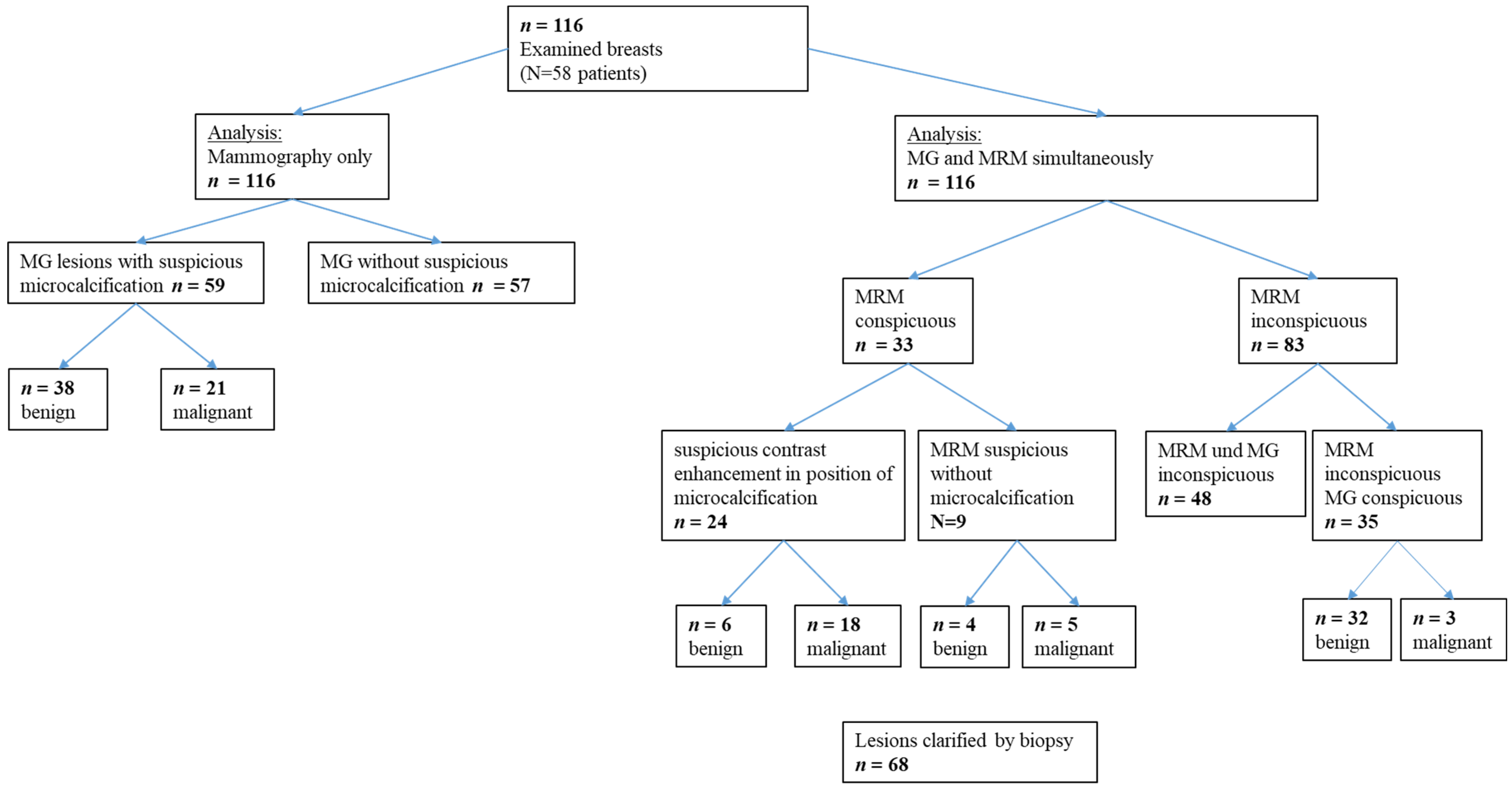
A two-sided independent t-test and a chi-square test were carried out to differentiate the mean values of the descriptive and image-morphological characteristics for patients with benign or malignant findings. A significance level of 5% was used (p-value < 0.05). The Mann–Whitney U-test was performed as a non-parametric test. To determine the additional diagnostic value of breast MRI, the sensitivity, specificity, positive predictive value (PPV), negative predictive value (NPV) and accuracy (ACC) of the combined examination of breast MRI and MG were evaluated. The statistical software IBM SPSS Statistics Version 23.0 (IBM Corp., Armonk, NY, USA) was used for the statistical analysis.

3. Results

3.1. Breast MRI and MG Examinations

A total of 116 breasts of 58 women were evaluated with breast MRI and MG. A total of 59 were suspicious (BI-RADS® category IV) in the MG and 21 turned out to be histologically malignant. Breast MRI evaluation resulted in 33 suspicious findings (Figure 1).

3.2. Characteristics of Trial Population

A total of 58 women were eligible for this prospective study. The mean age was 56 years ranging from 45 to 80 years, and the standard deviation (SD) 7.46 years. All women presented with suspicious MCs and one presented with two clusters. The statistical analysis between the groups of women with benign and malignant pathological findings showed no significant differences in terms of age, menopausal status, average BMI (body mass index) and family history. A significant deviation was only found in terms of breast density (Table 1).

3.3. Histology

In total, 68 breast findings from 58 patients were analyzed histologically. A total of 59 of these findings in the 58 women contained suspicious MCs in the MG. Out of 68 histological findings, 42 were benign and 26 were malignant. One woman had bilateral suspicious MCs. Nine additional findings were without MCs, four unilateral findings and five contralateral findings only detected on the MRI are described separately (see histology breast MRI section and Table 2).

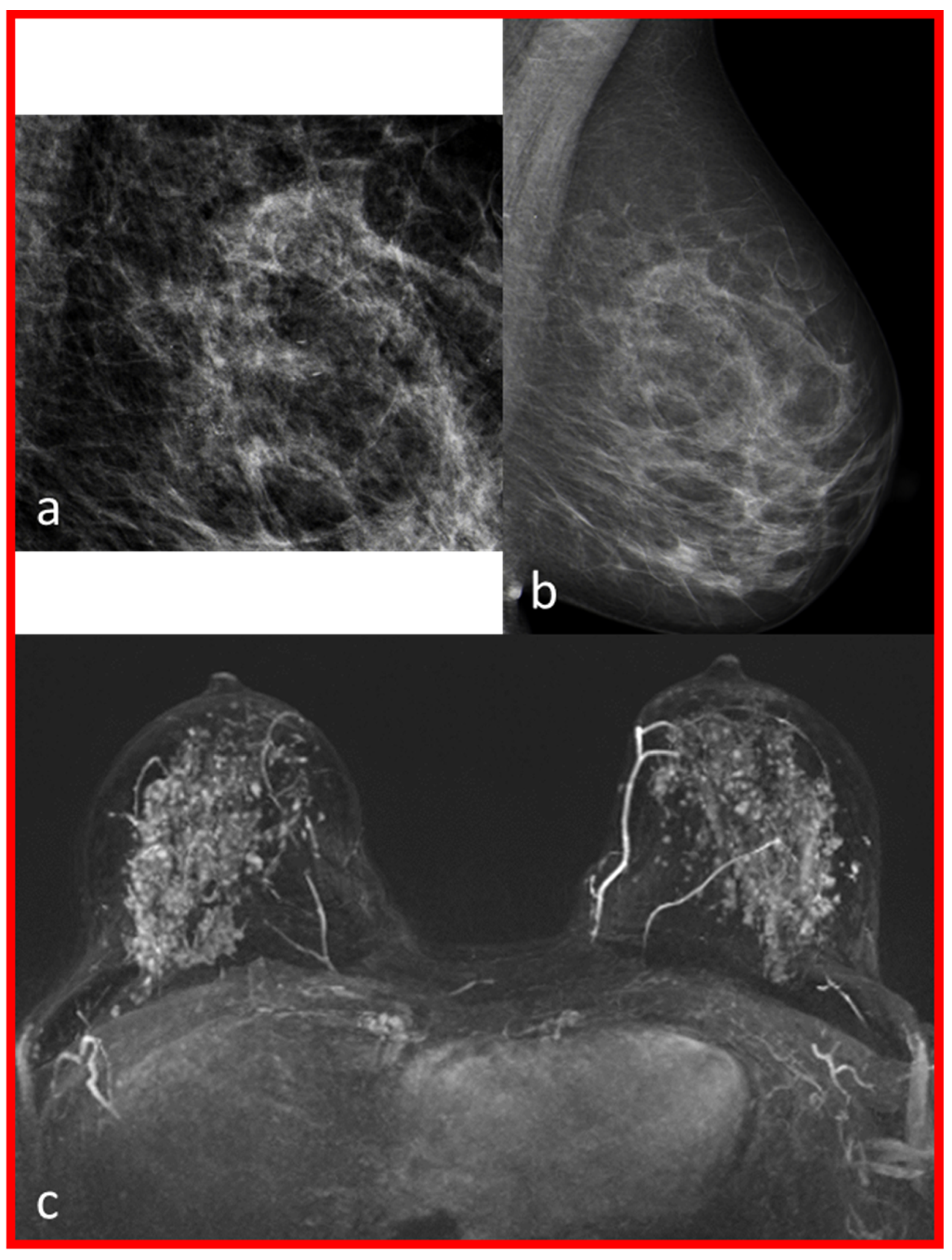
3.4. Histology of Lesions with Suspicious MCs in MG

Focusing on the suspicious MCs in the MG (n = 59), the histopathological results showed 38/59 (64%) benign findings and 21/59 (36%) malignant findings.
The 38 benign findings consisted of fibrocystic mastopathy in ten (26%), common ductal hyperplasia (UDH) in seven (18%), sclerosing adenosis in six (14%), fibroadenoma in six (14%), flat epithelial atypia (FEA) in five (13%), papilloma in two (5%) and atypical ductal hyperplasia (ADH) in one case (2%). In addition, a grade three lobular neoplasia (LN) associated with a papilloma (2%) was found. Six of these lesions were categorized as B3 lesions after biopsy, and the final histology resulted in four FEA, one LN3 and one UDH.
A total of 21 malignancies were diagnosed in 20/58 (34%) of the women, with one woman with bilateral findings:
-
A total of 14/21 (67%) of the malignant lesions represented a DCIS: high-grade in 7/14 cases (50%), intermediate-grade in one case (7%) and low-grade in six cases (43%).
-
A total of 7/21 (33%) malignant findings showed an invasive carcinoma. Two exclusively invasive carcinomas (no special type (NST), one G1 and one G2). A total of 5/7 of the invasive carcinomas were associated with DCIS (one NST G1 with low-grade DCIS, two NST G2 each with an intermediate-grade and high-grade DCIS, one NST G3 with a high-grade DCIS and one invasive tubular carcinoma one associated with a high-grade DCIS).
In 35 cases, no suspicious contrast enhancement in the MRI was found at the site of the MCs. A total of 32 of these cases turned out to be benign and in three cases a DCIS was detected (see Table 3 and Figure 2). In Figure 2, the 53-year-old patient number three of the following Table 3 is shown.

3.5. Histology of Lesions Additionally Detected in Breast MRI

Nine additional findings without MCs were detected in the breast MRI with consecutive histological evaluation. A total of 5/9 of the additional findings resulted in a malignant and 4/9 a benign finding.
Of these nine additional findings, five were on the contralateral side to the MCs (three malignant, two benign) and four (two malignant, two benign) of these were ipsilateral to the MCs, but at a different location. In the following Table 2, these nine additional findings are listed, together with the resulting histology, the MG finding, the breast MRI finding and the density of the mammary gland tissue. The MG and breast MR images of patient number five in Table 2 are shown in Figure 3. The histology of that patient shows on the right breast a 4.9 cm low-grade DCIS, the left breast shows a 7 mm invasive ductal carcinoma (G2 NST and a 5 cm intermediate-grade DCIS.

3.6. Comparison of the Diagnostic Accuracy of MG and MG Plus Breast MRI in Evaluation of MCs

The diagnostic performance of the MG alone in terms of MC evaluation was as follows: false positive in 38 cases, true positive in 21 cases. This resulted in a positive predictive value (PPV) of 36%.
The diagnostic accuracy of breast MRI evaluation of these 59 MCs was as follows: false positive in 6 cases, true positive in 18 cases, false negative in 3 cases and true negative in 32 cases. This resulted in a sensitivity of 86%, a specificity of 84%, a positive predictive value (PPV) of 75% and a negative predictive value (NPV) of 91% for differentiation between benign and malignant in these 59 MCs.
Breast MRI in addition to MG could increase the PPV from 36% to 75% by 39% compared to MG alone.

3.7. Results of the Additionally Performed Breast MRI

As a consequence of the additional performed breast MRI, nine additional biopsies were conducted. This resulted in four further benign and five malignant findings (see Table 2). Four of these findings were on the contralateral side of the MCs and five were unilaterally in a different position to the MCs. The diagnostic accuracy of breast MRI was as follows based on histopathological results (n = 68): false positive in 10 cases, true positive in 23 cases, false negative in 3 cases and true negative in 32 cases. This results in a sensitivity of 88%, a specificity of 76%, an NPV of 91%, a PPV of 70% and an accuracy of 81%.

4. Discussion

In this single-center trial with 58 patients presenting with 59 suspicious MCs in the MG, the additional performed breast MRI was able to improve the diagnostic accuracy for the differentiation of the MCs. The PPV increased from 36 % to 75 % by performing an additional breast MRI in these patients. Breast MRI led to nine additional biopsies, resulting in five accidentally diagnosed breast cancers in our study.
Bennani-Baiti et al. found an additional diagnostic benefit from breast MRI as well, especially for patients with mammographically detected MCs (BI-RADS® category four). In their meta-analysis of 20 studies, they described 1843 findings with suspected MCs, of which 748 (40.6%) were malignant. Pooled sensitivity and specificity were 87% and 81% for all lesions [3]. This corresponds to our results where the additional breast MRI resulted in a sensitivity of 86%, a specificity of 84%, a positive predictive value (PPV) of 75% and a negative predictive value (NPV) of 91% in lesions presenting with MCs. In other comparable studies on breast MRI and MCs in mammography, collectives of patients with an increased risk of breast cancer, for example in dense FGT or family high-risk situations, were examined. One of the first studies that addressed this question evaluated the role of breast MRI in 167 patients diagnosed with DCIS in the final surgery. The preoperatively performed MRI showed 98% of the DCIS lesions by only missing two, which were diagnosed in the MG [16]. Another study about women in a screening program for women with a higher risk of breast cancer showed longer survival of women taking part in MRI screening than those without. They reported a sensitivity of MRI plus mammography of 93% and a specificity of 63% [18]. Saadatmand et al. showed that annual breast MRI and mammography combined could improve breast cancer metastasis-free survival for women with genetic or familial predisposition [17]. Diagnostic accuracy is comparable to our study, but the study cohort is significantly different from the patient collective studied here.
In our study, MG showed 59 suspicious MC findings in 58 patients (BI-RADS® category four). Of these, 21 were malignant, corresponding to a PPV of 35%. This is comparable to the literature, where the PPV for suspicious MCs in MG is between 18.7% and 28.8% [24]. Around 64% of the histologically clarified suspicious MCs turn out to be benign [25,26]. Li et al. described a specificity of 42.9% and an accuracy of 69.2% for BI-RADS® category 3–5 MCs [27]. Cilotti et al. describe a specificity of 59% and a PPV of 63% [6] for BI-RADS® category four and five findings. In a study by Akita et al., MG showed a specificity of 24% and an accuracy of 44% [28] for all patients with suspicious MCs. In our study, 38 out of 59 MC findings were benign.
Surprisingly, we found five additional mammographically occult carcinomas in our cohort. This might be because of a close-to-the-chest wall localization outside the area recorded by the MG (2 cases). In the other three cases, women had extremely dense breasts, which is known to reduce the sensitivity of MG to under 30% [29]. Women with a breast density above 75% have a 4.7 times increased risk of breast cancer and carcinoma is detected 3.5 times worse in screening [30,31]. The five invasive carcinomas additionally detected by the breast MRI were hormone receptor positive and human epidermal growth factor receptor (HER) two negative. All patients were lymph-node-negative. Compared to the literature, the breast MRI detects especially early, node-negative, ductal and almost exclusively hormone-receptor-positive breast cancers [11,32]. In particular, two of the four patients with a high proliferation index (ki67) (20% and 60%) and lymph node negativity have benefited predictably from the earlier discovery by breast MRI [33]. In the literature, MRI screening of the contralateral breast in women with newly diagnosed breast cancer could improve overall survival with a hazard ratio of 2.51 [34]. One of the discussed limitations of breast MRI is overdiagnosis and therapy. We found nine additional lesions resulting in nine additional biopsies showing five invasive carcinomas. Nevertheless, four biopsies resulted in benign findings. However, in comparison, there is also a large overdiagnosis in MG with a PPV of BI-RADS category four microcalcifications described between 20 and 63% in the literature [4,6]. In the cohort analysis of Bennani-Baiti and coworkers, benign histology resulted in 60% of women who underwent a biopsy. In comparison, pooled PPV for BI-RADS® category four microcalcifications in MRI is 81% [3].
On the other hand, for the MC evaluation, when performing an additional MRI, 32 biopsies could have been avoided, when both image modalities would have been considered. This corresponded to a possible avoidance of 54%, in absolute numbers 32 out of 59, of biopsies. Without biopsies, 3/21 (14%) of the DCIS would have been overlooked (two low-grade and one intermediate-grade DCIS). High-grade DCIS or invasive carcinomas have not been overlooked. In 2018, Baltzer et al. showed that 39.5% of 81 biopsies in patients with suspect MCs could have been avoided in 858 retrospectively evaluated patients. A total of 2/71 intermediate-grade DCIS were not found, representing 2.8%—compared to 3/21 (14%) in the present study [35]. Narod et al. showed a very small impact on long-term mortality of 3.3% in 20 years of DCIS alone [36]. Feinberg et al. and Sagara et al. also describe a frequent overdiagnosis and therapy of DCIS [37,38]. In addition, it has been shown that there is no survival benefit of surgical therapy for patients with low-grade DCIS [38].
One limitation of our study might be the size of the study, which also limits the power of statistical results as PPV. The size of the cohort was strongly regulated by the scanning time of the MRI but it corresponds to the study sizes used in the literature for similar study setups [3]. This could lead to a limitation of the significance of the statistics. Larger studies with a larger patient collective would be desirable.

5. Conclusions

In our study, an additional performed breast MRI could have increased the diagnostic reliability in the assessment of MC in screening MG. The PPV increased from 36% to 75% if both modalities had been considered for the final report. By performing an additional breast MRI for MC evaluation, 54% of the biopsies could have been avoided retrospectively in our study, by overlooking only two low-grade and one intermediate-grade DCIS. Further, the benefit of these MRIs in our small cohort was that a not inconsiderable number of malignant lesions without mammographically visible MCs was revealed.

Author Contributions

P.P., E.W. and S.O. made substantial contributions to the conception and design of the study, to the acquisition, analysis, and interpretation of data, and to the critical revision of the manuscript. P.P. drafted the manuscript and was involved in revising it critically for important intellectual content. P.P. and E.W. made substantial contributions to statistical analyses, and M.W.B., M.U., C.C.H. and S.O. were involved in suggestions for manuscript revision. E.W. and P.P. were involved in the acquisition of data (patient and tumor characteristics, histopathological assessment, carrying out surgical procedures or systemic treatment). All authors gave their final approval of the version to be published, revised the manuscript critically and should have participated sufficiently in the work to take public responsibility for appropriate portions of the content. All authors have read and agreed to the published version of the manuscript.

Funding

We acknowledge financial support from Deutsche Forschungsgemeinschaft and Friedrich-Alexander-Universität Erlangen-Nürnberg within the funding program “Open Access Publication Funding”.

Institutional Review Board Statement

The study was conducted in accordance with the Declaration of Helsinki, and approved by the Ethics Committee of the University of Erlangen-Nürnberg, 14 January 2014 and the number is 305_13 B.

Informed Consent Statement

Informed consent was obtained from all subjects involved in the study.

Data Availability Statement

The data presented in this study are available upon request from the corresponding author.

Conflicts of Interest

The authors declare no conflict of interest.

References

  1. Krebs in Deutschland 2017/2018. Robert Koch Institut—Zentrum für Krebsregisterdaten. Available online: https://www.krebsdaten.de/Krebs/DE/Content/Publikationen/Krebs_in_Deutschland/kid_2021/kid_2021_c50_brust.pdf?__blob=publicationFile (accessed on 20 February 2023).
  2. Siu, A.L.; U.S. Preventive Services Task Force. Screening for Breast Cancer: U.S. Preventive Services Task Force Recommendation Statement. Ann. Intern. Med. 2016, 164, 279–296. [Google Scholar] [CrossRef] [PubMed]
  3. Bennani-Baiti, B.; Baltzer, P.A. MR Imaging for Diagnosis of Malignancy in Mammographic Microcalcifications: A Systematic Review and Meta-Analysis. Radiology 2017, 283, 692–701. [Google Scholar] [CrossRef] [PubMed]
  4. Rominger, M.; Wisgickl, C.; Timmesfeld, N. Breast microcalcifications as type descriptors to stratify risk of malignancy: A systematic review and meta-analysis of 10665 cases with special focus on round/punctate microcalcifications. Rofo 2012, 184, 1144–1152. [Google Scholar] [CrossRef] [PubMed]
  5. Bluekens, A.M.; Holland, R.; Karssemeijer, N.; Broeders, M.J.; den Heeten, G.J. Comparison of digital screening mammography and screen-film mammography in the early detection of clinically relevant cancers: A multicenter study. Radiology 2012, 265, 707–714. [Google Scholar] [CrossRef]
  6. Cilotti, A.; Iacconi, C.; Marini, C.; Moretti, M.; Mazzotta, D.; Traino, C.; Naccarato, A.G.; Piagneri, V.; Giaconi, C.; Bevilacqua, G.; et al. Contrast-enhanced MR imaging in patients with BI-RADS 3-5 microcalcifications. Radiol. Med. 2007, 112, 272–286. [Google Scholar] [CrossRef]
  7. Stomper, P.C.; Davis, S.P.; Weidner, N.; Meyer, J.E. Clinically occult ductal carcinoma in situ detected with mammography: Analysis of 100 cases with radiologic-pathologic correlation. Radiology 1989, 172, 235–241. [Google Scholar] [CrossRef]
  8. Verschuur-Maes, A.H.; van Gils, C.H.; van den Bosch, M.A.; De Bruin, P.C.; van Diest, P.J. Digital mammography: More microcalcifications, more columnar cell lesions without atypia. Mod. Pathol. 2011, 24, 1191–11977. [Google Scholar] [CrossRef]
  9. Feeley, L.; Kiernan, D.; Mooney, T.; Flanagan, F.; Hargaden, G.; Kell, M.; Stokes, M.; Kennedy, M. Digital mammography in a screening programme and its implications for pathology: A comparative study. J. Clin. Pathol. 2011, 64, 215–219. [Google Scholar] [CrossRef]
  10. Jobsen, J.J.; Scheijmans, L.J.E.E.; Smit, W.G.J.M.; Stenfert Kroese, M.C.; Struikmans, H.; van der Palen, J. Breast-conserving therapy for primary Ductal Carcinoma in Situ in The Netherlands: A multi-center study and population-based analysis. Breast 2018, 42, 3–9. [Google Scholar] [CrossRef]
  11. Kuhl, C.K.; Strobel, K.; Bieling, H.; Leutner, C.; Schild, H.H.; Schrading, S. Supplemental Breast MR Imaging Screening of Women with Average Risk of Breast Cancer. Radiology 2017, 283, 361–370. [Google Scholar] [CrossRef]
  12. Stehouwer, B.L.; Merckel, L.G.; Verkooijen, H.M.; Peters, N.H.; Mann, R.M.; Duvivier, K.M.; Mali, W.P.; Peeters, P.H.; Veldhuis, W.B.; van den Bosch, M.A. 3-T breast magnetic resonance imaging in patients with suspicious microcalcifications on mammography. Eur. Radiol. 2014, 24, 603–609. [Google Scholar] [CrossRef]
  13. Greenwood, H.I.; Heller, S.L.; Kim, S.; Sigmund, E.E.; Shaylor, S.D.; Moy, L. Ductal carcinoma in situ of the breasts: Review of MR imaging features. Radiographics 2013, 33, 1569–1588. [Google Scholar] [CrossRef]
  14. Peters, N.H.; Borel Rinkes, I.H.; Zuithoff, N.P.; Mali, W.P.; Moons, K.G.; Peeters, P.H. Meta-analysis of MR imaging in the diagnosis of breast lesions. Radiology 2008, 246, 116–124. [Google Scholar] [CrossRef]
  15. Lehman, C.D.; Gatsonis, C.; Kuhl, C.K.; Hendrick, R.E.; Pisano, E.D.; Hanna, L.; Peacock, S.; Smazal, S.F.; Maki, D.D.; Julian, T.B.; et al. MRI evaluation of the contralateral breast in women with recently diagnosed breast cancer. N. Engl. J. Med. 2007, 356, 1295–1303. [Google Scholar] [CrossRef]
  16. Kuhl, C.K.; Schrading, S.; Bieling, H.B.; Wardelmann, E.; Leutner, C.C.; Koenig, R.; Kuhn, W.; Schild, H.H. MRI for diagnosis of pure ductal carcinoma in situ: A prospective observational study. Lancet 2007, 370, 485–492. [Google Scholar] [CrossRef]
  17. Saadatmand, S.; Obdeijn, I.M.; Rutgers, E.J.; Oosterwijk, J.C.; Tollenaar, R.A.; Woldringh, G.H.; Bergers, E.; Verhoef, C.; Heijnsdijk, E.A.; Hooning, M.J.; et al. Survival benefit in women with BRCA1 mutation or familial risk in the MRI screening study (MRISC). Int. J. Cancer 2015, 137, 1729–1738. [Google Scholar] [CrossRef]
  18. Evans, D.G.; Kesavan, N.; Lim, Y.; Gadde, S.; Hurley, E.; Massat, N.J.; Maxwell, A.J.; Ingham, S.; Eeles, R.; Leach, M.O.; et al. MRI breast screening in high-risk women: Cancer detection and survival analysis. Breast Cancer Res. Treat 2014, 145, 663–672. [Google Scholar] [CrossRef]
  19. Passaperuma, K.; Warner, E.; Causer, P.A.; Hill, K.A.; Messner, S.; Wong, J.W.; Jong, R.A.; Wright, F.C.; Yaffe, M.J.; Ramsay, E.A.; et al. Long-term results of screening with magnetic resonance imaging in women with BRCA mutations. Br. J. Cancer 2012, 107, 24–30. [Google Scholar] [CrossRef]
  20. Mann, R.M.; Athanasiou, A.; Baltzer, P.A.T.; Camps-Herrero, J.; Clauser, P.; Fallenberg, E.M.; Forrai, G.; Fuchsjäger, M.H.; Helbich, T.H.; Killburn-Toppin, F.; et al. Breast Cancer Screening in Women with Extremely Dense Breasts Recommendations of the European Society of Breast Imaging (EUSOBI). Eur. Radiol. 2022, 32, 4036–4045. [Google Scholar] [CrossRef]
  21. Sickles, E.; D’Orsi, C.J.; Bassett, L.W. ACR BI-RADS® Mammography. In ACR BI-RADS® Atlas, Breast Imaging Reporting and Data System; American College of Radiology: Reston, VA, USA, 2013. [Google Scholar]
  22. Vereinbarung von Qualitätssicherungsmaßnahmen nach § 135 Abs. 2 SGB V zur kurativen Mammographie (Mammographie-Vereinbarung). 2011: Deutsches Ärzteblatt International Jg. 108 Heft 14. A-791–A-796. Available online: https://www.referenzzentrum-ms.de/site/assets/files/2132/kurative_mammographie_endversion.pdf (accessed on 21 February 2023).
  23. Morris, E.A.; Lee, C.H. ACR BI-RADS® Magnetic Resonance Imaging. In ACR BI-RADS® Atlas, Breast Imaging Reporting and Data System; American College of Radiology: Reston, VA, USA, 2013. [Google Scholar]
  24. Bent, C.K.; Bassett, L.; D’Orsi, C.J.; Sayre, J. The positive predictive value of BI-RADS microcalcification descriptors and final assessment categories. AJR Am. J. Roentgenol. 2010, 194, 1378–1383. [Google Scholar] [CrossRef]
  25. Aminololama-Shakeri, S.; Abbey, C.K.; Gazi, P.; Prionas, N.D.; Nosratieh, A.; Li, C.S.; Boone, J.M.; Lindfors, K.K. Differentiation of ductal carcinoma in-situ from benign micro-calcifications by dedicated breast computed tomography. Eur. J. Radiol. 2016, 85, 297–303. [Google Scholar] [CrossRef] [PubMed]
  26. Strobel, K.; Schrading, S.; Hansen, N.L.; Barabasch, A.; Kuhl, C.K. Assessment of BI-RADS category 4 lesions detected with screening mammography and screening US: Utility of MR imaging. Radiology 2015, 274, 343–351. [Google Scholar] [CrossRef] [PubMed]
  27. Lee, N.C.; Wong, F.L.; Jamison, P.M.; Jones, S.F.; Galaska, L.; Brady, K.T.; Wethers, B.; Stokes-Townsend, G.A. Implementation of the National Breast and Cervical Cancer Early Detection Program: The beginning. Cancer 2014, 120 (Suppl. S16), 2540–2548. [Google Scholar] [CrossRef] [PubMed]
  28. Akita, A.; Tanimoto, A.; Jinno, H.; Kameyama, K.; Kuribayashi, S. The clinical value of bilateral breast MR imaging: Is it worth performing on patients showing suspicious microcalcifications on mammography? Eur. Radiol. 2009, 19, 2089–2096. [Google Scholar] [CrossRef] [PubMed]
  29. Mandelson, M.T. Breast Density as a Predictor of Mammographic Detection: Comparison of Interval- and Screen-Detected Cancers. J. Natl. Cancer Inst. 2000, 92, 1081–1087. [Google Scholar] [CrossRef]
  30. Boyd, N.F.; Guo, H.; Martin, L.J.; Sun, L.; Stone, J.; Fishell, E.; Jong, R.A.; Hislop, G.; Chiarelli, A.; Minkin, S.; et al. Mammographic density and the risk and detection of breast cancer. N. Engl. J. Med. 2007, 356, 227–236. [Google Scholar] [CrossRef]
  31. Straub, L.; Riedel, J.; Luppa, P.B.; Wissing, J.; Artmann, A.; Kiechle, M.; Seifert-Klauss, V.R. Mammographic Density and Vitamin D Levels—A Cross-sectional Study. Geburtshilfe Frauenheilkd. 2017, 77, 257–267. [Google Scholar] [CrossRef]
  32. Bakker, M.F.; de Lange, S.V.; Pijnappel, R.M.; Mann, R.M.; Peeters, P.H.M.; Monninkhof, E.M.; Emaus, M.J.; Loo, C.E.; Bisschops, R.H.C.; Lobbes, M.B.I.; et al. Supplemental MRI Screening for Women with Extremely Dense Breast Tissue. N. Engl. J. Med. 2019, 381, 2091–2102. [Google Scholar] [CrossRef]
  33. Fasching, P.A.; Gass, P.; Häberle, L.; Volz, B.; Hein, A.; Hack, C.C.; Lux, M.P.; Jud, S.M.; Hartmann, A.; Beckmann, M.W.; et al. Prognostic effect of Ki-67 in common clinical subgroups of patients with HER2-negative, hormone receptor-positive early breast cancer. Breast Cancer Res. Treat 2019, 175, 617–625. [Google Scholar] [CrossRef]
  34. Freitas, V.; Li, X.; Amitai, Y.; Au, F.; Kulkarni, S.; Ghai, S.; Mulligan, A.M.; Bromley, M.; Siepmann, T. Contralateral Breast Screening with Preoperative MRI: Long-Term Outcomes for Newly Diagnosed Breast Cancer. Radiology 2022, 304, 297–307. [Google Scholar] [CrossRef]
  35. Baltzer, P.A.T.; Bennani-Baiti, B.; Stöttinger, A.; Bumberger, A.; Kapetas, P.; Clauser, P. Is breast MRI a helpful additional diagnostic test in suspicious mammographic microcalcifications? Magn. Reson. Imaging 2018, 46, 70–74. [Google Scholar] [CrossRef]
  36. Narod, S.A.; Iqbal, J.; Giannakeas, V.; Sopik, V.; Sun, P. Breast Cancer Mortality After a Diagnosis of Ductal Carcinoma In Situ. JAMA Oncol. 2015, 1, 888–896. [Google Scholar] [CrossRef]
  37. Feinberg, J.; Wetstone, R.; Greenstein, D.; Borgen, P. Is DCIS Overrated? Cancer Treat Res. 2018, 173, 53–72. [Google Scholar] [CrossRef]
  38. Sagara, Y.; Julia, W.; Golshan, M.; Toi, M. Paradigm Shift toward Reducing Overtreatment of Ductal Carcinoma In Situ of Breast. Front. Oncol. 2017, 7, 192. [Google Scholar] [CrossRef]
Figure 1. Results of the clinical evaluation as an overview. Breast MRI (magnetic resonance imaging), mammography (MG), n (number).
Diagnostics 13 01197 g001
Figure 2. The mammography and breast magnetic resonance images (MRI) of patient number two of Table 3 are shown. Amorphous, regional MCs of the left breast on the left in MLO (mediolateral-oblique)—projection (images (a,b)). Early post-contrast phase MIP (maximum intensity projection): no suspicious lesion identifiable on both sides, possibly due to the strong BPE (background parenchymal enhancement) assessed as pronounced (image (c)).
Diagnostics 13 01197 g002
Figure 3. The mammography and breast magnetic resonance images (MRI) of patient number five of Table 2 are shown. Grouped, coarse, heterogeneous microcalcifications (MCs) in the mammography (MG) of the right breast (subfigure (b), enlargement of the microcalcifications of subfigure (a), histology: low-grade DCIS). Inconspicuous MG of the left breast. In breast magnetic resonance imaging, no suspicious lesion was detected in the right breast. A non-mass enhancement was found in the left breast (image (c), histology: breast cancer, NST G2).
Diagnostics 13 01197 g003
Table 1. Characteristics of women with benign and malignant findings in mammography.
Benign Lesions (n = 36)Malignant Lesions (n = 22)(p-Value)
Age in years
  Median (range)

57 (45–74)

57.5 (45–80)

0.290
Menopausal status
  Premenopausal n (%)
  Postmenopausal n (%)

6 (15.4%)
30 (84.6%)

6 (28.6%)
16 (71.4%)


0.306
Average BMI (Kg/m²) (SD)25.72 (5.26)25.98 (3.35)0.855
Breast density n (%)
a
b
c
d

3 (8.33%)
11 (30.56%)
19 (52.78%)
3 (8.33%)

3 (13.64%)
12 (54.55%)
6 (27.27%)
1 (4.55%)
0.048 *
Average number of children (SD)1.67 (0.92)1.67 (1.07)1.000
Characteristics of women with benign and malignant findings in mammography, kg (kilograms), m (meters), SD (standard deviation), n (number), BMI (body mass index), ACR (American College of Radiology), * (statistically significant).
Table 2. Additional findings detected by breast magnetic resonance imaging (MRI.).
PatientMABPEBreast MRIDMGModalityHistology/IHC/TNM
1IMildMass, circumscribed, ovalcLocalization outside the MG coverage near the chest wallCore biopsy guided
by ultrasound
1.4 cm G1 NST/ER 90%, PR 30%, Ki-67 5%, HER2 negative/pT1c pN0 L0 V0 Pn1 G1
2IIModerateNME, segmental, heterogeneousbNo MCsMRI-guided
vacuum-assisted biopsy
13.4 cm low-grade DCIS
3IIModerateNME, linearbUnilateral MCs in different positionMRI-guided
vacuum-assisted biopsy
0.6 cm G1 tubular carcinoma/ER 90%, PR 30%, KI-67 10%, HER2 negative/pT1b pN0 L0 V0 Pn0 G1 associated with 3.2 cm low-grade DCIS
4IMinimalMass, irregular, not circumscribed-spiculated, heterogeneousbUnilateral MCs in different positionCore needle biopsy0.5 cm G3 NST/ER 90%, PR negative, Ki-67 60%, HER2 negative/ypT0 pN0 L0 V0 Pn0
5IIModerateNME, grouped, regionalcNo MCs Partial excision0.7 cm G2 NST//ER 90%, PR 40% Ki-67 20% HER2 negative/pT1b pN0 L0 V0 Pn0 G2 associated with 5.0 cm low-grade DCIS
6IMildNME, linear, heterogeneousbNo MCsDuct excisionFibrocystic changes
7IMildNME, diffuse, heterogeneousbNo MCsCore needle biopsy
guided by ultrasound
UDH
8IMinimalNME, linear, homogeneouscUnilateral MCs in different positionPartial excisionFibrocystic changes
9IModerateNME, focal, homogeneouscUnilateral MCs in different positionPartial excisionFEA
Additional findings detected by breast magnetic resonance imaging (MRI). MA (motion artifact), BPE (background parenchymal enhancement), NME (non-mass-enhancement), MG (mammography), ICH (immunohistochemistry), TNM (tumor/nodal/metastasis), ER (estrogen receptor), PR (progesterone receptor), KI-67 (proliferation marker), HER2 (human epidermal growth factor receptor 2), p (pathological), L (lymphatic), V (vessel), Pn (perineural sheath), UDH (usual ductal hyperplasia), FEA (flat epithelial atypia), DCIS (ductal carcinoma in situ), NST (breast cancer no special type), D: mammographic density.
Table 3. False negative findings in the combined analysis of mammography (MG) and breast magnetic resonance imaging (MRI).
PatientMABPEACRHistology and SizeShape and Distribution
12ModerateBIntermediate-grade DCIS (6.5 cm)No contrast enhancement in the position of macrocalcifications, multiple small foci on both sides.
21MarkedCLow-grade DCIS (0.15 cm)No contrast enhancement in the position of macrocalcifications.
31MarkedCLow-grade DCIS (5.6 cm)No contrast enhancement in the position of macrocalcifications, multiple cysts on both sides.
False negative findings in the combined analysis of mammography and breast magnetic resonance imaging (MRI), motion artifact (MA), BPE (background parenchymal enhancement), ACR (American College of Radiology), DCIS (ductal carcinoma in situ) and breast MRI (magnetic resonance imaging).
Disclaimer/Publisher’s Note: The statements, opinions and data contained in all publications are solely those of the individual author(s) and contributor(s) and not of MDPI and/or the editor(s). MDPI and/or the editor(s) disclaim responsibility for any injury to people or property resulting from any ideas, methods, instructions or products referred to in the content.

