

High-Frequency Ultrasound in Diagnosis and Treatment of Non-Melanoma Skin Cancer in the Head and Neck Region

 , , ,
, , ,  ,
,
Abstract
1. Introduction
2. Materials and Methods
Statistical Analyses
3. Results
4. Discussion
5. Conclusions
Author Contributions
Funding
Institutional Review Board Statement
Informed Consent Statement
Data Availability Statement
Conflicts of Interest
References
- Altamura, D.; Menzies, S.W.; Argenziano, G.; Zalaudek, I.; Soyer, H.P.; Sera, F.; Avramidis, M.; DeAmbrosis, K.; Fargnoli, M.C.; Peris, K. Dermatoscopy of basal cell carcinoma: Morphologic variability of global and local features and accuracy of diagnosis. J. Am. Acad. Dermatol. 2010, 62, 67–75. [Google Scholar] [CrossRef] [PubMed]
- Didona, D.; Paolino, G.; Bottoni, U.; Cantisani, C. Non Melanoma Skin Cancer Pathogenesis Overview. Biomedicines 2018, 6, 6. [Google Scholar] [CrossRef] [PubMed]
- Nassiri-Kashani, M.; Sadr, B.; Fanian, F.; Kamyab, K.; Noormohammadpour, P.; Shahshahani, M.M.; Zartab, H.; Naghizadeh, M.M.; Sarraf-Yazdy, M.; Firooz, A. Pre-operative assessment of basal cell carcinoma dimensions using high frequency ultrasonography and its correlation with histopathology. Skin Res. Technol. 2013, 19, e132–e138. [Google Scholar] [CrossRef] [PubMed]
- Leiter, U.; Eigentler, T.; Garbe, C. Epidemiology of skin cancer. Adv. Exp. Med. Biol. 2014, 810, 120–140. [Google Scholar] [CrossRef] [PubMed]
- Katalinic, A.; Kunze, U.; Schäfer, T. Epidemiology of cutaneous melanoma and non-melanoma skin cancer in Schleswig-Holstein, Germany: Incidence, clinical subtypes, tumour stages and localization (epidemiology of skin cancer). Br. J. Dermatol. 2003, 149, 1200–1206. [Google Scholar] [CrossRef]
- Tamas, T.; Dinu, C.; Lenghel, M.; Băciuț, G.; Bran, S.; Stoia, S.; Băciuț, M. The role of ultrasonography in head and neck Non-Melanoma Skin Cancer approach: An update with a review of the literature. Med. Ultrason. 2021, 23, 83–88. [Google Scholar] [CrossRef]
- Barcaui, E.d.O.; Carvalho, A.C.; Valiante, P.M.; Barcaui, C.B. High-frequency ultrasound associated with dermoscopy in pre-operative evaluation of basal cell carcinoma. An. Bras. Dermatol. 2014, 89, 828–831. [Google Scholar] [CrossRef]
- Calzavara-Pinton, P.; Ortel, B.; Venturini, M. Non-melanoma skin cancer, sun exposure and sun protection. G. Ital. Di Dermatol. E Venereol. Organo Uff. Soc. Ital. Di Dermatol. E Sifilogr. 2015, 150, 369–378. [Google Scholar]
- Crişan, D.; Badea, A.F.; Crişan, M.; Rastian, I.; Gheuca Solovastru, L.; Badea, R. Integrative analysis of cutaneous skin tumours using ultrasonogaphic criteria. Preliminary results. Med. Ultrason. 2014, 16, 285–290. [Google Scholar] [CrossRef]
- Crisan, M.; Crisan, D.; Sannino, G.; Lupsor, M.; Badea, R.; Amzica, F. Ultrasonographic staging of cutaneous malignant tumors: An ultrasonographic depth index. Arch. Dermatol. Res. 2013, 305, 305–313. [Google Scholar] [CrossRef]
- Badea, R.; Crişan, M.; Lupşor, M.; Fodor, L. Diagnosis and characterization of cutaneous tumors using combined ultrasonographic procedures (conventional and high resolution ultrasonography). Med. Ultrason. 2010, 12, 317–322. [Google Scholar]
- Alfageme Roldán, F. Elastography in Dermatology. Elastografía en dermatología. Actas Dermo-Sifiliogr. 2016, 107, 652–660. [Google Scholar] [CrossRef]
- Wortsman, X. Sonography of facial cutaneous basal cell carcinoma: A first-line imaging technique. J. Ultrasound Med. 2013, 32, 567–572. [Google Scholar] [CrossRef]
- Song, W.J.; Choi, H.J.; Lee, Y.M.; Tark, M.S.; Nam, D.H.; Han, J.K.; Cho, H.D. Clinical analysis of an ultrasound system in the evaluation of skin cancers: Correlation with histology. Ann. Plast. Surg. 2014, 73, 427–433. [Google Scholar] [CrossRef]
- Wortsman, X.; Vergara, P.; Castro, A.; Saavedra, D.; Bobadilla, F.; Sazunic, I.; Zemelman, V.; Wortsman, J. Ultrasound as predictor of histologic subtypes linked to recurrence in basal cell carcinoma of the skin. J. Eur. Acad. Dermatol. Venereol. JEADV 2015, 29, 702–707. [Google Scholar] [CrossRef]
- Schmid-Wendtner, M.H.; Burgdorf, W. Ultrasound scanning in dermatology. Arch. Dermatol. 2005, 141, 217–224. [Google Scholar] [CrossRef]
- Bhatt, K.D.; Tambe, S.A.; Jerajani, H.R.; Dhurat, R.S. Utility of high-frequency ultrasonography in the diagnosis of benign and malignant skin tumors. Indian J. Dermatol. Venereol. Leprol. 2017, 83, 162–182. [Google Scholar] [CrossRef]
- Jasaitiene, D.; Valiukeviciene, S.; Linkeviciute, G.; Raisutis, R.; Jasiuniene, E.; Kazys, R. Principles of high-frequency ultrasonography for investigation of skin pathology. J. Eur. Acad. Dermatol. Venereol. JEADV 2011, 25, 375–382. [Google Scholar] [CrossRef]
- Bezugly, A. High frequency ultrasound study of skin tumors in dermatological and aesthetic practice. Med. Ultrason. 2015, 17, 541–544. [Google Scholar] [CrossRef]
- Dasgeb, B.; Morris, M.A.; Mehregan, D.; Siegel, E.L. Quantified ultrasound elastography in the assessment of cutaneous carcinoma. Br. J. Radiol. 2015, 88, 20150344. [Google Scholar] [CrossRef]
- Markowitz, O.; Schwartz, M.; Feldman, E.; Bienenfeld, A.; Bieber, A.K.; Ellis, J.; Alapati, U.; Lebwohl, M.; Siegel, D.M. Evaluation of Optical Coherence Tomography as a Means of Identifying Earlier Stage Basal Cell Carcinomas while Reducing the Use of Diagnostic Biopsy. J. Clin. Aesthetic Dermatol. 2015, 8, 14–20. [Google Scholar]
- Olmedo, J.M.; Warschaw, K.E.; Schmitt, J.M.; Swanson, D.L. Optical coherence tomography for the characterization of basal cell carcinoma in vivo: A pilot study. J. Am. Acad. Dermatol. 2006, 55, 408–412. [Google Scholar] [CrossRef] [PubMed]
- Gambichler, T.; Orlikov, A.; Vasa, R.; Moussa, G.; Hoffmann, K.; Stücker, M.; Altmeyer, P.; Bechara, F.G. In vivo optical coherence tomography of basal cell carcinoma. J. Dermatol. Sci. 2007, 45, 167–173. [Google Scholar] [CrossRef] [PubMed]
- Sauermann, K.; Gambichler, T.; Wilmert, M.; Rotterdam, S.; Stücker, M.; Altmeyer, P.; Hoffmann, K. Investigation of basal cell carcinoma [correction of carcionoma] by confocal laser scanning microscopy in vivo. Skin Res. Technol. 2002, 8, 141–147. [Google Scholar] [CrossRef]
- Nori, S.; Rius-Díaz, F.; Cuevas, J.; Goldgeier, M.; Jaen, P.; Torres, A.; González, S. Sensitivity and specificity of reflectance-mode confocal microscopy for in vivo diagnosis of basal cell carcinoma: A multicenter study. J. Am. Acad. Dermatol. 2004, 51, 923–930. [Google Scholar] [CrossRef] [PubMed]
- Patalay, R.; Talbot, C.; Alexandrov, Y.; Lenz, M.O.; Kumar, S.; Warren, S.; Munro, I.; Neil, M.A.; König, K.; French, P.M.; et al. Multiphoton multispectral fluorescence lifetime tomography for the evaluation of basal cell carcinomas. PLoS ONE 2012, 7, e43460. [Google Scholar] [CrossRef] [PubMed]
- Koo, T.K.; Li, M.Y. A Guideline of Selecting and Reporting Intraclass Correlation Coefficients for Reliability Research. J. Chiropr. Med. 2016, 15, 155–163. [Google Scholar] [CrossRef]
- Portney, L.G.; Watkins, M.P. Foundations of Clinical Research: Applications to Practice, 3rd ed.; Prentice Hall: Upper Saddle River, NJ, USA, 2009; p. 892. [Google Scholar]
- Gamer, M.; Lemon, J.; Singh, I.F.P. Various Coefficients of Interrater Reliability and Agreement. 2019. Available online: https://CRAN.R-project.org/package=irr (accessed on 12 October 2022).
- McHugh, M.L. Interrater reliability: The kappa statistic. Biochem. Med. 2012, 22, 276–282. [Google Scholar] [CrossRef]
- Altman, D.G. Practical Statistics for Medical Research, 1st ed.; Chapman and Hall/CRC: Boca Raton, FL, USA, 1990; 624p. [Google Scholar]
- Sim, J.; Wright, C.C. The kappa statistic in reliability studies: Use, interpretation, and sample size requirements. Phys. Ther. 2005, 85, 257–268. [Google Scholar] [CrossRef]
- Tamas, T.; Baciut, M.; Nutu, A.; Bran, S.; Armencea, G.; Stoia, S.; Manea, A.; Crisan, L.; Opris, H.; Onisor, F.; et al. Is miRNA Regulation the Key to Controlling Non-Melanoma Skin Cancer Evolution? Genes 2021, 12, 1929. [Google Scholar] [CrossRef]
- Pampena, R.; Parisi, G.; Benati, M.; Borsari, S.; Lai, M.; Paolino, G.; Cesinaro, A.M.; Ciardo, S.; Farnetani, F.; Bassoli, S.; et al. Clinical and Dermoscopic Factors for the Identification of Aggressive Histologic Subtypes of Basal Cell Carcinoma. Front. Oncol. 2021, 10, 630458. [Google Scholar] [CrossRef]
- Uhara, H.; Hayashi, K.; Koga, H.; Saida, T. Multiple hypersonographic spots in basal cell carcinoma. Dermatol. Surg. Off. Publ. Am. Soc. Dermatol. Surg. 2007, 33, 1215–1219. [Google Scholar] [CrossRef]
- Alfageme Roldán, F. Ultrasound skin imaging. Actas Dermo-Sifiliogr. 2014, 105, 891–899. [Google Scholar] [CrossRef]
- Morris, M.A.; Ring, C.M.; Managuli, R.; Saboury, B.; Mehregan, D.; Siegel, E.; Dasgeb, B. Feature analysis of ultrasound elastography image for quantitative assessment of cutaneous carcinoma. Skin Res. Technol. 2018, 24, 242–247. [Google Scholar] [CrossRef]
- Alfageme, F.; Salgüero, I.; Nájera, L.; Suarez, M.L.; Roustan, G. Increased Marginal Stiffness Differentiates Infiltrative from Noninfiltrative Cutaneous Basal Cell Carcinomas in the Facial Area: A Prospective Study. J. Ultrasound Med. 2019, 38, 1841–1845. [Google Scholar] [CrossRef]
- Tanaka, T.; Tada, Y.; Ohnishi, T.; Watanabe, S. Usefulness of real-time tissue elastography for detecting the border of basal cell carcinomas. J. Dermatol. 2017, 44, 438–443. [Google Scholar] [CrossRef]
- National Comprehensive Cancer Network. NCCN Clinical Practice Guidelines in Oncology; Merkel Cell Carcinoma. Available online: http://www.nccn.org (accessed on 15 January 2017).
- Tai, P. A practical update of surgical management of Merkel cell carcinoma of the skin. ISRN Surg. 2013, 2013, 850797. [Google Scholar] [CrossRef]
- European Dermatology Forum. Guideline on the Diagnosis and Treatment of Merkel Cell Carcinoma. Available online: http://www.euroderm.org (accessed on 3 January 2016).
- Prickett, K.A.; Ramsey, M.L. Mohs Micrographic Surgery. In StatPearls; StatPearls Publishing: Treasure Island, FL, USA, 2022. Available online: https://www.ncbi.nlm.nih.gov/books/NBK441833/ (accessed on 25 July 2022).
- Nolan, G.S.; Wormald, J.C.R.; Kiely, A.L.; Totty, J.P.; Jain, A. Global incidence of incomplete surgical excision in adult patients with non-melanoma skin cancer: Study protocol for a systematic review and meta-analysis of observational studies. Syst. Rev. 2020, 9, 83. [Google Scholar] [CrossRef]
- Vilas-Sueiro, A.; Alfageme, F.; Salgüero, I.; De Las Heras, C.; Roustan, G. Ex Vivo High-Frequency Ultrasound for Assessment of Basal Cell Carcinoma. J. Ultrasound Med. 2019, 38, 529–531. [Google Scholar] [CrossRef]


| Ultrasonography | Dermoscopy | OCT | Confocal Microscopy | |
|---|---|---|---|---|
| Assessing the size of the tumor | It can assess the size in all three dimensions including the thickness [14] | It can assess the size except the thickness [1,6] | It can assess the size in all three dimensions for tumors with a thickness less than 2 mm [21,22,23,24] | It can assess the size in all three dimensions for tumors with a thickness less than 3 mm [25,26] |
| Type of subjacent tissue involvement | It can precisely identify the type of subjacent tissue involved (muscle, fascia, perichondrium) | It cannot precisely identify the type of subjacent tissue involved | Because the depth of penetration is less than 2 mm, we do not recommend using it for evaluation of subjacent tissue involvement | Because the depth of penetration is less than 3 mm, we do not recommend to using it for evaluation of subjacent tissue involvement |
| Surgical margins involvement detection | Yes (postoperative evaluation of the excised specimen) | Only preoperatively | Only preoperatively. Very limited data in the literature | Yes (Ex vivo confocal microscopy) |
| Vascularity | Yes | Yes | Yes (Dynamic Optical Coherence Tomography) | Yes |
| Specificity is diagnosis | 85–90% for both BCC and SCC | 95–99% for BCC Insufficient data for SCC | 80–100% for SCC 70–80% for BCC | 92–98% for SCC 93% for BCC |
| Sensitivity is diagnosis | 93–95% for both BCC and SCC | 90–93% for BCC 75–77% for SCC | 92–93% for SCC 92–95% for BCC | 74–77% for SCC 92% for BCC |
| Method | Tumor Thickness (mm), Median (IQR) | Difference (95% CI) | p-Value | ICC Consistency (95% CI) | p-Value | ICC Agreement (95% CI) | p-Value |
|---|---|---|---|---|---|---|---|
| Pathology | 2.5 (1.98–4.5) | ||||||
| Preoperative | |||||||
| 13 MHz | 2.5 (1.75–4.4) | 0 (0–0.32) | 0.063 | 0.982 (0.962–0.991) | <0.001 | 0.98 (0.957–0.99) | <0.001 |
| 20 MHz | 2.3 (1.75–4.4) | 0.2 (−0.09–0.18) | 0.574 | 0.981 (0.96–0.991) | <0.001 | 0.981 (0.961–0.991) | <0.001 |
| 40 MHz | 2.2 (1.9–4.5) | 0.3 (−0.07–0.15) | 0.394 | 0.992 (0.984–0.996) | <0.001 | 0.992 (0.985–0.996) | <0.001 |
| Postoperative | |||||||
| 13 MHz | 2.3 (1.45–4) | 0.2 (0.13–0.65) | 0.008 | 0.927 (0.853–0.964) | <0.001 | 0.909 (0.776–0.96) | <0.001 |
| 20 MHz | 2.35 (1.6–4.05) | 0.15 (0.1–0.6) | 0.011 | 0.927 (0.855–0.964) | <0.001 | 0.911 (0.787–0.96) | <0.001 |
| 40 MHz | 2.4 (1.6–3.95) | 0.1 (0.07–0.58) | 0.012 | 0.93 (0.859–0.965) | <0.001 | 0.916 (0.806–0.962) | <0.001 |
| Histopathological Diagnosis | Basal Cell Carcinoma | Squamous Cell Carcinoma | Total |
|---|---|---|---|
| Echographic diagnosis | |||
| Basal cell carcinoma | 25 | 0 | 25 |
| Squamous carcinoma | 1 | 5 | 6 |
| Total | 26 | 5 | 31 |
| Hyperechoic spots | |||
| Present | 25 | 0 | 25 |
| Absent | 1 | 5 | 6 |
| Total | 26 | 5 | 31 |
| Diagnostic Histopathologic | Cc Basal cell (n = 25) | Cc Squamous (n = 5) | Difference (95% CI) | p |
|---|---|---|---|---|
| Pulsatility index, median (IQR) | 0.75 (0.6–0.94) | 0.82 (0.54–0.95) | 0.07 (−0.3–0.31) | 0.889●[n1 = 25, n2 = 5] |
| Resistive index, median (IQR) | 0.5 (0.4–0.6) | 0.52 (0.42–0.63) | 0.02 (−0.13–0.18) | 0.933●[n1 = 25, n2 = 5] |
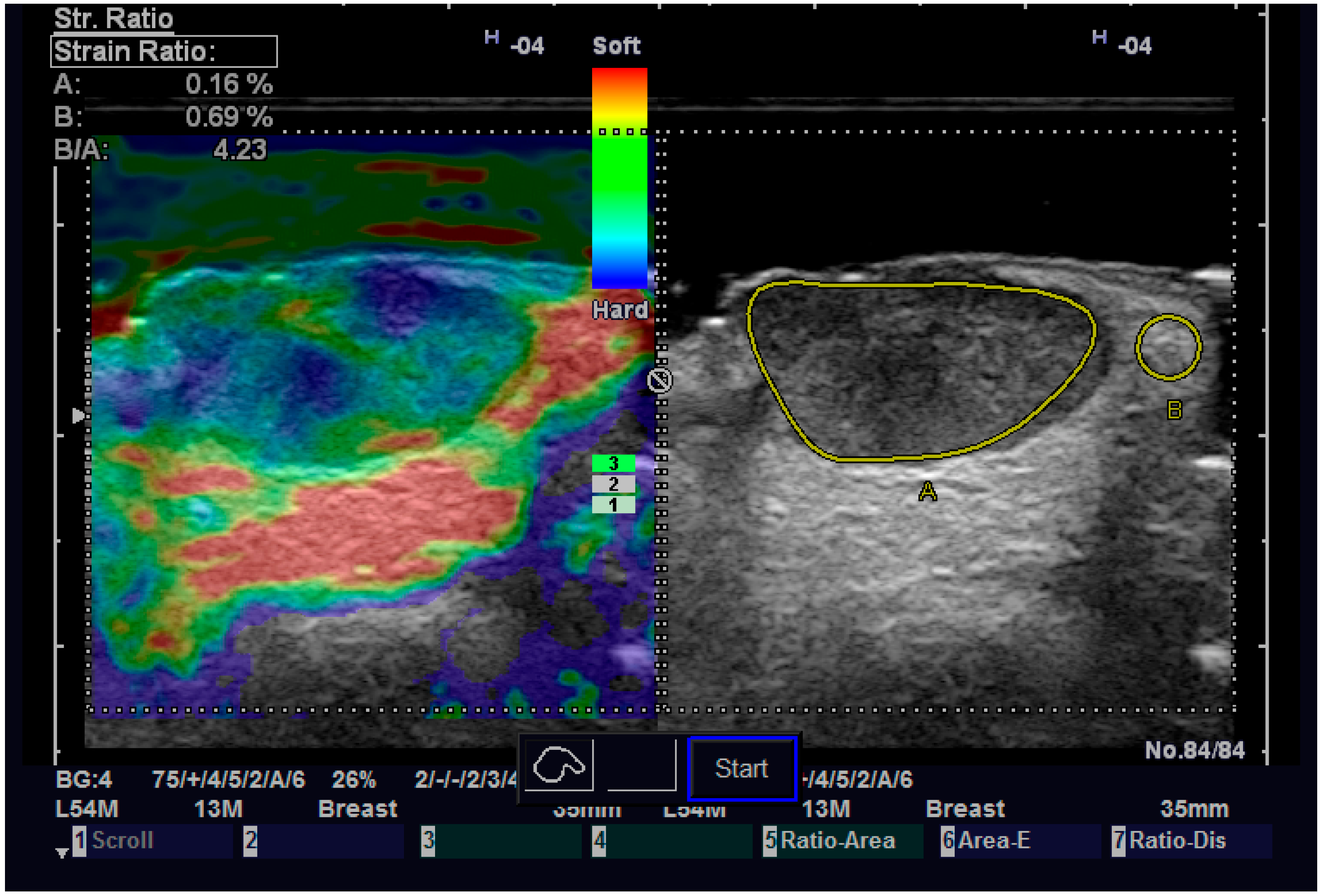
| Strain ratio, median (IQR) | 1.94 (1.38–2.62) | 3.2 (2.9–3.6) | 1.26 (−2.98–0.35) | 0.126●[n1 = 26, n2 = 5] |
| Diastolic speed, median (IQR) | 3.9 (3.9–4.1) | 4.3 (2.5–4.9) | 0.4 (−1–1.6) | 0.822●[n1 = 25, n2 = 5] |
| Systolic speed, median (IQR) | 6.3 (6.1–9) | 6.8 (6.4–8.2) | 0.5 (−1.9–2.2) | 0.758●[n1 = 25, n2 = 5] |
| Method | Tumor Margin (mm), Median (IQR) | Difference (95% CI) | p-Value | ICC Consistency (95% CI) | p-Value | ICC Agreement (95% CI) | p-Value |
|---|---|---|---|---|---|---|---|
| Pathology | 2 (1–2.75) | ||||||
| 13 MHz | 2 (1–2.65) | 0 (−0.05–0.15) | 0.367 | 0.993 (0.985–0.997) | <0.001 | 0.993 (0.985–0.997) | <0.001 |
| 20 MHz | 1.9 (1.15–3) | −0.1 (−0.05–0.25) | 0.203 | 0.961 (0.921–0.981) | <0.001 | 0.96 (0.92–0.981) | <0.001 |
| 40 MHz | 2 (1.1–2.8) | 0 (−0.05–0.27) | 0.195 | 0.972 (0.942–0.986) | <0.001 | 0.971 (0.941–0.986) | <0.001 |
Disclaimer/Publisher’s Note: The statements, opinions and data contained in all publications are solely those of the individual author(s) and contributor(s) and not of MDPI and/or the editor(s). MDPI and/or the editor(s) disclaim responsibility for any injury to people or property resulting from any ideas, methods, instructions or products referred to in the content. |
© 2023 by the authors. Licensee MDPI, Basel, Switzerland. This article is an open access article distributed under the terms and conditions of the Creative Commons Attribution (CC BY) license (https://creativecommons.org/licenses/by/4.0/).
Share and Cite
Tamas, T.; Dinu, C.; Lenghel, L.M.; Boțan, E.; Tamas, A.; Stoia, S.; Leucuta, D.C.; Bran, S.; Onisor, F.; Băciuț, G.; et al. High-Frequency Ultrasound in Diagnosis and Treatment of Non-Melanoma Skin Cancer in the Head and Neck Region. Diagnostics 2023, 13, 1002. https://doi.org/10.3390/diagnostics13051002
Tamas T, Dinu C, Lenghel LM, Boțan E, Tamas A, Stoia S, Leucuta DC, Bran S, Onisor F, Băciuț G, et al. High-Frequency Ultrasound in Diagnosis and Treatment of Non-Melanoma Skin Cancer in the Head and Neck Region. Diagnostics. 2023; 13(5):1002. https://doi.org/10.3390/diagnostics13051002
Chicago/Turabian StyleTamas, Tiberiu, Cristian Dinu, Lavinia Manuela Lenghel, Emil Boțan, Adela Tamas, Sebastian Stoia, Daniel Corneliu Leucuta, Simion Bran, Florin Onisor, Grigore Băciuț, and et al. 2023. "High-Frequency Ultrasound in Diagnosis and Treatment of Non-Melanoma Skin Cancer in the Head and Neck Region" Diagnostics 13, no. 5: 1002. https://doi.org/10.3390/diagnostics13051002
APA StyleTamas, T., Dinu, C., Lenghel, L. M., Boțan, E., Tamas, A., Stoia, S., Leucuta, D. C., Bran, S., Onisor, F., Băciuț, G., Armencea, G., & Băciuț, M. (2023). High-Frequency Ultrasound in Diagnosis and Treatment of Non-Melanoma Skin Cancer in the Head and Neck Region. Diagnostics, 13(5), 1002. https://doi.org/10.3390/diagnostics13051002