
Procedural Software Toolkit in the Armamentarium of Interventional Therapies: A Review of Additive Usefulness and Current Evidence

 ,
,
Abstract
:1. Introduction
2. Materials and Methods
Data Abstraction
3. Discussion
3.1. Planning and Guidance
3.1.1. SmartCT
SmartCT Vaso
SmartCT Soft Tissue
SmartCT Angio
3.1.2. AdvantageSim MD
3.1.3. AngioCARD
3.1.4. AngioViz
3.1.5. Autobone and VesselIQ Xpress
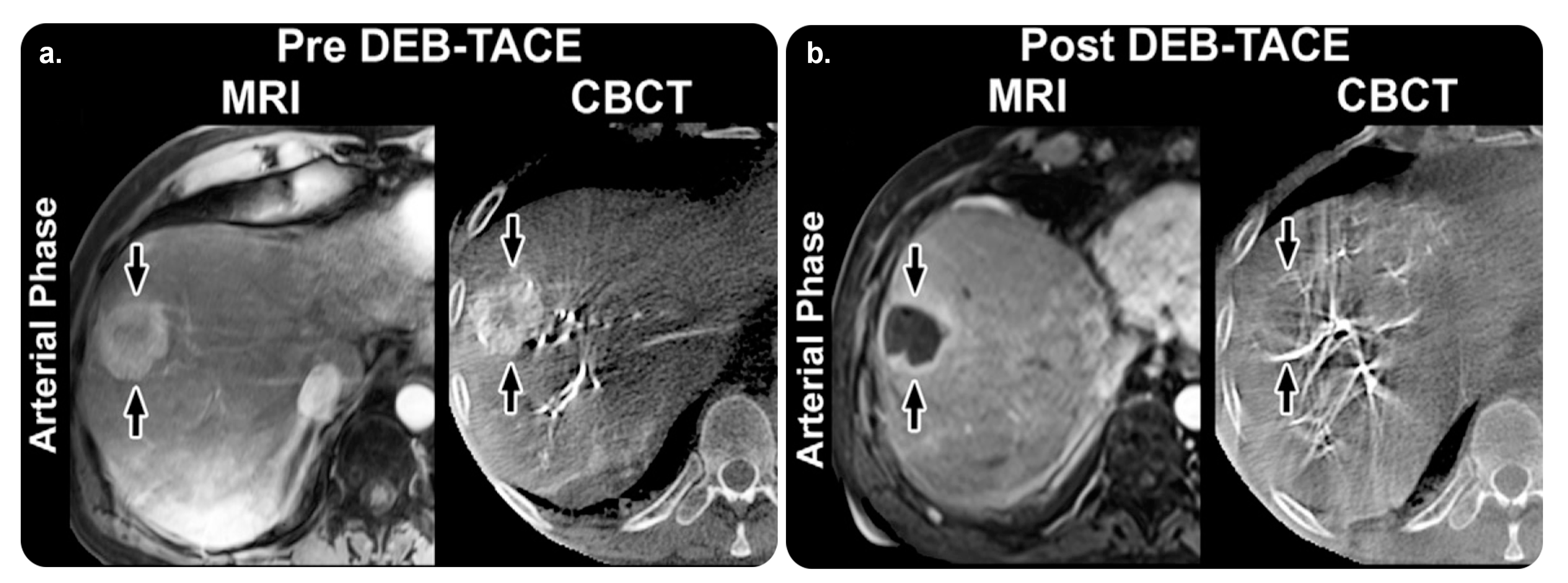
3.1.6. Hepatic VCAR
3.1.7. Innova CT HD and Innova 3D
3.1.8. MR VesselIQ Xpress
3.1.9. TEVAR Assist Solutions
3.2. Targeting
3.2.1. StentBoost Live
StentBoost Mobile
3.2.2. XperGuide
XperGuide Ablation
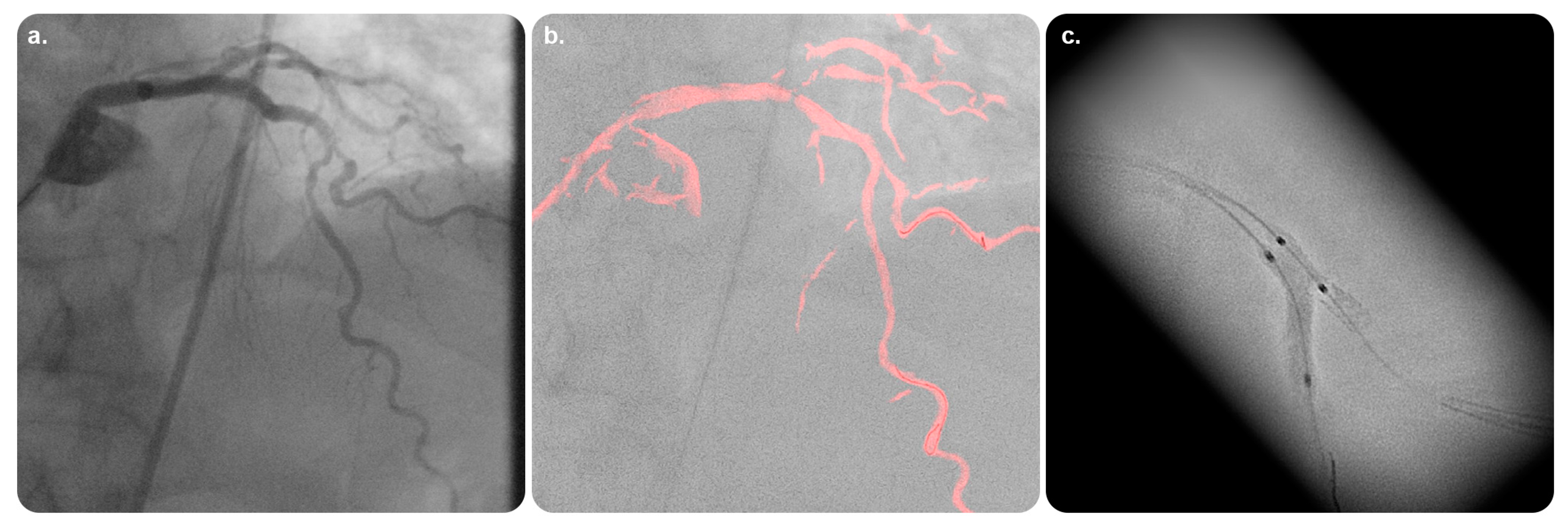
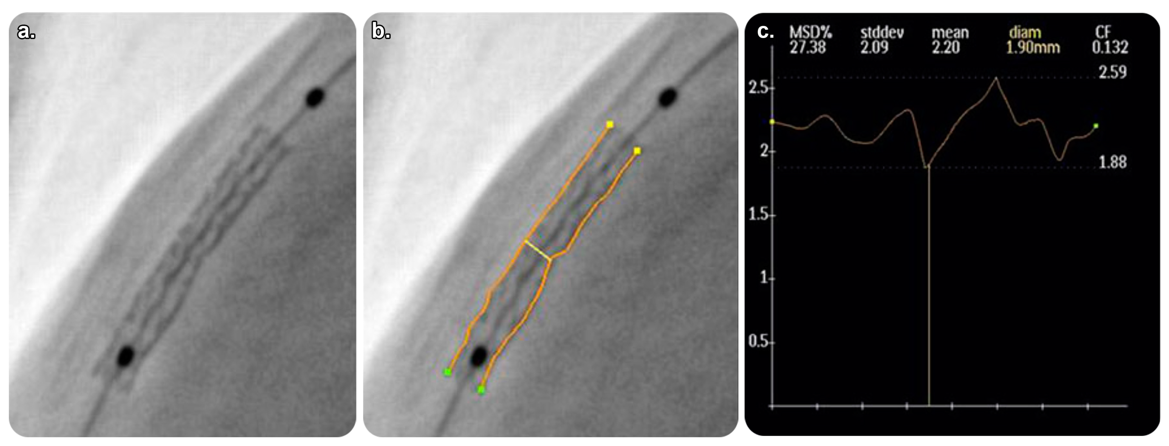
3.2.3. EmboGuide
3.2.4. Vision 2
TrackVision 2
3.3. Monitoring
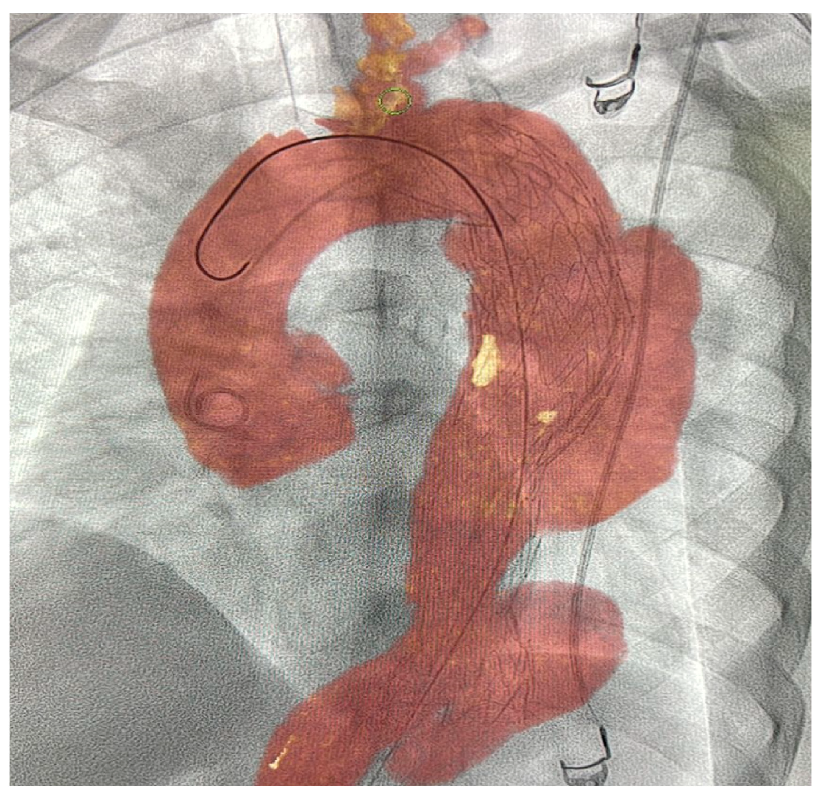
3.3.1. AneurysmFlow
3.3.2. Volume Viewer
3.3.3. Volume Viewer Innova
3.4. Intuitive Display
3.4.1. SmartCT Roadmap
3.4.2. VesselNavigator
3.4.3. HeartNavigator
3.4.4. EP Navigator
3.4.5. EchoNavigator
3.4.6. FlightPlan for EVAR
3.4.7. FlightPlan for Liver
3.4.8. Advantage 4D
3.4.9. Embo ASSIST with Virtual Injection
3.4.10. EVARVision
3.4.11. Stereo 3D
3.5. Postprocedural Check
3.5.1. SmartPerfusion
3.5.2. Innova 3D
3.5.3. Innova CT HD
4. Conclusions
Funding
Conflicts of Interest
Abbreviations
References
- Tsoumakidou, G.; Saltiel, S.; Villard, N.; Duran, R.; Meuwly, J.Y.; Denys, A. Image-guided marking techniques in interventional radiology: A review of current evidence. Diagn. Interv. Imaging 2021, 102, 699–707. [Google Scholar] [CrossRef] [PubMed]
- Cui, Z.; Shukla, P.A.; Habibollahi, P.; Park, H.S.; Fischman, A.; Kolber, M.K. A systematic review of automated feeder detection software for locoregional treatment of hepatic tumors. Diagn. Interv. Imaging 2020, 101, 439–449. [Google Scholar] [CrossRef] [PubMed]
- Cornelis, F.H.; Solomon, S.B. Image guidance in interventional radiology: Back to the future? Diagn. Interv. Imaging 2020, 101, 429–430. [Google Scholar] [CrossRef] [PubMed]
- Ji, J.; Fang, S.; Minjiang Chen Liyun Zheng Chen, W.; Zhao, Z.; Cheng, Y. Precision interventional radiology. J. Interv. Med. 2021, 4, 155–158. [Google Scholar] [CrossRef]
- Vercauteren, T.; Unberath, M.; Padoy, N.; Navab, N. CAI4CAI: The rise of contextual artificial intelligence in computer assisted interventions. Proc. IEEE 2020, 108, 198–214. [Google Scholar] [CrossRef] [Green Version]
- Chehab, M.A.; Brinjikji, W.; Copelan, A.; Venkatesan, A.M. Navigational tools for interventional radiology and interventional oncology applications. Semin. Interv. Radiol. 2015, 32, 416–427. [Google Scholar] [CrossRef] [Green Version]
- Wood, B.J.; Locklin, J.K.; Viswanathan, A.; Kruecker, J.; Haemmerich, D.; Cebral, J.; Sofer, A.; Cheng, R.; McCreedy, E.; Cleary, K.; et al. Technologies for guidance of radiofrequency ablation in the multimodality interventional suite of the future. J. Vasc. Interv. Radiol. 2007, 18, 9–24. [Google Scholar] [CrossRef] [Green Version]
- Linte, C.A.; Moore, J.T.; Chen, E.C.; Peters, T.M. Image-guided procedures: Tools, techniques; clinical applications. In Bioengineering for Surgery; Walid, A.F., James, D., Eds.; Elsevier: Amsterdam, The Netherlands; Chandos Publishing: Oxford, UK, 2016; pp. 59–90. ISBN 9780081001233. [Google Scholar] [CrossRef]
- Floridi, C.; Cellina, M.; Irmici, G.; Bruno, A.; Rossini, N.; Borgheresi, A.; Agostini, A.; Bruno, F.; Arrigoni, F.; Arrichiello, A.; et al. Precision imaging guidance in the era of precision oncology: An update of imaging tools for interventional procedures. J. Clin. Med. 2022, 11, 4028. [Google Scholar] [CrossRef]
- Philips SMARTCT, Release 1.0. The Netherlands. 2020; Volume 4522, p. 66871. Available online: https://www.philips.co.in/healthcare/product/HCNCVC846/smartct-3d-visualization-and-measurement-solution (accessed on 19 November 2022).
- Philips Smart, C.T. The Next Leap in Simplifying and Advancing 3D Imaging To enhance Interventional Confidence. Netherlands MAR 2021. Contract No.: 452299167591. 2021. Available online: https://www.documents.philips.com/assets/SmartCT (accessed on 19 November 2022).
- Philips SMARTCT, Release 1.0 Instructions for Use. Netherlands, p 2020–2002. Contract No.: 4522 203 66871. 2020.
- Philips SMARTCT, Release 1.0 Image Guided Therapy—Philips Customer Services Education—Quick Start Guide, Netherlands, p 2020–2003. Contract No.: 452220410211. 2020. Available online: https://www.philips.co.in/healthcare/product/smartct-3d-visualization-and-measurement-solution (accessed on 19 November 2022).
- Loffroy, R.; Lin, M.; Yenokyan, G.; Rao, P.P.; Bhagat, N.; Noordhoek, N.; Radaelli, A.; Blijd, J.; Liapi, E.; Geschwind, J.F. Intraprocedural C-arm dual-phase cone-beam CT: Can it be used to predict short-term response to TACE with drug-eluting beads in patients with hepatocellular carcinoma? Radiology 2013, 266, 636–648. [Google Scholar] [CrossRef] [Green Version]
- Higashihara, H.; Osuga, K.; Onishi, H.; Nakamoto, A.; Tsuboyama, T.; Maeda, N.; Hori, M.; Kim, T.; Tomiyama, N. Diagnostic accuracy of C-arm CT during selective transcatheter angiography for hepatocellular carcinoma: Comparison with intravenous contrast-enhanced, biphasic, dynamic MDCT. Eur. Radiol. 2012, 22, 872–879. [Google Scholar] [CrossRef]
- Miyayama, S.; Yamashiro, M.; Hashimoto, M.; Hashimoto, N.; Ikuno, M.; Okumura, K.; Yoshida, M.; Matsui, O. Comparison of local control in transcatheter arterial chemoembolization of hepatocellular carcinoma ≤6 cm with or without intraprocedural monitoring of the embolized area using cone-beam computed tomography. Cardiovasc. Interv. Radiol. 2014, 37, 388–395. [Google Scholar] [CrossRef] [PubMed]
- GE Healthcare. AdvantageSim MD. Adaptive Virtual Simulation Planning. 2013. Available online: https://www.gehealthcare.com/products/advanced-visualization/all-applications/advantagesim-md (accessed on 19 November 2022).
- GE Healthcare. Angio CARD. Setting the Standard for Vascular Reporting. 2012. Available online: https://www.gehealthcare.sa/-/jssmedia/widen/2018/01/25/0204/gehealthcarecom/migrated/2018/02/19/0837/ducts-advanced-visualization-product-spec-sheets-angiocard-gehc-datasheet_aw-angiocard_pdf.pdf?rev=-1 (accessed on 19 November 2022).
- GE Healthcare. AngioViz. Visualize Vascular Flow. 2012. Available online: https://www.gehealthcare.com/-/jssmedia/widen/2018/01/25/0204/gehealthcarecom/migrated/2018/02/19/0837/roducts-advanced-visualization-product-spec-sheets-angioviz-gehc-datasheet_aw-angioviz_pdf.pdf?rev=-1 (accessed on 19 November 2022).
- Monti, L.; Donati, D.; Menci, E.; Cioni, S.; Bellini, M.; Grazzini, I.; Leonini, S.; Galluzzi, P.; Bracco, S.; Severi, S.; et al. Cerebral circulation time is prolonged and not correlated with EDSS in multiple sclerosis patients: A study using digital subtracted angiography. PLoS ONE 2015, 10, e0116681. [Google Scholar] [CrossRef] [PubMed]
- GE Healthcare. Autobone, Vessel IQ Xpress—Fast Routine Analyses and Simplified Studies of Routine CT Angiography Exams. Available online: https://www.gehealthcare.com//spec-sheets-autobone-vessellq-xpress-ct (accessed on 19 November 2022).
- GE Healthcare. Hepatic VCAR—Providing Automated Segmentation and Assessment of Liver, Liver Lesions, and Vasculature; Enabling an Efficient and Consistent Workflow. Contract No.: DOC1448778. 2019. Available online: https://www.gehealthcare.com/products/advanced-visualization/all-applications/hepatic-vcar (accessed on 19 November 2022).
- Ho, J.L.; Konda, A.; Rahman, J.; Harris, E.; Korn, R.; Sabir, A.; Bawany, B.; Gulati, R.; Harris, G.J.; Boswell, W.D.; et al. Comparative analysis of three-dimensional volume rendering and maximum intensity projection for preoperative planning in liver cancer. Eur. J. Radiol. Open 2020, 7, 100259. [Google Scholar] [CrossRef] [PubMed]
- GE Healthcare. Innova. 3D Imaging in the Interventional Lab, vol 3d. 2012. Available online: https://www.gehealthcare.com/-/jssmedia/widen/2018/01/25/0204/gehealthcarecom/migrated/2018/02/19/0837/ducts-advanced-visualization-product-spec-sheets-innova-3d-gehc-datasheet_aw-innova-3d_pdf.pdf?rev=-1 (accessed on 19 November 2022).
- GE Healthcare. Innova CT HD. Next-Generation 3D Imaging. 2012. Available online: https://www.gehealthcare.com/-/jssmedia/widen/2018/01/25/0204/gehealthcarecom/migrated/2018/02/19/0837/advanced-visualization-product-spec-sheets-innova-ct-hd-gehc-datasheet_aw-innova-ct-hd_pdf.pdf?rev=-1 (accessed on 19 November 2022).
- GE Healthcare. MR VesselIQ Xpress. Vessel Analysis Made Flexible. 2012. Available online: https://www.gehealthcare.com/-/jssmedia/widen/2018/01/25/0204/gehealthcarecom/migrated/2018/02/19/0837/ualization-product-spec-sheets-mr-vesseliq-xpress-gehc-datasheet_aw-mr-vesseliq-xpress_pdf.pdf?rev=-1 (accessed on 19 November 2022).
- GE Healthcare. TEVAR Assist Solutions– Feel Confident. 2012. Available online: https://www.gehealthcare.com/-/jssmedia/widen/2018/01/25/0204/gehealthcarecom/migrated/2018/02/19/0837/on-product-spec-sheets-tevar-assist-solutions-gehc-datasheet_aw-tevar-assist-solutions_pdf.pdf?rev=-1 (accessed on 19 November 2022).
- Philips StentBoost, Release 4.2 Instructions for Use Netherlands, p 2017–2006. Contract No.: 4522 203 65881. 2017. Available online: https://www.philips.sa/en/healthcare/product/stentboost-enhanced-visualization-software (accessed on 19 November 2022).
- StentBoost, Release 4.4—Image Guided Therapy—Philips Customer Services Education—Quick Start Guide Netherlands 2020 2020-09. Contract No.: 452220410171. Available online: https://www.philips.co.in/healthcare/product/stentboost-enhanced-visualization-software (accessed on 19 November 2022).
- Shinde, R.S.; Hardas, S.; Grant, P.K.; Makhale, C.N.; Shinde, S.N.; Durairaj, M. Stent fracture detected with a novel fluoroscopic stent visualization technique—StentBoost. Can. J. Cardiol. 2009, 25, 487. [Google Scholar] [CrossRef] [Green Version]
- Mishell, J.M.; Vakharia, K.T.; Ports, T.A.; Yeghiazarians, Y.; Michaels, A.D. Determination of adequate coronary stent expansion using StentBoost, a novel fluoroscopic image processing technique. Catheter. Cardiovasc. Interv. 2007, 69, 84–93. [Google Scholar] [CrossRef]
- Dannenberg, L.; Polzin, A.; Bullens, R.; Kelm, M.; Zeus, T. On the road: First-in-man bifurcation percutaneous coronary intervention with the use of a dynamic coronary road map and StentBoost Live imaging system. Int. J. Cardiol. 2016, 215, 7–8. [Google Scholar] [CrossRef]
- Tanaka, N.; Pijls, N.H.; Koolen, J.J.; Botman, K.J.; Michels, H.R.; Brueren, B.R.; Peels, K.; Shindo, N.; Yamashita, J.; Yamashina, A. Assessment of optimum stent deployment by stent boost imaging: Comparison with intravascular ultrasound. Heart Vessels 2013, 28, 1–6. [Google Scholar] [CrossRef]
- Cura, F.; Albertal, M.; Candiello, A.; Nau, G.; Bonvini, V.; Tricherri, H.; Padilla, L.T.; Belardi, J.A. StentBoost visualization for the evaluation of coronary stent expansion during percutaneous coronary interventions. Cardiol. Ther. 2013, 2, 171–180. [Google Scholar] [CrossRef] [Green Version]
- Philips XperGuide, Release 1.3 Instructions for Use. Netherlands, p 2020–2001. Contract No.: 452220381051. 2020. Available online: https://www.documents.philips.com/assets/PhilipsXperGuide (accessed on 19 November 2022).
- Orlov, M.V.; Ansari, M.M.; Akrivakis, S.T.; Jadidi, A.; Nijhof, N.; Natan, S.R.; Wylie, J.V.; Hicks, A.; Armstrong, J.; Jais, P. First experience with rotational angiography of the right ventricle to guide ventricular tachycardia ablation. Heart Rhythm 2011, 8, 207–211. [Google Scholar] [CrossRef]
- Philips EmboGuide, Release 1.0 Instructions for Use. Neth 2021, vol 03. Contract No.: 452300103081. 2021. Available online: https://www.philips.co.in/healthcare/product/HCNCVC321/embo-guide (accessed on 19 November 2022).
- Philips EmboGuide, Release 1.2—Image Guided Therapy—Philips Customer Services Education—Quick Start Guide Netherlands, p 2019–2011. Contract No.: 452220410111. 2019. Available online: https://www.usa.philips.com/healthcare/product/HCNCVC321/embo-guide (accessed on 19 November 2022).
- Levy, E.B.; Krishnasamy, V.P.; Lewis, A.L.; Willis, S.; Macfarlane, C.; Anderson, V.; van der Bom, I.M.; Radaelli, A.; Dreher, M.R.; Sharma, K.V.; et al. First human experience with directly image-able iodinated embolization microbeads. Cardiovasc. Interv. Radiol. 2016, 39, 1177–1186. [Google Scholar] [CrossRef]
- GE Healthcare. Vision 2. Contract No.: DOC2673272:2–3D Live Guidance—GE Healthcare. 2012. Available online: https://www.gehealthcare.com/products/advanced-visualization/all-applications (accessed on 19 November 2022).
- GE Healthcare. TrackVision I. Progress with Confidence. 2022. Available online: https://www.gehealthcare.com/-/media/282a25d4270e44e1b6a32cbe861e4735.pdf (accessed on 19 November 2022).
- Augsburger, L.; Reymond, P.; Fonck, E.; Kulcsar, Z.; Farhat, M.; Ohta, M.; Stergiopulos, N.; Rüfenacht, D.A. Methodologies to assess blood flow in cerebral aneurysms: Current state of research and perspectives. J. Neuroradiol. 2009, 36, 270–277. [Google Scholar] [CrossRef] [PubMed] [Green Version]
- Sforza, D.M.; Putman, C.M.; Cebral, J.R. Hemodynamics of cerebral aneurysms. Annu. Rev. Fluid Mech. 2009, 41, 91–107. [Google Scholar] [CrossRef] [PubMed] [Green Version]
- AneurysmFlow, Release 1.2 Instructions for Use. Netherlands, vol 2019(12/2019). Contract No.: 4522 203 82801. Available online: https://www.documents.philips.com/assets/AneurysmFlow (accessed on 19 November 2022).
- AneurysmFlow. AUG. Contract No.: 452299117561 Making the difference with Live Image Guidance—Interventional X-ray. Netherlands. 2016. Available online: https://www.documents.philips.com/assets/AneurysmFlow2016 (accessed on 19 November 2022).
- AneurysmFlow, Release 1.2—Image Guided Therapy—Philips Customer Services Education—Quick Start Guide. Netherlands, p 2020–2012. Contract No.: 452220411411. 2020. Available online: https://www.philips.com.tr/healthcare/product/aneurysmflow-cerebral-aneurysm-flow-quantification (accessed on 19 November 2022).
- Frölich, A.M.; Nawka, M.T.; Ernst, M.; Frischmuth, I.; Fiehler, J.; Buhk, J.H. Intra-aneurysmal flow disruption after implantation of the Medina® embolization device depends on aneurysm neck coverage. PLoS ONE 2018, 13, e0191975. [Google Scholar] [CrossRef] [Green Version]
- Cancelliere, N.M.; Nicholson, P.; Radovanovic, I.; Mendes, K.M.; Orru, E.; Krings, T.; Pereira, V.M. Comparison of intra-aneurysmal flow modification using optical flow imaging to evaluate the performance of Evolve and Pipeline flow diverting stents. J. Neurointerv. Surg. 2020, 12, 814–817. [Google Scholar] [CrossRef] [PubMed]
- Volume Viewer Innova—Enhanced Workflow to Process X-ray, CT and MR 3D Models in Order to Assist the User during Clinical Practice. Contract No.: DOC1660741. GE Healthcare, 283 Rue de la Miniere 78530 Buc France. 2015. Available online: https://www.gehealthcare.com/volumeviewer_datasheet (accessed on 19 November 2022).
- Volume Viewer—GE Healthcare’s Premium Advanced Visualization and Image Processing Platform Brings You Powerful 3D Capabilities to Help You Visualize and Interpret Your Multimodality Imaging Data with Confidence and Ease. Contract No.: DOC1460639. GE Healthcare, 283 Rue de la Miniere 78530 Buc France. 2015. Available online: https://www.gehealthcare.com/products/advanced-visualization/all-applications/volume-viewer (accessed on 19 November 2022).
- Tacher, V.; Lin, M.; Desgranges, P.; Deux, J.F.; Grünhagen, T.; Becquemin, J.P.; Luciani, A.; Rahmouni, A.; Kobeiter, H. Image guidance for endovascular repair of complex aortic aneurysms: Comparison of two-dimensional and three-dimensional angiography and image fusion. J. Vasc. Interv. Radiol. 2013, 24, 1698–1706. [Google Scholar] [CrossRef] [PubMed] [Green Version]
- Sailer, A.M.; De Haan, M.W.; Peppelenbosch, A.G.; Jacobs, M.J.; Wildberger, J.E.; Schurink, G.W. CTA with fluoroscopy image fusion guidance in endovascular complex aortic aneurysm repair. Eur. J. Vasc. Endovasc. Surg. 2014, 47, 349–356. [Google Scholar] [CrossRef] [PubMed] [Green Version]
- VesselNavigator Making the Difference with Live Image Guidance—Endovascular Procedures Netherlands; 2015 OCT 2015. Contract No.: 452299115111. Available online: https://www.documents.philips.com/assets/VesselNavigator (accessed on 19 November 2022).
- VesselNavigator, Release 1.2—Image Guided Therapy—Philips Customer Services Education—Quick Start Guide Netherlands, p 2020–2008. Contract No.: 452220410141. 2020. Available online: https://www.usa.philips.com/healthcare/product/HCNCVC465/vessel-navigator (accessed on 19 November 2022).
- de Ruiter, Q.M.; Reitsma, J.B.; Moll, F.L.; van Herwaarden, J.A. Meta-analysis of cumulative radiation duration and dose during EVAR using mobile, fixed, or fixed/3D fusion C-arms. J. Endovasc. Ther. 2016, 23, 944–956. [Google Scholar] [CrossRef]
- Stangenberg, L.; Shuja, F.; Carelsen, B.; Elenbaas, T.; Wyers, M.C.; Schermerhorn, M.L. A novel tool for three-dimensional roadmapping reduces radiation exposure and contrast agent dose in complex endovascular interventions. J. Vasc. Surg. 2015, 62, 448–455. [Google Scholar] [CrossRef] [Green Version]
- Philips. HeartNavigator, Release 3.2 Instructions for Use. Netherlands, p 2019–2012. Contract No.: 452220383501. 2019. Available online: https://www.documents.philips.com/assets/HeartNavigator (accessed on 19 November 2022).
- Philips HeartNavigator Making the Difference with Live Image Guidance—Structural Heart Disease Procedures Netherlands 2016 JUL 2016. Contract No.: 452299120771. Available online: https://www.documents.philips.com/assets/PhilipsHeartNavigator (accessed on 19 November 2022).
- Jilaihawi, H.; Kashif, M.; Fontana, G.; Furugen, A.; Shiota, T.; Friede, G.; Makhija, R.; Doctor, N.; Leon, M.B.; Makkar, R.R. Cross-sectional computed tomographic assessment improves accuracy of aortic annular sizing for transcatheter aortic valve replacement and reduces the incidence of paravalvular aortic regurgitation. J. Am. Coll. Cardiol. 2012, 59, 1275–1286. [Google Scholar] [CrossRef] [Green Version]
- Coti, I.; Maierhofer, U.; Rath, C.; Werner, P.; Loewe, C.; Kocher, A.; Laufer, G.; Andreas, M. Effect of conventional and rapid-deployment aortic valve replacement on the distance from the aortic annulus to coronary arteries. Interact. Cardiovasc. Thorac. Surg. 2021, 32, 196–203. [Google Scholar] [CrossRef]
- Kočka, V.; Bártová, L.; Valošková, N.; Laboš, M.; Weichet, J.; Neuberg, M.; Toušek, A.P. Fully automated measurement of aortic root anatomy using Philips HeartNavigator computed tomography software: Fast, accurate, or both? Eur. Heart J. Suppl. B 2022, 24, B36–B41. [Google Scholar] [CrossRef] [PubMed]
- Vaitkus, P.T.; Wang, D.D.; Greenbaum, A.; Guerrero, M.; O’Neill, W. Assessment of a novel software tool in the selection of aortic valve prosthesis size for transcatheter aortic valve replacement. J. Invasive Cardiol. 2014, 26, 328–332. Available online: https://www.hmpgloballearningnetwork.com/site/jic/articles/assessment-novel-software-tool (accessed on 19 November 2022). [PubMed]
- Gerds-Li, J.H.; Tang, M.; Kriatselis, C.; Roser, M.; Goetze, S.; He, D.; Fleck, E. Rapid ventricular pacing to optimize rotational angiography in atrial fibrillation ablation. J. Interv. Card Electrophysiol. 2009, 26, 101–107. [Google Scholar] [CrossRef] [PubMed]
- Philips EP Navigator, Release 5.4 Instructions for Use. Netherlands, p 2019–2012. Contract No.: 452220383151. 2019. Available online: https://www.documents.philips.com/assets/EPNavigator (accessed on 19 November 2022).
- Philips EP Navigator, Release 5.4—Image Guided Therapy—Philips Customer Services Education—Quick Start Guide Netherlands, p 2019–2011. Contract No.: 452220410061. 2019. Available online: https://www.philips.sa/en/healthcare/product/ep-navigator (accessed on 19 November 2022).
- Knecht, S.; Skali, H.; O’Neill, M.D.; Wright, M.; Matsuo, S.; Chaudhry, G.M.; Haffajee, C.I.; Nault, I.; Gijsbers, G.H.; Sacher, F.; et al. Computed tomography–fluoroscopy overlay evaluation during catheter ablation of left atrial arrhythmia. Europace 2008, 10, 931–938. [Google Scholar] [CrossRef] [PubMed]
- Fujita, S.; Fujii, E.; Kagawa, Y.; Inoue, K.; Yamada, T.; Ito, M. The seamless integration of three-dimensional rotational angiography images into electroanatomical mapping systems to guide catheter ablation of atrial fibrillation. Heart Vessels 2018, 33, 1373–1380. [Google Scholar] [CrossRef] [PubMed]
- Buzaev, I.V.; Khalikova, G.; Plechev, V.V.; Onorato, E.M. EchoNavigator® technology facilitates transapical mitral paravalvular leak closure: A case report. Eur. Heart Case Rep. 2021, 5, ytab190. [Google Scholar] [CrossRef]
- Philips EchoNavigator, Release 3.0 Instructions for Use. Netherlands, p 2019–2012. Contract No.: 452300100051. 2019. Available online: https://www.documents.philips.com/assets/EchoNavigator_Release_3 (accessed on 19 November 2022).
- Kim, B.H.; Koh, Y.S.; Lee, K.Y.; Chung, W.B. Three-dimensional EchoNavigator system guided transcatheter closure of paravalvular leakage. J. Cardiovasc. Imaging 2019, 27, 227–229. [Google Scholar] [CrossRef]
- Philips EchoNavigator Making the difference with Live Image—Interventional Cardiology Netherlands; 2014 JUN 2014. Contract No.: 452299104031. Available online: https://www.documents.philips.com/assets/EchoNavigator2014 (accessed on 19 November 2022).
- Jone, P.N.; Ross, M.M.; Bracken, J.A.; Mulvahill, M.J.; Di Maria, M.V.; Fagan, T.E. Feasibility and safety of using a fused echocardiography/fluoroscopy imaging system in patients with congenital heart disease. J. Am. Soc. Echocardiogr. 2016, 29, 513–521. [Google Scholar] [CrossRef]
- Jungen, C.; Zeus, T.; Balzer, J.; Eickholt, C.; Petersen, M.; Kehmeier, E.; Veulemans, V.; Kelm, M.; Willems, S.; Meyer, C. Left atrial appendage closure guided by integrated echocardiography and fluoroscopy imaging reduces radiation exposure. PLoS ONE 2015, 10, e0140386. [Google Scholar] [CrossRef]
- Ternacle, J.; Gallet, R.; Nguyen, A.; Deux, J.F.; Fiore, A.; Teiger, E.; Dubois-Randé, J.L.; Riant, E.; Lim, P. Usefulness of echocardiographic-fluoroscopic fusion imaging in adult structural heart disease. Arch. Cardiovasc. Dis. 2018, 111, 441–448. [Google Scholar] [CrossRef]
- Hadeed, K.; Hascoët, S.; Karsenty, C.; Ratsimandresy, M.; Dulac, Y.; Chausseray, G.; Alacoque, X.; Fraisse, A.; Acar, P. Usefulness of echocardiographic-fluoroscopic fusion imaging in children with congenital heart disease. Arch. Cardiovasc. Dis. 2018, 111, 399–410. [Google Scholar] [CrossRef] [PubMed]
- GE Healthcare FlightPlan for EVAR—Simplified Sizing & Planning for EVAR. Contract No.: DOC1761261. 2015. Available online: https://apps.gehealthcare.com/app-products/flightplan-for-evar (accessed on 19 November 2022).
- GE Healthcare. FlightPlan for Liver—A Breakthrough in Liver Embolization. 2012. Available online: https://www.gehealthcare.com/-/jssmedia/widen/2018/01/25/0204/gehealthcarecom/migrated/2018/02/19/0837/zation-product-spec-sheets-flightplan-for-liver-gehc-datasheet_aw-flightplan-for-liver_pdf.pdf?rev=-1 (accessed on 19 November 2022).
- Durack, J.C.; Brown, K.T.; Avignon, G.; Brody, L.A.; Sofocleous, C.T.; Erinjeri, J.P.; Solomon, S.B. Assessment of automated cone-beam CT vessel identification software during transarterial hepatic embolization: Radiation dose, contrast medium volume, processing time, and operator perspectives compared to digital subtraction angiography. Clin. Radiol. 2018, 73, e1–e1057. [Google Scholar] [CrossRef] [PubMed]
- Joo, S.M.; Kim, Y.P.; Yum, T.J.; Eun, N.L.; Lee, D.; Lee, K.H. Optimized performance of flightplan during chemoembolization for hepatocellular carcinoma: Importance of the proportion of segmented tumor area. Korean J. Radiol. 2016, 17, 771–778. [Google Scholar] [CrossRef] [PubMed] [Green Version]
- Iwazawa, J.; Ohueo, S.; Hashimoto, N.; Mitani, T. Feasibility of using vessel detection software for the endovascular treatment of visceral arterial bleeding. Diagn. Interv. Radiol. 2014, 20, 160–163. [Google Scholar] [CrossRef] [PubMed] [Green Version]
- Ronot, M.; Abdel-Rehim, M.; Hakimé, A.; Kuoch, V.; Roux, M.; Chiaradia, M.; Vilgrain, V.; de Baere, T.; Deschamps, F. Cone-beam CT angiography for determination of tumor-feeding vessels during chemoembolization of liver tumors: Comparison of conventional and dedicated-software analysis. J. Vasc. Interv. Radiol. 2016, 27, 32–38. [Google Scholar] [CrossRef] [PubMed]
- GE Healthcare. Advantage 4D—Efficient and Accurate Visualization and Assessment of Respiratory-Induced Motion with Fast 4D Image Reconstruction. 2012. Available online: https://www.gehealthcare.com/-/jssmedia/widen/2018/01/25/0204/gehealthcarecom/migrated/2018/02/19/0837/advanced-visualization-product-spec-sheets-advantage-4d-gehc-datasheet_aw-advantage-4d_pdf.pdf?rev=-1 (accessed on 19 November 2022).
- GE Healthcare. Embo ASSIST—Simulate Your Embolization with Virtual Injection. 2019. Available online: https://www.gehealthcare.com/products/interventional-image-guided-systems/embo-assist (accessed on 19 November 2022).
- EVAR Vision—3D Fusion Guidance; Contract No.: DOC2673727; GE Healthcare: Chicago, IL, USA, 2022.
- GE Healthcare. Stereo 3D. 3D Localization from Two Projections. Contract No.: DOC2673727. 2022.
- Philips SMART Perfusion, Release 1.1 Instructions for Use. Neth 2019, vol 12. Contract No.: 452220383851. 2019. Available online: https://www.documents.philips.com/assets/SMARTPerfusion (accessed on 19 November 2022).
- Philips SMART Perfusion, Release 1.1- Image Guided Therapy—Philips Customer Services Education—Quick Start Guide Netherlands, p 2019–2012. Contract No.: 452220410091. 2019. Available online: https://www.philips.com.au/healthcare/product/smartperfusion-imaging-technology (accessed on 19 November 2022).


| Category | Software | Main Features |
| PLANNING and GUIDANCE | SmartCT |
|
| SmartCT Vaso |
| |
| SmartCT Soft Tissue |
| |
| SmartCT Angio |
| |
| AdvantageSim MD |
| |
| AngioCARD |
| |
| AngioViz |
| |
| Autobone and VesselIQ Xpress |
| |
| Hepatic VCAR |
| |
| Innova CT HD and Innova 3D |
| |
| MR VesselIQ Xpress |
| |
| TEVAR Assist Solutions |
|
| Category | Software | Main Features |
| TARGETING | StentBoost Live |
|
| StentBoost Mobile |
| |
| XperGuide |
| |
| XperGuide ablation |
| |
| EmboGuide |
| |
| Vision 2 |
| |
| TrackVision 2 |
|
| Category | Software | Main Features |
| MONITORING | AneurysmFlow |
|
| Volume Viewer |
| |
| Volume Viewer Innova |
|
| Category | Software | Main Features |
| INTUITIVE DISPLAY | SmartCT Roadmap |
|
| VesselNavigator |
| |
| HeartNavigator |
| |
| EP Navigator |
| |
| EchoNavigator |
| |
| FlightPlan for EVAR |
| |
| FlightPlan for Liver |
| |
| Advantage 4D |
| |
| Embo ASSIST with Virtual Injection |
| |
| EVARVision |
| |
| Stereo 3D |
|
| Category | Software | Main Features |
| POSTPROCEDURAL CHECK | SmartPerfusion |
|
| Innova 3D |
| |
| Innova CT HD |
|
Disclaimer/Publisher’s Note: The statements, opinions and data contained in all publications are solely those of the individual author(s) and contributor(s) and not of MDPI and/or the editor(s). MDPI and/or the editor(s) disclaim responsibility for any injury to people or property resulting from any ideas, methods, instructions or products referred to in the content. |
© 2023 by the authors. Licensee MDPI, Basel, Switzerland. This article is an open access article distributed under the terms and conditions of the Creative Commons Attribution (CC BY) license (https://creativecommons.org/licenses/by/4.0/).
Share and Cite
Al-Sharydah, A.M.; BinShaiq, F.K.; Aloraifi, R.I.; Almefleh, A.A.; Alessa, S.A.; Alobud, A.S.; AlSharidah, A.M.; Bin Dahmash, A.; Al-Aftan, M.S.; Al-Dhaferi, B.F. Procedural Software Toolkit in the Armamentarium of Interventional Therapies: A Review of Additive Usefulness and Current Evidence. Diagnostics 2023, 13, 765. https://doi.org/10.3390/diagnostics13040765
Al-Sharydah AM, BinShaiq FK, Aloraifi RI, Almefleh AA, Alessa SA, Alobud AS, AlSharidah AM, Bin Dahmash A, Al-Aftan MS, Al-Dhaferi BF. Procedural Software Toolkit in the Armamentarium of Interventional Therapies: A Review of Additive Usefulness and Current Evidence. Diagnostics. 2023; 13(4):765. https://doi.org/10.3390/diagnostics13040765
Chicago/Turabian StyleAl-Sharydah, Abdulaziz M., Faisal Khalid BinShaiq, Rayan Ibrahim Aloraifi, Abdulrahman Abdulaziz Almefleh, Saud Abdulaziz Alessa, Adi Saud Alobud, Abdulmonem Mohammed AlSharidah, Abdulmajeed Bin Dahmash, Mohammad S. Al-Aftan, and Bander Fuhaid Al-Dhaferi. 2023. "Procedural Software Toolkit in the Armamentarium of Interventional Therapies: A Review of Additive Usefulness and Current Evidence" Diagnostics 13, no. 4: 765. https://doi.org/10.3390/diagnostics13040765
APA StyleAl-Sharydah, A. M., BinShaiq, F. K., Aloraifi, R. I., Almefleh, A. A., Alessa, S. A., Alobud, A. S., AlSharidah, A. M., Bin Dahmash, A., Al-Aftan, M. S., & Al-Dhaferi, B. F. (2023). Procedural Software Toolkit in the Armamentarium of Interventional Therapies: A Review of Additive Usefulness and Current Evidence. Diagnostics, 13(4), 765. https://doi.org/10.3390/diagnostics13040765