Single-Energy Metal Artifact Reduction (SEMAR) in Ultra-High-Resolution CT Angiography of Patients with Intracranial Implants
Abstract
1. Introduction
2. Materials and Methods
2.1. Study Design
2.2. Ultra-High-Resolution Computed Tomography Angiography (UHR-CT)
2.3. Post-Processing and Image Reconstruction (SEMAR)
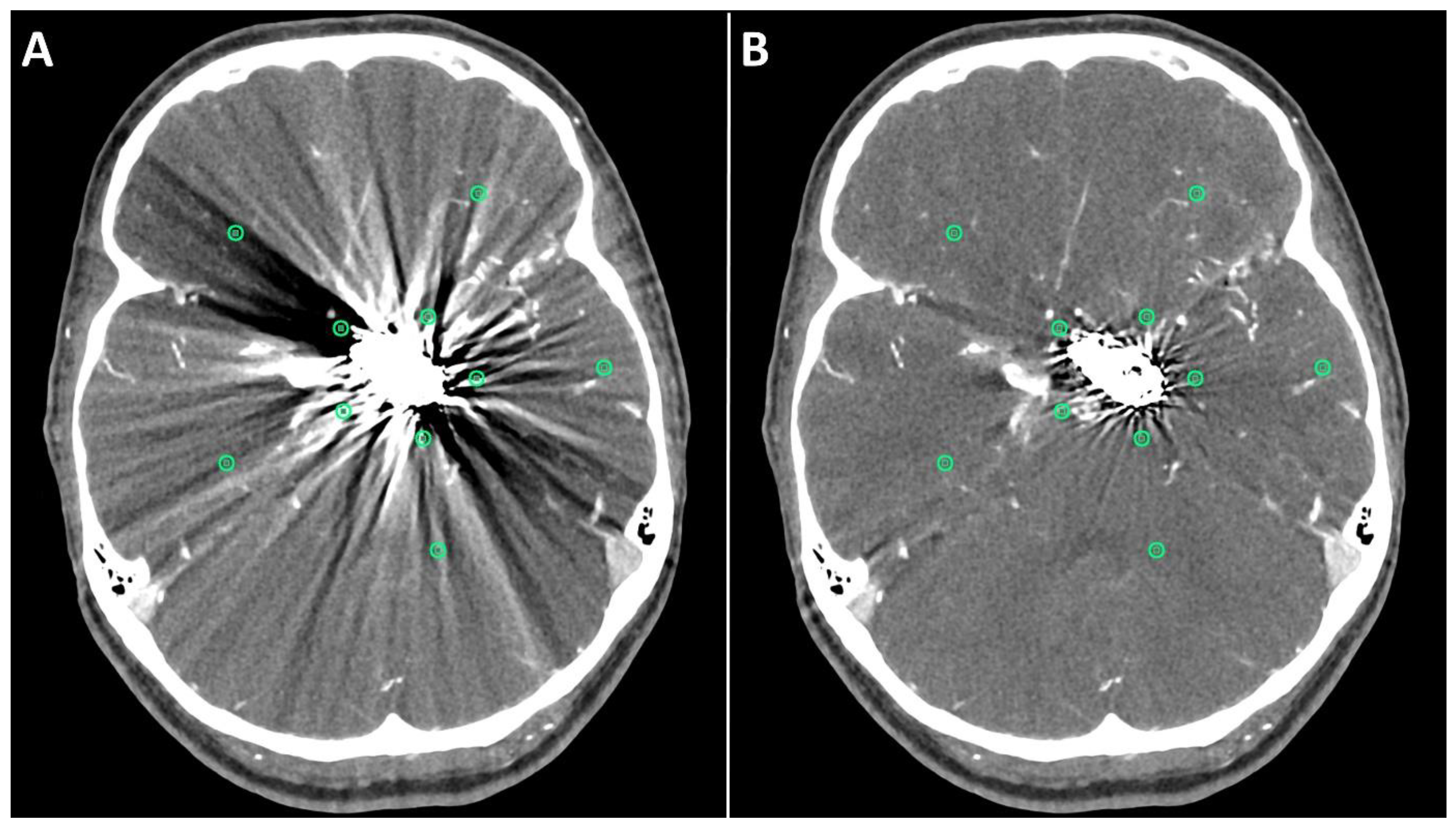
2.4. Quantitative Evaluation
2.5. Qualitative Evaluation
2.6. Statistical Analysis
3. Results
3.1. Patients
3.2. Quantitative Evaluation
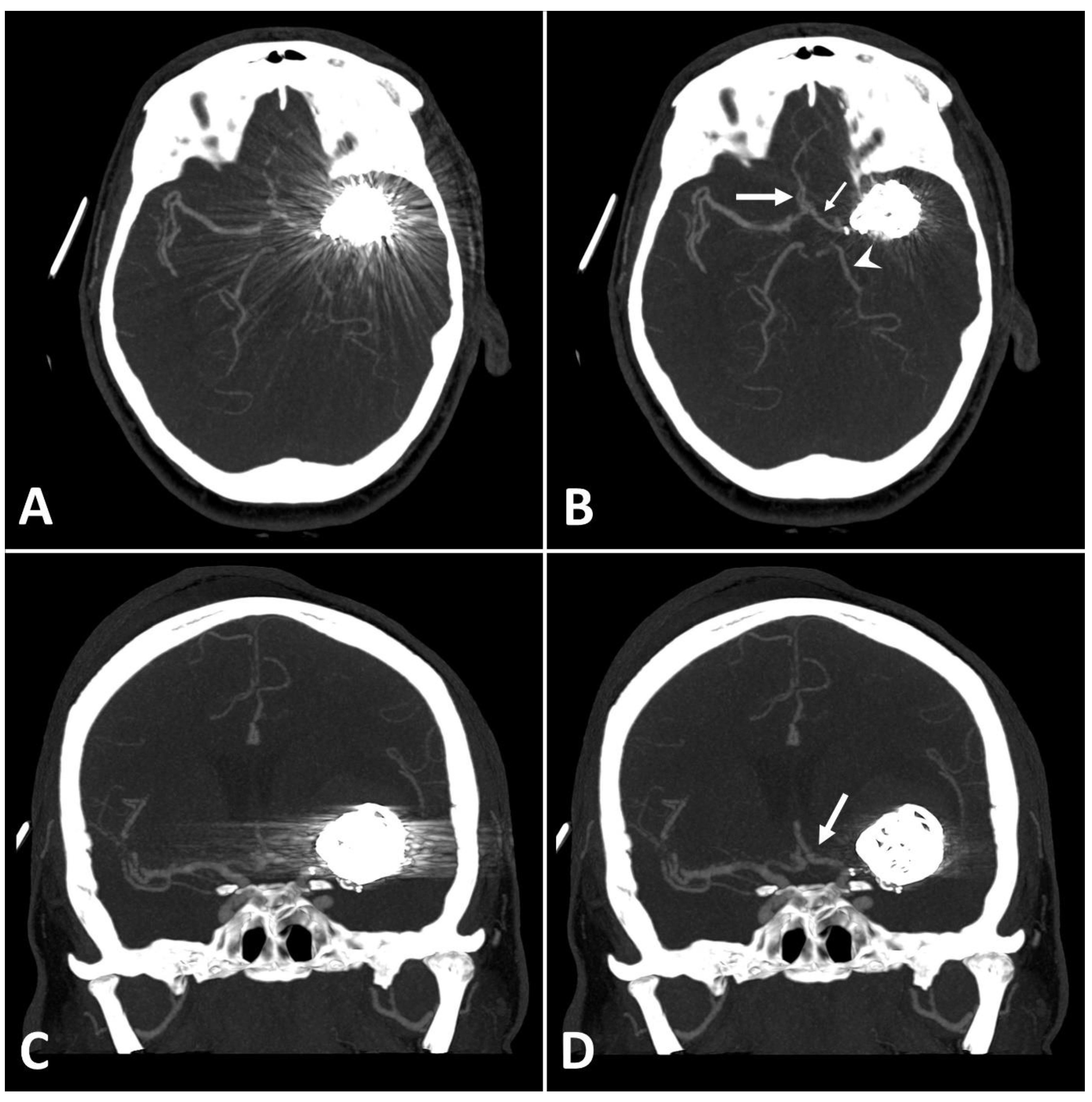
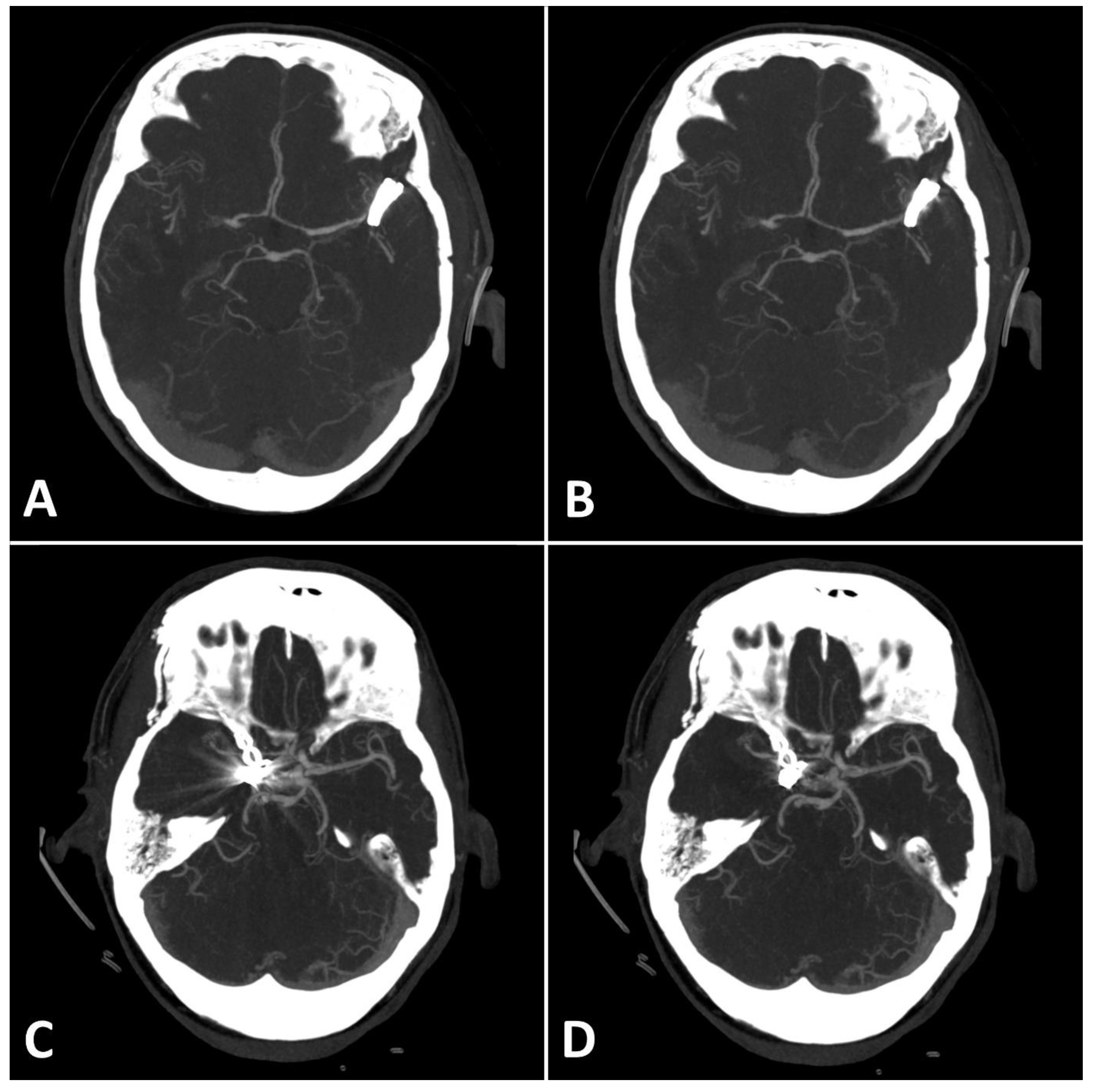
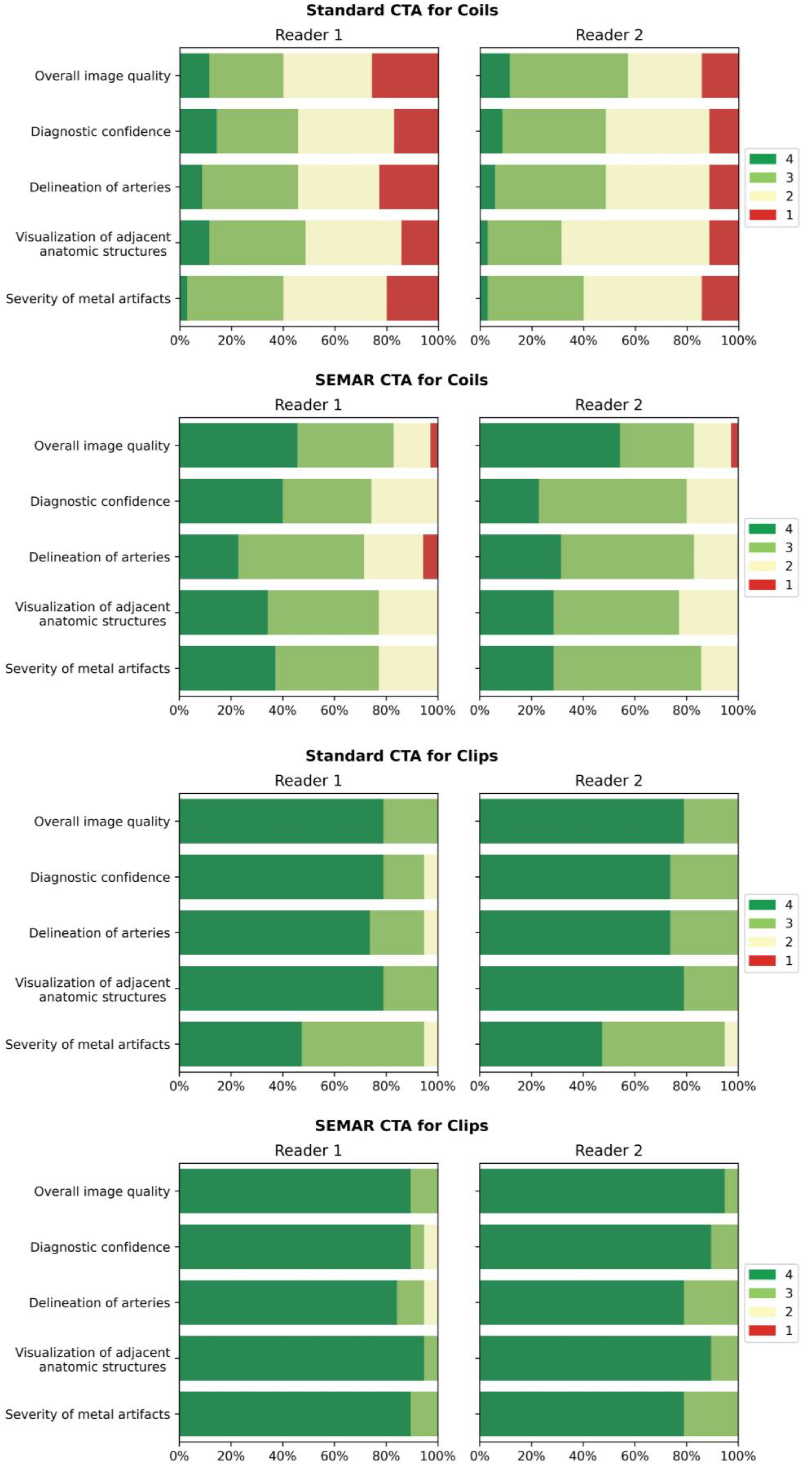
3.3. Qualitative Evaluation
4. Discussion
5. Conclusions
Author Contributions
Funding
Institutional Review Board Statement
Informed Consent Statement
Data Availability Statement
Conflicts of Interest
Abbreviations
| ACA | Anterior Cerebral Artery |
| ACI | Internal Carotid Artery |
| Acom | Anterior Communicating Artery |
| CNR | Contrast-Noise-Ration |
| CTA | Computed Tomography Angiography |
| DSA | Digital Subtraction Angiography |
| HU | Hounsfield-Units |
| MAI | Metal Artifact Index |
| MAR | Metal Artifact Reduction algorithm |
| MCA | Middle Cerebral Artery |
| MRI | Magnetic Resonance Imaging |
| Pcom | Posterior Communicating Artery |
| PICA | Posterior Inferior Cerebellar Artery |
| ROI | Region of Interest |
| SAH | Subarachnoid Hemorrhage |
| SEMAR | Single-Energy Metal Artifact Reduction algorithm |
| SNR | Signal-Noise-Ratio |
| UHR-CT | Ultra-High-Resolution Computed Tomography |
| UHR-CTA | Ultra-High-Resolution Computed Tomography Angiography |
References
- Rinkel, G.J.E.; Djibuti, M.; Algra, A.; Van Gijn, J. Prevalence and Risk of Rupture of Intracranial Aneurysms. Stroke 1998, 29, 251–256. [Google Scholar] [CrossRef] [PubMed]
- Steiner, T.; Juvela, S.; Unterberg, A.; Jung, C.; Forsting, M.; Rinkel, G. European Stroke Organization Guidelines for the Management of Intracranial Aneurysms and Subarachnoid Haemorrhage. Cerebrovasc. Dis. 2013, 35, 93–112. [Google Scholar] [CrossRef] [PubMed]
- D’Souza, S. Aneurysmal Subarachnoid Hemorrhage. J. Neurosurg. Anesthesiol. 2015, 27, 222–240. [Google Scholar] [CrossRef] [PubMed]
- Alakbarzade, V.; Pereira, A.C. Cerebral catheter angiography and its complications. Pract. Neurol. 2018, 18, 393–398. [Google Scholar] [CrossRef]
- Hänsel, N.H.; Schubert, G.A.; Scholz, B.; Nikoubashman, O.; Othman, A.E.; Wiesmann, M.; Pjontek, R.; Brockmann, M.A. Implant-specific follow-up imaging of treated intracranial aneurysms: TOF-MRA vs. metal artifact reduced intravenous flat panel computed tomography angiography (FPCTA). Clin. Radiol. 2018, 73, 218.e219–218.e215. [Google Scholar] [CrossRef]
- Friedrich, B.; Wostrack, M.; Ringel, F.; Ryang, Y.M.; Förschler, A.; Waldt, S.; Zimmer, C.; Nittka, M.; Preibisch, C. Novel Metal Artifact Reduction Techniques with Use of Slice-Encoding Metal Artifact Correction and View-Angle Tilting MR Imaging for Improved Visualization of Brain Tissue near Intracranial Aneurysm Clips. Clin. Neuroradiol. 2016, 26, 31–37. [Google Scholar] [CrossRef]
- Katsura, M.; Sato, J.; Akahane, M.; Kunimatsu, A.; Abe, O. Current and Novel Techniques for Metal Artifact Reduction at CT: Practical Guide for Radiologists. RadioGraphics 2018, 38, 450–461. [Google Scholar] [CrossRef]
- Katsura, M.; Sato, J.; Akahane, M.; Tajima, T.; Furuta, T.; Mori, H.; Abe, O. Single-energy metal artifact reduction technique for reducing metallic coil artifacts on post-interventional cerebral CT and CT angiography. Neuroradiology 2018, 60, 1141–1150. [Google Scholar] [CrossRef]
- Kidoh, M.; Utsunomiya, D.; Ikeda, O.; Tamura, Y.; Oda, S.; Funama, Y.; Yuki, H.; Nakaura, T.; Kawano, T.; Hirai, T.; et al. Reduction of metallic coil artefacts in computed tomography body imaging: Effects of a new single-energy metal artefact reduction algorithm. Eur. Radiol. 2016, 26, 1378–1386. [Google Scholar] [CrossRef]
- Ragusi, M.A.A.D.; Van Der Meer, R.W.; Joemai, R.M.S.; Van Schaik, J.; Van Rijswijk, C.S.P. Evaluation of CT Angiography Image Quality Acquired with Single-Energy Metal Artifact Reduction (SEMAR) Algorithm in Patients After Complex Endovascular Aortic Repair. CardioVascular Interv. Radiol. 2018, 41, 323–329. [Google Scholar] [CrossRef]
- Ucar, F.A.; Frenzel, M.; Abello Mercado, M.A.; Altmann, S.; Reder, S.; Brockmann, C.; Brockmann, M.A.; Othman, A.E. Feasibility of Ultra-High Resolution Supra-Aortic CT Angiography: An Assessment of Diagnostic Image Quality and Radiation Dose. Tomography 2021, 7, 711–720. [Google Scholar] [CrossRef] [PubMed]
- Hata, A.; Yanagawa, M.; Honda, O.; Kikuchi, N.; Miyata, T.; Tsukagoshi, S.; Uranishi, A.; Tomiyama, N. Effect of Matrix Size on the Image Quality of Ultra-high-resolution CT of the Lung. Acad. Radiol. 2018, 25, 869–876. [Google Scholar] [CrossRef] [PubMed]
- Pan, Y.-N.; Chen, G.; Li, A.-J.; Chen, Z.-Q.; Gao, X.; Huang, Y.; Mattson, B.; Li, S. Reduction of Metallic Artifacts of the Post-treatment Intracranial Aneurysms: Effects of Single Energy Metal Artifact Reduction Algorithm. Clin. Neuroradiol. 2019, 29, 277–284. [Google Scholar] [CrossRef] [PubMed]
- Bongers, M.N.; Schabel, C.; Thomas, C.; Raupach, R.; Notohamiprodjo, M.; Nikolaou, K.; Bamberg, F. Comparison and Combination of Dual-Energy- and Iterative-Based Metal Artefact Reduction on Hip Prosthesis and Dental Implants. PLoS ONE 2015, 10, e0143584. [Google Scholar] [CrossRef]
- Mangold, S.; Gatidis, S.; Luz, O.; König, B.; Schabel, C.; Bongers, M.N.; Flohr, T.G.; Claussen, C.D.; Thomas, C. Single-Source Dual-Energy Computed Tomography: Use of Monoenergetic Extrapolation for a Reduction of Metal Artifacts. Investig. Radiol. 2014, 49, 788–793. [Google Scholar] [CrossRef] [PubMed]
- Luo, S.; Zhang, L.J.; Meinel, F.G.; Zhou, C.S.; Qi, L.; McQuiston, A.D.; Schoepf, U.J.; Lu, G.M. Low tube voltage and low contrast material volume cerebral CT angiography. Eur. Radiol. 2014, 24, 1677–1685. [Google Scholar] [CrossRef]
- Jiang, L.; He, Z.H.; Zhang, X.D.; Lin, B.; Yin, X.H.; Sun, X.C. Value of noninvasive imaging in follow-up of intracranial aneurysm. Acta Neurochir. Suppl. 2011, 110, 227–232. [Google Scholar] [CrossRef]
- Obermueller, K.; Hostettler, I.; Wagner, A.; Boeckh-Behrens, T.; Zimmer, C.; Gempt, J.; Meyer, B.; Wostrack, M. Frequency and risk factors for postoperative aneurysm residual after microsurgical clipping. Acta Neurochir. 2021, 163, 131–138. [Google Scholar] [CrossRef]
- Gallas, S.; Januel, A.C.; Pasco, A.; Drouineau, J.; Gabrillargues, J.; Gaston, A.; Cognard, C.; Herbreteau, D. Long-Term Follow-Up of 1036 Cerebral Aneurysms Treated by Bare Coils: A Multicentric Cohort Treated between 1998 and 2003. Am. J. Neuroradiol. 2009, 30, 1986–1992. [Google Scholar] [CrossRef]
- Molyneux, A.J.; Kerr, R.S.; Birks, J.; Ramzi, N.; Yarnold, J.; Sneade, M.; Rischmiller, J. Risk of recurrent subarachnoid haemorrhage, death, or dependence and standardised mortality ratios after clipping or coiling of an intracranial aneurysm in the International Subarachnoid Aneurysm Trial (ISAT): Long-term follow-up. Lancet Neurol. 2009, 8, 427–433. [Google Scholar] [CrossRef]
- Johnston, S.C.; Dowd, C.F.; Higashida, R.T.; Lawton, M.T.; Duckwiler, G.R.; Gress, D.R. Predictors of Rehemorrhage After Treatment of Ruptured Intracranial Aneurysms. Stroke 2008, 39, 120–125. [Google Scholar] [CrossRef]
- Eisenhut, F.; Schmidt, M.A.; Kalik, A.; Struffert, T.; Feulner, J.; Schlaffer, S.-M.; Manhart, M.; Doerfler, A.; Lang, S. Clinical Evaluation of an Innovative Metal-Artifact-Reduction Algorithm in FD-CT Angiography in Cerebral Aneurysms Treated by Endovascular Coiling or Surgical Clipping. Diagnostics 2022, 12, 1140. [Google Scholar] [CrossRef] [PubMed]
- Kimura, Y.; Mikami, T.; Miyata, K.; Suzuki, H.; Hirano, T.; Komatsu, K.; Mikuni, N. Vascular assessment after clipping surgery using four-dimensional CT angiography. Neurosurg. Rev. 2019, 42, 107–114. [Google Scholar] [CrossRef] [PubMed]
- Pjontek, R.; Önenköprülü, B.; Scholz, B.; Kyriakou, Y.; Schubert, G.A.; Nikoubashman, O.; Othman, A.; Wiesmann, M.; Brockmann, M.A. Metal artifact reduction for flat panel detector intravenous CT angiography in patients with intracranial metallic implants after endovascular and surgical treatment. J. NeuroInterv. Surg. 2016, 8, 824–829. [Google Scholar] [CrossRef] [PubMed]
- Zheng, H.; Yang, M.; Jia, Y.; Zhang, L.; Sun, X.; Zhang, Y.; Nie, Z.; Wu, H.; Zhang, X.; Lei, Z.; et al. A Novel Subtraction Method to Reduce Metal Artifacts of Cerebral Aneurysm Embolism Coils. Clin. Neuroradiol. 2022, 32, 687–694. [Google Scholar] [CrossRef] [PubMed]
- Gupta, A.; Subhas, N.; Primak, A.N.; Nittka, M.; Liu, K. Metal artifact reduction: Standard and advanced magnetic resonance and computed tomography techniques. Radiol. Clin. N. Am. 2015, 53, 531–547. [Google Scholar] [CrossRef]
- Otsuka, T.; Nishihori, M.; Izumi, T.; Uemura, T.; Sakai, T.; Nakano, M.; Kato, N.; Kanamori, F.; Tsukada, T.; Uda, K.; et al. Streak Metal Artifact Reduction Technique in Cone Beam Computed Tomography Images after Endovascular Neurosurgery. Neurol. Med. Chir. 2021, 61, 468–474. [Google Scholar] [CrossRef]
- Meijer, F.J.A.; Schuijf, J.D.; de Vries, J.; Boogaarts, H.D.; van der Woude, W.J.; Prokop, M. Ultra-high-resolution subtraction CT angiography in the follow-up of treated intracranial aneurysms. Insights Imaging 2019, 10, 2. [Google Scholar] [CrossRef]
- Bier, G.; Bongers, M.N.; Hempel, J.M.; Örgel, A.; Hauser, T.K.; Ernemann, U.; Hennersdorf, F. Follow-up CT and CT angiography after intracranial aneurysm clipping and coiling-improved image quality by iterative metal artifact reduction. Neuroradiology 2017, 59, 649–654. [Google Scholar] [CrossRef]
- Brown, J.H.; Lustrin, E.S.; Lev, M.H.; Ogilvy, C.S.; Taveras, J.M. Reduction of aneurysm clip artifacts on CT angiograms: A technical note. AJNR Am. J. Neuroradiol. 1999, 20, 694–696. [Google Scholar]
- Nagayama, Y.; Tanoue, S.; Oda, S.; Sakabe, D.; Emoto, T.; Kidoh, M.; Uetani, H.; Sasao, A.; Nakaura, T.; Ikeda, O.; et al. Metal Artifact Reduction in Head CT Performed for Patients with Deep Brain Stimulation Devices: Effectiveness of a Single-Energy Metal Artifact Reduction Algorithm. Am. J. Neuroradiol. 2019, 41, 231–237. [Google Scholar] [CrossRef] [PubMed]
- Pechlivanis, I.; Koenen, D.; Engelhardt, M.; Scholz, M.; Koenig, M.; Heuser, L.; Harders, A.; Schmieder, K. Computed tomographic angiography in the evaluation of clip placement for intracranial aneurysm. Acta Neurochir. 2008, 150, 669. [Google Scholar] [CrossRef]
- Kuroda, H.; Toyota, S.; Kumagai, T.; Iwata, T.; Kobayashi, M.; Mori, K.; Taki, T. Feasibility of Smart Metal Artifact Reduction Algorithm on Computed Tomography Angiography for Clipping of Recurrent Aneurysms After Coil Embolization. World Neurosurg. 2019, 127, e1249–e1254. [Google Scholar] [CrossRef] [PubMed]
- Murai, S.; Hiramatsu, M.; Takasugi, Y.; Takahashi, Y.; Kidani, N.; Nishihiro, S.; Shinji, Y.; Haruma, J.; Hishikawa, T.; Sugiu, K.; et al. Metal artifact reduction algorithm for image quality improvement of cone-beam CT images of medium or large cerebral aneurysms treated with stent-assisted coil embolization. Neuroradiology 2020, 62, 89–96. [Google Scholar] [CrossRef] [PubMed]
- Olsrud, J.; Lätt, J.; Brockstedt, S.; Romner, B.; Björkman-Burtscher, I.M. Magnetic resonance imaging artifacts caused by aneurysm clips and shunt valves: Dependence on field strength (1.5 and 3 T) and imaging parameters. J. Magn. Reson. Imaging 2005, 22, 433–437. [Google Scholar] [CrossRef] [PubMed]


| Overall Image Quality | Diagnostic Confidence | Delineation of Arteries | Visualization of Adjacent Anatomic Structures | Severity of Metal Artifacts | |
|---|---|---|---|---|---|
| 4 | excellent | exact diagnosis possible | perfect vessel delineation | excellent | minimal artifacts |
| 3 | good | sufficient for diagnosis | good vessel delineation | good | mild artifacts |
| 2 | average | limited diagnostic confidence | average vessel delineation | average | strong artifacts |
| 1 | poor | insufficient for diagnosis | poor vessel delineation | poor | extensive artifacts |
| Coils | ||||
| Difference of Intensity in: | 1 mm | 5 mm | 10 mm | 15 mm |
| low-frequency field (bin 2 to 4) | 114 ± 23.6 | 18 ± 7.9 | 5.9 ± 3.1 | 3.8 ± 3.3 |
| middle-frequency field (bin 10 to 12) | 12.1 ± 9.3 | 14 ± 3.5 | 5.6 ± 2 | 0.5 ± 0.7 |
| high-frequency field (bin 20 to 22) | 1 ± 0.3 | 3 ± 1.9 | 1.8 ± 1.4 | 1.3 ± 0.4 |
| very high-frequency field (bin 40 to 42) | 0.1 ± 0.2 | 0.1 ± 0.1 | 0.4 ± 0.4 | 0.5 ± 0.4 |
| Clips | ||||
| Difference of Intensity in: | 1 mm | 5 mm | 10 mm | 15 mm |
| low-frequency field (bin 2 to 4) | 2.9 ± 13.9 | −1 ± 1.8 | 1.2 ± 1.7 | 0.9 ± 1.2 |
| middle-frequency field (bin 10 to 12) | 4.1 ± 3.5 | 1.2 ± 1 | 0.6 ± 0.3 | 0.6 ± 0.3 |
| high-frequency field (bin 20 to 22) | 0.3 ± 0.8 | 0.3 ± 0.2 | 0.3 ± 0.2 | 0.2 ± 0.2 |
| very high-frequency field (bin 40 to 42) | 0.4 ± 0.6 | 0.1 ± 0.1 | 0.1 ± 0.1 | 0.2 ± 0.1 |
| Coils | ||||||||
|---|---|---|---|---|---|---|---|---|
| Reader 1 | Reader 2 | |||||||
| Frequency (1/2/3/4) | Non-SEMAR | Median (IQR) | SEMAR | Median (IQR) | Non-SEMAR | Median (IQR) | SEMAR | Median (IQR) |
| Overall image Quality | 9/12/10/4 | 2 (2) | 1/5/13/16 | 3 (1) | 5/10/16/4 | 3 (1) | 1/5/10/19 | 4 (1) |
| Diagnostic confidence | 6/13/11/5 | 2 (1) | 0/9/12/14 | 3 (2) | 4/14/14/3 | 2 (1) | 0/7/20/8 | 3 (0) |
| Delineation of arteries | 8/11/13/3 | 2 (1) | 2/8/17/8 | 3 (1) | 4/14/15/2 | 2 (1) | 0/6/18/11 | 3 (1) |
| Visualization of adjacent anatomic structures | 5/13/13/4 | 2 (1) | 0/8/15/12 | 3 (1) | 4/20/10/1 | 2 (1) | 0/8/17/10 | 3 (1) |
| Severity of metal artifacts | 7/14/13/1 | 2 (1) | 0/8/14/13 | 3 (1) | 5/16/13/1 | 2 (1) | 0/5/20/10 | 3 (1) |
| Clips | ||||||||
| Reader 1 | Reader 2 | |||||||
| Overall image Quality | 0/0/4/15 | 4 (0) | 0/0/2/17 | 4 (0) | 0/0/4/15 | 4 (0) | 0/0/1/18 | 4 (0) |
| Diagnostic confidence | 0/1/3/15 | 4 (0) | 0/1/1/17 | 4 (0) | 0/0/5/14 | 4 (1) | 0/0/2/17 | 4 (0) |
| Delineation of arteries | 0/1/4/14 | 4 (1) | 0/1/2/16 | 4 (0) | 0/0/5/14 | 4 (1) | 0/0/4/15 | 4 (0) |
| Visualization of adjacent anatomic structures | 0/0/4/15 | 4 (0) | 0/0/1/18 | 4 (0) | 0/0/4/15 | 4 (0) | 0/0/2/17 | 4 (0) |
| Severity of metal artifacts | 0/1/9/9 | 3 (1) | 0/0/2/17 | 4 (0) | 0/1/9/9 | 3 (1) | 0/0/4/15 | 4 (0) |
Disclaimer/Publisher’s Note: The statements, opinions and data contained in all publications are solely those of the individual author(s) and contributor(s) and not of MDPI and/or the editor(s). MDPI and/or the editor(s) disclaim responsibility for any injury to people or property resulting from any ideas, methods, instructions or products referred to in the content. |
© 2023 by the authors. Licensee MDPI, Basel, Switzerland. This article is an open access article distributed under the terms and conditions of the Creative Commons Attribution (CC BY) license (https://creativecommons.org/licenses/by/4.0/).
Share and Cite
Jabas, A.; Abello Mercado, M.A.; Altmann, S.; Ringel, F.; Booz, C.; Kronfeld, A.; Sanner, A.P.; Brockmann, M.A.; Othman, A.E. Single-Energy Metal Artifact Reduction (SEMAR) in Ultra-High-Resolution CT Angiography of Patients with Intracranial Implants. Diagnostics 2023, 13, 620. https://doi.org/10.3390/diagnostics13040620
Jabas A, Abello Mercado MA, Altmann S, Ringel F, Booz C, Kronfeld A, Sanner AP, Brockmann MA, Othman AE. Single-Energy Metal Artifact Reduction (SEMAR) in Ultra-High-Resolution CT Angiography of Patients with Intracranial Implants. Diagnostics. 2023; 13(4):620. https://doi.org/10.3390/diagnostics13040620
Chicago/Turabian StyleJabas, Abdullah, Mario Alberto Abello Mercado, Sebastian Altmann, Florian Ringel, Christian Booz, Andrea Kronfeld, Antoine P. Sanner, Marc A. Brockmann, and Ahmed E. Othman. 2023. "Single-Energy Metal Artifact Reduction (SEMAR) in Ultra-High-Resolution CT Angiography of Patients with Intracranial Implants" Diagnostics 13, no. 4: 620. https://doi.org/10.3390/diagnostics13040620
APA StyleJabas, A., Abello Mercado, M. A., Altmann, S., Ringel, F., Booz, C., Kronfeld, A., Sanner, A. P., Brockmann, M. A., & Othman, A. E. (2023). Single-Energy Metal Artifact Reduction (SEMAR) in Ultra-High-Resolution CT Angiography of Patients with Intracranial Implants. Diagnostics, 13(4), 620. https://doi.org/10.3390/diagnostics13040620


