Interesting Images

Active Lumbar Spondylodiscitis on [68Ga]Ga-PSMA-11 PET/CT Mimicking Bone Metastasis

Department of Nuclear Medicine, Saarland University, 66421 Homburg, Germany
*
Author to whom correspondence should be addressed.
Diagnostics 2023, 13(24), 3616; https://doi.org/10.3390/diagnostics13243616
Submission received: 30 October 2023 / Revised: 5 December 2023 / Accepted: 6 December 2023 / Published: 7 December 2023
(This article belongs to the Section Medical Imaging and Theranostics)

Abstract

:
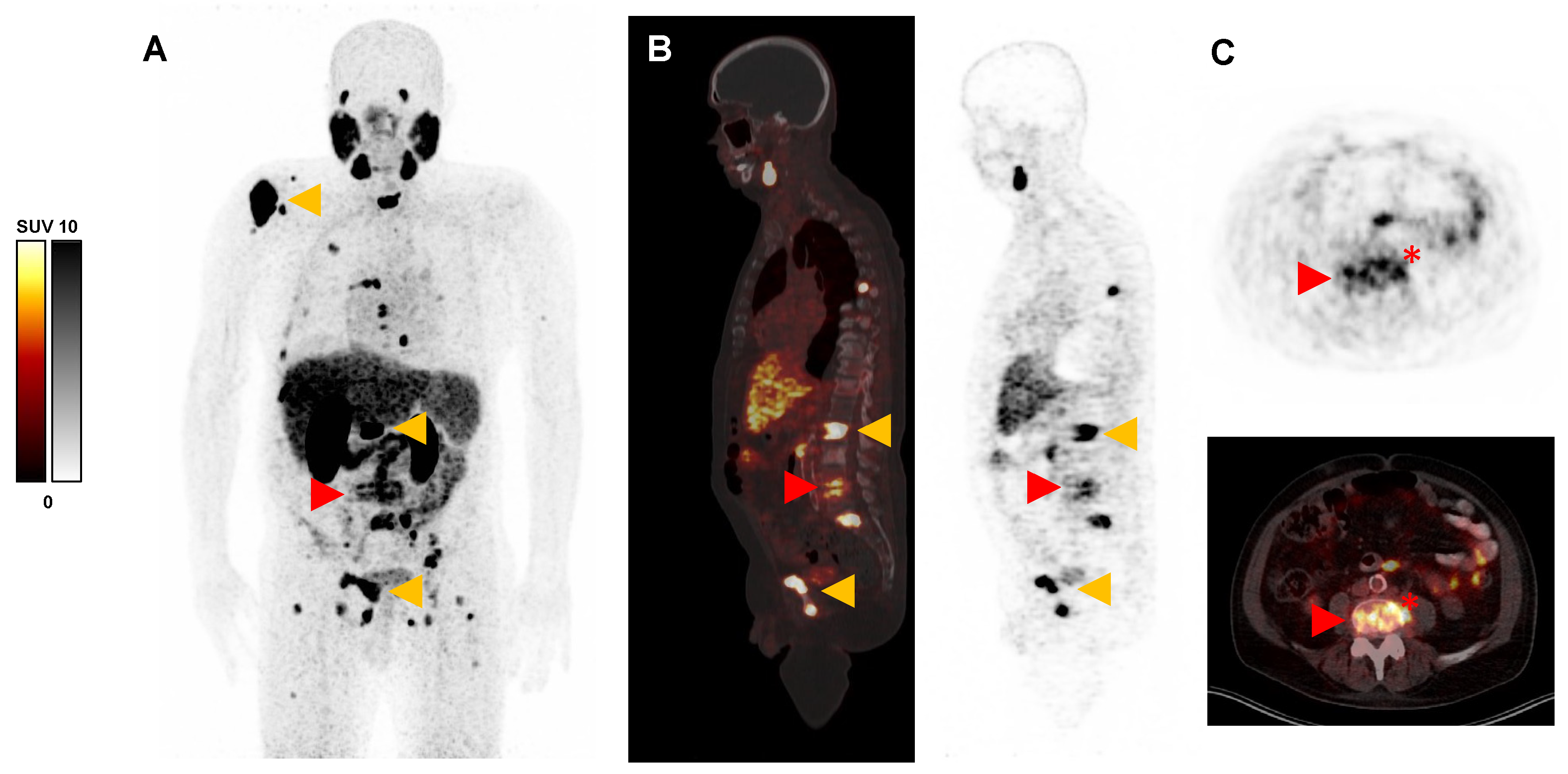
We report a [68Ga]Ga-PSMA-11 positron emission tomography/computed tomography (PET/CT) scan of a 71-year-old man with metastatic castration-resistant prostate cancer (mCRPC) and concomitant active lumbar spondylodiscitis, both PSMA-positive on a PET/CT scan. This interesting image should advise colleagues to consider spondylodiscitis as a differential diagnosis of PSMA-positive findings in the spine, particularly if intervertebral space and soft tissue are involved.

Figure 1. Prostate-specific membrane antigen (PSMA)-targeted positron emission tomography/computed tomography (PET/CT) scan with [68Ga]Ga-PSMA-11 (acquired 60 min post injection of 130 MBq) of a 71-year-old man with metastatic castration-resistant prostate cancer (mCRPC) referred for PSMA-targeted radioligand therapy. (A): Maximum intensity projection (MIP), (B): sagittal slices, and (C): transversal slices of [68Ga]Ga-PSMA-11 PET/CT, showing multiple intense PSMA-positive metastases (maximum standardized uptake value [SUVmax] up to 48.0, exemplary orange arrows) in the skeleton but also a moderate uptake (SUVmax 10.8) in the ground and upper plate of vertebral body L4 and L5, respectively (red arrow), also with involvement of the intervertebral space and surrounding soft tissue (red asterisk). Due to the distribution pattern, inflammatory etiology, i.e., spondylodiscitis, seemed more likely than additional metastasis, which could not be excluded at this point. Matching this differential diagnosis, the patient also reported newly developed pain in the lower back, and the inflammatory blood parameter, C-reactive protein (CRP), was highly elevated (61.8 mg/L). Neurological symptoms were not observed. Subsequently performed magnetic resonance imaging (MRI) confirmed the suspicion of active spondylodiscitis by showing low T1 and high T2 signal in disc space L4/5 and adjacent endplates, respectively. In contrast-enhanced T1 sequence vertebral endplates and paravertebral soft tissue revealed substantial enhancement. After the microbiological identification of streptococcus gallolyticus and subsequent appropriate antibiotic treatment, both pain and blood inflammation levels regressed. Spondylodiscitis mainly occurs in the elderly and is usually diagnosed on the basis of clinical, laboratory, and microbiological examinations, as well as imaging techniques such as CT, MRI, and PET with 18F-Fluorodeoxyglucose ([18F]FDG) [1,2,3,4,5]. In contrast, the finding of spondylodiscitis is rarely described on a PSMA PET/CT [6,7]. However, it is known that inflammatory processes may be related to the increased uptake of PSMA ligands, in line with our observed case of spondylodiscitis [8,9]. This interesting image should draw attention to spondylodiscitis as a differential diagnosis of PSMA-positive findings in the spine, especially when the intervertebral space and soft tissue are involved.
Diagnostics 13 03616 g001

Author Contributions

Conceptualization, F.R., C.B., R.G. and S.E.; investigation, F.R., C.B., R.G. and S.E.; writing—original draft preparation, F.R. and C.B.; writing—review and editing, R.G. and S.E.; visualization, F.R. and C.B.; supervision, R.G. and S.E. All authors have read and agreed to the published version of the manuscript.

Funding

This research received no external funding.

Institutional Review Board Statement

Ethical review and approval were waived for this study, due to the retrospective case report.

Informed Consent Statement

Written informed consent has been obtained from the patient to publish this paper.

Data Availability Statement

The datasets used and analyzed in this paper are available from the corresponding author on reasonable request.

Conflicts of Interest

The authors declare no conflict of interest.

References

  1. Zimmerli, W. Clinical practice. Vertebral osteomyelitis. N. Engl. J. Med. 2010, 362, 1022–1029. [Google Scholar] [CrossRef] [PubMed]
  2. Gouliouris, T.; Aliyu, S.H.; Brown, N.M. Spondylodiscitis: Update on diagnosis and management. J. Antimicrob. Chemother. 2010, 65, iii11–iii24. [Google Scholar] [CrossRef] [PubMed]
  3. Kouijzer, I.J.E.; Scheper, H.; de Rooy, J.W.J.; Bloem, J.L.; Janssen, M.J.R.; Hoven, L.v.D.; Hosman, A.J.F.; Visser, L.G.; Oyen, W.J.G.; Bleeker-Rovers, C.P.; et al. The diagnostic value of 18F-FDG-PET/CT and MRI in suspected vertebral osteomyelitis—A prospective study. Eur. J. Nucl. Med. Mol. Imaging 2018, 45, 798–805. [Google Scholar] [CrossRef] [PubMed]
  4. Prodromou, M.L.; Ziakas, P.D.; Poulou, L.S.; Karsaliakos, P.; Thanos, L.; Mylonakis, E. FDG PET is a robust tool for the diagnosis of spondylodiscitis: A meta-analysis of diagnostic data. Clin. Nucl. Med. 2014, 39, 330–335. [Google Scholar] [CrossRef] [PubMed]
  5. Smids, C.; Kouijzer, I.J.E.; Vos, F.J.; Sprong, T.; Hosman, A.J.F.; de Rooy, J.W.J.; Aarntzen, E.H.J.G.; de Geus-Oei, L.-F.; Oyen, W.J.G.; Bleeker-Rovers, C.P. A comparison of the diagnostic value of MRI and 18F-FDG-PET/CT in suspected spondylodiscitis. Infection 2017, 45, 41–49. [Google Scholar] [CrossRef] [PubMed]
  6. Klingenberg, S.; Jochumsen, M.R.; Pedersen, B.G.; Bouchelouche, K. 68Ga-PSMA Uptake in Escherichia coli Spondylodiscitis. Clin. Nucl. Med. 2019, 44, 916–919. [Google Scholar] [CrossRef] [PubMed]
  7. Gupta, N.; Elumalai, R.K.; Verma, R.; Belho, E.S.; Dhawan, S. Spinal Tuberculosis Mimicking as Prostate Cancer Metastases in Ga-68 Prostate-specific Membrane Antigen Positron-emission Tomography/Computed Tomography. Indian J. Nucl. Med. 2020, 35, 271–273. [Google Scholar] [CrossRef] [PubMed]
  8. Barbosa, F.d.G.; Queiroz, M.A.; Nunes, R.F.; Costa, L.B.; Zaniboni, E.C.; Marin, J.F.G.; Cerri, G.G.; Buchpiguel, C.A. Nonprostatic diseases on PSMA PET imaging: A spectrum of benign and malignant findings. Cancer Imaging 2020, 20, 23. [Google Scholar] [CrossRef] [PubMed]
  9. Malan, N.; Vangu, M.-T. Normal Variants, Pitfalls, and Artifacts in Ga-68 Prostate Specific Membrane Antigen (PSMA) PET/CT Imaging. Front. Nucl. Med. 2022, 2, 825512. [Google Scholar] [CrossRef]
Disclaimer/Publisher’s Note: The statements, opinions and data contained in all publications are solely those of the individual author(s) and contributor(s) and not of MDPI and/or the editor(s). MDPI and/or the editor(s) disclaim responsibility for any injury to people or property resulting from any ideas, methods, instructions or products referred to in the content.