Article

Characteristic Chest Computed Tomography Findings for Birt–Hogg–Dube Syndrome Indicating Requirement for Genetic Evaluation

1
Pulmonary Division, Department of Internal Medicine, Gangnam Severance Hospital, Yonsei University College of Medicine, Seoul 06273, Republic of Korea
2
Department of Radiology and the Research Institute of Radiological Science, Gangnam Severance Hospital, Yonsei University College of Medicine, Seoul 06273, Republic of Korea
3
Department of Laboratory Medicine, Gangnam Severance Hospital, Yonsei University College of Medicine, Seoul 06273, Republic of Korea
4
Department of Thoracic Surgery, Yonsei University College of Medicine, Seoul 06273, Republic of Korea
5
Department of Dermatology, Yonsei University College of Medicine, Seoul 06273, Republic of Korea
*
Author to whom correspondence should be addressed.
These authors contributed equally to this work.
Diagnostics 2023, 13(2), 198; https://doi.org/10.3390/diagnostics13020198
Submission received: 5 December 2022 / Revised: 26 December 2022 / Accepted: 28 December 2022 / Published: 5 January 2023
(This article belongs to the Section Medical Imaging and Theranostics)

Abstract

:
Background: Chest computed tomography (CT) findings are important for identifying Birt–Hogg–Dube (BHD) syndrome. However, the predictive power of classical criteria for chest CT findings is weak. Here, we aimed to identify more specific chest CT findings necessitating genetic examination for FLCN gene mutations. Methods: From June 2016 to December 2017, we prospectively enrolled 21 patients with multiple bilateral and basally located lung cysts on chest CT with no other apparent cause, including cases with and without spontaneous primary pneumothorax. All enrolled patients underwent FLCN mutation testing for diagnosis confirmation. Results: BHD was diagnosed in 10 of 21 enrolled patients (47.6%). There were no differences in clinical features between the BHD and non-BHD groups. Maximal cyst diameter was significantly greater in the BHD group (mean ± standard deviation; 4.1 ± 1.1 cm) than in the non-BHD group (1.6 ± 0.9 cm; p < 0.001). Diversity in cyst size was observed in 100.0% of BHD cases and 18.2% of non-BHD cases (p = 0.001). Morphological diversity was observed in 100.0% of BHD cases and 54.6% of non-BHD cases (p = 0.054). Areas under the receiver operating characteristic curves for predicting FLCN gene mutations were 0.955 and 0.909 for maximal cyst diameter and diversity in size, respectively. The optimal cut-off value for maximal diameter FLCN mutations prediction was 2.1 cm (sensitivity: 99%; specificity: 82%). Conclusions: Reliable chest CT features suggesting the need for FLCN gene mutations screening include variations in cyst size and the presence of cysts > 2.1 cm in diameter, predominantly occurring in the bilateral basal lungs.

1. Introduction

First reported in 1977, Birt–Hogg–Dube syndrome (BHD) is a rare genetic disorder caused by mutations in the folliculin gene (FLCN) [1,2,3]. Although BHD incidence remains unknown, the estimated prevalence of BHD in the general population is approximately 1/200,000 [4,5,6]. Recently, BHD is increasingly been diagnosed [6,7] which may be related to advancements in diagnostic technologies and increased awareness of the disease.
The clinical presentations of BHD include cutaneous manifestations (fibrofolliculoma, acrochordons; 84%), pulmonary cysts (70–85%), spontaneous recurrent pneumothorax (~25%), and renal cell carcinoma (19–35%) [3,8,9,10]. As these clinical manifestations can differ according to age group and patient characteristics, BHD remains difficult to diagnose, likely resulting in many cases of missed diagnosis [7,11]. Especially in Asia, skin manifestations and renal cell carcinoma are not predominant features of BHD, Therefore, in order to diagnose a disease that does not show symptoms or signs at an early stage, highlighting the importance of chest computed tomography (CT) findings in suspected cases [12].
In 2009, Menko et al. proposed modified diagnostic criteria for BHD based on the detection of DNA mutations in FLCN, citing chest CT features of BHD as an important minor criterion. Such features include multiple bilateral lung cysts in the basal region with no other apparent cause, presenting with or without spontaneous primary pneumothorax [7]. Using these widely accepted criteria, our previous study identified 17 patients with characteristic chest CT findings, and only six patients were finally diagnosed with BHD (35.3%) [11]. Therefore, characteristic chest CT findings beyond the classical criteria are required to improve the predictive power of CT for BHD. In this study, we aimed to identify which chest CT findings necessitate genetic examination for FLCN mutations.

2. Methods

2.1. Patients

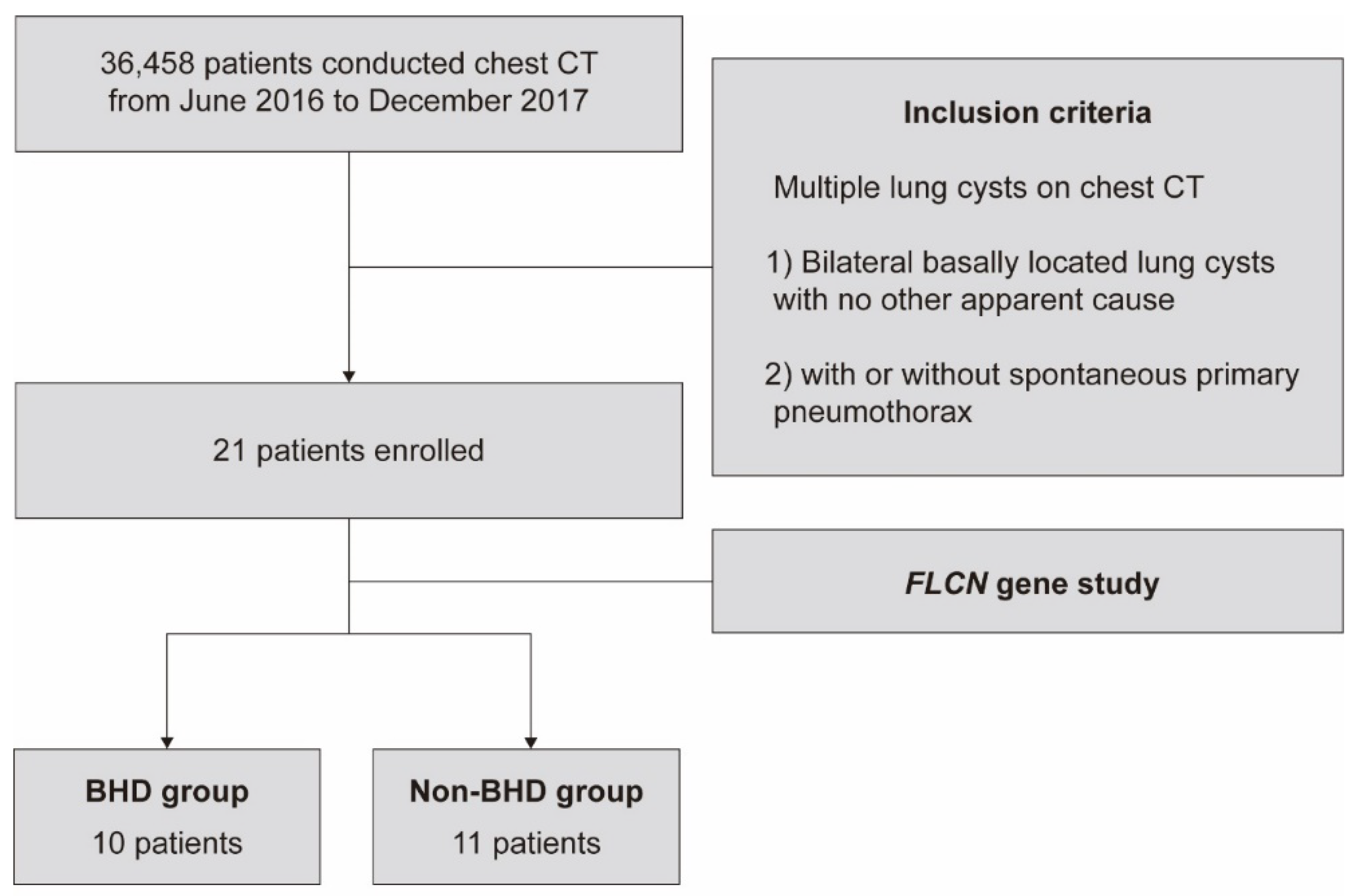
From June 2016 to December 2017, we screened all patients who underwent chest CT and prospectively enrolled patients with multiple bilateral and basally located lung cysts on chest CT, which are minor criteria for the diagnosis of BHD in previous studies [7,13]. Patients exhibiting cysts with other apparent causes were excluded such as chronic obstructive pulmonary disease, lymphangioleiomyomatosis, pulmonary Langerhans cell histiocytosis, and lymphocytic interstitial pneumonia, although cases both with and without spontaneous primary pneumothorax were included. All enrolled patients underwent screening for FLCN gene mutations to confirm their diagnosis (see Appendix A, Figure A1).
The following clinical data were collected for all enrolled patients: age, sex, smoking status, comorbidities, urinalysis, pulmonary function test (PFT) results, personal and family history of spontaneous pneumothorax, presence of skin lesions (evaluated by dermatologists), and renal lesions (evaluated by radiologists).

2.2. CT protocol and Analysis

Chest CT was performed using either a 64-slice MDCT scanner (Somatom Sensation 64; Siemens Medical Solutions, Erlangen, Germany) or a 128-slice MDCT scanner (Somatom Sensation AS+; Siemens Medical Solutions, Erlangen, Germany or Ingenuity Core 128, Philips Healthcare, Cleveland, OH, USA). After acquiring the scout image to determine the field-of-view, conventional CT scanning was performed with a 1–3 mm reconstruction interval in the mediastinal window setting. The exposure parameters for the CT scans were: 120 kVp, 100–200 mA, 1–3 mm slice thickness. Image reconstruction for conventional CT scans was performed using the scanner workstation. All CT images were retrieved using a picture archiving and communication system (PACS) (Centricity 4.0; GE Medical Systems, Mountain Prospect, Chicago, USA). Two radiologists (C.H.P. and T.H.K.) with >10 years of experience in chest radiology interpretation assessed the CT images in consensus. We prospectively screened all chest CT images which performed for any causes during study periods in Gangnam Severance hospital. The chest CT was interpreted without any genetic information associated with the BHD syndrome. A lung cyst was defined as an air-filled lesion with a perceptible wall but not more than 3 mm in thickness, and multiple/diffuse cysts were defined as multiple or numerous cysts distributed in both lungs by two experienced radiologists [14]. If the perceptible wall is not visible in the air-filled lesion, it is classified as emphysema, and if the thickness of the lesion wall exceeds 3 mm, it is classified as cavity. The number, size, morphology, and distribution of the lung cysts on chest CT were carefully evaluated. Cases with >40 cysts and those with <40 cysts were categorized separately. Cyst sizes were considered diverse when the coexistence of cysts <2 cm or >2 cm in diameter was observed. Cyst morphology was categorized as round, oval, or irregular, and diverse morphology was defined as the presence of all three types of cyst morphology in one lung. If there was a discrepancy in CT interpretation, a consensus was achieved through a discussion. If a consensus was not achieved for discrepancy, the third independent reader (J.M.S) mediated.

2.3. FLCN Gene Mutation Analysis

All enrolled patients underwent DNA testing for FLCN gene mutations. Whole blood samples were collected in ethylenediaminetetraacetic acid-containing tubes, and genomic DNA was extracted from each sample using an Easy-DNA™ Kit (Invitrogen, Carlsbad, CA, USA). The concentration and quality of the genomic DNA were assessed using Nanodrop (ND-1000, Thermo Scientific, Wilmington, DE, USA). Primers designed to amplify FLCN exons 4–14 and their flanking introns were used for polymerase chain reaction (PCR) amplification. PCR products were purified using a QIA-quick Gel Extraction Kit (Qiagen, Dusseldorf, Germany), and cycle-sequencing was performed using relevant PCR primers and a Big Dye Terminator Cycle Sequencing Ready Reaction Kit (Applied Biosystems, Foster City, CA, USA).
The sequences obtained were compared with the reference sequence using Sequencher® software (Gene Codes, Ann Arbor, MI, USA). Pathogenic variants were detected using Sanger sequencing followed by multiple ligation probe amplification to confirm that there were no large deletions in FLCN when no pathogenic variants were detected.

2.4. Statistical Analysis

Categorical variables are presented as frequencies (percentages). Continuous variables are presented as mean ± standard deviation (SD) for normally distributed variables and as median (interquartile range [IQR]) for non-normally distributed variables. Normality assumptions for continuous variables were confirmed using the Shapiro–Wilk test. Baseline characteristics were compared between patients with and without BHD using an independent t-test or Mann–Whitney U test. Fisher’s exact test was used to evaluate potential correlations between various chest CT features, using FLCN gene mutation results as a reference. Logistic regression analyses were used to evaluate the association between radiological features and BHD. Receiver operating characteristic (ROC) curve analysis was performed to identify the ability of CT features to aid in BHD diagnosis. The area under the curve (AUC) was calculated to assess the sensitivity, specificity, positive predictive value, and negative predictive value of CT findings. The optimal cut-off was defined using Youden’s J statistic [15,16]. A p value of 0.05 or less was considered statistically significant.
All statistical analyses were performed using R software version 4.0.2. (R v4.0.2. https://cloud.r-project.org/ (accessed on 31 October 2022)). For ROC curve analysis, the “ROCR” (version 1.0-11) and “pROC” packages (version 1.18.0) were used.

3. Results

3.1. Baseline Characteristics of Enrolled Patients

Among the 21 enrolled patients, 10 (47%) were diagnosed with BHD based on the presence of FLCN gene mutations (Table 1). Eleven exhibited negative results in the FLCN gene test. Age, male-to-female ratio, smoking status, comorbidities, proteinuria, haematuria, and PFT did not significantly differ between the two groups. Spontaneous pneumothorax was more frequent in the BHD group (70.0% and 30.0%, respectively) than in the non-BHD group (18.2% and 30.0%, respectively); however, the difference was insignificant (p = 0.051 and 0.181, respectively). Similarly, there were no differences in the prevalence of renal cell carcinoma or cutaneous lesions such as fibrofolliculoma or acrochordons between the groups. The prevalence of renal cysts was higher in the BHD group than in the non-BHD group; however, the difference was insignificant (50.0% and 12.5%, respectively; p = 0.240).

3.2. Clinical Characteristics of BHD

Table 2 shows the detailed clinical characteristics of BHD in each patient. No typical skin or kidney lesions were observed. Forty percent of all BHD patients were assessed for skin lesions by a dermatologist, and non-specific skin lesion (verruca) was observed in one patient (Patient C in Table 2). Renal imaging was available for all BHD patients, half of whom exhibited renal cysts. All BHD patients had normal renal function. In lung function, one patient exhibited impaired lung function with a mild obstructive pattern (Patient F, forced vital capacity [FVC]: 109%, forced expiratory volume in 1 s [FEV1]: 91%, FEV1/FVC: 59%, no response in the bronchodilator test). Seven patients (70.0%) had a personal history of pneumothorax, and three patients had a family history of pneumothorax.

3.3. Characteristics of Lung Cysts in BHD Patients

Lung cysts on chest CT exhibited different characteristics in patients with and without BHD (Figure 1). Over 80.0% and 72.7% of patients in the BHD and non-BHD groups exhibited >40 lung cysts, respectively, without significant differences between the groups (p = 1.000; Table 3). The maximal diameter of the lung cysts was significantly greater in the BHD group than in the non-BHD group (4.1 ± 1.1 cm vs. 1.6 ± 0.9 cm, p < 0.001). Lung cysts were diverse in size and morphology in all BHD cases, and diversity was more prevalent in the BHD group than in the non-BHD group (100% vs. 18.2 %, p = 0.001 and 100% vs. 54.5%, p = 0.054, respectively).

3.4. Correlation between Cystic Features and FLCN Gene Mutations

In the univariate analysis, the maximal diameter of lung cysts was positively correlated with the presence of FLCN gene mutations (odds ratio [OR], 95% confidence interval [CI]: 6.352, 2.112–53.223, respectively; Table 4). Contrarily, the number of cysts, diversity in size, and diversity in morphology were not significantly correlated with the presence of FLCN mutations (OR [95% CI], 1.500 [0.195–13.860], 999.999 [0.000–999.999], and 999.999 [0.000–999.999], respectively). Similarly, in the multivariate analysis, only the maximal cyst diameter was significantly correlated with the presence of FLCN gene mutations (OR [95% CI], 6.884 [1.745–117.748]).

3.5. ROC Analysis for BHD Diagnosis

Figure 2 shows the ROC curves for BHD diagnosis. The AUC values for cyst number, maximal diameter, diversity of size, and diversity of morphology were 0.536 (95% CI, 0.346–0.726), 0.955 (95% CI, 0.873–1.000), 0.909 (95% CI, 0.790–1.000), and 0.727 (95% CI, 0.573–0.882), respectively. In the multivariate model combining cyst number, maximal diameter, and morphological diversity, the AUC value was best at 0.964 (0.887–1.000), which was not significantly different from the value observed for maximal diameter alone. The optimum cut-off value for maximal diameter was 2.1 cm, with a sensitivity of 99% and a specificity of 82%.

4. Discussion

Characteristics of lung cysts in chest CT are important clues to suspect BHD syndrome and to conduct genetic testing. Classical criteria for BHD based on CT include the presence of multiple bilateral lung cysts that are basally located. In Asia, criteria other than chest CT imaging, including skin manifestations and renal cell cancer, are not prominent [11,12]. Therefore, chest CT findings play an important role for BHD detection in Asia. In addition, our previous prospective study confirmed that classical chest CT criteria only predict 33.3% of BHD cases [11], highlighting the need to identify more detailed chest CT findings that can improve sensitivity and specificity. In the present study, maximal cyst diameter was the single potent radiologic feature of BHD in both the univariate and multivariate logistic regression analyses. Furthermore, maximal cyst diameter exhibited the highest AUC for predicting FLCN gene mutation in the ROC analysis.
Previous studies have reported that lung cysts occur in 80–100% of BHD patients, which has been associated with a higher prevalence of lower lung-predominant cysts (100%), elliptical (floppy) paramediastinal cysts (88–94%), and disproportionate number of paramediastinal cysts (69–88%) than other cystic lung diseases [8,17,18,19,20].
In the current study, the maximal diameter of lung cysts was larger in BHD patients than in those with other diseases characterized by multiple lung cysts, and diversity in the size and morphology of lung cysts differed between patients with and without BHD. These results are consistent with those of previous studies [21,22]. It is assumed that these characteristics are associated with the “stretch hypothesis,” although the precise mechanisms underlying this association remain unclear [23]. Maximal cyst diameter was the single potent radiologic feature of BHD in both the univariate and multivariate logistic regression analyses of our study. Additionally, in the ROC curve analysis for predicting FLCN gene mutations, the maximal cyst diameter had the highest AUC value, with an optimal cut-off value of 2.1 cm. However, given the small number of patients included in this study and the lack of validation in other cohorts, further research is required to generalize this finding.
The number of lung cysts in BHD patients is reported to vary from 0 to >400 [24,25]. Several studies have reported differences in the number of lung cysts between BHD patients and those with other multiple cystic lung diseases. Tobino et al. [21] reported that the number of lung cysts was smaller in BHD cases than in lymphangioleiomyomatosis cases (28.7 ± 66.9 and 148.6 ± 102.7, respectively, p < 0.01). Park et al. [22] reported similar results. However, in this study, the number of lung cysts did not differ between the two groups, which may have been due to the small number of patients and the inability to subdivide the non-BHD group based on specific diagnoses.
There were no significant differences in clinical characteristics between the BHD and non-BHD groups in this study. In non-Asian regions, typical skin lesions are found in >80% of BHD patients. As one of the major criteria of diagnosis for BHD by the European BHD consortium, typical skin lesion (at least 5 fibrofolliculomas or trichodiscomas with onset in adult life and with at least 1 confirmed histopathologically) is a characteristic clinical manifestation that can be diagnosed for BHD without FLCN mutation [8,26]. However, typical skin lesions tend to be less frequently reported in Asian patients than in those of other races, and several studies have demonstrated that only 20% of Asian BHD patients present with typical skin lesions [3,11,27,28]. In the current study, there were also no patients with a typical skin lesion. The reason for the regional difference in skin lesion prevalence may be a racial difference or a bias caused by a retrospective study design, but further research is needed.
Notably, no BHD patients in our study presented with typical renal lesions. In previous studies, renal tumors were observed in approximately 14–34% of BHD patients [29,30]. However, typical renal lesions are also reported less frequently in Asia than in other regions. Yang et al. [31] and Park et al. [11] also reported no typical renal lesions among Asian patients with BHD. Atypical renal cysts were identified in approximately half of the BHD patients in this study; however, as the prevalence of renal cysts in the general population is 27%, renal cysts may not be associated with BHD [32]. Therefore, it suggests that skin and renal lesions in the diagnosis of BHD in Asians will be less important than those in non-Asian people; however, further research is required to evaluate the relationship between race and typical skin or renal lesions in patients with BHD.
In this study, we observed no significant difference in the history of pneumothorax according to BHD. Previous studies have reported a relationship between a history of pneumothorax and BHD. Houweling et al. [9] reported that 24% of patients carrying FLCN gene mutations had a past history of pneumothorax; contrarily, Johannesma et al. [33] reported that primary spontaneous pneumothorax is related to BHD in 10% of cases. We speculate that the insignificance of the difference was due to the small sample size of the current study.
This study had several limitations. First, the number of patients was small; therefore, there is a possibility that statistical significance may not have been achieved. Second, the final diagnoses in patients who were not diagnosed with BHD remained unclear, which may have affected the results. Third, there are technical limitations such as the resolution of chest CT. Recently, CT with higher resolution such as 256-slice MDCT has been used widely. However, in this study, 64-slice MDCT or 128-slice MDCT were used as the main modality. A relatively low-resolution CT technique may have influenced the results. This study also has strengths as a prospective study, as it can provide more accurate insights into the prevalence and characteristics of BHD than retrospective analyses.

5. Conclusions

Chest CT may be useful as an initial screening modality to suggest the possibility of BHD, necessitating the identification of specific radiological criteria for BHD diagnosis. Our findings indicate that reliable chest CT features suggesting the need to screen for FLCN gene mutations include variations in cyst size and the presence of cysts >2.1 cm in diameter, predominantly occurring in the bilateral basal lung zones.

Author Contributions

Conceptualization, Y.J.C., H.J.P., K.-A.L. and M.K.B.; Data curation, H.J.P., J.M.S., T.H.K., D.H.M., S.L., S.E.L. and M.K.B.; Formal analysis, Y.J.C., C.H.P., T.H.K. and M.K.B.; Funding acquisition, M.K.B.; Investigation, Y.J.C., T.H.K., S.E.L. and M.K.B.; Methodology, Y.J.C., C.H.P., H.J.P., S.L. and M.K.B.; Project administration, H.J.P., D.H.M. and M.K.B.; Resources, M.K.B.; Software, Y.J.C., C.H.P. and M.K.B.; Supervision, H.J.P., J.M.S., K.-A.L., D.H.M. and M.K.B.; Validation, Y.J.C. and M.K.B.; Visualization, Y.J.C., K.-A.L. and M.K.B.; Writing—original draft, Y.J.C. and C.H.P.; Writing—review and editing, Y.J.C. and M.K.B. All authors have read and agreed to the published version of the manuscript.

Funding

This research was supported by the Basic Science Research Program through the National Research Foundation of Korea (NRF), funded by the Ministry of Science and ICT (NRF-2018R1C1B5083292).

Institutional Review Board Statement

This study was approved by the Institutional Review Board of Gangnam Severance Hospital, Yonsei University Health System (approval number: 3-2016-0285).

Informed Consent Statement

MKB: the corresponding author, has been identified as the guarantor of the paper, taking responsibility for the integrity of the work from inception to published articles.

Data Availability Statement

The data that support the findings of this study are available on request from the corresponding author. The data are not publicly available due to privacy or ethical restrictions.

Conflicts of Interest

The authors declare no conflict of interest.

Abbreviations

CT: computed tomography; BHD: Birt–Hogg–Dube syndrome; FLCN: folliculin; PFT: pulmonary function testing; PACS: picture archiving and communication system; PCR: polymerase chain reaction; SD: standard deviation; IQR: interquartile range; ROC: receiver operating characteristic; AUC: area under the curve; FVC: forced vital capacity; FEV1: forced expiratory volume in 1 s; OR: odds ratio; CI: confidence interval

Appendix A

Figure A1. Flow chart of patient selection.
Diagnostics 13 00198 g0a1

References

  1. Birt, A.R.; Hogg, G.R.; Dubé, W.J. Hereditary multiple fibrofolliculomas with trichodiscomas and acrochordons. Arch. Dermatol. 1977, 113, 1674–1677. [Google Scholar] [CrossRef] [PubMed]
  2. Nickerson, M.L.; Warren, M.B.; Toro, J.R.; Matrosova, V.; Glenn, G.; Turner, M.L.; Duray, P.; Merino, M.; Choyke, P.; Pavlovich, C.P.; et al. Mutations in a novel gene lead to kidney tumors, lung wall defects, and benign tumors of the hair follicle in patients with the Birt-Hogg-Dubé syndrome. Cancer Cell. 2002, 2, 157–164. [Google Scholar] [CrossRef] [PubMed] [Green Version]
  3. Kunogi, M.; Kurihara, M.; Ikegami, T.S.; Kobayashi, T.; Shindo, N.; Kumasaka, T.; Gunji, Y.; Kikkawa, M.; Iwakami, S.I.; Hino, O.; et al. Clinical and genetic spectrum of Birt-Hogg-Dube syndrome patients in whom pneumothorax and/or multiple lung cysts are the presenting feature. J. Med. Genet. 2010, 47, 281–287. [Google Scholar] [CrossRef] [PubMed]
  4. Steinlein, O.K.; Ertl-Wagner, B.; Ruzicka, T.; Sattler, E.C. Birt-Hogg-Dubé syndrome: An underdiagnosed genetic tumor syndrome. J. Dtsch. Dermatol. Ges. 2018, 16, 278–283. [Google Scholar] [CrossRef] [Green Version]
  5. Näf, E.; Laubscher, D.; Hopfer, H.; Streit, M.; Matyas, G. Birt-Hogg-Dubé syndrome: Novel FLCN frameshift deletion in daughter and father with renal cell carcinomas. Fam. Cancer 2016, 15, 127–132. [Google Scholar] [CrossRef] [Green Version]
  6. Published BHD Families. January, 2017. Available online: https://www.bhdsyndrome.org/for-researchers/what-is-bhd/introduction/published-bhd-families/ (accessed on 1 July 2020).
  7. Menko, F.H.; Van Steensel, M.A.; Giraud, S.; Friis-Hansen, L.; Richard, S.; Ungari, S.; Nordenskjöld, M.; vOHansen, T.; Solly, J.; Maher, E.R.; et al. Birt-Hogg-Dubé syndrome: Diagnosis and management. Lancet Oncol. 2009, 10, 1199–1206. [Google Scholar] [CrossRef]
  8. Toro, J.R.; Wei, M.H.; Glenn, G.M.; Weinreich, M.; Toure, O.; Vocke, C.; Turner, M.; Choyke, P.; Merino, M.J.; Pinto, P.A.; et al. BHD mutations, clinical and molecular genetic investigations of Birt-Hogg-Dubé syndrome: A new series of 50 families and a review of published reports. J. Med. Genet. 2008, 45, 321–331. [Google Scholar] [CrossRef]
  9. Houweling, A.C.; Gijezen, L.M.; Jonker, M.A.; van Doorn, M.B.; Oldenburg, R.A.; van Spaendonck-Zwarts, K.Y.; Leter, E.M.; van Os, T.A.; van Grieken, N.C.; Jaspars, E.H.; et al. Renal cancer and pneumothorax risk in Birt-Hogg-Dubé syndrome; an analysis of 115 FLCN mutation carriers from 35 BHD families. Br. J. Cancer 2011, 105, 1912–1919. [Google Scholar] [CrossRef]
  10. Johannesma, P.C.; van de Beek, I.; van der Wel, T.J.; Reinhard, R.; Rozendaal, L.; Starink, T.M.; van Waesberghe, J.H.T.; Horenblas, S.; Gille, H.J.; Jonker, M.A.; et al. Renal imaging in 199 Dutch patients with Birt-Hogg-Dubé syndrome: Screening compliance and outcome. PLoS ONE 2019, 14, e0212952. [Google Scholar] [CrossRef]
  11. Park, H.J.; Park, C.H.; Lee, S.E.; Lee, G.D.; Byun, M.K.; Lee, S.; Lee, K.A.; Kim, T.H.; Kim, S.H.; Yang, S.Y.; et al. Birt-Hogg-Dube syndrome prospectively detected by review of chest computed tomography scans. PLoS ONE 2017, 12, e0170713. [Google Scholar] [CrossRef]
  12. Guo, T.; Shen, Q.; Ouyang, R.; Song, M.; Zong, D.; Shi, Z.; Long, Y.; Chen, P.; Peng, H. The clinical characteristics of East Asian patients with Birt-Hogg-Dube syndrome. Ann. Transl. Med. 2020, 8, 1436. [Google Scholar] [CrossRef] [PubMed]
  13. Gupta, N.; Seyama, K.; McCormack, F.X. Pulmonary manifestations of Birt-Hogg-Dubé syndrome. Fam. Cancer 2013, 12, 387–396. [Google Scholar] [CrossRef] [PubMed]
  14. Lee, K.C.; Kang, E.Y.; Yong, H.S.; Kim, C.; Lee, K.Y.; Hwang, S.H.; Oh, Y.W. A Stepwise Diagnostic Approach to Cystic Lung Diseases for Radiologists. Korean J. Radiol. 2019, 20, 1368–1380. [Google Scholar] [CrossRef] [PubMed]
  15. Perkins Neil, J.; Schisterman Enrique, F. The inconsistency of “optimal” cut-points using two ROC based criteria. Am. J. Epidemiol. 2006, 163, 670–675. [Google Scholar] [CrossRef] [PubMed] [Green Version]
  16. Youden, W.J. Index for rating diagnostic tests. Cancer 1950, 3, 32–35. [Google Scholar] [CrossRef]
  17. Zbar, B.; Alvord, W.G.; Glenn, G.; Turner, M.; Pavlovich, C.P.; Schmidt, L.; Walther, M.; Choyke, P.; Weirich, G.; Hewitt, S.M.; et al. Risk of renal and colonic neoplasms and spontaneous pneumothorax in the Birt-Hogg-Dubé syndrome. Cancer Epidemiol. Biomark. Prev. 2002, 11, 393–400. [Google Scholar]
  18. Furuya, M.; Tanaka, R.; Koga, S.; Yatabe, Y.; Gotoda, H.; Takagi, S.; Hsu, Y.H.; Fujii, T.; Okada, A.; Kuroda, N.; et al. Pulmonary cysts of Birt-Hogg-Dubé syndrome: A clinicopathologic and immunohistochemical study of 9 families. Am. J. Surg. Pathol. 2012, 36, 589–600. [Google Scholar] [CrossRef] [Green Version]
  19. Gupta, N.; Vassallo, R.; Wikenheiser-Brokamp, K.A.; McCormack, F.X. Diffuse Cystic Lung Disease. Part II. Am. J. Respir. Crit. Care Med. 2015, 192, 17–29. [Google Scholar] [CrossRef] [Green Version]
  20. Escalon, J.G.; Richards, J.C.; Koelsch, T.; Downey, G.P.; Lynch, D.A. Isolated Cystic Lung Disease: An Algorithmic Approach to Distinguishing Birt-Hogg-Dubé Syndrome, Lymphangioleiomyomatosis, and Lymphocytic Interstitial Pneumonia. AJR Am. J. Roentgenol. 2019, 212, 1260–1264. [Google Scholar] [CrossRef]
  21. Tobino, K.; Hirai, T.; Johkoh, T. Differentiation between Birt-Hogg-Dube syndrome and lymphangioleiomyomatosis: Quantitative analysis of pulmonary cysts on computed tomography of the chest in 66 females. Eur. J. Radiol. 2012, 81, 1340. [Google Scholar] [CrossRef]
  22. Park, H.J.; Chae, E.J.; Do, K.-H.; Lee, S.M.; Song, J.W. Differentiation between Lymphangioleiomyomatosis and Birt-Hogg-Dubé Syndrome: Analysis of Pulmonary Cysts on CT Images. Am. J. Roentgenol. 2019, 212, 766–772. [Google Scholar] [CrossRef] [PubMed]
  23. Kennedy, J.C.; Khabibullin, D.; Henske, E.P. Mechanisms of pulmonary cyst pathogenesis in Birt-Hogg-Dube syndrome: The stretch hypothesis. Semin. Cell Dev. Biol. 2016, 52, 47–52. [Google Scholar] [CrossRef] [PubMed]
  24. Tobino, K.; Gunji, Y.; Kurihara, M.; Kunogi, M.; Koike, K.; Tomiyama, N.; Johkoh, T.; Kodama, Y.; Iwakami, S.I.; Kikkawa, M.; et al. Characteristics of pulmonary cysts in Birt-Hogg-Dubé syndrome: Thin-section CT findings of the chest in 12 patients. Eur. J. Radiol. 2011, 77, 403–409. [Google Scholar] [CrossRef] [PubMed]
  25. Agarwal, P.P.; Gross, B.H.; Holloway, B.J.; Seely, J.; Stark, P.; Kazerooni, E.A. Thoracic CT findings in Birt-Hogg-Dube syndrome. AJR Am. J. Roentgenol. 2011, 196, 349–352. [Google Scholar] [CrossRef] [PubMed] [Green Version]
  26. Schmidt, L.S.; Nickerson, M.L.; Warren, M.B.; Glenn, G.M.; Toro, J.R.; Merino, M.J.; Turner, M.L.; Choyke, P.L.; Sharma, N.; Peterson, J.; et al. Germline BHD-mutation spectrum and phenotype analysis of a large cohort of families with Birt-Hogg-Dubé syndrome. Am. J. Hum. Genet. 2005, 76, 1023–1033. [Google Scholar] [CrossRef] [Green Version]
  27. Furuya, M.; Nakatani, Y. Birt-Hogg-Dube syndrome: Clinicopathological features of the lung. J. Clin. Pathol. 2013, 66, 178–186. [Google Scholar] [CrossRef] [Green Version]
  28. Murakami, Y.; Wataya-Kaneda, M.; Tanaka, M.; Takahashi, A.; Tsujimura, A.; Inoue, K.; Nonomura, N.; Katayama, I. Two Japanese cases of birt-hogg-dubé syndrome with pulmonary cysts, fibrofolliculomas, and renal cell carcinomas. Case Rep. Dermatol. 2014, 6, 20–28. [Google Scholar] [CrossRef]
  29. Gupta, S.; Kang, H.C.; Ganeshan, D.M.; Bathala, T.K.; Kundra, V. Diagnostic Approach to Hereditary Renal Cell Carcinoma. Am. J. Roentgenol. 2015, 204, 1031–1041. [Google Scholar] [CrossRef]
  30. Gupta, S.; Kang, H.C.; Ganeshan, D.; Morani, A.; Gautam, R.; Choyke, P.L.; Kundra, V. The ABCs of BHD: An In-Depth Review of Birt-Hogg-Dubé Syndrome. AJR Am. J. Roentgenol. 2017, 209, 1291–1296. [Google Scholar] [CrossRef]
  31. Ding, Y.; Zhu, C.; Zou, W.; Ma, D.; Min, H.; Chen, B.; Ye, M.; Pan, Y.; Cao, L.; Wan, Y.; et al. FLCN intragenic deletions in Chinese familial primary spontaneous pneumothorax. Am. J. Med. Genet. A 2015, 167A, 1125–1133. [Google Scholar] [CrossRef]
  32. Mensel, B.; Kühn, J.P.; Kracht, F.; Völzke, H.; Lieb, W.; Dabers, T.; Lorbeer, R. Prevalence of renal cysts and association with risk factors in a general population: An MRI-based study. Abdom. Radiol. 2018, 43, 3068–3074. [Google Scholar] [CrossRef] [PubMed]
  33. Johannesma, P.C.; Reinhard, R.; Kon, Y.; Sriram, J.D.; Smit, H.J.; van Moorselaar, R.J.A.; Menko, F.H.; Postmus, P.E. Prevalence of Birt-Hogg-Dubé syndrome in patients with apparently primary spontaneous pneumothorax. Eur. Respir. J. 2015, 45, 1191–1194. [Google Scholar] [CrossRef] [PubMed]
Figure 1. Differences in the characteristics of lung cysts between the BHD and non-BHD groups. (A): Transverse plane view of chest computed tomography for BHD patients. (B): Transverse plane view of chest computed tomography for non-BHD. (C): Gross pathology images of lung cysts in BHD patient, The number of cyst is same as marked number in chest computed tomography image (A). BHD: Birt–Hogg–Dube syndrome.
Diagnostics 13 00198 g001
Figure 2. Receiver operating characteristic analysis for diagnosis of BHD. BHD: Birt–Hogg–Dube syndrome.
Diagnostics 13 00198 g002
Table 1. Baseline characteristics of patients according to the results of screening for FLCN gene mutations.
BHDNon-BHDp-Value
(N = 10)(N = 11)
Sex 0.730
Male2 (20.0%)4 (36.4%)
Female8 (80.0%)7 (63.6%)
Age (years; mean ± standard deviation)49.4 ± 16.250.4 ± 12.40.879
Smoking status 0.961
Never smoker9 (90.0%)11 (100.0%)
Current smoker1 (10.0%)0 (0.0%)
Comorbidities
Hypertension3 (30.0%)0 (0.0%)0.181
Diabetes0 (0.0%)1 (9.1%)1.000
Tuberculosis0 (0.0%)0 (0.0%)
Proteinuria0 (0.0%)0 (0.0%)
Hematuria2 (28.6%)1 (10.0%)0.732
Pulmonary function test
FVC (%; mean ± standard deviation)94.8 ± 11.591.8 ± 10.10.603
FEV1 (%; median [IQR])91.0 [9]93.0 [17]0.796
FEV1/FVC (%; mean ± standard deviation)72.8 ± 7.879.5 ± 16.70.384
History of spontaneous pneumothorax7 (70.0%)2 (18.2%)0.051
Family history of spontaneous pneumothorax3 (30.0%)0 (0.0%)0.181
Typical skin lesions †0 (0.0%)0 (0.0%)NA
Typical renal lesions ‡0 (0.0%)0 (0.0%)NA
Renal cyst5 (50.0%)1 (12.5%)0.240
BHD, Birt–Hogg–Dube syndrome; FVC, forced vital capacity; FEV1, forced expiratory volume in 1 s; IQR, interquartile range; NA, not available; † Fibrofolliculomas or trichodiscomas; ‡ Early onset renal cancer (<50 years), multifocal or bilateral location, or mixed chromophobe and oncocytic histology.
Table 2. Clinical and FLCN gene characteristics of BHD patients.
PatientAgeSexSkin LesionRenal LesionRenal Function ImpairmentLung Function ImpairmentHistory of PneumothoraxFLCN Gene Mutation
Personal HistoryFamily History ‡
A50MNoneCortical single cyst †, 1.1 cmNoneNonePositiveNegativec.1285dupC
B31FNoneNoneNoneNonePositivePositivec.1177-5_1177-3delCTC
C56FVerruca †Cortical multiple cyst †, tinyNoneNoneNegativeNegativec.1023_1024insTCTTC
D54FNoneCortical single cyst †, 2.1 cmNoneNonePositiveNegativec.1177-5_1177-3delCTC
E53FNoneCortical multiple cyst †, tinyNoneN/APositivePositivec.507G>A
F70FNoneMedullary single cyst †, 0.9 cmNoneObstructiveNegativeNegativec.1128G>A
G35MNoneNoneNoneN/APositiveNegativec.469_471delTTC
H24FNoneNoneNoneN/APositiveNegativec.1285delC
I46FNoneNoneNoneNoneNegativePositivec.1285dupC
J75FNoneNoneNoneN/APositiveNegativec.469_471delTTC
BHD, Birt–Hogg–Dube syndrome; † Atypical features of BHD; ‡ Family history within second-degree relatives.
Table 3. Characteristic chest CT findings according to FLCN gene mutation status.
Features of Lung CystsBHDNon-BHDp-Value
(N = 10)(N = 11)
Number > 408 (80%)8 (72.7%)1.000
Maximal diameter (Centimeters; mean ± standard deviation)4.1 ± 1.11.6 ± 0.9<0.001 **
Diversity of size10 (100%)2 (18.2%)0.001 *
Diversity of morphology10 (100%)6 (54.5%)0.054
BHD, Birt–Hogg–Dube syndrome; CT, computed tomography; * p < 0.05, ** p < 0.001.
Table 4. Logistic regression between radiologic features and BHD.
Features of Lung CystsUnivariate AnalysisMultivariate Analysis †
Odds Ratio (95% Confidence Interval)p-ValueOdds Ratio (95% Confidence Interval)p-Value
Number > 401.500 (0.195–13.860)0.6971.306 (0.001–6.392)0.341
Maximum diameter (cm)6.352 (2.112–53.223)0.0146.884 (1.745–117.748)0.048 *
Diversity of size999.999 (0.000–999.999)0.995Omitted
Diversity of morphology999.999 (0.000–999.999)0.995999.999 (0.000–999.999)0.997
AUC, area under the curve; ROC curve, receiver operating characteristic curve; BHD, Birt–Hogg–Dube syndrome; CT, computed tomography; IQR, interquartile range; * < 0.05; † Diversity of size was omitted from the multivariate analysis because it was highly correlated with the maximum diameter.
Disclaimer/Publisher’s Note: The statements, opinions and data contained in all publications are solely those of the individual author(s) and contributor(s) and not of MDPI and/or the editor(s). MDPI and/or the editor(s) disclaim responsibility for any injury to people or property resulting from any ideas, methods, instructions or products referred to in the content.