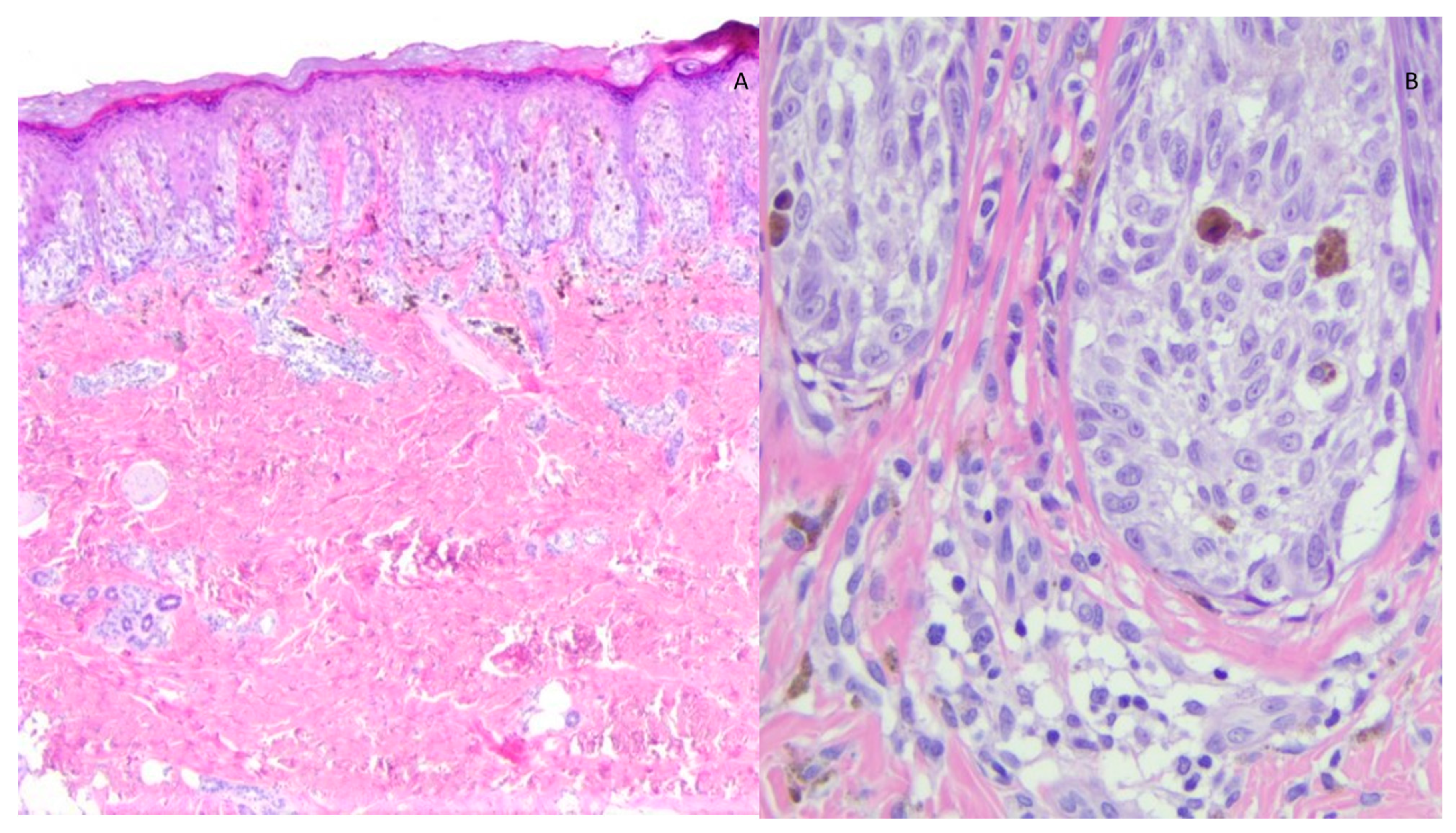
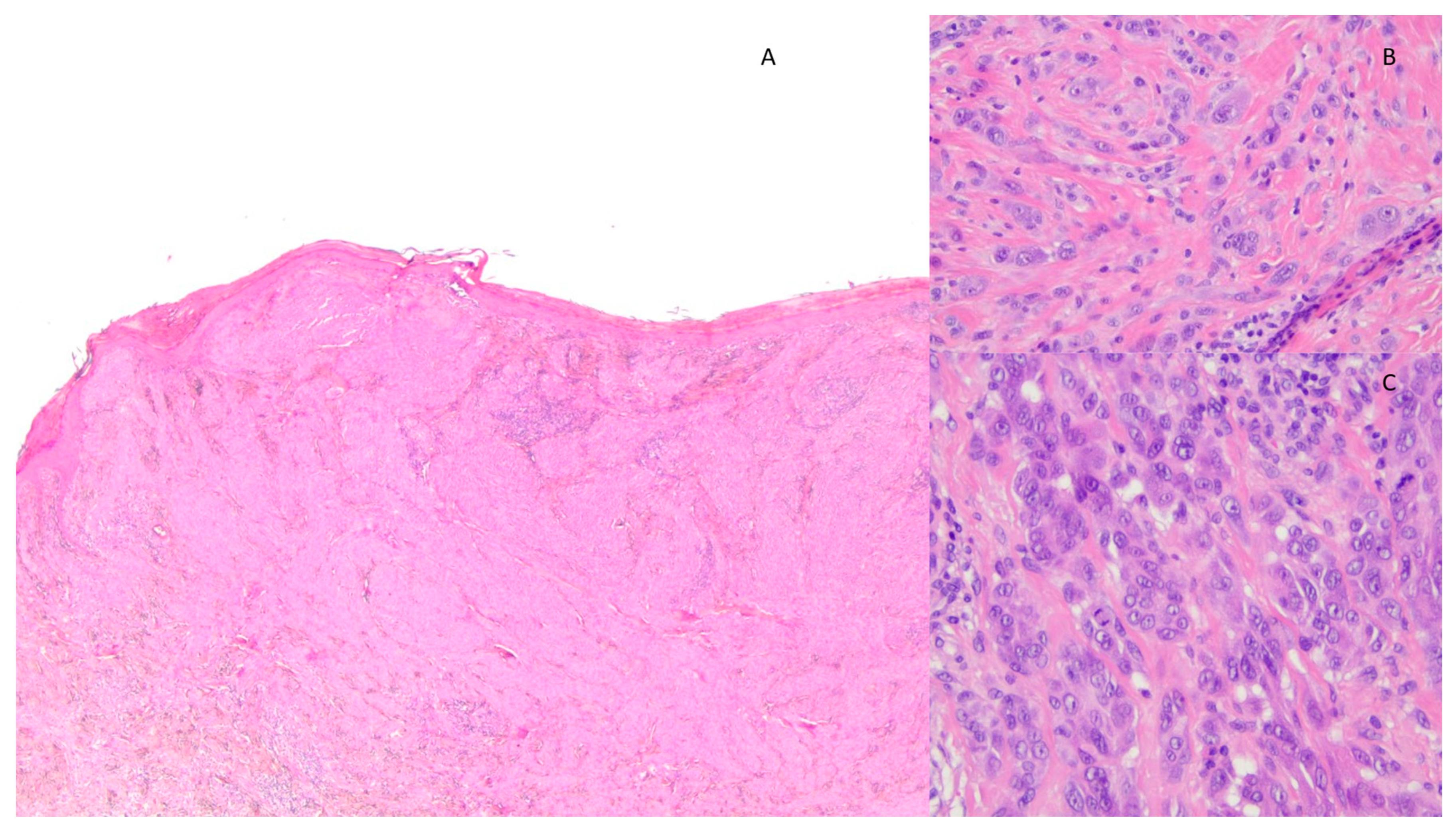
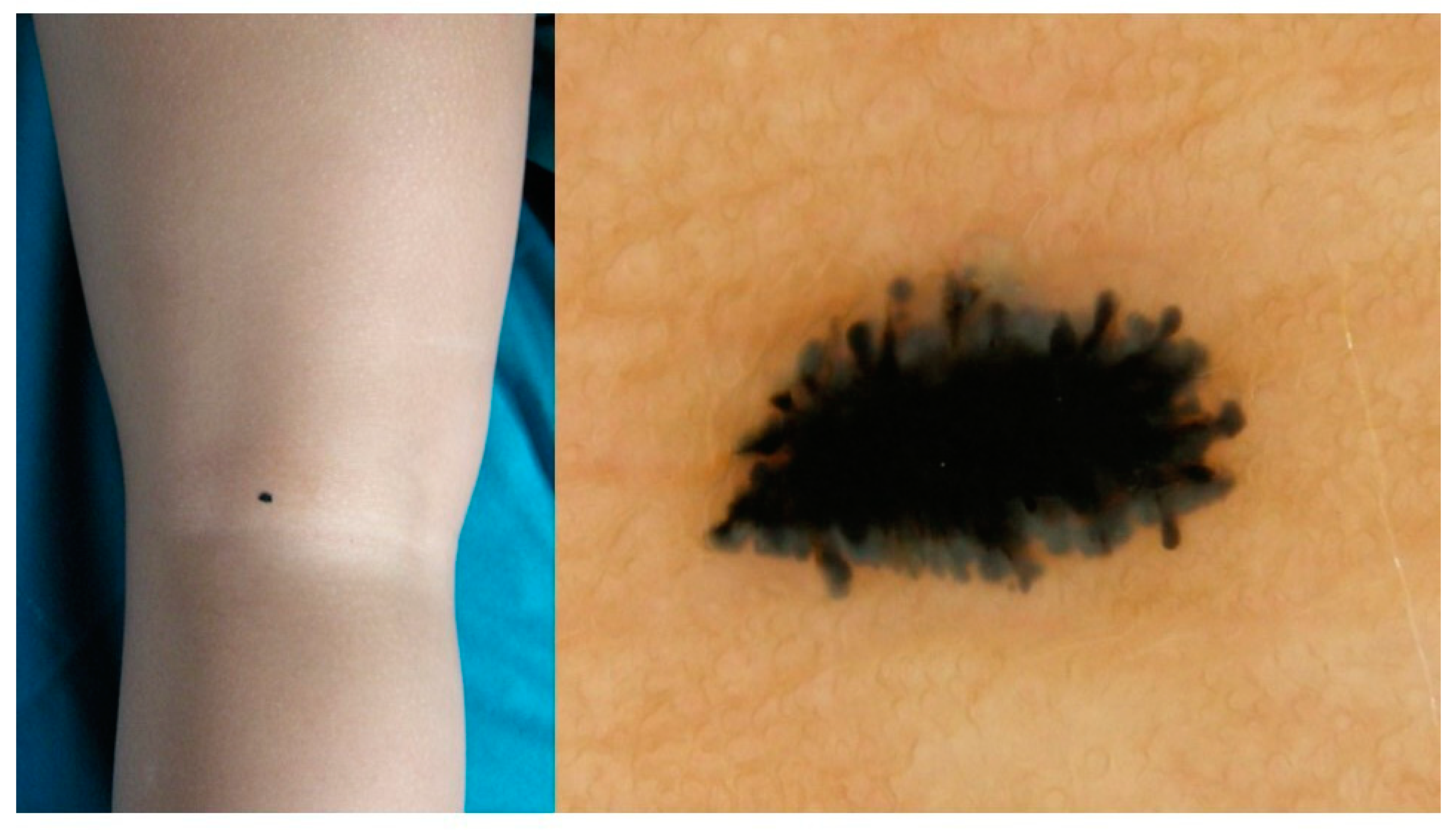
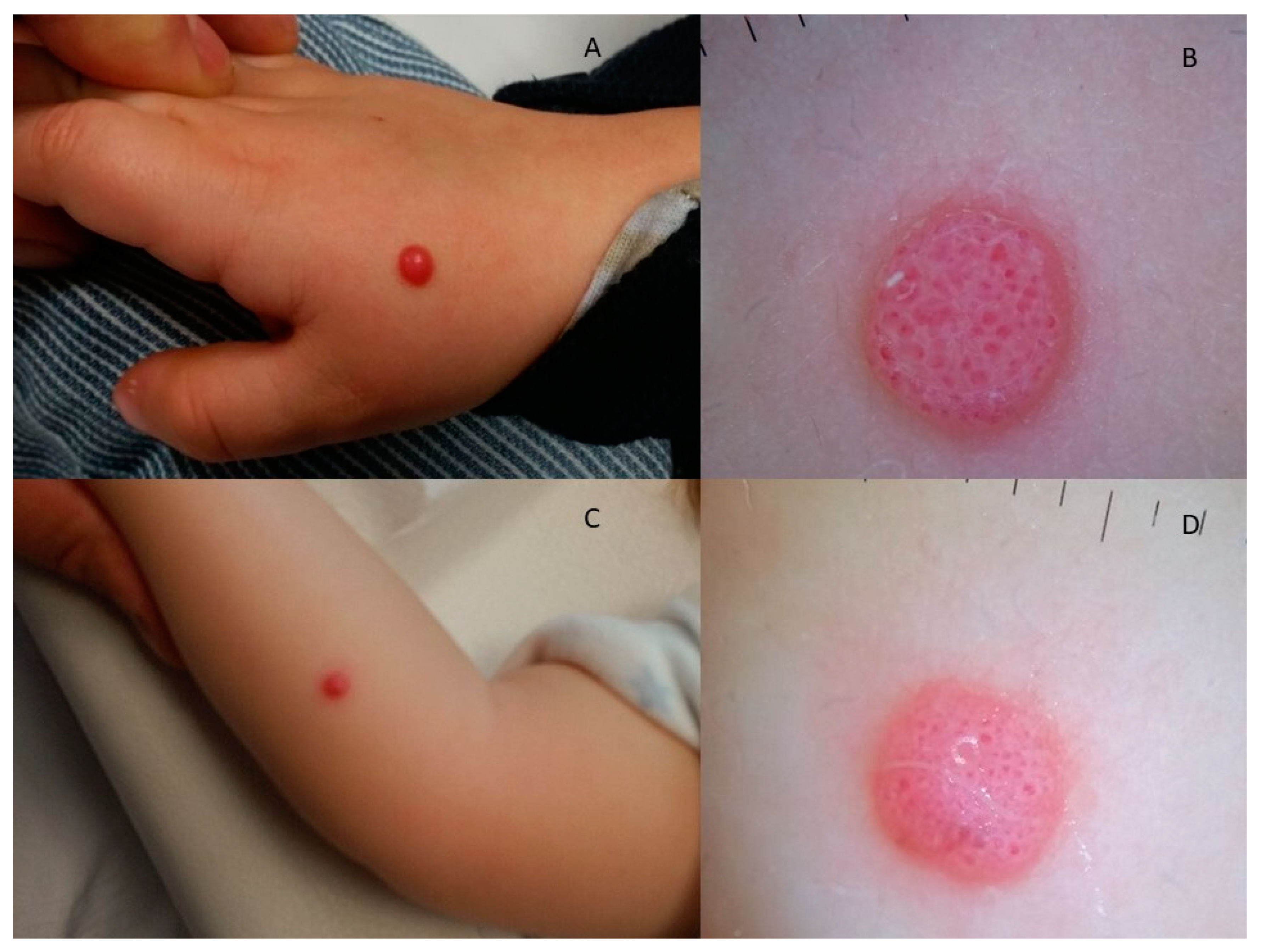
Paediatric Spitzoid Neoplasms: 10-Year Retrospective Study Characterizing Histological, Clinical, Dermoscopic Presentation and FISH Test Results
Abstract
1. Introduction
2. Materials and Methods
Statistical Analysis
3. Results
4. Discussion
5. Conclusions
Author Contributions
Funding
Institutional Review Board Statement
Informed Consent Statement
Data Availability Statement
Conflicts of Interest
References
- Dika, E.; Ravaioli, G.M.; Fanti, P.A.; Neri, I.; Patrizi, A. Spitz Nevi and Other Spitzoid Neoplasms in Children: Overview of Incidence Data and Diagnostic Criteria. Pediatr. Dermatol. 2017, 34, 25–32. [Google Scholar] [CrossRef] [PubMed]
- Cheng, T.W.; Ahern, M.C.; Giubellino, A. The Spectrum of Spitz Melanocytic Lesions: From Morphologic Diagnosis to Molecular Classification. Front. Oncol. 2022, 12, 889223. [Google Scholar] [CrossRef] [PubMed]
- Schaffer, J.V. Update on melanocytic nevi in children. Clin. Dermatol. 2015, 33, 368–386. [Google Scholar] [CrossRef] [PubMed]
- Ludgate, M.W.; Fullen, D.R.; Lee, J.; Lowe, L.; Bradford, C.; Geiger, J.; Schwartz, J.; Johnson, T.M. The atypical Spitz tumor of uncertain biologic potential: A series of 67 patients from a single institution. Cancer 2009, 115, 631–641. [Google Scholar] [CrossRef] [PubMed]
- Abboud, J.; Stein, M.; Ramien, M.; Malic, C. The diagnosis and management of the Spitz nevus in the pediatric population: A systematic review and meta-analysis protocol. Syst. Rev. 2017, 6, 81. [Google Scholar] [CrossRef] [PubMed]
- De Giorgi, V.; Venturi, F.; Silvestri, F.; Trane, L.; Savarese, I.; Scarfì, F.; Cencetti, F.; Pecenco, S.; Tramontana, M.; Maio, V.; et al. Atypical Spitz tumours: An epidemiological, clinical and dermoscopic multicentre study with 16 years of follow-up. Clin. Exp. Dermatol. 2022, 47, 1464–1471. [Google Scholar] [CrossRef]
- Bartenstein, D.; Fisher, J.; Stamoulis, C.; Weldon, C.; Huang, J.; Gellis, S.; Liang, M.; Schmidt, B.; Hawryluk, E. Clinical features and outcomes of spitzoid proliferations in children and adolescents. Br. J. Dermatol. 2019, 181, 366–372. [Google Scholar] [CrossRef]
- Davies, O.M.; Majerowski, J.; Segura, A.; Kelley, S.W.; Sokumbi, O.; Humphrey, S.R. A sixteen-year single-center retrospective chart review of Spitz nevi and spitzoid neoplasms in pediatric patients. Pediatr. Dermatol. 2020, 37, 1073–1082. [Google Scholar] [CrossRef]
- Afanasiev, O.K.; Tu, J.H.; Chu, D.H.; Swetter, S.M. Characteristics of melanoma in white and nonwhite children, adolescents, and young adults: Analysis of a pediatric melanoma institutional registry, 1995–2018. Pediatr. Dermatol. 2019, 36, 448–454. [Google Scholar] [CrossRef]
- Lallas, A.; Apalla, Z.; Ioannides, D.; Lazaridou, E.; Kyrgidis, A.; Broganelli, P.; Alfano, R.; Zalaudek, I.; Argenziano, G.; Bakos, R.; et al. Update on dermoscopy of Spitz/Reed naevi and management guidelines by the International Dermoscopy Society. Br. J. Dermatol. 2017, 177, 645–655. [Google Scholar] [CrossRef]
- Malignant Spitz tumour and Spitz Naevus. In WHO Classification of Skin Tumors, 4th ed.; Elder, D.E., Massi, D., Scolyer, R.A., Willemze, R., Eds.; World Health Organization: Geneva, Switzerland, 2018. [Google Scholar]
- Lallas, A.; Kyrgidis, A.; Ferrara, G.; Kittler, H.; Apalla, Z.; Castagnetti, F.; Longo, C.; Moscarella, E.; Piana, S.; Zalaudek, I.; et al. Atypical Spitz tumours and sentinel lymph node biopsy: A systematic review. Lancet Oncol. 2014, 15, e178–e183. [Google Scholar] [CrossRef] [PubMed]
- Massi, D.; Tomasini, C.; Senetta, R.; Paglierani, M.; Salvianti, F.; Errico, M.E.; Donofrio, V.; Collini, P.; Tragni, G.; Sementa, A.R.; et al. Atypical Spitz tumors in patients younger than 18 years. J. Am. Acad. Dermatol. 2014, 72, 37–46. [Google Scholar] [CrossRef] [PubMed]
- Requena, C.; Requena, L.; Kutzner, H.; Sánchez Yus, E. Spitz nevus: A clinicopathological study of 349 cases. Am. J. Dermatopathol. 2009, 31, 107–116. [Google Scholar] [CrossRef]
- Hill, S.J.; Delman, K.A. Pediatric melanomas and the atypical spitzoid melanocytic neoplasms. Am. J. Surg. 2012, 203, 761–767. [Google Scholar] [CrossRef] [PubMed]
- Herreid, P.A.; Shapiro, P.E. Age distribution of Spitz nevus vs malignant melanoma. Arch. Dermatol. 1996, 132, 352–353. [Google Scholar] [CrossRef] [PubMed]
- Sainz-Gaspar, L.; Sánchez-Bernal, J.; Noguera-Morel, L.; Hernández-Martín, A.; Colmenero, I.; Torrelo, A. Spitz Nevus and Other Spitzoid Tumors in Children—Part 1: Clinical, Histopathologic, and Immunohistochemical Features. Actas Dermo-Sifiliogr. (Engl. Ed.) 2020, 111, 7–19, English, Spanish. [Google Scholar] [CrossRef]
- Argenziano, G.; Agozzino, M.; Bonifazi, E.; Broganelli, P.; Brunetti, B.; Ferrara, G.; Fulgione, E.; Garrone, A.; Zalaudek, I. Natural evolution of Spitz nevi. Dermatology 2011, 222, 256–260. [Google Scholar] [CrossRef]
- Cesinaro, A.M.; Foroni, M.; Sighinolfi, P.; Migaldi, M.; Trentini, G.P. Spitz nevus is relatively frequent in adults: A clinico-pathologic study of 247 cases related to patient’s age. Am. J. Dermatopathol. 2005, 2, 469–475. [Google Scholar] [CrossRef]
- Barnhill, R.L.; Barnhill, M.A.; Berwick, M.; Mihm, M.C., Jr. The histologic spectrum of pigmented spindle cell nevus: A review of 120 cases with emphasis on atypical variants. Hum. Pathol. 1991, 22, 52–58. [Google Scholar] [CrossRef]
- Luo, S.; Sepehr, A.; Tsao, H. Spitz nevi and other Spitzoid lesions part I. Background and diagnoses. J. Am. Acad. Dermatol. 2011, 65, 1073–1084. [Google Scholar] [CrossRef]
- Paradela, S.; Fonseca, E.; Pita-Fernández, S.; Prieto, V.G. Spitzoid and non-spitzoid melanoma in children: A prognostic comparative study. J. Eur. Acad. Dermatol. Venereol. 2013, 27, 1214–1221. [Google Scholar] [CrossRef] [PubMed]
- Menezes, F.D.; Mooi, W.J. Spitz Tumors of the Skin. Surg. Pathol. Clin. 2017, 10, 281–298. [Google Scholar] [CrossRef]
- Kamino, H.; Misheloff, E.; Ackerman, A.B.; Flotte, T.J.; Greco, M.A. Eosinophilic globules in Spitz’s nevi: New findings and a diagnostic sign. Am. J. Dermatopathol. 1979, 1, 323–324. [Google Scholar] [CrossRef] [PubMed]
- Spatz, A.; Calonje, E.; Handfield-Jones, S.; Barnhill, R.L. Spitz tumors in children: A grading system for risk stratification. Arch. Dermatol. 1999, 135, 282–285. [Google Scholar] [CrossRef] [PubMed]
- Dika, E.; Neri, I.; Fanti, P.; Barisani, A.; Ravaioli, G.M.; Patrizi, A. Spitz nevi: Diverse clinical, dermatoscopic and histopathological features in childhood. J. Dtsch. Dermatol. Ges. 2017, 15, 70–75. [Google Scholar] [CrossRef]
- Zalaudek, I.; Grinschgl, S.; Argenziano, G.; Marghoob, A.A.; Blum, A.; Richtig, E.; Wolf, I.H.; Fink-Puches, R.; Kerl, H.; Soyer, H.P.; et al. Age-related prevalence of dermoscopy patterns in acquired melanocytic naevi. Br. J. Dermatol. 2006, 154, 299–304. [Google Scholar] [CrossRef]
- Moscarella, E.; Lallas, A.; Kyrgidis, A.; Ferrara, G.; Longo, C.; Scalvenzi, M.; Staibano, S.; Carrera, C.; Díaz, M.; Broganelli, P.; et al. Clinical and dermoscopic features of atypical Spitz tumors: A multicenter, retrospective, case-control study. J. Am. Acad. Dermatol. 2015, 73, 777–784. [Google Scholar] [CrossRef]
- Lallas, A.; Apalla, Z.; Papageorgiou, C.; Evangelou, G.; Ioannides, D.; Argenziano, G. Management of Flat Pigmented Spitz and Reed Nevi in Children. JAMA Dermatol. 2018, 154, 1353–1354. [Google Scholar] [CrossRef]
- Lallas, A.; Moscarella, E.; Longo, C.; Kyrgidis, A.; de Mestier, Y.; Vale, G.; Guida, S.; Pellacani, G.; Argenziano, G. Likelihood of finding melanoma when removing a Spitzoid-looking lesion in patients aged 12 years or older. J. Am. Acad. Dermatol. 2015, 72, 47–53. [Google Scholar] [CrossRef]
- Lange, J.R.; Palis, B.E.; Chang, D.C.; Soong, S.J.; Balch, C.M. Melanoma in children and teenagers: An analysis of patients from the National Cancer Data Base. J. Clin. Oncol. 2007, 25, 1363–1368. [Google Scholar] [CrossRef]
- Viglizzo, G.; Herzum, A.; Gariazzo, L.; Garibeh, E.; Occella, C. Pediatric Spitzoid Lesions of the Ear: A Single Center Experience and Review of Literature. Dermatol. Rep. 2023. Available online: https://www.pagepress.org/journals/index.php/dr/article/view/9642 (accessed on 12 July 2023).
- Cazzato, G.; Massaro, A.; Colagrande, A.; Lettini, T.; Cicco, S.; Parente, P.; Nacchiero, E.; Lospalluti, L.; Cascardi, E.; Giudice, G.; et al. Dermatopathology of Malignant Melanoma in the Era of Artificial Intelligence: A Single Institutional Experience. Diagnostics 2022, 12, 1972. [Google Scholar] [CrossRef]
- Cazzato, G.; Colagrande, A.; Cimmino, A.; Arezzo, F.; Loizzi, V.; Caporusso, C.; Marangio, M.; Foti, C.; Romita, P.; Lospalluti, L.; et al. Artificial Intelligence in Dermatopathology: New Insights and Perspectives. Dermatopathology 2021, 8, 418–425. [Google Scholar] [CrossRef] [PubMed]
- Requena, C.; Rubio, L.; Traves, V.; Sanmartín, O.; Nagore, E.; Llombart, B.; Serra, C.; Fernández-Serra, A.; Botella, R.; Guillén, C. Spitz Nevus with Features of Clark Nevus, So-Called SPARK Nevus: Case Series Presentation with Emphasis on Cytological and Histological Features. Dermatopathology 2021, 8, 525–530. [Google Scholar] [CrossRef]
- Requena, C.; Rubio, L.; Traves, V.; Sanmartín, O.; Nagore, E.; Llombart, B.; Serra, C.; Fernández-Serra, A.; Botella, R.; Guillén, C. Fluorescence in situ hybridization for the differential diagnosis between Spitz naevus and spitzoid melanoma. Histopathology 2012, 61, 899–909. [Google Scholar] [CrossRef] [PubMed]
- Gerami, P.; Jewell, S.S.; Morrison, L.E.; Blondin, B.B.; Schulz, J.B.; Ruffalo, T.B.; Matushek, P.I.; Legator, M.B.; Jacobson, K.M.; Dalton, S.R.; et al. Fluorescence in situ hybridization (FISH) as an ancillary diagnostic tool in the diagnosis of melanoma. Am. J. Surg. Pathol. 2009, 33, 1146–1156, Erratum in Am. J. Surg. Pathol. 2010, 34, 688. [Google Scholar] [CrossRef]
- Morey, A.L.; Murali, R.; McCarthy, S.W.; Mann, G.J.; Scolyer, R.A. Diagnosis of cutaneous melanocytic tumours by four-colour fluorescence in situ hybridisation. Pathology 2009, 41, 383–387. [Google Scholar] [CrossRef]
- Newman, M.D.; Lertsburapa, T.; Mirzabeigi, M.; Mafee, M.; Guitart, J.; Gerami, P. Fluorescence in situ hybridization as a tool for microstaging in malignant melanoma. Mod. Pathol. 2009, 22, 989–995. [Google Scholar] [CrossRef]
- Massi, D.; Cesinaro, A.M.; Tomasini, C.; Paglierani, M.; Bettelli, S.; Maso, L.D.; Simi, L.; Salvianti, F.; Pinzani, P.; Orlando, C.; et al. Atypical Spitzoid melanocytic tumors: A morphological, mutational, and FISH analysis. J. Am. Acad. Dermatol. 2011, 64, 919–935. [Google Scholar] [CrossRef]
- Gerami, P.; Li, G.; Pouryazdanparast, P.; Blondin, B.; Beilfuss, B.; Slenk, C.; Du, J.; Guitart, J.; Jewell, S.; Pestova, K. A highly specific and discriminatory FISH assay for distinguishing between benign and malignant melanocytic neoplasms. Am. J. Surg. Pathol. 2012, 36, 808–817. [Google Scholar] [CrossRef]
- Gerami, P.; Scolyer, R.A.; Xu, X.; Elder, D.E.; Abraham, R.M.; Fullen, D.; Prieto, V.G.; Leboit, P.E.; Barnhill, R.L.; Cooper, C.; et al. Risk assessment for atypical spitzoid melanocytic neoplasms using FISH to identify chromosomal copy number aberrations. Am. J. Surg. Pathol. 2013, 37, 676–684. [Google Scholar] [CrossRef]


| Total of Lesions (%) | Benign (%) | Atypical (%) | Melanoma (%) | p-Value | OR (95% CI) | |
|---|---|---|---|---|---|---|
| NUMBER OF LESIONS | 255 (100) | 210 (82) | 43 (17) | 2 (1) | ||
| SEX | ||||||
| female | 123 (48) | 97 | 25 | 1 | ||
| male | 132 (52) | 113 | 18 | 1 | ||
| mean (median) age at diagnosis in years | 7.9 (7) | 8 (7) | 7.1 (7) | 11.5 (11.5) | >0.05 | |
| age ≤ 11 years | 198 | 160 (76) | 37 (86) | 1 (50) | ||
| age ≥ 12 years | 57 | 50 (24) | 6 (14) | 1 (50) | ||
| mean (median) lesion dimension in mm) | 5.3 (5) | 5 (5) | 6.3 (6) | 7.5 (7.5) | ||
| Diameter 1–5 | 158 (62) | 142 (68) | 16 (37) | 0 (0) | <0.0001 | 3.7849 (1.9264–7.4364) |
| Diameter 6–12 | 97 (38) | 68 (32) | 27 (63) | 2 (100) | ||
| LOCALIZATION | 255 (100) | 210 (82) | 43 (17) | 2 (1) | ||
| Hands and feet | 19 (7) | 17 (8) | 2 (5) | 0 (0) | >0.05 | |
| Head and neck | 53 (21) | 35 (17) | 17 (40) | 1 (50) | 0.0005 | 3.3333 (1.6583–6.7001) |
| Trunk | 30 (12) | 27 (13) | 2 (5) | 1 (50) | >0.05 | |
| Upper Limb | 53 (21) | 42 (20) | 11 (25) | 0 (0) | >0.05 | |
| Lower Limb | 97 (38) | 86 (41) | 11 (25) | 0 (0) | 0.0385 | 2.1437 (1.0296–4.4635) |
| Perineum | 3 (1) | 3 (1) | 0 (0) | 0 (0) | >0.05 | |
| SHAPE | 255 (100) | 210 (82) | 43 (17) | 2 (1) | <0.0001 | 17.1921 (2.3154–127.6519) |
| Raised | 195 (76) | 151 (72) | 42 (98) | 2 (100) | ||
| Flat | 60 (24) | 59 (28) | 1 (2) | 0 (0) | ||
| COLOR | 255 (100) | 210 (82) | 43 (17) | 2 (1) | <0.0001 | 16.6250 (4.9955–55.3284) |
| Pigmented | 117 (46) | 114 (54) | 2 (5) | 1 (50) | ||
| Hypo-/non-pigmented (=pink) | 138 (54) | 96 (46) | 41 (95) | 1 (50) | ||
| FISH | 85 (33) | 42 (20) | 43 (100) | 0 (0) | 0.0038 | 4.0421 (1.5935–10.2534) |
| Positive | 34 | 10 (29) | 24 (71) | 0 (0) | ||
| Negative | 51 | 32 (63) | 19 (37) | 0 (0) |
| Total of Benign SN Lesions | Hypo-/Non-Pigmented (=Pink Lesion, Pigmentation Involving <50% of the Lesion) | Pigmented (Pigmentation Involving ≥50% of the Lesion) | p-Value | OR (95% CI) | |
|---|---|---|---|---|---|
| NUMBER OF LESIONS | 210 | 96 | 114 | ||
| Head and neck | 35 (17) | 26 | 9 | 0.0003 | 4.33 (1.9159–9.8008) |
| Trunk | 27 (13) | 14 | 13 | >0.05 | |
| Limbs | 148 (70) | 56 | 92 | 0.0005 | 2.9870 (1.6112–5.5375) |
| Total of Lesions (%) | Benign (%) | Atypical (%) | Melanoma (%) | p-Value | OR (95% CI) | |
|---|---|---|---|---|---|---|
| GLOBAL DERMATOSCOPIC PATTERNS | 100 (100) | 62 (62) | 36 (36) | 2 (2) | ||
| VASCULAR Dotted or clods vessels on pink background | 20 (20) | 14 (23) | 6 (17) | 0 (0) | p > 0.05 | |
| STARBURST (REED) Symmetric centrifugal streaks/large globules | 16 (16) | 16 (26) | 0 (0) | 0 (0) | p = 0.004 | 27.3226 (1.5872–470.3542) |
| GLOBULAR Brown/black globules mainly | 8 (8) | 6 (10) | 2 (6) | 0 (0) | p > 0.05 | |
| MULTICOMPONENT Combination of ≥2 different predominant features | 37 (37) | 16 (26) | 20 (56) | 1 (50) | p = 0.0052 | 3.5515 (1.5091–8.3582) |
| RETICULAR Pigment network mainly | 4 (4) | 4 (6) | 0 (0) | 0 (0) | p > 0.05 | |
| ASPECIFIC Main pattern not referable to any category | 8 (8) | 2 (3) | 6 (17) | 0 (0) | p > 0.05 | |
| HOMOGENEUOS Homogeneous pigmentation mainly | 7(7) | 4 (6) | 2 (6) | 1 (50) | p > 0.05 |
Disclaimer/Publisher’s Note: The statements, opinions and data contained in all publications are solely those of the individual author(s) and contributor(s) and not of MDPI and/or the editor(s). MDPI and/or the editor(s) disclaim responsibility for any injury to people or property resulting from any ideas, methods, instructions or products referred to in the content. |
© 2023 by the authors. Licensee MDPI, Basel, Switzerland. This article is an open access article distributed under the terms and conditions of the Creative Commons Attribution (CC BY) license (https://creativecommons.org/licenses/by/4.0/).
Share and Cite
Herzum, A.; Occella, C.; Vellone, V.G.; Gariazzo, L.; Pastorino, C.; Ferro, J.; Sementa, A.; Mazzocco, K.; Vercellino, N.; Viglizzo, G. Paediatric Spitzoid Neoplasms: 10-Year Retrospective Study Characterizing Histological, Clinical, Dermoscopic Presentation and FISH Test Results. Diagnostics 2023, 13, 2380. https://doi.org/10.3390/diagnostics13142380
Herzum A, Occella C, Vellone VG, Gariazzo L, Pastorino C, Ferro J, Sementa A, Mazzocco K, Vercellino N, Viglizzo G. Paediatric Spitzoid Neoplasms: 10-Year Retrospective Study Characterizing Histological, Clinical, Dermoscopic Presentation and FISH Test Results. Diagnostics. 2023; 13(14):2380. https://doi.org/10.3390/diagnostics13142380
Chicago/Turabian StyleHerzum, Astrid, Corrado Occella, Valerio Gaetano Vellone, Lodovica Gariazzo, Carlotta Pastorino, Jacopo Ferro, Angela Sementa, Katia Mazzocco, Nadia Vercellino, and Gianmaria Viglizzo. 2023. "Paediatric Spitzoid Neoplasms: 10-Year Retrospective Study Characterizing Histological, Clinical, Dermoscopic Presentation and FISH Test Results" Diagnostics 13, no. 14: 2380. https://doi.org/10.3390/diagnostics13142380
APA StyleHerzum, A., Occella, C., Vellone, V. G., Gariazzo, L., Pastorino, C., Ferro, J., Sementa, A., Mazzocco, K., Vercellino, N., & Viglizzo, G. (2023). Paediatric Spitzoid Neoplasms: 10-Year Retrospective Study Characterizing Histological, Clinical, Dermoscopic Presentation and FISH Test Results. Diagnostics, 13(14), 2380. https://doi.org/10.3390/diagnostics13142380


