
Retrospective Cohort Study of Shear-Wave Elastography and Computed Tomography Enterography in Crohn’s Disease

Abstract
:1. Introduction
2. Materials and Methods
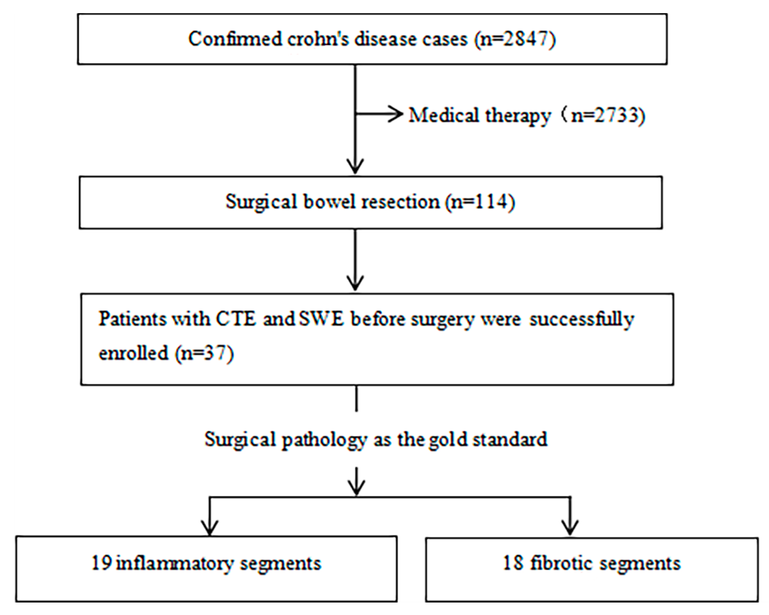
2.1. Patients
2.2. Traditional Intestinal Ultrasound and 2D-SWE Imaging
2.3. CT Enterographic Techniques
2.4. Histological Evaluation
2.5. Statistical Analysis
3. Results
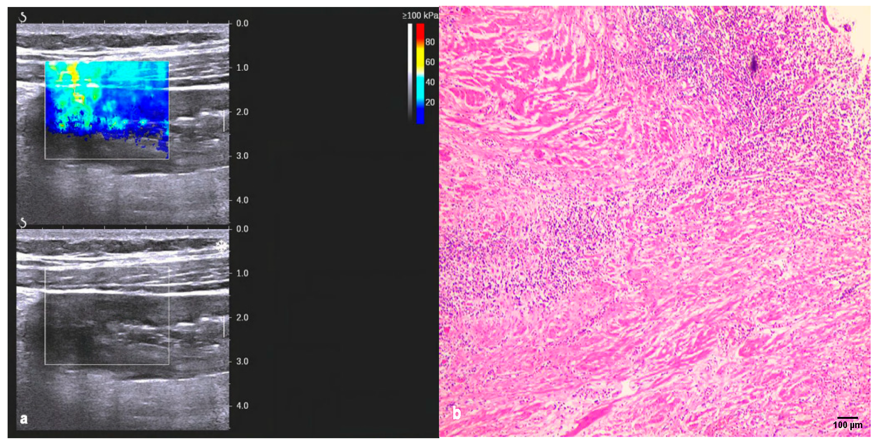
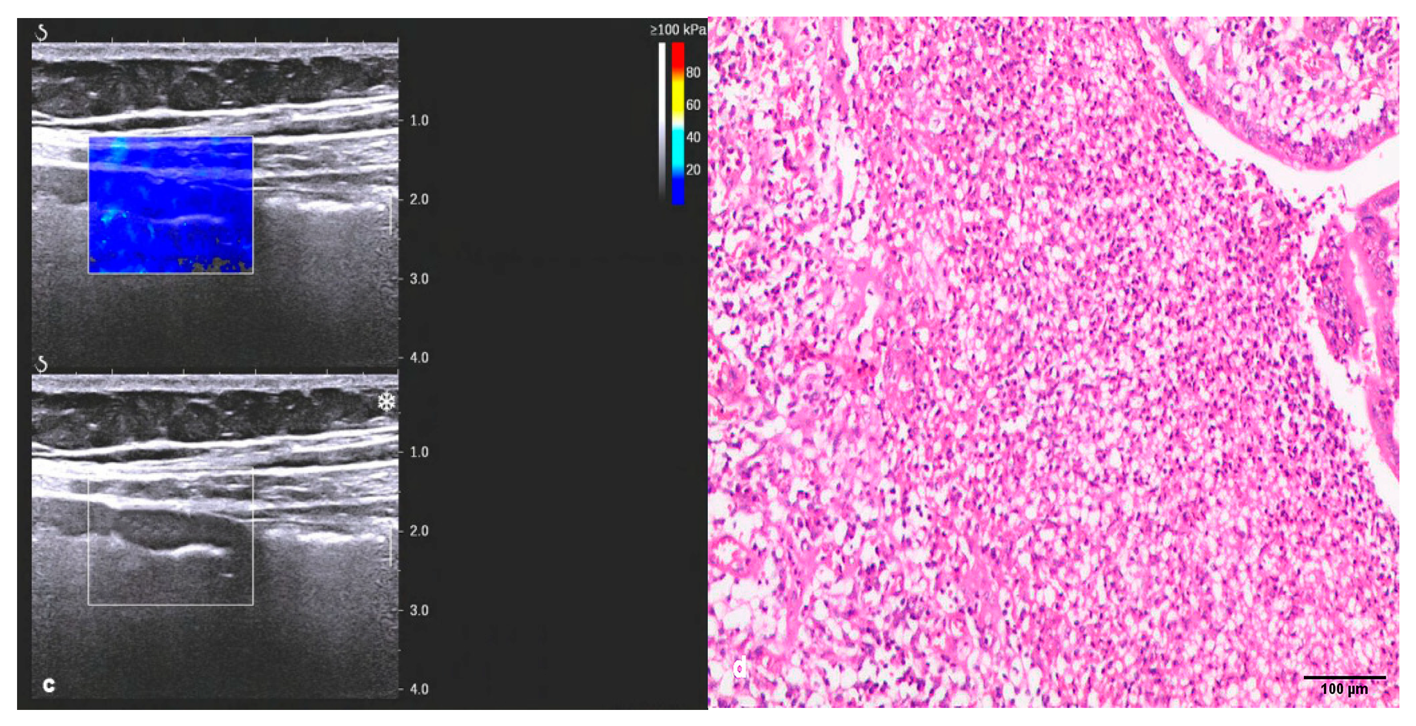
3.1. Conventional Ultrasound and Two-Dimensional Shear-Wave Elastography (2D-SWE)
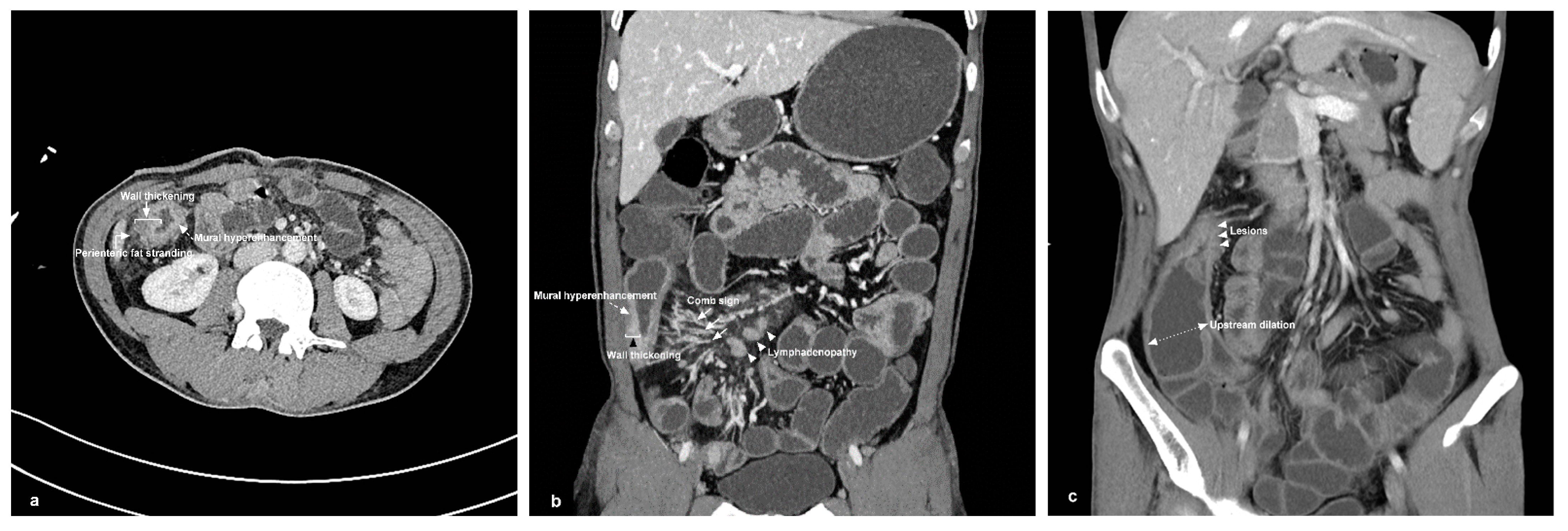
3.2. CTE Findings and Diagnostic Yield of CD Phenotypes
3.3. CTE Combined with 2D-SWE to Distinguish between Inflammatory and Fibrotic Lesions
4. Discussion
5. Conclusions
Author Contributions
Funding
Institutional Review Board Statement
Informed Consent Statement
Data Availability Statement
Acknowledgments
Conflicts of Interest
References
- Ng, S.C.; Shi, H.Y.; Hamidi, N.; Underwood, F.E.; Tang, W.; Benchimol, E.I.; Panaccione, R.; Ghosh, S.; Wu, J.C.Y.; Chan, F.K.L.; et al. Worldwide incidence and prevalence of inflammatory bowel disease in the 21st century: A systematic review of population-based studies. Lancet 2017, 390, 2769–2778. [Google Scholar] [CrossRef] [PubMed]
- Gajendran, M.; Loganathan, P.; Catinella, A.P.; Hashash, J.G. A comprehensive review and update on Crohn’s disease. Dis. Mon. 2018, 64, 20–57. [Google Scholar] [CrossRef] [PubMed]
- Torres, J.; Bonovas, S.; Doherty, G.; Kucharzik, T.; Gisbert, J.P.; Raine, T.; Adamina, M.; Armuzzi, A.; Bachmann, O.; Bager, P.; et al. ECCO Guidelines on Therapeutics in Crohn’s Disease: Medical Treatment. J. Crohns Colitis 2020, 14, 4–22. [Google Scholar] [CrossRef] [Green Version]
- Rieder, F.; Bettenworth, D.; Ma, C.; Parker, C.E.; Williamson, L.A.; Nelson, S.A.; van Assche, G.; Di Sabatino, A.; Bouhnik, Y.; Stidham, R.W.; et al. An expert consensus to standardise definitions, diagnosis and treatment targets for anti-fibrotic stricture therapies in Crohn’s disease. Aliment. Pharmacol. Ther. 2018, 48, 347–357. [Google Scholar] [CrossRef] [Green Version]
- Rieder, F.; Latella, G.; Magro, F.; Yuksel, E.S.; Higgins, P.D.; Di Sabatino, A.; de Bruyn, J.R.; Rimola, J.; Brito, J.; Bettenworth, D.; et al. European Crohn’s and Colitis Organisation Topical Review on Prediction, Diagnosis and Management of Fibrostenosing Crohn’s Disease. J. Crohns Colitis 2016, 10, 873–885. [Google Scholar] [CrossRef] [Green Version]
- Bruining, D.H.; Zimmermann, E.M.; Loftus, E.V., Jr.; Sandborn, W.J.; Sauer, C.G.; Strong, S.A. Consensus Recommendations for Evaluation, Interpretation, and Utilization of Computed Tomography and Magnetic Resonance Enterography in Patients with Small Bowel Crohn’s Disease. Gastroenterology 2018, 154, 1172–1194. [Google Scholar] [CrossRef]
- Panés, J.; Bouzas, R.; Chaparro, M.; García-Sánchez, V.; Gisbert, J.P.; Martínez de Guereñu, B.; Mendoza, J.L.; Paredes, J.M.; Quiroga, S.; Ripollés, T.; et al. Systematic review: The use of ultrasonography, computed tomography and magnetic resonance imaging for the diagnosis, assessment of activity and abdominal complications of Crohn’s disease. Aliment. Pharmacol. Ther. 2011, 34, 125–145. [Google Scholar] [CrossRef]
- Bettenworth, D.; Bokemeyer, A.; Baker, M.; Mao, R.; Parker, C.E.; Nguyen, T.; Ma, C.; Panés, J.; Rimola, J.; Fletcher, J.G.; et al. Stenosis Therapy and Anti-Fibrotic Research (STAR) Consortium. Assessment of Crohn’sdisease-associated small bowel strictures and fibrosis on cross-sectional imaging: A systematic review. Gut 2019, 68, 1115–1126. [Google Scholar] [CrossRef] [Green Version]
- Gordon, I.O.; Bettenworth, D.; Bokemeyer, A.; Srivastava, A.; Rosty, C.; de Hertogh, G.; Robert, M.E.; Valasek, M.A.; Mao, R.; Kurada, S.; et al. Stenosis Therapy and Anti-Fibrotic Research (STAR) Consortium. Histopathology Scoring Systems of Stenosis Associated With Small Bowel Crohn’s Disease: A Systematic Review. Gastroenterology 2020, 158, 137–150.e1. [Google Scholar] [CrossRef]
- Gomollón, F.; Dignass, A.; Annese, V.; Tilg, H.; Van Assche, G.; Lindsay, J.O.; Peyrin-Biroulet, L.; Cullen, G.J.; Daperno, M.; Kucharzik, T.; et al. ECCO. 3rd European Evidence-based Consensus on the Diagnosis and Management of Crohn’s Disease 2016: Part 1: Diagnosis and Medical Management. J. Crohns Colitis 2017, 11, 3–25. [Google Scholar] [CrossRef] [Green Version]
- Maconi, G.; Nylund, K.; Ripolles, T.; Calabrese, E.; Dirks, K.; Dietrich, C.F.; Hollerweger, A.; Sporea, I.; Saftoiu, A.; Maaser, C.; et al. EFSUMB Recommendations and Clinical Guidelines for Intestinal Ultrasound (GIUS) in Inflammatory Bowel Diseases. Ultraschall Der Med. 2018, 39, 304–317. [Google Scholar] [CrossRef] [PubMed] [Green Version]
- Bamber, J.; Cosgrove, D.; Dietrich, C.F.; Fromageau, J.; Bojunga, J.; Calliada, F.; Cantisani, V.; Correas, J.M.; D’Onofrio, M.; Drakonaki, E.E.; et al. EFSUMB guidelines and recommendations on the clinical use of ultrasound elastography. Part 1: Basic principles and technology. Ultraschall Der Med. 2013, 34, 169–184. [Google Scholar] [CrossRef] [PubMed] [Green Version]
- Cosgrove, D.O.; Berg, W.A.; Doré, C.J.; Skyba, D.M.; Henry, J.P.; Gay, J.; Cohen-Bacrie, C.; BE1 Study Group. Shear wave elastography for breast masses is highly reproducible. Eur. Radiol. 2012, 22, 1023–1032. [Google Scholar] [CrossRef] [PubMed] [Green Version]
- Ferraioli, G.; Wong, V.W.; Castera, L.; Berzigotti, A.; Sporea, I.; Dietrich, C.F.; Choi, B.I.; Wilson, S.R.; Kudo, M.; Barr, R.G. Liver Ultrasound Elastography: An Update to the World Federation for Ultrasound in Medicine and Biology Guidelines and Recommendations. Ultrasound Med. Biol. 2018, 44, 2419–2440. [Google Scholar] [CrossRef] [Green Version]
- Dillman, J.R.; Stidham, R.W.; Higgins, P.D.R.; Moons, D.S.; Johnson, L.A.; Rubin, J.M. US elastography-derived shear wave velocity helps distinguish acutely inflamed from fibrotic bowel in a Crohn disease animal model. Radiology 2013, 267, 757–766. [Google Scholar] [CrossRef] [Green Version]
- Chen, Y.-J.; Mao, R.; Li, X.-H.; Cao, Q.-H.; Chen, Z.-H.; Liu, B.-X.; Chen, S.-L.; Chen, B.-L.; He, Y.; Zeng, Z.-R.; et al. Real-Time Shear Wave Ultrasound Elastography Differentiates Fibrotic from Inflammatory Strictures in Patients with Crohn’s Disease. Inflamm. Bowel. Dis. 2018, 24, 2183–2190. [Google Scholar] [CrossRef]
- Thimm, M.A.; Cuffari, C.; Garcia, A.; Sidhu, S.; Hwang, M. Contrast-Enhanced Ultrasound and Shear Wave Elastography Evaluation of Crohn’s Disease Activity in Three Adolescent Patients. Pediatr. Gastroenterol. Hepatol. Nutr. 2019, 22, 282–290. [Google Scholar] [CrossRef]
- SLichtenstein, G.R.; Loftus, E.V.; Isaacs, K.L.; Regueiro, M.D.; Gerson, L.B.; Sands, B.E. ACG Clinical Guideline: Management of Crohn’s Disease in Adults. Am. J. Gastroenterol. 2018, 113, 481–517. [Google Scholar] [CrossRef]
- Choi, D.; Lee, S.J.; Cho, Y.A.; Lim, H.K.; Kim, S.H.; Lee, W.J.; Lim, J.H.; Park, H.; Lee, Y.R. Bowel wall thickening in patients with Crohn’s disease: CT patterns and correlation with inflammatory activity. Clin. Radiol. 2003, 58, 68–74. [Google Scholar] [CrossRef]
- Paulsen, S.R.; Huprich, J.E.; Fletcher, J.G.; Booya, F.; Young, B.M.; Fidler, J.L.; Johnson, C.D.; Barlow, J.M.; Earnest, F., 4th. CT enterography as a diagnostic tool in evaluating small bowel disorders: Review of clinical experience with over 700 cases. Radiographics 2006, 26, 641–657. [Google Scholar] [CrossRef] [Green Version]
- Expert Panel on Gastrointestinal Imaging; Kim, D.H.; Chang, K.J.; Fowler, K.J.; Cash, B.D.; Garcia, E.M.; Kambadakone, A.R.; Levy, A.D.; Liu, P.S.; Mace, S.E.; et al. ACR Appropriateness Criteria® Crohn Disease. J. Am. Coll. Radiol. 2020, 17, S81–S99. [Google Scholar] [CrossRef]
- Bruining, D.H.; Bhatnagar, G.; Rimola, J.; Taylor, S.; Zimmermann, E.M.; Fletcher, J.G. CT and MR enterography in Crohn’s disease: Current and future applications. Abdom. Imaging 2015, 40, 965–974. [Google Scholar] [CrossRef] [PubMed]
- Limberg, B. Diagnosis of chronic inflammatory bowel disease by ultrasonography. Z. Gastroenterol. 1999, 37, 495–508. [Google Scholar]
- Li, X.; Feng, S.; Huang, L.; Zhou, J.; Zhou, Z.; Huang, S.; Mao, R.; He, Y.; Liu, W.; Xue, H.; et al. Expert guideline on imaging examination and report specification of inflammatory bowel disease in China. Chin. J. Inflamm. Bowel. Dis. 2021, 5, 109–113. [Google Scholar] [CrossRef]
- Guglielmo, F.F.; Anupindi, S.A.; Fletcher, J.G.; Al-Hawary, M.M.; Dillman, J.R.; Grand, D.J.; Bruining, D.H.; Chatterji, M.; Darge, K.; Fidler, J.L.; et al. Small Bowel Crohn Disease at CT and MR Enterography: Imaging Atlas and Glossary of Terms. Radiographics 2020, 40, 354–375. [Google Scholar] [CrossRef]
- Chiorean, M.V.; Sandrasegaran, K.; Saxena, R.; Maglinte, D.D.; Nakeeb, A.; Johnson, C.S. Correlation of CT enteroclysis with surgical pathology in Crohn’s disease. Am. J. Gastroenterol. 2007, 102, 2541–2550. [Google Scholar] [CrossRef] [PubMed]
- Adler, J.; Punglia, D.R.; Dillman, J.R.; Polydorides, A.D.; Dave, M.; Al-Hawary, M.M.; Platt, J.F.; McKenna, B.J.; Zimmermann, E.M. Computed tomography enterography findings correlate with tissue inflammation, not fibrosis in resected small bowel Crohn’s disease. Inflamm. Bowel. Dis. 2012, 18, 849–856. [Google Scholar] [CrossRef] [Green Version]
- Macaluso, F.S.; Papi, C.; Orlando, A.; Festa, S.; Pugliese, D.; Bonovas, S.; Pansieri, C.; Piovani, D.; Fiorino, G.; Fantini, M.C.; et al. Italian Group for the Study of Inflammatory Bowel Disease (IG-IBD); Working panel; Review panel. Use of biologics for the management of Crohn’s disease: IG-IBD clinical guidelines based on the GRADE methodology. Dig. Liver Dis. 2023, 55, 442–453. [Google Scholar] [CrossRef]
- Adamina, M.; Bonovas, S.; Raine, T.; Spinelli, A.; Warusavitarne, J.; Armuzzi, A.; Bachmann, O.; Bager, P.; Biancone, L.; Bokemeyer, B.; et al. ECCO Guidelines on Therapeutics in Crohn’s Disease: Surgical Treatment. J. Crohn’s Colitis 2020, 14, 155–168. [Google Scholar] [CrossRef] [Green Version]
- Lu, C.; Gui, X.; Chen, W.; Fung, T.; Novak, K.; Wilson, S.R. Ultrasound Shear Wave Elastography and Contrast Enhancement: Effective Biomarkers in Crohn’s Disease Strictures. Inflamm. Bowel Dis. 2017, 23, 421–430. [Google Scholar] [CrossRef]
- Rieder, F.; Zimmermann, E.M.; Remzi, F.H.; Sandborn, W.J. Crohn’s disease complicated by strictures: A systematic review. Gut 2013, 62, 1072–1084. [Google Scholar] [CrossRef] [PubMed] [Green Version]
- Munger, J.S.; Harpel, J.G.; Gleizes, P.E.; Mazzieri, R.; Nunes, I.; Rifkin, D.B. Latent transforming growth factor-beta: Structural features and mechanisms of activation. Kidney Int. 1997, 51, 1376–1382. [Google Scholar] [CrossRef] [PubMed] [Green Version]
- Vaday, G.G.; Hershkoviz, R.; Rahat, M.A.; Lahat, N.; Cahalon, L.; Lider, O. Fibronectin-bound TNF-alpha stimulates monocyte matrix metalloproteinase-9 expression and regulates chemotaxis. J. Leukoc. Biol. 2000, 68, 737–747. [Google Scholar] [CrossRef] [PubMed]
- Wells, R.G. The role of matrix stiffness in regulating cell behavior. Hepatology 2008, 47, 1394–1400. [Google Scholar] [CrossRef]
- Ura, H.; Obara, T.; Yokota, K.; Shibata, Y.; Okamura, K.; Namiki, M. Effects Of Transforming Growth-Factor-Beta Relasead From Gastric-Carcinoma Cells On The Contraction Of Collagen-Matrix Gels Containing Fibroblasts. Cancer Res. 1991, 51, 3550–3554. [Google Scholar] [CrossRef] [PubMed]
- Dillman, J.R.; Stidham, R.W.; Higgins, P.D.; Moons, D.S.; Johnson, L.A.; Keshavarzi, N.R.; Rubin, J.M. Ultrasound shear wave elastography helps discriminate low-grade from high-grade bowel wall fibrosis in ex vivo human intestinal specimens. J. Ultrasound Med. 2014, 33, 2115–2123. [Google Scholar] [CrossRef] [Green Version]
- Siddiki, H.A.; Fidler, J.L.; Fletcher, J.G.; Burton, S.S.; Huprich, J.E.; Hough, D.M.; Johnson, C.D.; Bruining, D.H.; Loftus, E.V.; Sandborn, W.J.; et al. Prospective comparison of state-of-the-art MR enterography and CT enterography in small-bowel Crohn’s disease. AJR Am. J. Roentgenol. 2009, 193, 113–121. [Google Scholar] [CrossRef] [Green Version]
- Peyrin-Biroulet, L.; Chamaillard, M.; Gonzalez, F.; Beclin, E.; Decourcelle, C.; Antunes, L.; Gay, G.; Neut, C.; Colombel, J.-F.; Desreumaux, P. Mesenteric fat in Crohn’s disease: A pathogenetic hallmark or an innocent bystander? Gut 2007, 56, 577–583. [Google Scholar] [CrossRef] [Green Version]
- Ma, L.; Shen, X.; Chen, Y.J.; Zhang, Q.; Li, B.; Zhang, W. Computed tomography enterography for crohn’s disease: Correlation between the imaging findings and histopathologic scoring system. Abdom. Radiol. 2021, 46, 4121–4129. [Google Scholar] [CrossRef]
- Higgins; Peter, D.R.; Joel, P.; Paul, S.; Ellen, Z. Inflammation vs. fibrosis: Evaluation of small bowel narrowing in Crohn’s disease with CT enterography. Am. J. Gastroenterol. 2003, 98, S241. [Google Scholar] [CrossRef] [Green Version]


| Parameters | Value |
|---|---|
| Age, Mean (SD), Years | 29.51 (11.52) |
| Gender, M/F (n) | 31/6 |
| Clinical characteristics | |
| Lower abdominal pain | 37 |
| Diarrhea | 22 |
| Abdominal mass | 16 |
| Weight loss | 11 |
| Fever | 7 |
| Hematochezia | 10 |
| White blood cell (109/L) | 7.89 ± 3.89 |
| Hemoglobin (g/L) | 109.84 ± 22.25 |
| Platelet (109/L) | 349.73 ± 122.64 |
| Neutrophil (109/L) | 6.53 ± 4.00 |
| Lymphocyte (109/L) | 0.96 ± 0.63 |
| Total protein (g/L) | 59.16 ± 9.41 |
| Albumin (g/L) | 31.38 ± 8.58 |
| Erythrocyte sedimentation rate (μg/g) | 33.70 ± 20.62 |
| C-reactive protein (mg/L) | 53.42 ± 39.10 |
| Inflammation | Fibrosis | p-Value | |
|---|---|---|---|
| Bowel wall thickness (mm) | 9.07 ± 4.14 | 10.53 ± 3.66 | NS |
| Limberg grading, n (%) | NS | ||
| I | 0 (0.00) | 1 (5.56) | |
| Ⅱ | 2 (10.53) | 8 (44.44) | |
| Ⅲ | 12 (63.16) | 7 (38.89) | |
| Ⅳ | 5 (26.32) | 2 (11.11) | |
| 2D-SWE, Emean, Kpa | 17.59 ± 7.21 | 35.24 ± 13.31 | 0.000 * |
| CTE findings, n (%) | |||
| Bowel wall thickening | 19 (100.00) | 18 (100.00) | NS |
| Mural hyperenhancement | 19 (100.00) | 18 (100.00) | NS |
| Comb sign | 11 (57.89) | 4 (22.22) | 0.020 * |
| Lymphadenopathy | 13 (68.42) | 12 (66.67) | NS |
| Perienteric fat stranding | 19 (100) | 9 (50.00) | 0.000 * |
| Upstream dilation | 11 (57.89) | 8 (44.44) | NS |
| CTE score | 4.84 ± 0.77 | 3.83 ± 1.15 | 0.004 * |
Disclaimer/Publisher’s Note: The statements, opinions and data contained in all publications are solely those of the individual author(s) and contributor(s) and not of MDPI and/or the editor(s). MDPI and/or the editor(s) disclaim responsibility for any injury to people or property resulting from any ideas, methods, instructions or products referred to in the content. |
© 2023 by the authors. Licensee MDPI, Basel, Switzerland. This article is an open access article distributed under the terms and conditions of the Creative Commons Attribution (CC BY) license (https://creativecommons.org/licenses/by/4.0/).
Share and Cite
Zhang, M.; Xiao, E.; Liu, M.; Mei, X.; Dai, Y. Retrospective Cohort Study of Shear-Wave Elastography and Computed Tomography Enterography in Crohn’s Disease. Diagnostics 2023, 13, 1980. https://doi.org/10.3390/diagnostics13111980
Zhang M, Xiao E, Liu M, Mei X, Dai Y. Retrospective Cohort Study of Shear-Wave Elastography and Computed Tomography Enterography in Crohn’s Disease. Diagnostics. 2023; 13(11):1980. https://doi.org/10.3390/diagnostics13111980
Chicago/Turabian StyleZhang, Minping, Enhua Xiao, Minghui Liu, Xilong Mei, and Yinghuan Dai. 2023. "Retrospective Cohort Study of Shear-Wave Elastography and Computed Tomography Enterography in Crohn’s Disease" Diagnostics 13, no. 11: 1980. https://doi.org/10.3390/diagnostics13111980
APA StyleZhang, M., Xiao, E., Liu, M., Mei, X., & Dai, Y. (2023). Retrospective Cohort Study of Shear-Wave Elastography and Computed Tomography Enterography in Crohn’s Disease. Diagnostics, 13(11), 1980. https://doi.org/10.3390/diagnostics13111980