
Ex Vivo Fluorescence Confocal Microscopy (FCM) of Prostate Biopsies Rethought: Opportunities of Intraoperative Examinations of MRI-Guided Targeted Biopsies in Routine Diagnostics

 ,
,
Abstract
:1. Introduction
2. Materials and Methods
2.1. Analyzed Patient Cohort
2.2. Data Set
2.3. Analysis of Clinically Relevant Carcinoma Infiltrates in the Biopsies
2.4. Definition of Carcinoma Requiring 1ntervention
2.5. Study Design
2.6. Statistical Analysis
3. Results
3.1. Biopsy Sampling Sites
3.2. MRI Findings
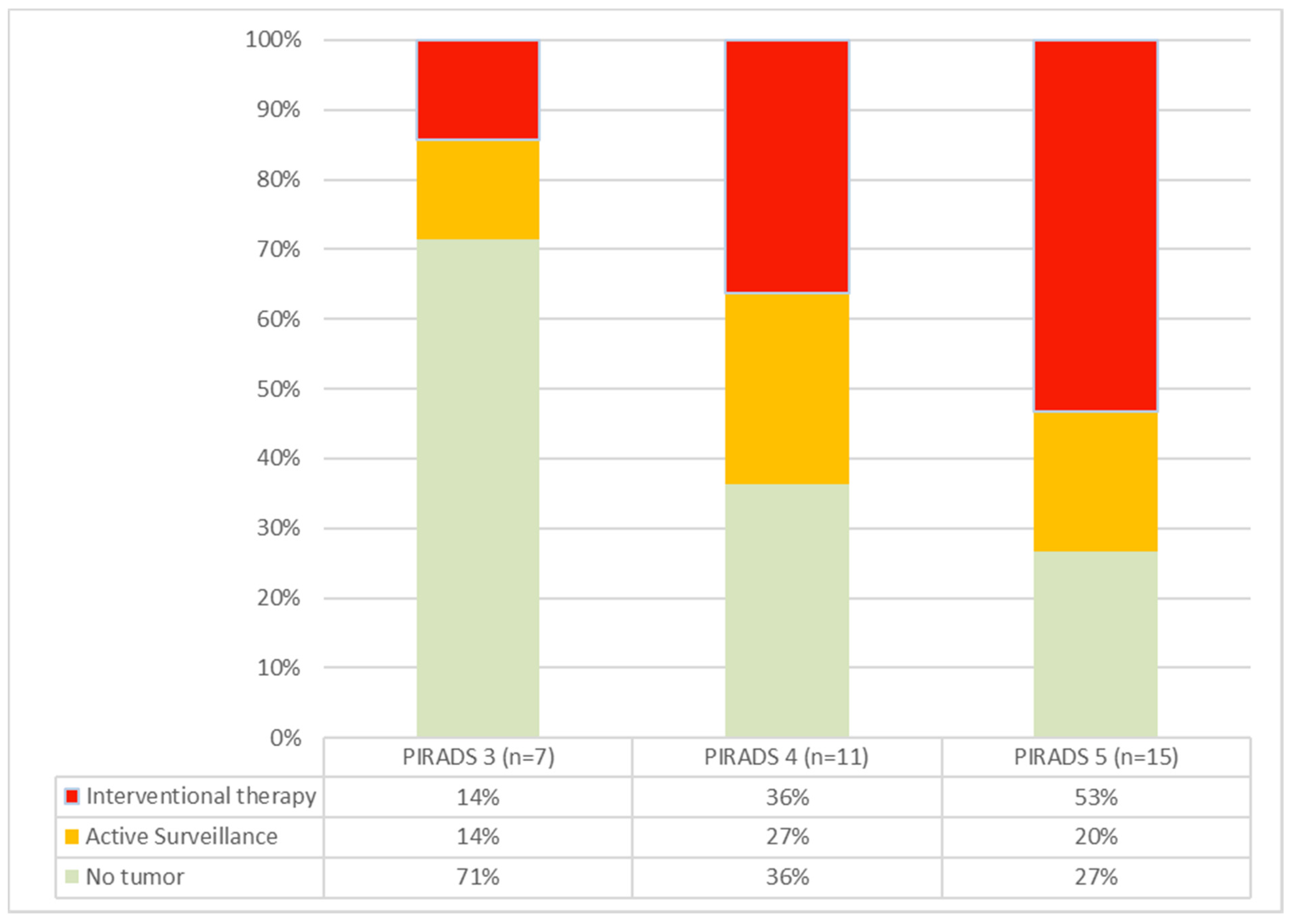
3.3. Detection Rates of Clinically Relevant Tumor Infiltrates
3.4. Therapy Decisions Based on the Targeted Biopsies
3.5. Therapy Decisions in Patients under Active Surveillance
3.6. Follow-Up Results
3.7. Summary
4. Discussion
Author Contributions
Funding
Institutional Review Board Statement
Informed Consent Statement
Data Availability Statement
Acknowledgments
Conflicts of Interest
References
- Sung, H.; Ferlay, J.; Siegel, R.L.; Laversanne, M.; Soerjomataram, I.; Jemal, A.; Bray, F. Global Cancer Statistics 2020: GLOBOCAN Estimates of Incidence and Mortality Worldwide for 36 Cancers in 185 Countries. CA Cancer J. Clin. 2021, 71, 209–249. [Google Scholar] [CrossRef] [PubMed]
- Hodge, K.K.; McNeal, J.E.; Terris, M.K.; Stamey, T.A. Random systematic versus directed ultrasound guided transrectal core biopsies of the prostate. J. Urol. 1989, 142, 71–74; discussion 74–75. [Google Scholar] [CrossRef]
- D’Amico, A.V. Risk-based management of prostate cancer. N. Engl. J. Med. 2011, 365, 169–171. [Google Scholar] [CrossRef]
- Parker, C.; Castro, E.; Fizazi, K.; Heidenreich, A.; Ost, P.; Procopio, G.; Tombal, B.; Gillessen, S.; ESMO Guidelines Committee. Prostate cancer: ESMO Clinical Practice Guidelines for diagnosis, treatment and follow-up. Ann. Oncol. 2020, 31, 1119–1134. [Google Scholar] [CrossRef] [PubMed]
- Hagmann, S.; Ramakrishnan, V.; Tamalunas, A.; Hofmann, M.; Vandenhirtz, M.; Vollmer, S.; Hug, J.; Niggli, P.; Nocito, A.; Kubik-Huch, R.A.; et al. Two Decades of Active Surveillance for Prostate Cancer in a Single-Center Cohort: Favorable Outcomes after Transurethral Resection of the Prostate. Cancers 2022, 14, 368. [Google Scholar] [CrossRef] [PubMed]
- Midiri, F.; Vernuccio, F.; Purpura, P.; Alongi, P.; Bartolotta, T.V. Multiparametric MRI and Radiomics in Prostate Cancer: A Review of the Current Literature. Diagnostics 2021, 11, 1829. [Google Scholar] [CrossRef]
- Turkbey, B.; Rosenkrantz, A.B.; Haider, M.A.; Padhani, A.R.; Villeirs, G.; Macura, K.J.; Tempany, C.M.; Choyke, P.L.; Cornud, F.; Margolis, D.J.; et al. Prostate Imaging Reporting and Data System Version 2.1: 2019 Update of Prostate Imaging Reporting and Data System Version 2. Eur. Urol. 2019, 76, 340–351. [Google Scholar] [CrossRef] [PubMed]
- D’Agostino, D.; Romagnoli, D.; Giampaoli, M.; Bianchi, F.M.; Corsi, P.; Del Rosso, A.; Schiavina, R.; Brunocilla, E.; Artibani, W.; Porreca, A. “In-Bore” MRI-Guided Prostate Biopsy for Prostate Cancer Diagnosis: Results from 140 Consecutive Patients. Curr. Urol. 2020, 14, 22–31. [Google Scholar] [CrossRef]
- Watts, S.; Leydon, G.; Birch, B.; Prescott, P.; Lai, L.; Eardley, S.; Lewith, G. Depression and anxiety in prostate cancer: A systematic review and meta-analysis of prevalence rates. BMJ Open 2014, 4, e003901. [Google Scholar] [CrossRef]
- Scandurra, C.; Muzii, B.; La Rocca, R.; Di Bello, F.; Bottone, M.; Califano, G.; Longo, N.; Maldonato, N.M.; Mangiapia, F. Social Support Mediates the Relationship between Body Image Distress and Depressive Symptoms in Prostate Cancer Patients. Int. J. Environ. Res. Public Health 2022, 19, 4825. [Google Scholar] [CrossRef]
- Helgeson, V.S.; Lepore, S.J.; Eton, D.T. Moderators of the benefits of psychoeducational interventions for men with prostate cancer. Health Psychol. 2006, 25, 348–354. [Google Scholar] [CrossRef] [Green Version]
- Appleton, L.; Wyatt, D.; Perkins, E.; Parker, C.; Crane, J.; Jones, A.; Moorhead, L.; Brown, V.; Wall, C.; Pagett, M.; et al. The impact of prostate cancer on men’s everyday life. Eur. J. Cancer Care 2014, 24, 71–84. [Google Scholar] [CrossRef]
- Steginga, S.K.; Occhipinti, S.; Gardiner, R.A.; Yaxley, J.; Heathcote, P. Prospective study of men’s psychological and decision-related adjustment after treatment for localized prostate cancer. Urology 2004, 63, 751–756. [Google Scholar] [CrossRef] [PubMed]
- Lofters, A.; Juffs, H.G.; Pond, G.R.; Tannock, I.F. “PSA-it is”: Knowledge of serum prostate specific antigen and other causes of anxiety in men with metaststic prostate cancer. J. Urol. 2002, 168, 2516–2520. [Google Scholar] [CrossRef]
- Klotz, L.H. PSAdynia and other PSA-related syndromes: A new epidemic—A case history and taxonomy. Urology 1997, 50, 831–832. [Google Scholar] [CrossRef]
- Awsare, N.S.; Green, J.S.; Aldwinckle, B.; Hanbury, D.C.; Boustead, G.B.; McNicholas, T.A. The measurement of psychological distress in men being investigated for the presence of prostate cancer. Prostate Cancer Prostatic Dis. 2008, 11, 384–389. [Google Scholar] [CrossRef] [PubMed]
- Essink-Bot, M.L.; de Koning, H.J.; Nijs, H.G.; Kirkels, W.J.; van der Maas, P.J.; Schroder, F.H. Short-term effects of population-based screening for prostate cancer on health-related quality of life. J. Natl. Cancer Inst. 1998, 90, 925–931. [Google Scholar] [CrossRef] [PubMed] [Green Version]
- Gustafsson, O.; Theorell, T.; Norming, U.; Perski, A.; Ohstrom, M.; Nyman, C.R. Psychological reactions in men screened for prostate cancer. Br. J. Urol. 1995, 75, 631–636. [Google Scholar] [CrossRef]
- Gareau, D.S.; Li, Y.; Huang, B.; Eastman, Z.; Nehal, K.S.; Rajadhyaksha, M. Confocal mosaicing microscopy in Mohs skin excisions: Feasibility of rapid surgical pathology. J. Biomed. Opt. 2008, 13, 054001. [Google Scholar] [CrossRef]
- Ragazzi, M.; Piana, S.; Longo, C.; Castagnetti, F.; Foroni, M.; Ferrari, G.; Gardini, G.; Pellacani, G. Fluorescence confocal microscopy for pathologists. Mod. Pathol. 2014, 27, 460–471. [Google Scholar] [CrossRef]
- Puliatti, S.; Bertoni, L.; Pirola, G.M.; Azzoni, P.; Bevilacqua, L.; Eissa, A.; Elsherbiny, A.; Sighinolfi, M.C.; Chester, J.; Kaleci, S.; et al. Ex vivo fluorescence confocal microscopy: The first application for real-time pathological examination of prostatic tissue. BJU Int. 2019, 124, 469–476. [Google Scholar] [CrossRef]
- Bertoni, L.; Puliatti, S.; Reggiani Bonetti, L.; Maiorana, A.; Eissa, A.; Azzoni, P.; Bevilacqua, L.; Spandri, V.; Kaleci, S.; Zoeir, A.; et al. Ex vivo fluorescence confocal microscopy: Prostatic and periprostatic tissues atlas and evaluation of the learning curve. Virchows Arch. 2020, 476, 511–520. [Google Scholar] [CrossRef] [Green Version]
- Rocco, B.; Sighinolfi, M.C.; Sandri, M.; Spandri, V.; Cimadamore, A.; Volavsek, M.; Mazzucchelli, R.; Lopez-Beltran, A.; Eissa, A.; Bertoni, L.; et al. Digital Biopsy with Fluorescence Confocal Microscope for Effective Real-time Diagnosis of Prostate Cancer: A Prospective, Comparative Study. Eur. Urol. Oncol. 2021, 4, 784–791. [Google Scholar] [CrossRef] [PubMed]
- Titze, U.; Hansen, T.; Titze, B.; Schulz, B.; Gunnemann, A.; Rocco, B.; Sievert, K.D. Feasibility study for ex vivo fluorescence confocal microscopy (FCM) on diagnostic prostate biopsies. Quant. Imaging Med. Surg. 2021, 11, 1322–1332. [Google Scholar] [CrossRef]
- Titze, U.; Hansen, T.; Brochhausen, C.; Titze, B.; Schulz, B.; Gunnemann, A.; Rocco, B.; Sievert, K.D. Diagnostic Performance of Ex Vivo Fluorescence Confocal Microscopy in the Assessment of Diagnostic Biopsies of the Prostate. Cancers 2021, 13, 5685. [Google Scholar] [CrossRef] [PubMed]
- Matoso, A.; Epstein, J.I. Defining clinically significant prostate cancer on the basis of pathological findings. Histopathology 2019, 74, 135–145. [Google Scholar] [CrossRef] [PubMed] [Green Version]
- Ahmed, H.U.; Hu, Y.; Carter, T.; Arumainayagam, N.; Lecornet, E.; Freeman, A.; Hawkes, D.; Barratt, D.C.; Emberton, M. Characterizing clinically significant prostate cancer using template prostate mapping biopsy. J. Urol. 2011, 186, 458–464. [Google Scholar] [CrossRef]
- Ahmed, H.U.; El-Shater Bosaily, A.; Brown, L.C.; Gabe, R.; Kaplan, R.; Parmar, M.K.; Collaco-Moraes, Y.; Ward, K.; Hindley, R.G.; Freeman, A.; et al. Diagnostic accuracy of multi-parametric MRI and TRUS biopsy in prostate cancer (PROMIS): A paired validating confirmatory study. Lancet 2017, 389, 815–822. [Google Scholar] [CrossRef] [Green Version]
- Fleiss, J.; Cohen, J. The equivalence of weighted kappa and the intraclass correlation coefficient as measures of reliability. Educ. Psychol. Meas. 1973, 33, 613–619. [Google Scholar] [CrossRef]
- Landis, J.R.; Koch, G.G. The measurement of observer agreement for categorical data. Biometrics 1977, 33, 159–174. [Google Scholar] [CrossRef] [Green Version]
- Drost, F.H.; Osses, D.F.; Nieboer, D.; Steyerberg, E.W.; Bangma, C.H.; Roobol, M.J.; Schoots, I.G. Prostate MRI, with or without MRI-targeted biopsy, and systematic biopsy for detecting prostate cancer. Cochrane Database Syst. Rev. 2019, 4, CD012663. [Google Scholar] [CrossRef] [PubMed]
- Drost, F.H.; Osses, D.; Nieboer, D.; Bangma, C.H.; Steyerberg, E.W.; Roobol, M.J.; Schoots, I.G. Prostate Magnetic Resonance Imaging, with or Without Magnetic Resonance Imaging-targeted Biopsy, and Systematic Biopsy for Detecting Prostate Cancer: A Cochrane Systematic Review and Meta-analysis. Eur. Urol. 2020, 77, 78–94. [Google Scholar] [CrossRef]
- Kasivisvanathan, V.; Stabile, A.; Neves, J.B.; Giganti, F.; Valerio, M.; Shanmugabavan, Y.; Clement, K.D.; Sarkar, D.; Philippou, Y.; Thurtle, D.; et al. Magnetic Resonance Imaging-targeted Biopsy Versus Systematic Biopsy in the Detection of Prostate Cancer: A Systematic Review and Meta-analysis. Eur. Urol. 2019, 76, 284–303. [Google Scholar] [CrossRef] [Green Version]
- Ahdoot, M.; Wilbur, A.R.; Reese, S.E.; Lebastchi, A.H.; Mehralivand, S.; Gomella, P.T.; Bloom, J.; Gurram, S.; Siddiqui, M.; Pinsky, P.; et al. MRI-Targeted, Systematic, and Combined Biopsy for Prostate Cancer Diagnosis. N. Engl. J. Med. 2020, 382, 917–928. [Google Scholar] [CrossRef] [PubMed]
- Egevad, L.; Ahmad, A.S.; Algaba, F.; Berney, D.M.; Boccon-Gibod, L.; Comperat, E.; Evans, A.J.; Griffiths, D.; Grobholz, R.; Kristiansen, G.; et al. Standardization of Gleason grading among 337 European pathologists. Histopathology 2013, 62, 247–256. [Google Scholar] [CrossRef] [PubMed]
- Matheus, W.E.; Ferreira, U.; Brandao, E.A.; Ferruccio, A.A.; Billis, A. The importance of histopathologic review of biopsies in patients with prostate cancer referred to a tertiary uro-oncology center. Int. Braz. J. Urol. 2019, 45, 32–37. [Google Scholar] [CrossRef] [PubMed] [Green Version]
- Eklund, M.; Jaderling, F.; Discacciati, A.; Bergman, M.; Annerstedt, M.; Aly, M.; Glaessgen, A.; Carlsson, S.; Gronberg, H.; Nordstrom, T.; et al. MRI-Targeted or Standard Biopsy in Prostate Cancer Screening. N. Engl. J. Med. 2021, 385, 908–920. [Google Scholar] [CrossRef] [PubMed]
- Derin, O.; Fonseca, L.; Sanchez-Salas, R.; Roberts, M.J. Infectious complications of prostate biopsy: Winning battles but not war. World J. Urol. 2020, 38, 2743–2753. [Google Scholar] [CrossRef]

| Patient Data | Targeted Biopsies | Standard Biopsies | |||||||
|---|---|---|---|---|---|---|---|---|---|
| Patient | Indication | PIRADS | Biopsies | Total | Tumor | Relevant | Total | Tumor | Relevant |
| P01 | AS | 4 | 24 | 8 | 4 | 1 | 16 | 3 | 0 |
| P02 | PRE | 4 | 35 | - | - | - | - | - | - |
| P03 | AS | 3 | 24 | 14 | 5 | 3 | 10 | 2 | 2 |
| P04 | PRE | 5 | 30 | - | - | - | - | - | - |
| P05 | PRE | 5 | 12 | 2 | 1 | 0 | 10 | 0 | 0 |
| P06 | AS | 3 | 12 | 3 | 0 | 0 | 9 | 0 | 0 |
| P07 | PRE | 5 | 12 | 6 | 4 | 4 | 6 | 1 | 0 |
| P08 | PRE | 5 | 12 | - | - | - | - | - | - |
| P09 | PRE | 2 | 12 | 6 | 5 | 5 | 6 | 0 | 0 |
| - | - | - | - | - | - | - | - | - | |
| P11 | PRE | 4 | 12 | 7 | 3 | 2 | 5 | 0 | 0 |
| P12 | PRE | 5 | 14 | 11 | 6 | 6 | 3 | 1 | 1 |
| P13 | PRE | 3 | 12 | 7 | - | - | - | - | - |
| P14 | PRE | 4 | 12 | 8 | 0 | 0 | 4 | 1 | 0 |
| P15 | PRE | 3 | 12 | 7 | - | - | 5 | - | - |
| P16 | PRE | 4 | 20 | - | - | - | - | - | - |
| P17 | PRE | 4 | 21 | 3 | 1 | 0 | 18 | 0 | 0 |
| P18 | PRE | 3 | 14 | - | - | - | - | - | - |
| P19 | PRE | 3 | 14 | 7 | 0 | 0 | 7 | 0 | 0 |
| P20 | AS | 5 | 12 | 9 | 1 | 1 | 3 | 0 | 0 |
| P21 | PRE | 3 | 15 | - | - | - | - | - | - |
| P22 | PRE | 5 | 13 | 8 | 6 | 6 | 5 | 3 | 2 |
| P23 | PRE | 5 | 12 | 8 | 5 | 5 | 4 | 0 | 0 |
| P24 | PRE | 4 | 12 | 7 | 3 | 1 | 5 | 2 | 1 |
| P25 | PRE | 5 | 14 | 4 | 3 | 1 | 10 | 0 | 0 |
| P26 | PRE | 5 | 14 | 9 | 9 | 9 | 5 | 3 | 3 |
| P27 | PRE | 5 | 20 | 6 | 3 | 3 | 14 | 0 | 0 |
| P28 | PRE | 4 | 16 | 6 | 5 | 0 | 10 | 1 | 1 |
| P29 | PRE | 5 | 14 | 10 | 1 | 0 | 4 | 0 | 0 |
| P30 | PRE | 5 | 15 | - | - | - | - | - | - |
| P31 | AS | 5 | 12 | 7 | 0 | 0 | 5 | 1 | 1 |
| P32 | PRE | 4 | 13 | - | - | - | - | - | - |
| P33 | PRE | 4 | 15 | - | - | - | - | - | - |
| P34 | AS | 4 | 16 | 10 | 0 | 0 | 6 | 0 | 0 |
| P35 | PRE | 5 | 15 | - | - | - | - | - | - |
| Ʃ | 173 | 65 | 47 | 170 | 18 | 11 | |||
| % | 38% | 72% | 11% | 61% | |||||
| Clinical Data | Resulting FCM-Diagnosis of MRI Targeted Biopsies | Tumor Board | Follow Up | ||||||||||
|---|---|---|---|---|---|---|---|---|---|---|---|---|---|
| Patient | Age | PSA | Length [mm] | ISUP Grade | Interv. (KDS) | Biopsies | Tumor | % | ISUP Grade | Intervention | Therapy | Stage | ISUP Grade |
| P01 + | 65 | 6.5 | 8 | 1 | 2 | 24 | 7 | 70 | 1 | 2 | RPE | pT2c pN0 R0 | 2 |
| P02 | 68 | 7.8 | - | - | 0 | 35 | 0 | - | - | 0 | - | - | - |
| P03 + | 77 | 3.2 | 4 | 4 | 2 | 24 | 7 | 30 | 3 | 2 | RT | n.a. | n.a. |
| P04 | 66 | 16 | - | - | 0 | 30 | 0 | - | - | 0 | - | - | - |
| P05 | 79 | 11.6 | 4 | 1 | 2 | 12 | 1 | 25 | 1 | 1 | AS | pT1c | 1 |
| P06 + | 60 | 8 | - | - | 0 | 12 | 0 | - | - | 1 | AS | pT1c | 1 |
| P07 | 57 | 9 | 8.5 | 2 | 2 | 12 | 5 | 65 | 2 | 2 | RPE | pT2c pN0 R0 | 3 |
| P08 | 49 | 1.1 | - | - | 0 | 12 | 0 | - | - | 0 | - | - | - |
| P09 | 57 | 6.4 | 10 | 4 | 2 | 12 | 5 | 90 | 3 | 2 | RPE | pT3a pN0 R0 | 3 |
| - | - | - | - | - | - | - | - | - | - | - | - | ||
| P11 | 59 | 9.6 | 9.5 | 2 | 2 | 12 | 3 | 65 | 2 | 2 | RPE | pT3a pNx R0 | 3 |
| P12 | 79 | 55 | 14.5 | 5 | 2 | 14 | 7 | 90 | 5 | 2 | CT | n.a. * | n.a. * |
| P13 | 64 | 11.8 | - | - | 0 | 12 | 0 | - | - | 0 | - | - | - |
| P14 | 64 | 6.6 | - | - | 0 | 12 | 1 | 5 | 1 | 1 | AS | pT1c | 1 |
| P15 | 61 | 3.55 | - | - | 0 | 12 | 0 | - | - | 0 | - | - | - |
| P16 | 66 | 18.6 | - | - | 0 | 20 | 0 | - | - | 0 | - | - | - |
| P17 | 78 | 4.68 | 0.1 | 1 | 1 | 21 | 1 | 10 | 1 | 1 | AS | pT1c | 1 |
| P18 | 72 | 16.4 | - | - | 0 | 14 | 0 | - | - | 0 | - | - | - |
| P19 | 74 | 14.55 | - | - | 0 | 14 | 0 | - | - | 0 | - | - | - |
| P20 + | 58 | 5.96 | 8 | 1 | 1 | 12 | 1 | 45 | 1 | 1 | AS | pT1c | 1 |
| P21 | 66 | 5.4 | - | - | 0 | 15 | 0 | - | - | 0 | - | - | - |
| P22 | 66 | 9.92 | 17 | 2 | 2 | 13 | 9 | 95 | 2 | 2 | RPE | pT3b pN1 R0 | 3 |
| P23 | 73 | 8.7 | 13 | 4 | 2 | 12 | 5 | 70 | 4 | 2 | RPE | pT2c pN0 R0 | 3 |
| P24 | 69 | 12.9 | 6.5 | 1 | 2 | 12 | 5 | 40 | 1 | 2 | RPE | pT2c pN0 R0 | 2 |
| P25 | 77 | 18 | 8.8 | 2 | 2 | 14 | 3 | 40 | 2 | 2 | RPE | pT2c pN0 R0 | 2 |
| P26 | 61 | 33 | 14 | 2 | 2 | 14 | 12 | 55 | 2 | 2 | RT | n.a. | n.a. |
| P27 | 64 | 32.7 | 7 | 2 | 2 | 20 | 3 | 95 | 2 | 2 | RPE | pT2c pN0 R0 | 2 |
| P28 | 52 | 6.39 | 3 | 1 | 2 | 16 | 6 | 45 | 1 | 2 | RPE | pT2c pN0 R0 | 2 |
| P29 | 63 | 5.89 | 2 | 1 | 1 | 14 | 1 | 10 | 1 | 2 | RPE | pT2a pNx R0 * | 1 |
| P30 | 61 | 3.31 | - | - | 0 | 15 | 0 | - | - | 0 | - | - | - |
| P31 + | 64 | 7.28 | - | - | 0 | 12 | 1 | 40 | 1 | 1 | AS | pT1c | 1 |
| P32 | 63 | 4.41 | - | - | 0 | 13 | 0 | - | - | 0 | - | - | - |
| P33 | 69 | 6.86 | - | - | 0 | 15 | 0 | - | - | 0 | - | - | - |
| P34 + | 68 | 7.83 | - | - | 0 | 16 | 0 | - | - | 1 | AS | pT1c | 1 |
| P35 | 55 | 3.81 | - | - | 0 | 15 | 0 | - | - | 0 | - | - | - |
| FCM Targeted-Biopsies | ||||
|---|---|---|---|---|
| No Tumor | Uncertain Relevance | Intervention | ||
| Tumorboard | No Tumor | 13 | 0 | 0 |
| 19 | 1 | |||
| Active Surveillance | ||||
| 4 | 2 | 1 | ||
| Intervention | 0 | 1 | 13 | |
| 1 | ||||
| Sensitivity | 93% | |||
| Specificity | 95% | |||
| Positive predictive value | 93% | |||
| Negative predictive value | 95% | |||
| Cohen’s Kappa | 0.88 | |||
| Level of agreement | very good | |||
Publisher’s Note: MDPI stays neutral with regard to jurisdictional claims in published maps and institutional affiliations. |
© 2022 by the authors. Licensee MDPI, Basel, Switzerland. This article is an open access article distributed under the terms and conditions of the Creative Commons Attribution (CC BY) license (https://creativecommons.org/licenses/by/4.0/).
Share and Cite
Sievert, K.-D.; Hansen, T.; Titze, B.; Schulz, B.; Omran, A.; Brockkötter, L.; Gunnemann, A.; Titze, U. Ex Vivo Fluorescence Confocal Microscopy (FCM) of Prostate Biopsies Rethought: Opportunities of Intraoperative Examinations of MRI-Guided Targeted Biopsies in Routine Diagnostics. Diagnostics 2022, 12, 1146. https://doi.org/10.3390/diagnostics12051146
Sievert K-D, Hansen T, Titze B, Schulz B, Omran A, Brockkötter L, Gunnemann A, Titze U. Ex Vivo Fluorescence Confocal Microscopy (FCM) of Prostate Biopsies Rethought: Opportunities of Intraoperative Examinations of MRI-Guided Targeted Biopsies in Routine Diagnostics. Diagnostics. 2022; 12(5):1146. https://doi.org/10.3390/diagnostics12051146
Chicago/Turabian StyleSievert, Karl-Dietrich, Torsten Hansen, Barbara Titze, Birte Schulz, Ahmad Omran, Lukas Brockkötter, Alfons Gunnemann, and Ulf Titze. 2022. "Ex Vivo Fluorescence Confocal Microscopy (FCM) of Prostate Biopsies Rethought: Opportunities of Intraoperative Examinations of MRI-Guided Targeted Biopsies in Routine Diagnostics" Diagnostics 12, no. 5: 1146. https://doi.org/10.3390/diagnostics12051146
APA StyleSievert, K.-D., Hansen, T., Titze, B., Schulz, B., Omran, A., Brockkötter, L., Gunnemann, A., & Titze, U. (2022). Ex Vivo Fluorescence Confocal Microscopy (FCM) of Prostate Biopsies Rethought: Opportunities of Intraoperative Examinations of MRI-Guided Targeted Biopsies in Routine Diagnostics. Diagnostics, 12(5), 1146. https://doi.org/10.3390/diagnostics12051146