Article

A Novel CDH1 Variant Identified in a Chinese Family with Blepharocheilodontic Syndrome

1
First Clinical Division, Peking University School and Hospital of Stomatology & National Center of Stomatology & National Clinical Research Center for Oral Diseases & National Engineering Research Center of Oral Biomaterials and Digital Medical Devices, Beijing 100081, China
2
Department of Prosthodontics, Peking University School and Hospital of Stomatology & National Center of Stomatology & National Clinical Research Center for Oral Diseases & National Engineering Research Center of Oral Biomaterials and Digital Medical Devices, Beijing 100081, China
*
Authors to whom correspondence should be addressed.
These authors contributed equally to this work.
Diagnostics 2022, 12(12), 2936; https://doi.org/10.3390/diagnostics12122936
Submission received: 28 October 2022 / Revised: 16 November 2022 / Accepted: 22 November 2022 / Published: 24 November 2022
(This article belongs to the Special Issue Diagnosis of Dental Diseases)

Abstract

:
The goal of the current study was to identify the pathogenic gene variant in a Chinese family with Blepharocheilodontic (BCD) syndrome. Whole-exome sequencing (WES) and Sanger sequencing were used to identify the pathogenic gene variant. The harmfulness of the variant was predicted by bioinformatics. We identified a novel heterozygous missense variant c.1198G>A (p.Asp400Asn) in the CDH1 gene in the proband and his mother with BCD syndrome. The sequencing results of three healthy individuals in this family are wild type. This result is consistent with familial co-segregation. According to ReVe, REVEL, CADD, gnomAD, dbSNP, and the classification of pathogenic variants with the standards of the 2015 American College of Medical Genetics and Genomics and the Association for Molecular Pathology (ACMG), c.1198G>A (p.Asp400Asn) is predicted to be a likely pathogenic. We observed that variant c.1198G>A (p.Asp400Asn) was located in the extracellular cadherin-type repeats in CDH1. Amino acid sequence alignment of the CDH1 protein among multiple species showed that Asp400 was highly evolutionarily conserved. The conformational analysis showed that this variant might cause structural damage to the CDH1 protein. Phenotypic analysis revealed unique dental phenotypes in patients with BCD syndrome, such as oligodontia, conical-shaped teeth, and notching of the incisal edges. Our results broaden the variation spectrum of BCD syndrome and phenotype spectrum of CDH1, which can help with the clinical diagnosis, treatment, and genetic counseling in relation to BCD syndrome.

1. Introduction

Tooth agenesis is one of the most common congenital malformations in humans, which causes clinically challenging problems, such as malocclusion, masticatory dysfunction, speech changes, and aesthetic problems [1]. The prevalence of tooth agenesis is approximately 2–10% in different regions of the population [2,3,4]. Tooth agenesis may appear as part of a recognized genetic syndrome or as a non-syndromic familial form, which occurs as an isolated trait. Tooth agenesis has been associated with many factors, including genetics, epigenetics, and environmental factors. Among them, genetic defects have been demonstrated to play a major role [1].
Blepharocheilodontic (BCD) syndrome (OMIM 119580) is a rare autosomal dominant disorder characterized by eyelid malformations, cleft lip/palate (CL/P), and dental anomalies [5,6]. Eyelid anomalies (i.e., ectropion, euryblepharon, lagophthalmy, and distichiasis) are typical symptoms [7]. Bilateral CL/P is also one of the main features [8]. Dental anomalies usually manifest as oligodontia and a conical tooth shape [5]. In addition, hypothyroidism, hair abnormalities, dysmorphic facial features, syndactyly, imperforate anus, dermoid cysts, and neural tube defects were also reported in a few cases [5,9,10].
Recently, studies have identified that the pathogenic genes of BCD syndrome are cadherin 1 (CDH1) and catenin delta 1 (CTNND1) [7,8]. CDH1 encodes E-cadherin, a single-pass transmembrane protein playing an essential role in epithelial cell adherence [11]. CTNND1 encodes catenin delta 1 (alias p120ctn), an armadillo repeat-containing protein that interacts with the juxta-membrane cytoplasmic tail of CDH1 and controls the stability of the complex [12]. Studies on mouse models presented the significant roles of CDH1 in eyelid, craniofacial, tooth, and hair developments [13,14,15,16]. Additionally, CDH1 gene variants were also reported to be associated with diffuse gastric and lobular breast cancer syndrome with CL/P (HDGC; OMIM 137215) or non-syndromic CL/P (NSCLP) [17,18,19,20,21]. In 2016, Nishi et al. identified a de novo CDH1 missense variant in a patient with symptoms similar to BCD syndrome [22]. In 2017, Ghoumid et al. identified five CDH1 deleterious missense variants and three CTNND1 truncating variants in 11 patients with BCD syndrome from 8 families and first pointed out that CDH1 and CTNND1 are the pathogenic genes of BCD syndrome [7]. In 2018, Kievit et al. further confirmed that CDH1 and CTNND1 are pathogenic for BCD syndrome, and they identified CDH1 or CTNND1 variants in 15 patients with BCD syndrome [8].
In this study, we performed whole-exome sequencing (WES) and clinical assessments of a family with inherited BCD syndrome to investigate the genetic causes and clinical phenotypes. A novel missense CDH1 variant was identified in the family. Then, we use bioinformatic and conservation analyses to predict the pathogenicity of the identified variant. In addition, we analyzed the number and morphological variation of teeth in patients with BCD syndrome.

2. Materials and Methods

2.1. Subjects

The proband and his family were recruited at the Department of Prosthodontics, Peking University School of Stomatology. Oral and maxillofacial examinations were performed by experienced dentists. Panoramic radiographs were taken to confirm the position of missing teeth. Eye and hair examinations were performed by ophthalmologists and dermatologists, respectively. Blood samples were provided by two patients and three unaffected family members. Informed consent was obtained from all the participants, and the proband provided consent for the publication of the intraoral clinical photographs. This study was conducted with permission from the Ethics Committee of Peking University School and Hospital of Stomatology (PKUSSIRB-202162021).

2.2. WES

Peripheral blood genomic DNA was extracted using the BioTek DNA Whole-Blood Mini Kit (BioTek, Beijing, China), referring to the instructions. WES was performed in all the five available family members by Beijing Angen Gene Medicine Technology (Beijing, China) using the Illumina-X10 platform to identify the potential pathogenic gene variants. Based on the WES results, orodental-related genes were annotated [23]. Then, we filtered all the nonsynonymous single-nucleotide variants and insertions/deletions according to the MAF ≤ 0.01 in the databases, including the single-nucleotide polymorphism database (dbSNP, http://www.ncbi.nlm.nih.gov/projects/SNP/snp_summary.cgi , accessed on 1 October 2020) and the genome aggregation database (gnomAD, http://gnomad.broadinstitute.org accessed on 1 October 2020). The variants that affected protein function were predicted based on the results obtained from ReVe [24], rare-exome-variant ensemble learner (REVEL) [25], and combined annotation-dependent depletion (CADD) [26].

2.3. Sanger Sequencing

To verify the variants and analyze the familial co-segregation, polymerase chain reaction (PCR) was employed to amplify the exon 9 of the CDH1 gene (NM_004360) (primers can be provided according to the requirements). The PCR products were sent to Beijing Angen Gene Medicine Technology for purification and Sanger sequencing.

2.4. Conservation Analysis

For conservation analysis, the amino acid sequences of CDH1 among different species were obtained from the NCBI database (https://www.ncbi.nlm.nih.gov/, accessed on 30 September 2022). Molecular Evolutionary Genetics Analysis Version 11 (MEGA 11.0) [20] was used to conduct the multiple sequence alignment.

2.5. Conformational Analysis

The three-dimensional structure of the CDH1 protein was predicted using the Alpha Fold Protein Structure Database (https://alphafold.ebi.ac.uk/, accessed on 1 October 2020). PyMol v2.1 (Molecular Graphics System, DeLano Scientific, San Carlos, CA, USA) was used to analyze the three-dimensional structural changes caused by the variant.

3. Results

3.1. Clinical Findings

This was a three generation family, in which the proband and his mother were BCD patients. The proband was a 9-year-old boy who presented with congenital missing of all primary teeth, congenital missing of 14 permanent teeth, abnormalities in tooth morphology, euryblepharon, ectropion, distichiasis, sparse hair, high frontal hairline, and broad forehead (Figure 1A–E and Table 1). The abnormality in the morphology of the teeth was the notching of the incisal edges. His mother was a 35-year-old female who presented with congenital missing of all primary teeth, congenital missing of 19 permanent teeth, abnormalities in tooth morphology, euryblepharon, ectropion, distichiasis, sparse hair, high frontal hairline, and broad forehead (Figure 1F,G and Table 1). The abnormality in the morphology of the teeth was that they were conical-shaped.

3.2. The Identification of the CDH1 Variant

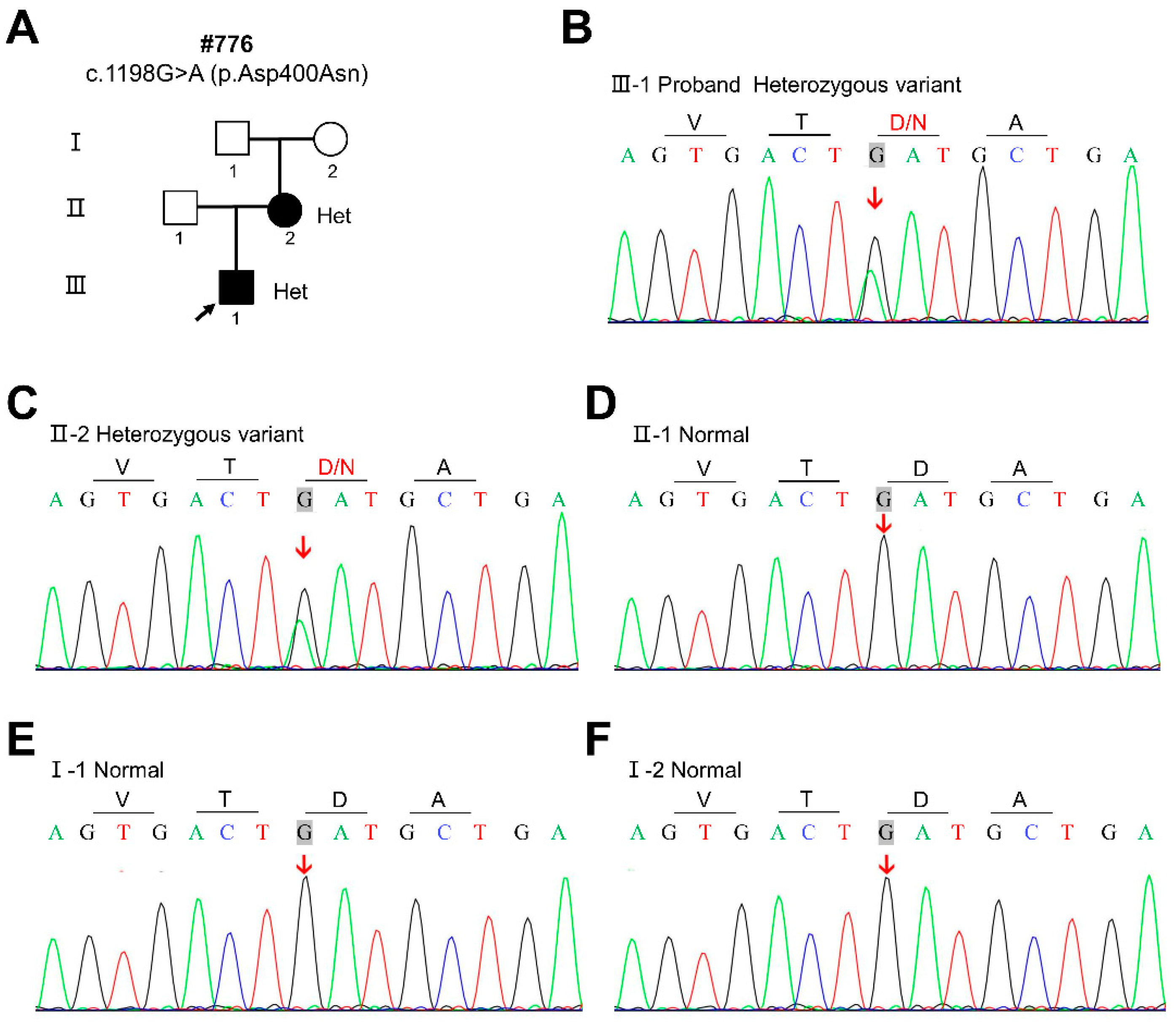
By WES and Sanger sequencing, we identified a novel heterozygous missense variant c.1198G>A (p.Asp400Asn) in the CDH1 gene (NM_004360) in the proband and his mother (Figure 2B,C). According to ReVe, REVEL, CADD, gnomAD, dbSNP, and the classification of pathogenic variants with the standards of the 2015 American College of Medical Genetics and Genomics and the Association for Molecular Pathology (ACMG), c.1198G>A (p.Asp400Asn) was predicted to be likely pathogenic (Table 2). The sequencing results of three healthy individuals (Ⅰ-1, Ⅰ-2, and Ⅱ-1) in this family are wild type (Figure 2D–F). This result is consistent with familial co-segregation.

3.3. Conservation and Conformational Analyses

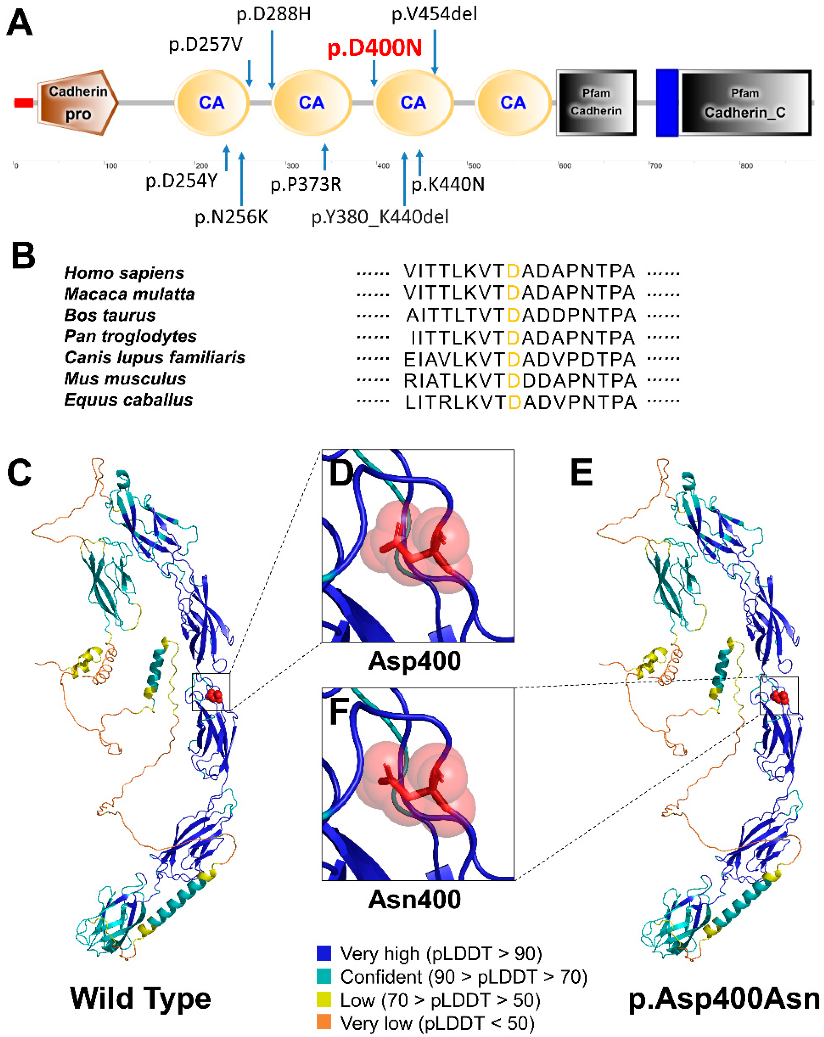
We conducted the conservation and conformational analyses to predict the effects of the identified novel CDH1 variant c.1198G>A (p.Asp400Asn). This variant resulted in the replacement of Asn at position 400 of CDH1 protein (NP_004351.1) with Asp. We observed that c.1198G>A (p.Asp400Asn) was located in the extracellular cadherin-type repeats in CDH1 (Figure 3A). The amino acid sequence alignment of the CDH1 protein among multiple species showed Asp400 was highly evolutionarily conserved (Figure 3B). The residue at sequence position 400 in this protein is an aspartic acid that has a negatively charged side chain, making it hydrophilic (i.e., preferring the surface of the protein to its interior) (Figure 3C,D). The variant residue is an asparagine that has a neutral side chain (Figure 3F,E).

4. Discussion

BCD syndrome is an extremely rare autosomal dominant genetic disease. Its main features are cleft lip/palate, eye-lid malformations, and ectodermal dysplasia. Few BCD cases have been reported in the Asian population; there was only one literature report on a Japanese girl who had a cleft lip and palate, choanal atresia, tetralogy of Fallot, and a neural tube defect, which was caused by a missense variant in the CDH1 gene [22]. This girl’s symptoms were similar to BCD syndrome, but unfortunately the authors did not describe the dental phenotypes associated with the symptoms.
In the present study, the proband and his mother were congenitally missing 14 and 19 permanent teeth, respectively, and both had missing primary teeth. The abnormality in the morphology of their teeth included conical-shaped teeth and teeth with the notching of the incisal edges. These phenotypes did not appear simultaneously in patients with tooth agenesis in our previous study [27,28,29,30,31,32]. Awadh et al. described the dental characteristics of patients with BCD syndrome in detail: the mean number of missing permanent teeth was 13.7; the location of missing teeth included maxillary central incisors, maxillary lateral incisors, maxillary canines, and maxillary premolars; teeth morphological abnormalities included conical-shaped teeth, taurodontism, and notching of the incisal edges [33]. Thus, the dental characteristics of the patients with BCD syndrome in our study were consistent with the study conducted by Awadh et al. [33]. Moreover, eye symptoms in this study included euryblepharon, ectropion, and distichiasis, which were also consistent with the diagnosis of BCD syndrome [5,34]. The patients in this study also had other symptoms of BCD syndrome, such as sparse hair, a high frontal hairline, and broad forehead. Although most of the reported patients with BCD syndrome have a bilateral cleft lip and palate [5,35], the patients in this study did not. By WES and Sanger sequencing, we segregated a novel heterozygous missense variant c.1198G>A (p.Asp400Asn) in the CDH1 gene in the proband and his mother. The sequencing results of three healthy individuals (Ⅰ-1, Ⅰ-2, and Ⅱ-1) in this family are wild type. This result is consistent with family co-separation. In 2017, Ghoumid et al. first reported that BCD syndrome was a CDH1 pathway-related disorder due to variants in CDH1 and CTNND1, and identified five CDH1 deleterious missense variants and three CTNND1 truncating variants in 11 patients with BCD syndrome from eight families [7]. In 2018, Kievit et al. identified CDH1 or CTNND1 variants in 15 patients with BCD syndrome, which further confirmed the relationship between CDH1 and BCD syndrome [8]. Combining the clinical phenotype and genetic test results, we diagnosed the patients in this study with BCD syndrome caused by the CDH1 variant.
The CDH1 gene encodes a classical cadherin of the cadherin superfamily. Five cadherin-type repetitions are joined together by Ca2+ ions to create rigid, rod-like proteins in the extracellular region of the CDH1 protein. In prior studies, all BCD-associated missense variations were found at highly conserved extracellular cadherin-type repeats of CDH1, which directly interact with Ca2+ ion [7,8]. In this study, the variant CDH1:p.Asp400Asn was also found in extracellular cadherin-type repeats. This result is consistent with the previous studies.
It has been previously reported that CDH1 gene variants may lead to hereditary diffuse gastric cancer (HDGC) [21], with or without CL/P, and invasive lobular breast cancer (ILC) [36]. The patients in this study did not have any history of cancer, and no CLP was detected. Ghoumid et al. concluded the phenotype spectrum of CDH1 variants is extensive, including asymptomatic carriers, NSCLP, HDGC with or without CL/P, BCD syndrome, etc. [7]. The phenotype of CDH1-associated BCD syndrome also varied widely. The patients in this study had dental and ocular symptoms, but not symptoms such as CLP, lagophthalmos, ankyloblepharon, malformed ears, everted lower lip, hypothyroidism, hypertelorism, imperforate anus, dermoid cysts, and neural tube defect. It has previously been reported that the CDH1 missense variant is likely to induce cell-specific biological behavior with distinct clinical impacts [37]. However, the specific pathogenic mechanisms require further study.
The tooth phenotype associated with BCD syndrome is noteworthy. The dental symptoms of the patients in this study included the congenital missing of primary and permanent teeth and abnormalities in the morphology of their teeth. The number of missing permanent teeth exceeded six (except for the third molar), which could be diagnosed as oligodontia. The patients’ molars and premolars had small, tapered crowns. In addition, the notching of the incisal edges was also a unique feature. At present, only the study conducted by Awadh et al. [33] described in detail the dental abnormalities of patients with BCD syndrome, which is consistent with the present study. These characteristic dental phenotypes could be used as a basis for the diagnosis of BCD syndrome in future research.
Dental development in humans is a lengthy and intricate process involving a series of reciprocal and sequential interactions between the embryonic oral epithelium and the neural crest-derived mesenchyme. Despite environmental interference, tooth development is mainly controlled by genes. So far, over 200 genes have been identified during tooth development, and these genes are involved in multiple signaling pathways, including the ectodysplasin A (EDA), bone morphogenetic protein (BMP), sonic hedgehog homolog (SHH), fibroblast growth factor (FGF), wingless-type MMTV integration site family (WNT), and transform growth factor (TGF) pathways [38]. Variants in any of these strictly balanced signaling cascades can lead to tooth agenesis and/or other organ defects. The causative gene for non-syndromic tooth agenesis has been a difficult area of research, and there are still many patients with undetected causative genetic variants. Extensive research of publicly available datasets identified 15 genes responsible for non-syndromic tooth agenesis, and genotype–phenotype association analysis indicated that the majority of these genes are also responsible for syndromic tooth agenesis or other diseases [39]. Therefore, the causative genes for non-syndromic tooth agenesis were found from syndromic tooth agenesis. The phenotypes of the different variants of CDH1 were extremely different. As a newly identified syndromic tooth agenesis causative gene, CDH1 can serve as a potential causative gene for non-syndromic tooth agenesis.

5. Conclusions

In summary, we reported a novel heterozygous missense variant c.1198G>A (p.Asp400Asn) in the CDH1 gene in a Chinese family with BCD syndrome. This is the first report of CDH1-associated BCD syndrome in the Chinese population. Our study analyzed the pathogenicity of this variation and summarized the characteristic tooth morphological variation of BCD syndrome. In addition, we also suggested that CDH1 can be used as a potential non-syndromic tooth agenesis pathogenic gene for screening. Our results broaden the variation spectrum of BCD syndrome and phenotype spectrum of CDH1, which could help with the clinical diagnosis, treatment, and genetic counseling of the disease. However, the pathogenic mechanism of CDH1-associated BCD syndrome remains unclear and needs to be studied more extensively.

Author Contributions

Conceptualization, M.Y. and H.L. (Haochen Liu); methodology, Y.L. and B.L.; software, H.L. (Hangbo Liu) and L.S.; validation, H.L. (Haochen Liu), B.L. and Y.L.; formal analysis, B.L. and H.L. (Hangbo Liu); investigation, Y.L. and B.L.; resources, H.L. (Haochen Liu) and Y.L.; data curation, L.S. and M.Y.; writing—original draft preparation, B.L., Y.L., M.Y. and H.L. (Haochen Liu); writing—review and editing, B.L., M.Y., H.L. (Haochen Liu) and Y.L.; visualization, M.Y.; supervision, H.F.; project administration, H.F.; funding acquisition, H.L. (Haochen Liu), M.Y., Y.L. and H.F. All authors have read and agreed to the published version of the manuscript.

Funding

This research was funded by National Natural Science Foundation of China, grant number 81970902, 82100976, 81600851 and 81600846.

Institutional Review Board Statement

The study was conducted in accordance with the Declaration of Helsinki and approved by the Ethics Committee of Peking University School and Hospital of Stomatology (PKUSSIRB-202162021, approved data: 22 February 2022).

Informed Consent Statement

Informed consent was obtained from all the participants, and the proband provided consent for the publication of the intraoral clinical photographs.

Data Availability Statement

The variations identified in this study were submitted to the ClinVar database (submission ID SCV000979048.2).

Conflicts of Interest

The authors declare no conflict of interest.

References

  1. Chhabra, N.; Goswami, M.; Chhabra, A. Genetic Basis of Dental Agenesis—Molecular Genetics Patterning Clinical Dentistry. Med. Oral 2014, 19, e112–e119. [Google Scholar] [CrossRef] [PubMed]
  2. Shimizu, T.; Maeda, T. Prevalence and Genetic Basis of Tooth Agenesis. Jpn. Dent. Sci. Rev. 2009, 45, 52–58. [Google Scholar] [CrossRef] [Green Version]
  3. Rølling, S.; Poulsen, S. Oligodontia in Danish Schoolchildren. Acta Odontol. Scand. 2001, 59, 111–112. [Google Scholar] [CrossRef]
  4. Polder, B.J.; Van’t Hof, M.A.; Van der Linden, F.P.G.M.; Kuijpers-Jagtman, A.M. A Meta-Analysis of the Prevalence of Dental Agenesis of Permanent Teeth. Community Dent. Oral Epidemiol. 2004, 32, 217–226. [Google Scholar] [CrossRef]
  5. Gorlin, R.J.; Zellweger, H.; Curtis, M.W.; Wiedemann, H.R.; Warburg, M.; Majewski, F.; Gillessen-Kaesbach, G.; Prahl-Andersen, B.; Zackai, E. Blepharo-Cheilo-Dontic (BCD) Syndrome. Am. J. Med. Genet. 1996, 65, 109–112. [Google Scholar] [CrossRef]
  6. Weaver, K.N.; Rutledge, K.D.; Grant, J.H.; Robin, N.H. Imperforate Anus Is a Rare Associated Finding in Blepharocheilodontic Syndrome. Am. J. Med. Genet. A 2010, 152A, 438–440. [Google Scholar] [CrossRef]
  7. Ghoumid, J.; Stichelbout, M.; Jourdain, A.-S.; Frenois, F.; Lejeune-Dumoulin, S.; Alex-Cordier, M.-P.; Lebrun, M.; Guerreschi, P.; Duquennoy-Martinot, V.; Vinchon, M.; et al. Blepharocheilodontic Syndrome Is a CDH1 Pathway-Related Disorder Due to Mutations in CDH1 and CTNND1. Genet. Med. 2017, 19, 1013–1021. [Google Scholar] [CrossRef] [PubMed] [Green Version]
  8. Kievit, A.; Tessadori, F.; Douben, H.; Jordens, I.; Maurice, M.; Hoogeboom, J.; Hennekam, R.; Nampoothiri, S.; Kayserili, H.; Castori, M.; et al. Variants in Members of the Cadherin-Catenin Complex, CDH1 and CTNND1, Cause Blepharocheilodontic Syndrome. Eur. J. Hum. Genet. 2018, 26, 210–219. [Google Scholar] [CrossRef] [PubMed] [Green Version]
  9. Gil da Silva Lopes, V.L.; Guion-Almeida, M.L.; de Oliveira Rodini, E.S. Blepharocheilodontic (BCD) Syndrome: Expanding the Phenotype? Am. J. Med. Genet. A 2003, 121A, 266–270. [Google Scholar] [CrossRef] [PubMed]
  10. Freitas, E.L.; Martinhago, C.D.; Ramos, E.S.; Murray, J.C.; Gil-da-Silva-Lopes, V.L. Preliminary Molecular Studies on Blepharocheilodontic Syndrome. Am. J. Med. Genet. A 2007, 143A, 2757–2759. [Google Scholar] [CrossRef] [PubMed]
  11. Patel, S.D.; Chen, C.P.; Bahna, F.; Honig, B.; Shapiro, L. Cadherin-Mediated Cell-Cell Adhesion: Sticking Together as a Family. Curr. Opin. Struct. Biol. 2003, 13, 690–698. [Google Scholar] [CrossRef] [PubMed]
  12. Gumbiner, B.M. Regulation of Cadherin-Mediated Adhesion in Morphogenesis. Nat. Rev. Mol. Cell Biol. 2005, 6, 622–634. [Google Scholar] [CrossRef]
  13. Smith, A.L.; Dohn, M.R.; Brown, M.V.; Reynolds, A.B. Association of Rho-Associated Protein Kinase 1 with E-Cadherin Complexes Is Mediated by P120-Catenin. Mol. Biol. Cell 2012, 23, 99–110. [Google Scholar] [CrossRef] [PubMed]
  14. Shimizu, Y.; Thumkeo, D.; Keel, J.; Ishizaki, T.; Oshima, H.; Oshima, M.; Noda, Y.; Matsumura, F.; Taketo, M.M.; Narumiya, S. ROCK-I Regulates Closure of the Eyelids and Ventral Body Wall by Inducing Assembly of Actomyosin Bundles. J. Cell Biol. 2005, 168, 941–953. [Google Scholar] [CrossRef] [Green Version]
  15. Li, C.-Y.; Cha, W.; Luder, H.-U.; Charles, R.-P.; McMahon, M.; Mitsiadis, T.A.; Klein, O.D. E-Cadherin Regulates the Behavior and Fate of Epithelial Stem Cells and Their Progeny in the Mouse Incisor. Dev. Biol. 2012, 366, 357–366. [Google Scholar] [CrossRef] [PubMed] [Green Version]
  16. Bartlett, J.D.; Dobeck, J.M.; Tye, C.E.; Perez-Moreno, M.; Stokes, N.; Reynolds, A.B.; Fuchs, E.; Skobe, Z. Targeted P120-Catenin Ablation Disrupts Dental Enamel Development. PLoS ONE 2010, 5, e12703. [Google Scholar] [CrossRef] [Green Version]
  17. Benusiglio, P.R.; Caron, O.; Consolino, E.; Duvillard, P.; Coulet, F.; Blayau, M.; Malka, D. Cleft Lip, Cleft Palate, Hereditary Diffuse Gastric Cancer and Germline Mutations in CDH1. Int. J. Cancer 2013, 132, 2470. [Google Scholar] [CrossRef]
  18. Brito, L.A.; Yamamoto, G.L.; Melo, S.; Malcher, C.; Ferreira, S.G.; Figueiredo, J.; Alvizi, L.; Kobayashi, G.S.; Naslavsky, M.S.; Alonso, N.; et al. Rare Variants in the Epithelial Cadherin Gene Underlying the Genetic Etiology of Nonsyndromic Cleft Lip with or without Cleft Palate. Hum. Mutat. 2015, 36, 1029–1033. [Google Scholar] [CrossRef]
  19. van der Post, R.S.; Vogelaar, I.P.; Carneiro, F.; Guilford, P.; Huntsman, D.; Hoogerbrugge, N.; Caldas, C.; Schreiber, K.E.C.; Hardwick, R.H.; Ausems, M.G.E.M.; et al. Hereditary Diffuse Gastric Cancer: Updated Clinical Guidelines with an Emphasis on Germline CDH1 Mutation Carriers. J. Med. Genet. 2015, 52, 361–374. [Google Scholar] [CrossRef] [Green Version]
  20. Kluijt, I.; Siemerink, E.J.M.; Ausems, M.G.E.M.; van Os, T.A.M.; de Jong, D.; Simões-Correia, J.; van Krieken, J.H.; Ligtenberg, M.J.; Figueiredo, J.; van Riel, E.; et al. CDH1-Related Hereditary Diffuse Gastric Cancer Syndrome: Clinical Variations and Implications for Counseling. Int. J. Cancer 2012, 131, 367–376. [Google Scholar] [CrossRef] [PubMed]
  21. Hansford, S.; Kaurah, P.; Li-Chang, H.; Woo, M.; Senz, J.; Pinheiro, H.; Schrader, K.A.; Schaeffer, D.F.; Shumansky, K.; Zogopoulos, G.; et al. Hereditary Diffuse Gastric Cancer Syndrome: CDH1 Mutations and Beyond. JAMA Oncol. 2015, 1, 23–32. [Google Scholar] [CrossRef] [Green Version]
  22. Nishi, E.; Masuda, K.; Arakawa, M.; Kawame, H.; Kosho, T.; Kitahara, M.; Kubota, N.; Hidaka, E.; Katoh, Y.; Shirahige, K.; et al. Exome Sequencing-Based Identification of Mutations in Non-Syndromic Genes among Individuals with Apparently Syndromic Features. Am. J. Med. Genet. A 2016, 170, 2889–2894. [Google Scholar] [CrossRef] [PubMed]
  23. Prasad, M.K.; Geoffroy, V.; Vicaire, S.; Jost, B.; Dumas, M.; Le Gras, S.; Switala, M.; Gasse, B.; Laugel-Haushalter, V.; Paschaki, M.; et al. A Targeted Next-Generation Sequencing Assay for the Molecular Diagnosis of Genetic Disorders with Orodental Involvement. J. Med. Genet. 2016, 53, 98–110. [Google Scholar] [CrossRef] [PubMed] [Green Version]
  24. Li, J.; Zhao, T.; Zhang, Y.; Zhang, K.; Shi, L.; Chen, Y.; Wang, X.; Sun, Z. Performance Evaluation of Pathogenicity-Computation Methods for Missense Variants. Nucleic Acids Res. 2018, 46, 7793–7804. [Google Scholar] [CrossRef] [PubMed] [Green Version]
  25. Ioannidis, N.M.; Rothstein, J.H.; Pejaver, V.; Middha, S.; McDonnell, S.K.; Baheti, S.; Musolf, A.; Li, Q.; Holzinger, E.; Karyadi, D.; et al. REVEL: An Ensemble Method for Predicting the Pathogenicity of Rare Missense Variants. Am. J. Hum. Genet. 2016, 99, 877–885. [Google Scholar] [CrossRef] [PubMed] [Green Version]
  26. Kircher, M.; Witten, D.M.; Jain, P.; O’Roak, B.J.; Cooper, G.M.; Shendure, J. A General Framework for Estimating the Relative Pathogenicity of Human Genetic Variants. Nat. Genet. 2014, 46, 310–315. [Google Scholar] [CrossRef] [PubMed] [Green Version]
  27. Song, S.; Han, D.; Qu, H.; Gong, Y.; Wu, H.; Zhang, X.; Zhong, N.; Feng, H. EDA Gene Mutations Underlie Non-Syndromic Oligodontia. J. Dent. Res. 2009, 88, 126–131. [Google Scholar] [CrossRef] [PubMed] [Green Version]
  28. Song, S.; Zhao, R.; He, H.; Zhang, J.; Feng, H.; Lin, L. WNT10A Variants Are Associated with Non-Syndromic Tooth Agenesis in the General Population. Hum. Genet. 2014, 133, 117–124. [Google Scholar] [CrossRef] [PubMed]
  29. Wong, S.-W.; Han, D.; Zhang, H.; Liu, Y.; Zhang, X.; Miao, M.Z.; Wang, Y.; Zhao, N.; Zeng, L.; Bai, B.; et al. Nine Novel PAX9 Mutations and a Distinct Tooth Agenesis Genotype-Phenotype. J. Dent. Res. 2018, 97, 155–162. [Google Scholar] [CrossRef] [PubMed]
  30. Yu, P.; Yang, W.; Han, D.; Wang, X.; Guo, S.; Li, J.; Li, F.; Zhang, X.; Wong, S.-W.; Bai, B.; et al. Mutations in WNT10B Are Identified in Individuals with Oligodontia. Am. J. Hum. Genet. 2016, 99, 195–201. [Google Scholar] [CrossRef] [PubMed]
  31. Zheng, J.; Yu, M.; Liu, H.; Cai, T.; Feng, H.; Liu, Y.; Han, D. Novel MSX1 Variants Identified in Families with Nonsyndromic Oligodontia. Int. J. Oral. Sci. 2021, 13, 2. [Google Scholar] [CrossRef] [PubMed]
  32. Liu, H.; Liu, H.; Su, L.; Zheng, J.; Feng, H.; Liu, Y.; Yu, M.; Han, D. Four Novel PAX9 Variants and the PAX9-Related Non-Syndromic Tooth Agenesis Patterns. Int. J. Mol. Sci. 2022, 23, 8142. [Google Scholar] [CrossRef] [PubMed]
  33. Awadh, W.; Kiukkonen, A.; Nieminen, P.; Arte, S.; Hurmerinta, K.; Rice, D.P. Blepharocheilodontic (BCD) Syndrome: New Insights on Craniofacial and Dental Features. Am. J. Med. Genet. A 2017, 173, 905–913. [Google Scholar] [CrossRef]
  34. Ababneh, F.K.; Al-Swaid, A.; Elhag, A.; Youssef, T.; Alsaif, S. Blepharo-Cheilo-Dontic (BCD) Syndrome: Expanding the Phenotype, Case Report and Review of Literature. Am. J. Med. Genet. A 2014, 164A, 1525–1529. [Google Scholar] [CrossRef]
  35. Korula, S.; Wilson, L.; Salomonson, J. Distinct Craniofacial Syndrome of Lagophthalmia and Bilateral Cleft Lip and Palate. Am. J. Med. Genet. 1995, 59, 229–233. [Google Scholar] [CrossRef] [PubMed]
  36. Petridis, C.; Shinomiya, I.; Kohut, K.; Gorman, P.; Caneppele, M.; Shah, V.; Troy, M.; Pinder, S.E.; Hanby, A.; Tomlinson, I.; et al. Germline CDH1 Mutations in Bilateral Lobular Carcinoma In Situ. Br. J. Cancer 2014, 110, 1053–1057. [Google Scholar] [CrossRef] [Green Version]
  37. Figueiredo, J.; Söderberg, O.; Simões-Correia, J.; Grannas, K.; Suriano, G.; Seruca, R. The Importance of E-Cadherin Binding Partners to Evaluate the Pathogenicity of E-Cadherin Missense Mutations Associated to HDGC. Eur. J. Hum. Genet. 2013, 21, 301–309. [Google Scholar] [CrossRef] [Green Version]
  38. Ye, X.; Attaie, A. Genetic Basis of Nonsyndromic and Syndromic Tooth Agenesis. J. Pediatr. Genet. 2016, 5, 198–208. [Google Scholar] [CrossRef] [Green Version]
  39. Yu, M.; Wong, S.-W.; Han, D.; Cai, T. Genetic Analysis: Wnt and Other Pathways in Nonsyndromic Tooth Agenesis. Oral Dis. 2019, 25, 646–651. [Google Scholar] [CrossRef]
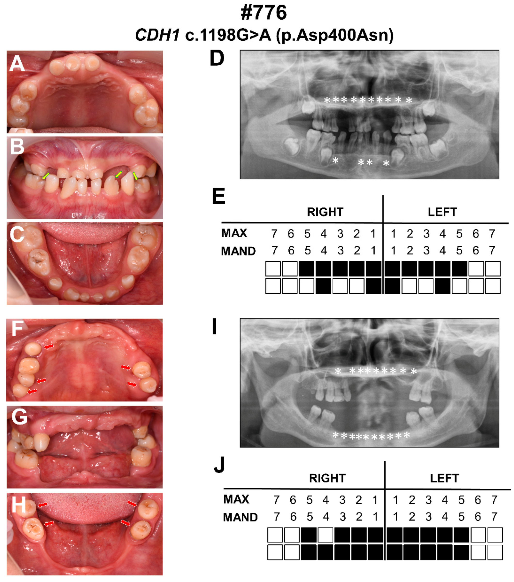
Figure 1. Clinical photographs and panoramic radiographs of the proband and his mother. (AC) Intro-oral photographs of the proband. (D) Panoramic radiograph of the proband. (E) Schematic of congenital missing permanent teeth of the proband. (FH) Intro-oral photographs of the proband’s mother. (I) Panoramic radiograph of the proband’s mother. (J) Schematic of congenital missing permanent teeth of the proband’s mother. Asterisks and solid squares indicate congenitally missing teeth; the yellow arrows indicate teeth with notching of the incisal edges; the red arrows indicate conical-shaped teeth. Max: maxillary; Mand: mandibular.
Diagnostics 12 02936 g001
Figure 2. Family pedigree and sequencing chromatograms of the proband and family members in this study. (A) Pedigree of the BCD family #776. (B) Sequencing chromatograms showing a heterozygous CDH1 variant c.1198G>A (p.Asp400Asn) in the proband (III-1). (C) Sequencing chromatograms showing a heterozygous CDH1 variant c.1198G>A (p.Asp400Asn) in the proband’s mother (II-2). (DF) Sequencing chromatograms showing a wild-type CDH1 genotype in the three healthy individuals (I-1, I-2, and II-1).
Diagnostics 12 02936 g002
Figure 3. Location and conservation and conformational analyses of CDH1 variant p.Asp400Asn. (A) Schematic diagram of the wild-type human CDH1 (NP_004351.1) protein and the distribution of the novel CDH1 variant identified in this study and reported variants identified in patients with BCD syndrome. (B) Conservation analysis of the affected amino acids in CDH1 protein. (C) Tertiary structure of the wild-type CDH1 protein predicted by AlphaFold. (E) Tertiary structure of the p.Asp400Asn variant CDH1 protein. (D,F) Predicted conformational consequences of CDH1: p.Asp400Asn.
Diagnostics 12 02936 g003
Table 1. Clinical features of patients in this study.
Clinical FeaturesThe ProbandⅡ-2
GenderMaleFemale
Age935
Number of missing permanent teeth1419
Congenital missing of primary teeth++
Abnormality in morphology of teeth++
CL/P--
Euryblepharon++
Lagophthalmos--
Ectropion++
Lacrimal duct abnormalities --
Distichiasis++
Ankyloblepharon --
Sparse hair++
High frontal hairline++
Broad forehead++
Malformed ears--
Everted lower lip--
Hypothyroidism--
Hypertelorism--
Imperforate anus --
Dermoid cysts --
Neural tube defect --
Table 2. Pathogenic prediction of the CDH1 variant identified in this study.
PatientsVariantVariant TypeDomainReVeREVELCADDdbSNP/
gnomAD
ACMG Classification
(Evidence of Pathogenicity)
#776 III-1;
#776 II-2
c.1198G>A/p.Asp400AsnHetero-zygous3rd CA0.680
Disease causing
0.643
Disease causing
26.3
Disease causing
-Likely pathogenic
PS2, PM2, PP3, PP4
PS2: De novo in a patient with the disease but no family history. PM2: Absent from controls in Exome Aggregation Consortium, Exome Sequencing Project, or 1000 Genomes Project. PP3: Multiple lines of computational evidence support the existence of a detrimental impact on the gene or gene product. PP4: The phenotypic or family history of a patient is very precise for a disease with a single genetic cause.
Publisher’s Note: MDPI stays neutral with regard to jurisdictional claims in published maps and institutional affiliations.