Review

Lymphatic Endothelial Markers and Tumor Lymphangiogenesis Assessment in Human Breast Cancer

1
Center of Oncology, Renmin Hospital of Wuhan University, Wuhan 430060, China
2
Department of Pathology, The Central Hospital of Wuhan, Tongji Medical College, Huazhong University of Science and Technology, Wuhan 430014, China
3
Department of Peritoneal Cancer Surgery, Beijing Shijitan Hospital of Capital Medical University, Beijing 100038, China
4
Department of Pathology, Beijing Shijitan Hospital, Capital Medical University, Beijing 100038, China
*
Authors to whom correspondence should be addressed.
These authors contributed equally to this work.
Diagnostics 2022, 12(1), 4; https://doi.org/10.3390/diagnostics12010004
Submission received: 18 October 2021 / Revised: 9 December 2021 / Accepted: 17 December 2021 / Published: 21 December 2021
(This article belongs to the Section Pathology and Molecular Diagnostics)

Abstract

:
Metastasis via lymphatic vessels or blood vessels is the leading cause of death for breast cancer, and lymphangiogenesis and angiogenesis are critical prerequisites for the tumor invasion–metastasis cascade. The research progress for tumor lymphangiogenesis has tended to lag behind that for angiogenesis due to the lack of specific markers. With the discovery of lymphatic endothelial cell (LEC) markers, growing evidence demonstrates that the LEC plays an active role in lymphatic formation and remodeling, tumor cell growth, invasion and intravasation, tumor–microenvironment remodeling, and antitumor immunity. However, some studies have drawn controversial conclusions due to the variation in the LEC markers and lymphangiogenesis assessments used. In this study, we review recent findings on tumor lymphangiogenesis, the most commonly used LEC markers, and parameters for lymphangiogenesis assessments, such as the lymphatic vessel density and lymphatic vessel invasion in human breast cancer. An in-depth understanding of tumor lymphangiogenesis and LEC markers can help to illustrate the mechanisms and distinct roles of lymphangiogenesis in breast cancer progression, which will help in exploring novel potential predictive biomarkers and therapeutic targets for breast cancer.

1. Introduction

The latest data show that breast cancer has become the most common malignant carcinoma, with 2.26 million new cases in 2020 worldwide [1]. Metastasis via lymphatic vessels or blood vessels is the leading cause of death for breast cancer, and tumor lymphangiogenesis and angiogenesis are critical prerequisites for the tumor invasion–metastasis cascade [2,3,4]. Whether breast cancer cells gain access to the systematic circulation via blood vessels or via lymphatic vessels at the primary tumor site remains an open question [3,5,6,7]. It is well known that angiogenesis is a critical process for cancer growth, and anti-angiogenic therapy has been incorporated into treatment guidelines for some solid tumors [8]. As early as 1996, an international consensus on the quantification of angiogenesis was established and was updated five years later [9]. Conversely, research progress for lymphangiogenesis has lagged behind that for angiogenesis due to a lack of specific markers. There were few studies about tumor lymphangiogenesis, until the discovery of lymphatic markers that can distinguish lymphatic vessels from blood vessels or other structures, which is now significantly driving the progression of tumor lymphangiogenesis research [10,11,12,13,14].
Studies reveal that tumor lymphangiogenesis is involved in cancer progression [6,7,11,12,13]. Mouse models have shown that the blood vessels in lymph nodes can serve as a short-cut route for tumor cells entering the systemic circulation, which also challenges the assumption that lymph node involvement in breast cancer is an epi-phenomenon [6,7]. Beyond providing a circulation conduit for tumor cells, growing evidence demonstrates that tumor lymphatic endothelial cells (LECs) are actively involved in lymphatic formation and remodeling, tumor cell growth, tumor- and immune-cell-directed invasion, antitumor immunity, the tumor microenvironment, and premetastatic niches remodeling [12,13,14,15]. A high lymphatic vessel density (LVD) was found to correlate with lymph node metastasis or poor survival in breast cancer [16]. Ndiaye et al. [17] indicated that the lymphatic vessel is a double-edged sword in tumor metastasis. In addition, Niemiec et al. [18] found that a high LVD identified a high risk of progression in pN0/chemotherapy-naive patients but low risk in pN+/chemotherapy-treated patients. Ginter et al. [19], however, found that lymphatic vessels were occasionally present in tumor cell nests or tumor-associated stroma and did not participate in metastasis. These discrepant findings may be partly attributable to the difference in the lymphatic markers and stains used, inconsistent counting methods for LVD, and the distinct subpopulations of patients across studies.
In this review, we summarized recent findings on tumor lymphangiogenesis, the most commonly used LEC markers, and parameters for lymphangiogenesis assessment, such as the LVD and lymphatic vessel invasion (LVI) in human breast cancer. A profound understanding of tumor lymphangiogenesis and LEC markers could help to delineate the mechanisms and distinct roles of lymphangiogenesis in breast cancer progression, facilitating the exploration of novel potential predictive biomarkers and therapeutic targets for breast cancer.

2. Tumor Lymphangiogenesis

Tumor lymphangiogenesis is the formation of new lymphatic vessels [4,20], that generally constitute lymphatic capillaries or initial collecting lymphatics, a crucial initiating step in tumor spread.

2.1. Lymphatic Capillaries and Collecting Lymphatics

Lymphatics are tree-shaped hierarchical networks composed of lymphatic capillaries, initial collecting lymphatics, afferent and efferent collecting lymphatics, and the thoracic duct [21] (Figure 1). Generally, lymphatic capillaries are expanded blind-ended irregular lumens lined with a single layer of oak-leaf-shaped LECs and anchored to the surrounding tissue via anchoring filaments [21,22]. In lymphatic capillaries, overlapping flaps (i.e., primary lymphatic valves) between adjacent LECs are anchored on the sides by discontinuous button-like junctions [23]. By contrast, the collecting lymphatics are lined with elongated spindle-shaped LECs with continuous zipper-like junctions and covered with continuous basement membrane (BM) and a smooth muscle cells layer, which helps to pump the lymph [23]. Both button- and zipper-like junctions are composed of vascular endothelial cell cadherin (VECD) and tight junction proteins [23]. In addition, the platelet/endothelial cell-adhesion molecule-1, namely CD31, is partially colocalized with VE-cadherin at zipper-like junctions, and at the tip of overlapping flaps where there is a lack of buttons [21,23]. Moreover, there are no pericytes or smooth muscle cells covering the LEC layer of lymphatic capillaries. In addition, the BM around the capillaries is always absent or discontinuous. The anatomical features mentioned above facilitate the entry of tissue fluid, proteins, antigens, tumor and immune cells into the capillaries through gaps between buttons located at the overlapping flaps [24,25,26].
Crosstalk between lymphatics and immune cells plays an important role in the uptake of cells into lymphatics [25]. Tumor cells can actively enter the lymphatics via chemokine gradients generated by LECs [26,27]. Multiple mechanisms of directed cell migration have been thoroughly reviewed [28]. Then, the afferent collecting lymphatics drain lymph into lymph nodes, where the adaptive immune response and immune tolerance are initiated. Overlapping flaps in lymphatic capillaries facilitate lymph entry, while valves (i.e., secondary lymphatic valves) in collecting vessels maintain the lymph flow in one direction and prevent backflow. In addition, initial collecting lymphatics or precollectors, located between lymphatic capillaries and collecting lymphatics, are composed of a single layer of endothelia, secondary valves, and continuous CD31-VECD junctions [21]. Through these mechanisms, the lymphatics play an important role in maintaining the homeostasis of body fluids and immunity.

2.2. Tumor Lymphangiogenesis and Lymphatic Endothelial Cellular Origins

The transcription factor prospero homeobox gene protein 1 (PROX-1), a homologue of the Drosophila melanogaster PROX-1, is known to be a master control gene for lymphatic differentiation and subsequent vasculature formation [29,30]. According to the venous origin theory, LECs could originate from a subpopulation of vascular endothelial cells (VECs) in the cardinal vein (CV) or the LEC progenitors in the intersegmental vessels during embryogenesis [31,32,33]. PROX-1-expressing endothelial cells (ECs) acquire a lymphatic identity resulted in down-regulation of VEC-specific markers and up-regulation of LEC-specific markers such as vascular endothelial growth factor receptor-3 (VEGFR-3) and lymphatic vessel endothelial hyaluronan receptor-1 (LYVE-1) [29,30] (Figure 2a). Subsequently, the differentiated LECs sprout out of the CV and migrate towards the signal site finely modulated by stimulators and suppressors. In PROX-1-null mice, the ECs in the CV did not express LEC markers or secrete secondary cytokines [34]. Regarding tumor lymphangiogenesis, earlier studies stated that tumor-associated LECs originate from the existing lymphatics. With the deepening of research, more and more studies demonstrated that some tumor-associated LECs showed non-venous origins, such as mesenchymal origin [32,35,36,37].
Tumor lymphangiogenesis is a complex process mediated by multi-functional cytokines secreted by tumor cells and cells in the tumor microenvironment [38,39] (Figure 2b). Among which, the vascular endothelial growth factor (VEGF)-C is the first isolated and the most well studied pro-lymphangiogenic factor [40]. VEGF-C/VEGF-D/VEGFR-3 is the central and classical signaling axis currently known to promote LECs proliferation, migration, and survival [41]. It was reported that podoplanin-expressing tumor-associated macrophages could stimulate lymphangiogenesis via activating VEGF-C/VEGFR-3 or integrin pathways [42,43]. In addition, the CCL21/CCR7 chemokine axis was shown to mediate VEGF-C secretion of breast tumor cells [44]. Some other signaling pathways are probably involved in breast cancer lymphangiogenesis. For instance, nectin-4 promoted lymphangiogenesis through the CXCR4/CXCL12-LYVE-1 axis [45]. Lysyl oxidase-like protein 2 enhanced LECs invasion and lymphatic vessel formation via the activation of AKT-Snail and ERK pathways directly, or stimulated VEGF-C and CXCL12 secretion by tumor-associated fibroblasts [46]. However, their exact role needs to be further explored, as studies are increasingly demonstrating that tumor lymphangiogenesis shows high heterogeneity in metabolic mechanism, functional plasticity, and cellular origins [32,47].
The majority of tumor LECs sprout from pre-existing lymphatic vessels, while a few LECs have been found to originate from bone marrow-derived cell progenitors [35,36] (Figure 2b). For example, markers of myeloid lymphatic endothelial cell progenitors (M-LECPs) were found to be expressed on a portion of lymphatic vessels in breast cancer tissue, but not in normal breast tissue, and M-LECPs co-expressed high levels of PROX1, LYVE-1, podoplanin, and VEGFR-3 [36]. Current evidence for M-LECPs-derived LECs in tumor lymphangiogenesis could be observed in reference [36,48]. In addition, the insertion of TIE-2-expressing monocytes into lymphatic vessels was observed in breast cancer tissue but not in adjacent normal tissues [49]. Though the mechanisms underlying breast tumor lymphangiogenesis and LEC origin have not yet been fully elucidated, it is certain that tumor-associated lymphatic vessels may comprise endothelial cells with heterogeneous phenotypes according to different tumor microenvironments. Thus, LECs may share some markers in common with VECs or with some bone-marrow-derived cell progenitors.
Figure 2. Lymphangiogenesis and lymphatic endothelial cellular origins in breast cancer. (a) LEC precursors are polarized by the specific expression of PROX-1 in nuclei; then, the expression of VEGFR-3, LYVE-1, and podoplanin on the cell membrane is up-regulated and the expression of VEC-specific genes is down-regulated. (b) Among multiple signaling axes, VEGF-C/VEGF-D/VEGFR-3 is the central axis promoting LEC proliferation, migration, and survival. Podoplanin-expressing TAMs [42] and the CCL21/CCR7 chemokine axis [44] might mediate VEGF-C secretion by tumor cells and stimulate lymphangiogenesis. Additionally, tumor cells expressing lysyl oxidase-like protein 2 might enhance lymphangiogenesis via stimulating VEGF-C and CXCL12 secretion by TAFs [46] and activating the CXCR4/CXCL12-LYVE-1 axis [45]. The majority of tumor LECs sprout from pre-existing lymphatic vessels; a few LECs were found to originate from bone marrow-derived cell progenitors, such as M-LECPs [36] and TEMs [49].
Diagnostics 12 00004 g002

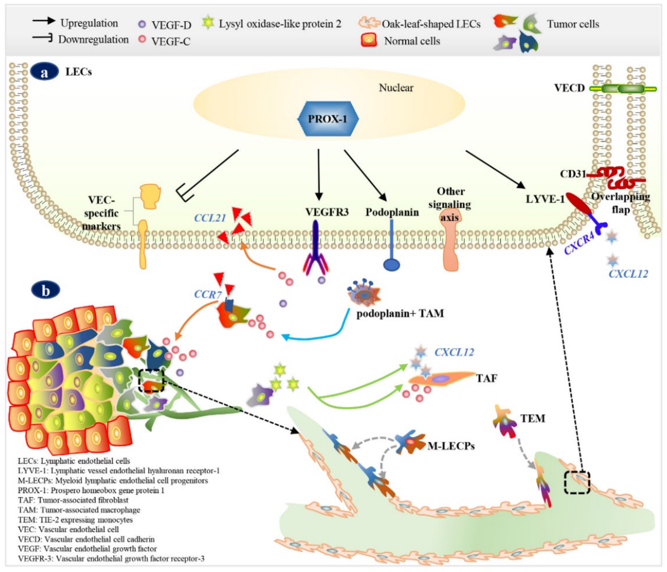
3. Markers of LECs or Lymphatic Vessels

Ideally, during lymphangiogenesis assessment, positive markers should be specifically and sensitively expressed by LECs with a good signal-to-noise ratio compared to surrounding tissues, be easily detectable at the histological level, and must be resistant to biologic chemical agents during histological processing.
Due to its key role in lymphangiogenesis, PROX-1 is considered as a constitutive marker of LECs, located in the nuclei of all LECs in the physiologic and pathologic state [50] (Figure 2a). However, PROX-1 is also expressed by normal breast ductal epithelial cells and tumor cells. Studies showed that the expression of PROX-1 in breast tumor cells could reduce the MMP14-dependent invasiveness of the tumor cells [51], and PROX-1 may be a suppressor gene in breast tumor cells [52]. Thus, PROX-1 alone is unsuitable as a specific marker for LVD and LVI assessments in breast cancer, but it is really an indispensable marker in identifying mechanisms underlying tumor lymphangiogenesis and origins of LEC when combined with other markers [36,49].
VEGFR-3, the receptor for VEGF-C and VEGF-D, is usually co-expressed on the surfaces of endothelial precursor cells and LECs with PROX-1, podoplanin, and LYVE-1 [53,54] (Figure 2a). VEGFR-3 is a membrane-anchored tyrosine kinase that is initially expressed on VECs during embryogenesis and early postnatal development but is restricted to LECs and some high endothelial venules during adulthood [54]. The key role of the VEGF-C/VEGF-D/VEGFR-3 signaling axis in lymphangiogenesis is currently unquestionable. However, the expression of VEGFR-3 is up-regulated on VECs of actively angiogenic blood vessels in breast cancer [55], which in turn leads to its poor lymphatic specificity. In addition, expression of VEGFR-3 is higher in tumor cells than in normal glandular cells [56,57].
LYVE-1 is a homologue of the CD44 hyaluronan receptor and an integral membrane glycoprotein involved in cell interactions [58]. LYVE-1 may not be essential for normal lymphatic development, as LYVE-1-gene-targeted mice develop normally and exhibit a functional network of lymphatic vessels [59]. The expression of LYVE-1 in normal breast ductal epithelial cells and tumor cells is not observed. Instead, LYVE-1 is selectively expressed on the overlapping flaps of LECs in initial lymphatic capillaries (Figure 2a) and significantly contributes to the transmigration of cells into lymphatics [60,61]. After its activation during cells transit, LYVE-1 can be endocytosed into or shed from LECs [62]. Thus, LYVE-1 on lymphatic capillaries presents the instability in immunostainings, and is absent on new single LECs or LECs bundles, which makes it inappropriate for the identification of intratumoral lymphangiogenesis. For example, Van der Auwera et al. [63] demonstrated that LYVE-1 showed weak or no immunoreactivity on intratumoral LECs.
Podoplanin is a transmembrane glycoprotein first detected on podocytes, which is also expressed on the luminal surface of LECs (Figure 2a), but not on VECs [64]. Podoplanin-null mice have defects in lymphatic vessels formation, which results in diminished lymphatic transport, congenital lymphedema, and the dilation of lymphatic vessels [65]; podoplanin-positive lymphatics were showed to be rate-limiting for breast cancer metastasis [66]. Stacker et al. [67] showed that podoplanin seemed to be expressed on small lymphatic vessels but not on lymphatic vessels covered with SMCs. D2-40 is one of the most widely used commercial monoclonal antibodies that binds to a fixation-resistant epitope of podoplanin [68]. It was reported that D2-40 showed higher sensitivity in distinguishing lymphatics than PROX-1 and LYVE-1 in breast cancer tissue [63,69,70] (Figure 3). D2-40 showed the strongest immunoreactivity in both intratumoral and peritumoral LECs, of D2-40 positive intratumoral vessels, 35.1% were positive for PROX-1 and 37.9% showed weak positive for LYVE-1 [63]. Notably, D2-40 is also positive in macrophages, cancer-associated fibroblasts [42,71], and myoepithelial cells [72] in breast tissue. Therefore, myoepithelial cell makers should be combined with D2-40 to differentiate LVI from ductal carcinoma in situ (DCIS).
Other molecules with poor specificity have limited role in identifying lymphatics. For instance, CD31 is a pan-endothelial marker expressed on the LECs and VECs, but it is found to be expressed more strongly in LYVE-1-negative VECs than in LYVE-1-positive LECs [73]. Neuropilin (NRP) 2 is not only a receptor for semaphorins in nerve tissue, but also a co-receptor of VEGFR-3 on LECs, which plays an important role in lymphangiogenesis via VEGF-C/NRP-2/VEGFR-3 axis [74]. However, NRP-2 is also expressed on breast cancer cells [75] and tumor-initiating cells [76]. In summary, all these markers used in combination reasonably with other biomarkers contribute to research in tumor lymphangiogenesis (Table 1). D2-40, LYVE-1, and PROX-1 are recognized LEC markers in human breast cancer, among which D2-40 has the best performance in lymphangiogenesis assessment from the current study. Meanwhile, with the intensive study of lymphatic structure and function, novel lymphatic markers are expected to be discovered in the near future.

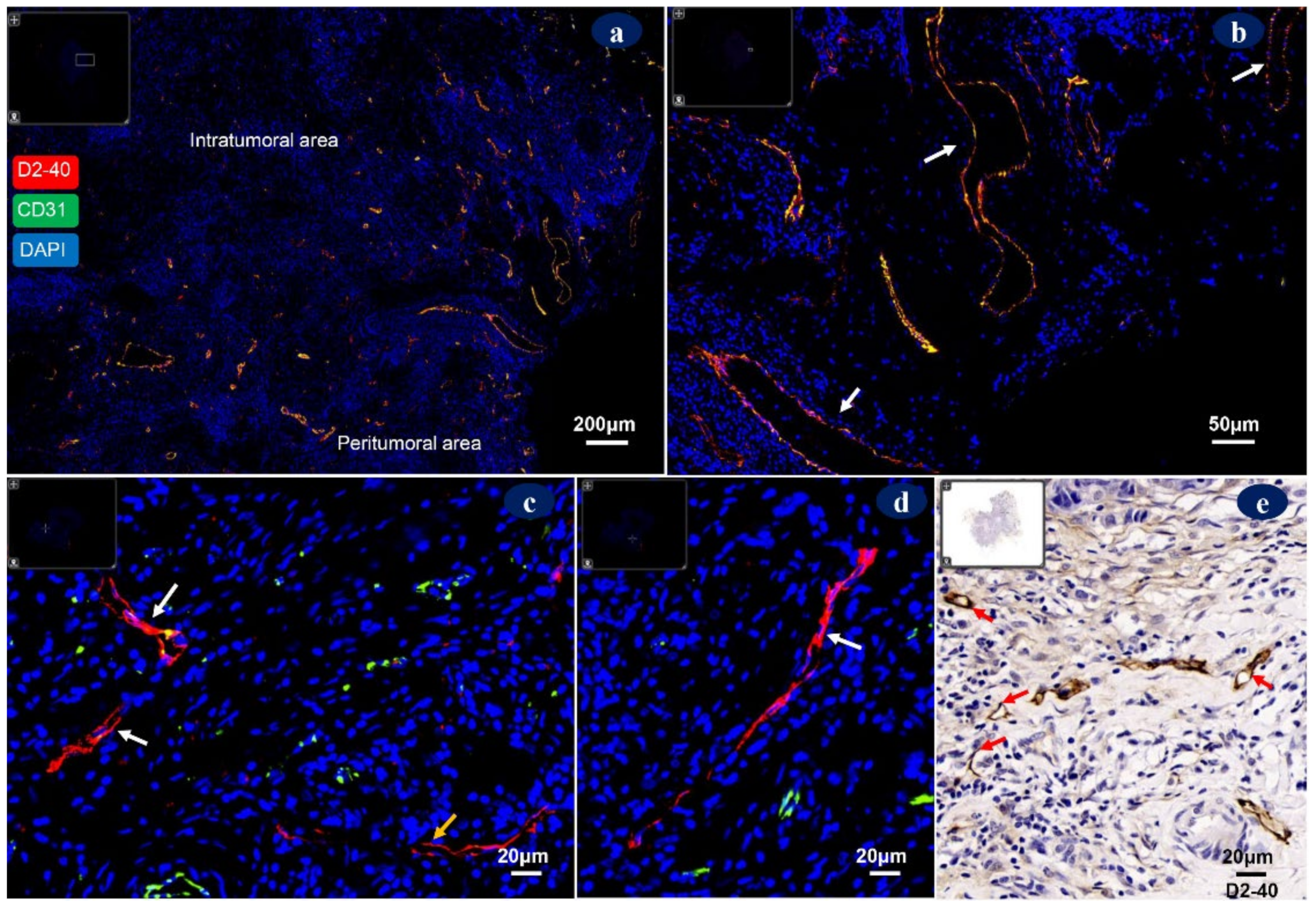
4. Morphology of Tumor-Associated Lymphatic Vessels

Tumor-associated lymphatic vessels exhibit highly heterogeneous morphology and function when compared to normal vessels. Dysregulated lymphatic vessels are organized in a non-hierarchical manner and consist of leaky vessels with irregular distributions. The role of intratumoral vs. peritumoral lymphatic vessels in the development of breast cancer has long been controversial. In peritumoral stroma, lymphatic vessels appear to be inflamed and dilated, which serve as the functional route for cancer-cell dissemination (Figure 4a,b). By contrast, the existence of lymphatic vessels in intratumoral areas is ambiguous, and the majority of the lymphatic vessels in intratumoral areas are sparse, dysfunctional, collapsed vessels with a long narrow or atretic shape (Figure 4c), which is unfavorable for lymph draining or cell migration [90]. These destructing lymphatics may contribute to increasing the compressive stress within tumor areas [91,92]. Some studies have even demonstrated that lymphangiogenesis is not evident in breast tissues and that breast cancer cells might invade and destroy intratumoral lymph vessels rather than stimulating them, and metastasis is mainly via pre-existing lymphatics [92,93].
However, LECs are actively involved in tumor cell growth and invasion, antitumor immunity, and tumor microenvironment remodeling [13,14,15]. We conjecture that the cancer-promoting role of intratumoral LECs could have been underestimated due to differences in assessments. Thus, more research is required to clarify the function of intratumoral lymphangiogenesis. A tumor-associated lymphatic vessels unit should preferably consider single immunoreactive LEC and positive LECs clusters or bundles separate from other lymphatic vessels (Figure 4d) irrespective of lumen status, but positive LECs of large vessels with BM were not included. It was demonstrated that in primary breast tumors, lymphatic vessels mainly present at the peritumoral stroma and only occasionally present in tumor nests and tumor-associated stroma, and LVD within tumor nests had no relationship with metastasis [19]. Whereas, Niemiec et al. [18] demonstrated that a high intratumoral LVD and LVI were adverse factors for disease-free survival in pN0/chemotherapy/trastuzumab-naïve patients.
Another pathologic phenomenon, peritumoral cleft, was defined as clear spaces without a ECs layer that separate tumor cells from the stroma (Figure 5a–c). It has long been considered as fibroblasts retraction [94] or another pathogenesis, such as the situation when ECs shed from the vessel wall during improper tissue processing. Kos et al. [94] found that peritumoral clefts occurred in 92% of invasive ductal breast carcinomas and were not associated with lymphangiogenesis. However, some researchers have suggested that peritumoral clefts seemed to be immature unfinished lymphatic channels with prognostic value or a reflection of epithelial–stromal interaction during lymphangiogenesis [95,96].

5. Lymphatic Vessel Density (LVD)

The first international consensus on the methodology of lymphangiogenesis quantification suggested that the extent of tumor lymphangiogenesis could be quantified according to the LVD or lymphatic endothelial proliferation [97]. The LVD is a commonly used parameter in research (Table 2), based on the assumption that a high LVD might indicate an increased risk of cancer cell intravasation and intensive communication between cancer cells and LECs, which plays an important role in tumor progression [98]. However, some studies have yielded inconsistent conclusions regarding the prognostic value of the LVD. On the one hand, Zhang et al. [16] showed that a high LVD and LVI were both unfavorable prognostic factors in breast cancer, and the peritumoral LVD was higher than the intratumoral LVD (77.9% vs. 40.07%), while both the peritumoral and intratumoral LVD were positively correlated with lymph node metastases. Chen et al. [66] showed that a reduced LVD could up-regulate the frequency of intratumoral macrophages and reduce lung metastasis in breast cancer. On the other hand, Britto et al. [99] indicated that though D2-40 improves the diagnostic accuracy of the LVD and LVI, neither the LVD nor LVI could be used as a predictor of sentinel lymph-node metastasis. Furthermore, Niemiec et al. [18] indicated that a high LVD might identify a low progression risk in pN+/chemotherapy-treated patients. That is to say, chemotherapy-treated breast cancer patients with a high LVD showed better survival. This is theoretically explicable; the tumor antigens, immune cells, and drug transported via lymphatic vessels and blood vessels may play a beneficial role in cancer therapy [17,100]. For example, Kruger et al. [100] showed that a high baseline microvessel density was significantly associated with the response to bevacizumab. However, tumor vasculatures are dysfunctional, leading to immunosuppression and therapy resistance in the vast majority of cases, and normalizing the tumor vasculature can optimize drug uptake [90].
A range of confounding factors may be responsible for the equivocal conclusions (Table 2). Firstly, different endothelial markers were utilized across the studies. Secondly, there is currently no clinical consensus for LVD assessment. The LVD is usually assessed using the Weidner method [101], which was first proposed in 1991 for quantifying blood microvessel density. Hotspot areas with the most lymphatic vessels are firstly chosen at low magnification; then vessels are counted at high magnification in representative fields within the hotspots [101,102]. However, the representative fields used in research were chosen with different magnifications, number, and acreage. Moreover, vessel counts were recorded with the maximum, median, or total counts resulting in varied cut-off values.
Table 2. Studies on the relationship between the lymphatic vessel density (LVD) and breast cancer survival over the last decade.
First Author(No.) PatientsAntibody (Corp, Dilution)LVD Evaluation
Areas
Counts (Magnification)Cutoff ValueHigh LVD
Norhisham et al. [83](58) Breast carcinomaD2-40 (Dako, 1:100)
CD 34 (Dako, 1:100)
Intra- and peritumoralSum of vessels/Sum of area (100×)Median valueDistant metastasis (peri-LVD)
Niemiec et al. [18](139) pT1-2N0M0 IDC
chemotherapy-naive
D2-40 (Cell Marque, 1:100)
CD34 (QBEnd 10, 1:50)
Intra- and peritumoral, peripheralThe highest of 20 hotspots (100×)Minimum p-valuePoor DFS and MFS
Niemiec et al. [18](215) pT1-3N+M0 IDC
chemotherapy-treated
D2-40 (Cell Marque, 1:100)
CD34 (QBEnd 10, 1:50)
Intra- and peritumoral, peripheralThe highest of 20 hotspots (100×)Minimum p-valueFavorable DFS and MFS
Abe et al. [87](91) IDCD2-40 (1:100)NAMean value of 5 hotspots (200×)Mean valuePoor OS and DFS
Wahal et al. [84](30) Invasive breast carcinomaD2-40 (Dako, NA)
CD31 (Dako, NA)
Intratumoral, peritumoralMean value of 5 hotspots (NA)NAHigh lymph node ratio
Zhao et al. [103](73) IDCD2-40 (Signet, 1:25)Intratumoral, peritumoralMean value of 5 hotspots (NA)Median
value
Poor OS and DFS (peri-LVD)
Mohammed et al. [86](197) N0 Basal-like BC (200) N0 Non-basal-like BCD2-40 (AngioBio, 1:100)
CD34 (Serotec, 1:500)
CD31 (Dako, 1:100)
Whole sectionSum of vessels/sum of area (100×)NANo association with 20-year OS
Abbreviations: Corp: corporation; DFS, disease-free survival; IDC, invasive ductal breast cancer; MFS, metastasis-free survival; NA, not available; OS, overall survival; peri-LVD, peritumoral lymphatic vessel density.

6. Lymphatic Vessel Invasion (LVI)

Lymphovascular invasion is the presence of tumor emboli in lymphatic vessels or blood vessels implying an increased risk of dissemination in breast cancer [16,104,105]. Nevertheless, the independent prognostic value of LVI in breast cancer remains unclear, as in many cases, lymphovascular invasion has been assessed through hematoxylin–eosin (HE) stains, which cannot distinguish between LVI and clefts, blood vessels invasion, or DCIS [70,83,106,107] (Figure 5). The concordance of lymphovascular invasion assessment between pathologists based on HE staining is variable, ranging from a median of 0.86 (0.54, 0.99) in a lymphovascular-invasion-positive group to 0.93 (0.52, 1.0) in the lymphovascular-invasion-negative group [106]. An immunohistochemistry (IHC) staining with endothelial markers can accurately distinguish lymphatic vessels [70,105,107]. Abbasi et al. [108] demonstrated that the kappa coefficient between HE and D2-40 staining for detecting lymphovascular invasion was 0.078; and the specificity and negative predictive values of HE staining were 66% and 54.8%, respectively.
Even so, there exists contradictory findings; some studies have demonstrated that the LVI was associated with poor outcomes [70,83] (Table 3). It was reported that though the blood vessel density was higher than the LVD in breast cancer, tumor emboli predominantly occurred in lymphatic vessels [83,85]. However, Kos et al. [94] demonstrated that in patients with axillary lymph node metastasis, the peritumoral LVI was twice and the intratumoral LVI was five times more than that in patients without lymph node metastasis. Fujii et al. [109] suggested that it was blood vessel invasion, not the LVI, which indicated high biological aggressiveness. One cause for the contradiction is the variation in the markers and stain methods utilized in detecting lymphovascular invasion. Another cause is the inherent microfocal nature of lymphovascular invasion. It was observed that only 20% lymphovascular invasions are consistently present in the tumor tissue; others present focal or inconsistent foci that are highly likely to be missed on tissue sections, leading to false negatives [106]. It is perhaps for this reason that IHC is not routinely used for the initial screening for lymphovascular invasion in most clinical institutions. Thus, the above factors limit the clinical utility of the LVI as a robust predictor for breast cancer.

7. Lymphangiogenesis in the Era of Precision Medicine

In the era of precision medicine, the treatment concept for early breast cancer has shifted from the maximum tolerated therapy to the minimum effective therapy [111]. Accurate risk stratification is essential for making a tailoring treatment to maximize survival while minimizing unnecessary interventions. The traditional prognostic tool, such as TNM staging, shows little or no prognostic value in early breast cancer without lymph node involvement or distant metastases. In this case, the characteristics of the tumor microenvironment, such as tumor lymphangiogenesis and angiogenesis, can be predictive or prognostic biomarkers, which well complements the TNM staging system and tumor molecular profiling. Compared with other molecular biotechnology, histomorphology has advantages in combining morphological characteristics with protein expression in tissue in situ and plays an irreplaceable role in lymphangiogenesis research.
In addition, the elucidation of the molecular mechanisms underlying the tumor lymphangiogenesis process will provide novel therapeutic strategies for anti-tumor immunomodulation. For instance, targeting VEGFR-3/-2 signaling pathways could be a potential strategy for combating lymphatic metastases [41,79]. Inhibiting NADPH oxidase 4 attenuated lymphangiogenesis in breast cancer [112], and the knockdown of lysyl oxidase-like protein 2 (LOXL2) suppressed lymphangiogenesis and lymph nodes metastases [46]. In addition, miR-128-3p may modulate the proliferation of LECs via directly targeting VEGF-C/VEGFR3 [113].

8. Conclusions

In summary, with the discovery of lymphatic markers, intensive studies on lymphangiogenesis reveal that LECs play an actively role in the invasion–metastasis cascade of breast cancer. However, we should bear in mind that tumor-associated lymphatic vessels comprise LECs with heterogeneous phenotypes, and there are no ideal markers that are restricted to the LECs or work robustly in various pathologic conditions hitherto. Thus, sensitive and specific LECs markers and standard lymphangiogenesis assessments are warranted to avoid controversial conclusions. In the meantime, in-depth research is progressively revealing the mechanisms and distinct roles of lymphangiogenesis in breast cancer progression, which is helping in the exploration of novel prognostic and predictive biomarkers and therapeutic targets for breast cancer.

Author Contributions

Conceptualization and supervision, Y.-S.C. and Y.L.; writing—original draft preparation, J.-M.C. and B.L.; writing—review and editing, Y.L.; investigation, R.M. and X.-X.L.; funding acquisition, J.-M.C., B.L. and Y.L. All authors have read and agreed to the published version of the manuscript.

Funding

This research was funded by the National Natural Science Foundation of China (82073376); Beijing Municipal Administration of Hospitals’ Ascent Plan (DFL20180701); the National Natural Science Foundation of China (81901820); and the Health commission of Hubei Province scientific research project (WJ2019H365).

Institutional Review Board Statement

Not applicable.

Informed Consent Statement

Not applicable.

Data Availability Statement

Not applicable.

Conflicts of Interest

The authors declare no conflict of interest.

Appendix A. Protocols

Parallel hematoxilin-eosin (HE), immunohistochemistry (IHC), and immunofluorescence (IF) stainings were performed in consecutive sections derived from patients with invasive breast cancer.
IHC staining: Firstly, serial paraffin sections of 4 μm thickness were deparaffinized in xylene (15 min each) and rehydrated in a series of descending ethanol concentrations (5 min each). Then antigen retrieval was performed in sodium citrate buffer (Ph 6.0) in a microwave (10 min at full power followed by 10 min at low power). And endogenous peroxidase activity was blocked with 3% H2O2 (room temperature for 25 min). Secondly, sections were incubated with the primary antibodies overnight at 4℃. Unbound primary antibody was washed with PBS (pH 7.4, 3 times, 5 min each) prior to the addition of HRP-labeled polymer (room temperature for 50 min). Finally, visualized with 3, 3′-diaminobenzidine chromogens (DAB), counterstained with hematoxylin (3 min), dehydrated in a series of ascending ethanol concentrations and xylene.
IF staining: Firstly, serial paraffin sections of 4 μm thickness were deparaffinized in xylene (15 min each) and rehydrated in a series of descending ethanol concentrations (5 min each). Then antigen retrieval was performed in sodium citrate buffer (Ph 6.0) in a microwave (10 min at full power followed by 10 min at low power). Secondly, sections were incubated with the anti-CD 31 antibody overnight at 4 °C. Unbound primary antibody was washed with PBS (pH 7.4, 3 times, 5 min each) prior to the addition of HRP-labeled polymer (room temperature for 50 min), followed by PBS (pH 7.4, 3 times, 5 min each) and incubated with TSA-488 (room temperature for 30 min). Then antigen retrieval was repeated as mentioned above. Thirdly, sections were incubated with the D2-40 overnight at 4 °C and incubated with CY3-tyramide (room temperature for 50 min). Finally, counterstained with DAPI (room temperature for 10 min).
Table A1. Antibodies used for immunohistochemistry and immunofluorescent stains.
AntibodyMainly ExpressedTypeDilutionCorp
Primary Antibodies
LYVE-1Lymph endotheliaRabbit, monoclonal1:200Bioss
CD31EndotheliaRabbit, monoclonal1:2000Abcam
D2-40Lymph endotheliaRabbit, monoclonal1:200Abways
PROX-1Lymph endotheliaRabbit, monoclonal1:500Abcam
Secondary Antibodies
Goat anti-rabbit-HRP antibody Ready-to-useDAKO
TSA-488 1:4000Proteintech Group
CY3-Goat anti-rabbit tyramide 1:500Jackson
DAB 1:100DAKO
DAPI 1:100Solarbio

References

  1. Latest Global Cancer Data: Cancer Burden Rises to 19.3 Million New Cases and 10.0 Million Cancer Deaths in 2020. Available online: https://www.iarc.fr/wp-content/uploads/2020/12/pr292_E.pdf (accessed on 15 December 2020).
  2. Lambert, A.W.; Pattabiraman, D.R.; Weinberg, R.A. Emerging biological principles of metastasis. Cell 2017, 168, 670–691. [Google Scholar] [CrossRef] [Green Version]
  3. Nathanson, S.D.; Krag, D.; Kuerer, H.M.; Newman, L.A.; Brown, M.; Kerjaschki, D.; Pereira, E.R.; Padera, T.P. Breast cancer metastasis through the lympho-vascular system. Clin. Exp. Metastasis 2018, 35, 443–454. [Google Scholar] [CrossRef] [PubMed]
  4. Paduch, R. The role of lymphangiogenesis and angiogenesis in tumor metastasis. Cell Oncol. 2016, 39, 397–410. [Google Scholar] [CrossRef] [PubMed] [Green Version]
  5. Barbieri, E.; Anghelone, C.A.P.; Gentile, D.; La Raja, C.; Bottini, A.; Tinterri, C. Metastases from occult breast cancer: A case report of carcinoma of unknown primary syndrome. Case Rep. Oncol. 2020, 13, 1158–1163. [Google Scholar] [CrossRef] [PubMed]
  6. Pereira, E.R.; Kedrin, D.; Seano, G.; Gautier, O.; Meijer, E.F.J.; Jones, D.; Chin, S.M.; Kitahara, S.; Bouta, E.M.; Chang, J.; et al. Lymph node metastases can invade local blood vessels, exit the node, and colonize distant organs in mice. Science 2018, 359, 1403–1407. [Google Scholar] [CrossRef] [PubMed] [Green Version]
  7. Brown, M.; Assen, F.P.; Leithner, A.; Abe, J.; Schachner, H.; Asfour, G.; Bago-Horvath, Z.; Stein, J.V.; Uhrin, P.; Sixt, M.; et al. Lymph node blood vessels provide exit routes for metastatic tumor cell dissemination in mice. Science 2018, 359, 1408–1411. [Google Scholar] [CrossRef] [Green Version]
  8. Al-Abd, A.M.; Alamoudi, A.J.; Abdel-Naim, A.B.; Neamatallah, T.A.; Ashour, O.M. Anti-angiogenic agents for the treatment of solid tumors: Potential pathways, therapy and current strategies—A review. J. Adv. Res. 2017, 8, 591–605. [Google Scholar] [CrossRef]
  9. Vermeulen, P.B.; Gasparini, G.; Fox, S.B.; Colpaert, C.; Marson, L.P.; Gion, M.; Belien, J.A.; de Waal, R.M.; Van Marck, E.; Magnani, E.; et al. Second international consensus on the methodology and criteria of evaluation of angiogenesis quantification in solid human tumours. Eur. J. Cancer 2002, 38, 1564–1579. [Google Scholar] [CrossRef]
  10. Zhou, H.; Lei, P.J.; Padera, T.P. Progression of metastasis through lymphatic system. Cells 2021, 10, 627. [Google Scholar] [CrossRef]
  11. Ma, Q.; Dieterich, L.C.; Detmar, M. Multiple roles of lymphatic vessels in tumor progression. Curr. Opin. Immunol. 2018, 53, 7–12. [Google Scholar] [CrossRef]
  12. Farnsworth, R.H.; Achen, M.G.; Stacker, S.A. The evolving role of lymphatics in cancer metastasis. Curr. Opin. Immunol. 2018, 53, 64–73. [Google Scholar] [CrossRef] [PubMed]
  13. He, M.; He, Q.; Cai, X.; Chen, Z.; Lao, S.; Deng, H.; Liu, X.; Zheng, Y.; Liu, X.; Liu, J.; et al. Role of lymphatic endothelial cells in the tumor microenvironment-a narrative review of recent advances. Transl. Lung. Cancer Res. 2021, 10, 2252–2277. [Google Scholar] [CrossRef] [PubMed]
  14. Van de Velde, M.; Ebroin, M.; Durre, T.; Joiret, M.; Gillot, L.; Blacher, S.; Geris, L.; Kridelka, F.; Noel, A. Tumor exposed-lymphatic endothelial cells promote primary tumor growth via IL6. Cancer Lett. 2021, 497, 154–164. [Google Scholar] [CrossRef] [PubMed]
  15. Randolph, G.J.; Ivanov, S.; Zinselmeyer, B.H.; Scallan, J.P. The lymphatic system: Integral roles in immunity. Annu. Rev. Immunol. 2017, 35, 31–52. [Google Scholar] [CrossRef] [PubMed] [Green Version]
  16. Zhang, S.; Yi, S.; Zhang, D.; Gong, M.; Cai, Y.; Zou, L. Intratumoral and peritumoral lymphatic vessel density both correlate with lymph node metastasis in breast cancer. Sci. Rep. 2017, 7, 40364–40370. [Google Scholar] [CrossRef]
  17. Ndiaye, P.D.; Pages, G. VEGF-C and lymphatic vessels: A double-edged sword in tumor development and metastasis. Med. Sci. 2019, 35, 132–137. [Google Scholar] [CrossRef]
  18. Niemiec, J.A.; Adamczyk, A.; Ambicka, A.; Mucha-Małecka, A.; Wysocki, W.M.; Biesaga, B.; Ziobro, M.; Cedrych, I.; Grela-Wojewoda, A.; Domagała-Haduch, M.; et al. Prognostic role of lymphatic vessel density and lymphovascular invasion in chemotherapy-naive and chemotherapy-treated patients with invasive breast cancer. Am. J. Transl. Res. 2017, 9, 1435–1447. [Google Scholar] [PubMed]
  19. Ginter, P.S.; Karagiannis, G.S.; Entenberg, D.; Lin, Y.; Condeelis, J.; Jones, J.G.; Oktay, M.H. Tumor microenvironment of metastasis (TMEM) doorways are restricted to the blood Vessel endothelium in both primary breast cancers and their lymph node metastases. Cancers 2019, 11, 1507. [Google Scholar] [CrossRef] [Green Version]
  20. Stacker, S.A.; Williams, S.P.; Karnezis, T.; Shayan, R.; Fox, S.B.; Achen, M.G. Lymphangiogenesis and lymphatic vessel remodelling in cancer. Nat. Rev. Cancer 2014, 14, 159–172. [Google Scholar] [CrossRef]
  21. Breslin, J.W.; Yang, Y.; Scallan, J.P.; Sweat, R.S.; Adderley, S.P.; Murfee, W.L. Lymphatic Vessel Network Structure and Physiology. Compr. Physiol. 2018, 9, 207–299. [Google Scholar] [CrossRef]
  22. Lokmic, Z. Utilizing lymphatic cell markers to visualize human lymphatic abnormalities. J. Biophotonics 2018, 11, e201700117. [Google Scholar] [CrossRef] [PubMed] [Green Version]
  23. Baluk, P.; Fuxe, J.; Hashizume, H.; Romano, T.; Lashnits, E.; Butz, S.; Vestweber, D.; Corada, M.; Molendini, C.; Dejana, E.; et al. Functionally specialized junctions between endothelial cells of lymphatic vessels. J. Exp. Med. 2007, 204, 2349–2362. [Google Scholar] [CrossRef]
  24. Pflicke, H.; Sixt, M. Preformed portals facilitate dendritic cell entry into afferent lymphatic vessels. J. Exp. Med. 2009, 206, 2925–2935. [Google Scholar] [CrossRef] [PubMed] [Green Version]
  25. Permanyer, M.; Bosnjak, B.; Forster, R. Dendritic cells, T cells and lymphatics: Dialogues in migration and beyond. Curr. Opin. Immunol. 2018, 53, 173–179. [Google Scholar] [CrossRef]
  26. Alitalo, A.; Detmar, M. Interaction of tumor cells and lymphatic vessels in cancer progression. Oncogene 2012, 31, 4499–4508. [Google Scholar] [CrossRef] [PubMed] [Green Version]
  27. Borsig, L.; Wolf, M.J.; Roblek, M.; Lorentzen, A.; Heikenwalder, M. Inflammatory chemokines and metastasis—Tracing the accessory. Oncogene 2014, 33, 3217–3224. [Google Scholar] [CrossRef] [PubMed] [Green Version]
  28. SenGupta, S.; Parent, C.A.; Bear, J.E. The principles of directed cell migration. Nat. Rev. Mol. Cell. Biol. 2021, 22, 529–547. [Google Scholar] [CrossRef]
  29. Wigle, J.T.; Harvey, N.; Detmar, M.; Lagutina, I.; Grosveld, G.; Gunn, M.D.; Jackson, D.G.; Oliver, G. An essential role for Prox1 in the induction of the lymphatic endothelial cell phenotype. EMBO J. 2002, 21, 1505–1513. [Google Scholar] [CrossRef]
  30. Hong, Y.K.; Detmar, M. Prox1, master regulator of the lymphatic vasculature phenotype. Cell Tissue Res. 2003, 314, 85–92. [Google Scholar] [CrossRef]
  31. Semo, J.; Nicenboim, J.; Yaniv, K. Development of the lymphatic system: New questions and paradigms. Development 2016, 143, 924–935. [Google Scholar] [CrossRef] [Green Version]
  32. Wong, B.W.; Zecchin, A.; Garcia-Caballero, M.; Carmeliet, P. Emerging Concepts in Organ-Specific Lymphatic Vessels and Metabolic Regulation of Lymphatic Development. Dev. Cell 2018, 45, 289–301. [Google Scholar] [CrossRef] [Green Version]
  33. Yang, Y.; Garcia-Verdugo, J.M.; Soriano-Navarro, M.; Srinivasan, R.S.; Scallan, J.P.; Singh, M.K.; Epstein, J.A.; Oliver, G. Lymphatic endothelial progenitors bud from the cardinal vein and intersomitic vessels in mammalian embryos. Blood 2012, 120, 2340–2348. [Google Scholar] [CrossRef]
  34. Wigle, J.T.; Oliver, G. Prox1 function is required for the development of the murine lymphatic system. Cell 1999, 98, 769–778. [Google Scholar] [CrossRef] [Green Version]
  35. Gutierrez-Miranda, L.; Yaniv, K. Cellular origins of the lymphatic endothelium: Implications for cancer lymphangiogenesis. Front. Physiol. 2020, 11, 577584. [Google Scholar] [CrossRef]
  36. Volk-Draper, L.; Patel, R.; Bhattarai, N.; Yang, J.; Wilber, A.; DeNardo, D.; Ran, S. Myeloid-derived lymphatic endothelial cell progenitors significantly contribute to lymphatic metastasis in clinical breast cancer. Am. J. Pathol. 2019, 189, 2269–2292. [Google Scholar] [CrossRef]
  37. Kazenwadel, J.; Harvey, N.L. Lymphatic endothelial progenitor cells: Origins and roles in lymphangiogenesis. Curr. Opin. Immunol. 2018, 53, 81–87. [Google Scholar] [CrossRef] [PubMed]
  38. Sainz-Jaspeado, M.; Claesson-Welsh, L. Cytokines regulating lymphangiogenesis. Curr. Opin. Immunol. 2018, 53, 58–63. [Google Scholar] [CrossRef] [PubMed]
  39. Varricchi, G.; Loffredo, S.; Galdiero, M.R.; Marone, G.; Cristinziano, L.; Granata, F.; Marone, G. Innate effector cells in angiogenesis and lymphangiogenesis. Curr. Opin. Immunol. 2018, 53, 152–160. [Google Scholar] [CrossRef] [PubMed]
  40. Joukov, V.; Pajusola, K.; Kaipainen, A.; Chilov, D.; Lahtinen, I.; Kukk, E.; Saksela, O.; Kalkkinen, N.; Alitalo, K. A novel vascular endothelial growth factor, VEGF-C, is a ligand for the Flt4 (VEGFR-3) and KDR (VEGFR-2) receptor tyrosine kinases. EMBO J. 1996, 15, 290–298. [Google Scholar] [CrossRef]
  41. Hsu, M.C.; Pan, M.R.; Hung, W.C. Two Birds, One Stone: Double Hits on Tumor Growth and Lymphangiogenesis by Targeting Vascular Endothelial Growth Factor Receptor 3. Cells 2019, 8, 270. [Google Scholar] [CrossRef] [Green Version]
  42. Bieniasz-Krzywiec, P.; Martin-Perez, R.; Ehling, M.; Garcia-Caballero, M.; Pinioti, S.; Pretto, S.; Kroes, R.; Aldeni, C.; Di Matteo, M.; Prenen, H.; et al. Podoplanin-expressing macrophages promote lymphangiogenesis and lymphoinvasion in breast cancer. Cell Metab. 2019, 30, 917–936. [Google Scholar] [CrossRef]
  43. Chen, W.S.; Cao, Z.; Sugaya, S.; Lopez, M.J.; Sendra, V.G.; Laver, N.; Leffler, H.; Nilsson, U.J.; Fu, J.; Song, J.; et al. Pathological lymphangiogenesis is modulated by galectin-8-dependent crosstalk between podoplanin and integrin-associated VEGFR-3. Nat. Commun. 2016, 7, 11302–11318. [Google Scholar] [CrossRef] [PubMed] [Green Version]
  44. Tutunea-Fatan, E.; Majumder, M.; Xin, X.; Lala, P.K. The role of CCL21/CCR7 chemokine axis in breast cancer-induced lymphangiogenesis. Mol. Cancer 2015, 14, 35–52. [Google Scholar] [CrossRef] [PubMed] [Green Version]
  45. Sethy, C.; Goutam, K.; Das, B.; Dash, S.R.; Kundu, C.N. Nectin-4 promotes lymphangiogenesis and lymphatic metastasis in breast cancer by regulating CXCR4-LYVE-1 axis. Vascul. Pharmacol. 2021, 140, 106865. [Google Scholar] [CrossRef] [PubMed]
  46. Wang, C.; Xu, S.; Tian, Y.; Ju, A.; Hou, Q.; Liu, J.; Fu, Y.; Luo, Y. Lysyl Oxidase-Like Protein 2 promotes tumor lymphangiogenesis and lymph node metastasis in breast cancer. Neoplasia 2019, 21, 413–427. [Google Scholar] [CrossRef]
  47. Hu, X.; Luo, J. Heterogeneity of tumor lymphangiogenesis: Progress and prospects. Cancer Sci. 2018, 109, 3005–3012. [Google Scholar] [CrossRef] [Green Version]
  48. Ran, S.; Volk-Draper, L. Lymphatic endothelial cell progenitors in the tumor microenvironment. Adv. Exp. Med. Biol. 2020, 1234, 87–105. [Google Scholar] [CrossRef]
  49. Bron, S.; Henry, L.; Faes-Van’t Hull, E.; Turrini, R.; Vanhecke, D.; Guex, N.; Ifticene-Treboux, A.; Marina Iancu, E.; Semilietof, A.; Rufer, N.; et al. TIE-2-expressing monocytes are lymphangiogenic and associate specifically with lymphatics of human breast cancer. Oncoimmunology 2016, 5, e1073882. [Google Scholar] [CrossRef]
  50. Wilting, J.; Papoutsi, M.; Christ, B.; Nicolaides, K.H.; von Kaisenberg, C.S.; Borges, J.; Stark, G.B.; Alitalo, K.; Tomarev, S.I.; Niemeyer, C.; et al. The transcription factor Prox1 is a marker for lymphatic endothelial cells in normal and diseased human tissues. FASEB J. 2002, 16, 1271–1273. [Google Scholar] [CrossRef]
  51. Gramolelli, S.; Cheng, J.; Martinez-Corral, I.; Vaha-Koskela, M.; Elbasani, E.; Kaivanto, E.; Rantanen, V.; Tuohinto, K.; Hautaniemi, S.; Bower, M.; et al. PROX1 is a transcriptional regulator of MMP14. Sci. Rep. 2018, 8, 9531–9543. [Google Scholar] [CrossRef]
  52. Sun, X.; Huang, T.; Zhang, C.; Zhang, S.; Wang, Y.; Zhang, Q.; Liu, Z. Long non-coding RNA LINC00968 reduces cell proliferation and migration and angiogenesis in breast cancer through up-regulation of PROX1 by reducing hsa-miR-423-5p. Cell Cycle 2019, 18, 1908–1924. [Google Scholar] [CrossRef]
  53. Salven, P.; Mustjoki, S.; Alitalo, R.; Alitalo, K.; Rafii, S. VEGFR-3 and CD133 identify a population of CD34+ lymphatic/vascular endothelial precursor cells. Blood 2003, 101, 168–172. [Google Scholar] [CrossRef] [Green Version]
  54. Kaipainen, A.; Korhonen, J.; Mustonen, T.; van Hinsbergh, V.W.; Fang, G.H.; Dumont, D.; Breitman, M.; Alitalo, K. Expression of the fms-like tyrosine kinase 4 gene becomes restricted to lymphatic endothelium during development. Proc. Natl. Acad. Sci. USA 1995, 92, 3566–3590. [Google Scholar] [CrossRef] [Green Version]
  55. Valtola, R.; Salven, P.; Heikkila, P.; Taipale, J.; Joensuu, H.; Rehn, M.; Pihlajaniemi, T.; Weich, H.; deWaal, R.; Alitalo, K. VEGFR-3 and its ligand VEGF-C are associated with angiogenesis in breast cancer. Am. J. Pathol. 1999, 154, 1381–1390. [Google Scholar] [CrossRef] [Green Version]
  56. Longatto Filho, A.; Martins, A.; Costa, S.M.; Schmitt, F.C. VEGFR-3 expression in breast cancer tissue is not restricted to lymphatic vessels. Pathol. Res. Pract. 2005, 201, 93–99. [Google Scholar] [CrossRef] [PubMed] [Green Version]
  57. Baker, E.; Whiteoak, N.; Hall, L.; France, J.; Wilson, D.; Bhaskar, P. Mammaglobin-A, VEGFR3, and Ki67 in human breast cancer pathology and five year survival. Breast Cancer 2019, 13, 1178223419858957. [Google Scholar] [CrossRef] [PubMed]
  58. Banerji, S.; Ni, J.; Wang, S.X.; Clasper, S.; Su, J.; Tammi, R.; Jones, M.; Jackson, D.G. LYVE-1, a new homologue of the CD44 glycoprotein, is a lymph-specific receptor for hyaluronan. J. Cell Biol. 1999, 144, 789–801. [Google Scholar] [CrossRef]
  59. Gale, N.W.; Prevo, R.; Espinosa, J.; Ferguson, D.J.; Dominguez, M.G.; Yancopoulos, G.D.; Thurston, G.; Jackson, D.G. Normal lymphatic development and function in mice deficient for the lymphatic hyaluronan receptor LYVE-1. Mol. Cell Biol. 2007, 27, 595–604. [Google Scholar] [CrossRef] [Green Version]
  60. Rinaldi, E.; Baggi, F. LYVE-1 is ’on stage’ now: An emerging player in dendritic cell docking to lymphatic endothelial cells. Cell Mol. Immunol. 2018, 15, 663–665. [Google Scholar] [CrossRef]
  61. Jackson, D.G. Lymphatic regulation of cellular trafficking. J. Clin. Cell Immunol. 2014, 5, 258. [Google Scholar] [CrossRef] [PubMed]
  62. Johnson, L.A.; Prevo, R.; Clasper, S.; Jackson, D.G. Inflammation-induced uptake and degradation of the lymphatic endothelial hyaluronan receptor LYVE-1. J. Biol. Chem. 2007, 282, 33671–33680. [Google Scholar] [CrossRef] [Green Version]
  63. Van der Auwera, I.; Van den Eynden, G.G.; Colpaert, C.G.; Van Laere, S.J.; van Dam, P.; Van Marck, E.A.; Dirix, L.Y.; Vermeulen, P.B. Tumor lymphangiogenesis in inflammatory breast carcinoma: A histomorphometric study. Clin. Cancer Res. 2005, 11, 7637–7642. [Google Scholar] [CrossRef] [PubMed] [Green Version]
  64. Breiteneder-Geleff, S.; Soleiman, A.; Kowalski, H.; Horvat, R.; Amann, G.; Kriehuber, E.; Diem, K.; Weninger, W.; Tschachler, E.; Alitalo, K.; et al. Angiosarcomas express mixed endothelial phenotypes of blood and lymphatic capillaries: Podoplanin as a specific marker for lymphatic endothelium. Am. J. Pathol. 1999, 154, 385–394. [Google Scholar] [CrossRef]
  65. Schacht, V.; Ramirez, M.I.; Hong, Y.K.; Hirakawa, S.; Feng, D.; Harvey, N.; Williams, M.; Dvorak, A.M.; Dvorak, H.F.; Oliver, G.; et al. T1alpha/podoplanin deficiency disrupts normal lymphatic vasculature formation and causes lymphedema. EMBO J. 2003, 22, 3546–3556. [Google Scholar] [CrossRef] [PubMed]
  66. Chen, Y.; Keskin, D.; Sugimoto, H.; Kanasaki, K.; Phillips, P.E.; Bizarro, L.; Sharpe, A.; LeBleu, V.S.; Kalluri, R. Podoplanin+ tumor lymphatics are rate limiting for breast cancer metastasis. PLoS Biol. 2018, 16, e2005907. [Google Scholar] [CrossRef] [PubMed]
  67. Stacker, S.A.; Achen, M.G.; Jussila, L.; Baldwin, M.E.; Alitalo, K. Lymphangiogenesis and cancer metastasis. Nat. Rev. Cancer 2002, 2, 573–583. [Google Scholar] [CrossRef]
  68. Sonne, S.B.; Herlihy, A.S.; Hoei-Hansen, C.E.; Nielsen, J.E.; Almstrup, K.; Skakkebaek, N.E.; Marks, A.; Leffers, H.; Rajpert-De Meyts, E. Identity of M2A (D2-40) antigen and gp36 (Aggrus, T1A-2, podoplanin) in human developing testis, testicular carcinoma in situ and germ-cell tumours. Virchows. Arch. 2006, 449, 200–206. [Google Scholar] [CrossRef]
  69. Evangelou, E.; Kyzas, P.A.; Trikalinos, T.A. Comparison of the diagnostic accuracy of lymphatic endothelium markers: Bayesian approach. Mod. Pathol. 2005, 18, 1490–1497. [Google Scholar] [CrossRef]
  70. Gujam, F.J.A.; Going, J.J.; Mohammed, Z.M.A.; Orange, C.; Edwards, J.; McMillan, D.C. Immunohistochemical detection improves the prognostic value of lymphatic and blood vessel invasion in primary ductal breast cancer. BMC Cancer 2014, 14, 676–686. [Google Scholar] [CrossRef] [Green Version]
  71. Tanaka, Y.; Ohno, T.; Kadonaga, T.; Kidokoro, Y.; Wakahara, M.; Nosaka, K.; Sakabe, T.; Suzuki, Y.; Nakamura, H.; Umekita, Y. Podoplanin expression in cancer-associated fibroblasts predicts unfavorable prognosis in node-negative breast cancer patients with hormone receptor-positive/HER2—negative subtype. Breast Cancer 2021, 28, 822–828. [Google Scholar] [CrossRef]
  72. Gudlaugsson, E.; Skaland, I.; Undersrud, E.; Janssen, E.A.; Soiland, H.; Baak, J.P. D2-40/p63 defined lymph vessel invasion has additional prognostic value in highly proliferating operable node negative breast cancer patients. Mod. Pathol. 2011, 24, 502–511. [Google Scholar] [CrossRef] [Green Version]
  73. Fiedler, U.; Christian, S.; Koidl, S.; Kerjaschki, D.; Emmett, M.S.; Bates, D.O.; Christofori, G.; Augustin, H.G. The sialomucin CD34 is a marker of lymphatic endothelial cells in human tumors. Am. J. Pathol. 2006, 168, 1045–1053. [Google Scholar] [CrossRef] [PubMed] [Green Version]
  74. Wang, J.; Huang, Y.; Zhang, J.; Xing, B.; Xuan, W.; Wang, H.; Huang, H.; Yang, J.; Tang, J. NRP-2 in tumor lymphangiogenesis and lymphatic metastasis. Cancer Lett. 2018, 418, 176–184. [Google Scholar] [CrossRef] [PubMed]
  75. Yasuoka, H.; Kodama, R.; Tsujimoto, M.; Yoshidome, K.; Akamatsu, H.; Nakahara, M.; Inagaki, M.; Sanke, T.; Nakamura, Y. Neuropilin-2 expression in breast cancer: Correlation with lymph node metastasis, poor prognosis, and regulation of CXCR4 expression. BMC Cancer 2009, 9, 220. [Google Scholar] [CrossRef] [PubMed] [Green Version]
  76. Goel, H.L.; Pursell, B.; Chang, C.; Shaw, L.M.; Mao, J.; Simin, K.; Kumar, P.; Vander Kooi, C.W.; Shultz, L.D.; Greiner, D.L.; et al. GLI1 regulates a novel neuropilin-2/alpha6beta1 integrin based autocrine pathway that contributes to breast cancer initiation. EMBO Mol. Med. 2013, 5, 488–508. [Google Scholar] [CrossRef] [PubMed]
  77. Wang, Q.S.; He, R.; Yang, F.; Kang, L.J.; Li, X.Q.; Fu, L.; Sun, B.; Feng, Y.M. FOXF2 deficiency permits basal-like breast cancer cells to form lymphangiogenic mimicry by enhancing the response of VEGF-C/VEGFR3 signaling pathway. Cancer Lett. 2018, 420, 116–126. [Google Scholar] [CrossRef] [PubMed]
  78. Varney, M.L.; Singh, R.K. VEGF-C-VEGFR3/Flt4 axis regulates mammary tumor growth and metastasis in an autocrine manner. Am. J. Cancer Res. 2015, 5, 616–628. [Google Scholar]
  79. Garcia-Caballero, M.; Paupert, J.; Blacher, S.; Van de Velde, M.; Quesada, A.R.; Medina, M.A.; Noel, A. Targeting VEGFR-3/-2 signaling pathways with AD0157: A potential strategy against tumor-associated lymphangiogenesis and lymphatic metastases. J. Hematol. Oncol. 2017, 10, 122. [Google Scholar] [CrossRef] [Green Version]
  80. Janiszewska, M.; Stein, S.; Metzger Filho, O.; Eng, J.; Kingston, N.L.; Harper, N.W.; Rye, I.H.; Alečković, M.; Trinh, A.; Murphy, K.C.; et al. The impact of tumor epithelial and microenvironmental heterogeneity on treatment responses in HER2+ breast cancer. JCI Insight 2021, 6. [Google Scholar] [CrossRef]
  81. Mitrofanova, I.; Zavyalova, M.; Riabov, V.; Cherdyntseva, N.; Kzhyshkowska, J. The effect of neoadjuvant chemotherapy on the correlation of tumor-associated macrophages with CD31 and LYVE-1. Immunobiology 2018, 223, 449–459. [Google Scholar] [CrossRef]
  82. Ivanov, I.; Popovska, S.; Betova, T.; Dineva, T. Single detection kit, double antibody cocktail (D2-40/p63) protocol for identification of lymphatic vascular invasion in breast tissues containing extensive intraductal/-lobular tumour growth. Virchows Archiv. 2011, 459, S74–S75. [Google Scholar] [CrossRef]
  83. Norhisham, N.F.; Chong, C.Y.; Safuan, S. Peritumoral lymphatic vessel density and invasion detected with immunohistochemical marker D240 is strongly associated with distant metastasis in breast carcinoma. BMC Clin. Pathol. 2017, 17, 2. [Google Scholar] [CrossRef] [PubMed] [Green Version]
  84. Wahal, S.P.; Goel, M.M.; Mehrotra, R. Lymphatic vessel assessment by podoplanin (D2-40) immunohistochemistry in breast cancer. J. Cancer Res. Ther. 2015, 11, 798–804. [Google Scholar] [CrossRef] [PubMed]
  85. Mohammed, R.A.A.; Martin, S.G.; Mahmmod, A.M.; MacMillan, R.D.; Green, A.R.; Paish, E.C.; Ellis, I.O. Objective assessment of lymphatic and blood vascular invasion in lymph node-negative breast carcinoma: Findings from a large case series with long-term follow-up. J. Pathol. 2011, 223, 358–365. [Google Scholar] [CrossRef]
  86. Mohammed, R.A.; Ellis, I.O.; Mahmmod, A.M.; Hawkes, E.C.; Green, A.R.; Rakha, E.A.; Martin, S.G. Lymphatic and blood vessels in basal and triple-negative breast cancers: Characteristics and prognostic significance. Mod. Pathol. 2011, 24, 774–785. [Google Scholar] [CrossRef] [PubMed] [Green Version]
  87. Abe, N.; Ohtake, T.; Saito, K.; Kumamoto, K.; Sugino, T.; Takenoshita, S. Clinicopathological significance of lymphangiogenesis detected by immunohistochemistry using D2-40 monoclonal antibody in breast cancer. Fukushima J. Med Sci. 2016, 62, 57–63. [Google Scholar] [CrossRef] [Green Version]
  88. Ordonez, N.G. Immunohistochemical endothelial markers: A review. Adv. Anat. Pathol. 2012, 19, 281–295. [Google Scholar] [CrossRef]
  89. Mastrantonio, R.; You, H.; Tamagnone, L. Semaphorins as emerging clinical biomarkers and therapeutic targets in cancer. Theranostics 2021, 11, 3262–3277. [Google Scholar] [CrossRef]
  90. Matuszewska, K.; Pereira, M.; Petrik, D.; Lawler, J.; Petrik, J. Normalizing tumor vasculature to reduce hypoxia, enhance perfusion, and optimize therapy uptake. Cancers 2021, 13, 4444. [Google Scholar] [CrossRef]
  91. Padera, T.P.; Kadambi, A.; di Tomaso, E.; Carreira, C.M.; Brown, E.B.; Boucher, Y.; Choi, N.C.; Mathisen, D.; Wain, J.; Mark, E.J.; et al. Lymphatic metastasis in the absence of functional intratumor lymphatics. Science 2002, 296, 1883–1886. [Google Scholar] [CrossRef]
  92. Padera, T.P.; Stoll, B.R.; Tooredman, J.B.; Capen, D.; di Tomaso, E.; Jain, R.K. Pathology: Cancer cells compress intratumour vessels. Nature 2004, 427, 695. [Google Scholar] [CrossRef] [PubMed]
  93. Agarwal, B.; Saxena, R.; Morimiya, A.; Mehrotra, S.; Badve, S. Lymphangiogenesis does not occur in breast cancer. Am. J. Surg. Pathol. 2005, 29, 1449–1455. [Google Scholar] [CrossRef] [PubMed]
  94. Kos, M.; Leniček, T. Lymphatic and small blood vessel density in the tumor and peritumoral tissue in invasive breast carcinoma of no special type. Libri Oncologici 2018, 46, 5–13. [Google Scholar] [CrossRef]
  95. Acs, G.; Paragh, G.; Rakosy, Z.; Laronga, C.; Zhang, P.J. The extent of retraction clefts correlates with lymphatic vessel density and VEGF-C expression and predicts nodal metastasis and poor prognosis in early-stage breast carcinoma. Mod. Pathol. 2012, 25, 163–177. [Google Scholar] [CrossRef] [Green Version]
  96. Acs, G.; Khakpour, N.; Kiluk, J.; Lee, M.C.; Laronga, C. The presence of extensive retraction clefts in invasive breast carcinomas correlates with lymphatic invasion and nodal metastasis and predicts poor outcome: A prospective validation study of 2742 consecutive cases. Am. J. Surg. Pathol. 2015, 39, 325–337. [Google Scholar] [CrossRef] [PubMed]
  97. Van der Auwera, I.; Cao, Y.; Tille, J.C.; Pepper, M.S.; Jackson, D.G.; Fox, S.B.; Harris, A.L.; Dirix, L.Y.; Vermeulen, P.B. First international consensus on the methodology of lymphangiogenesis quantification in solid human tumours. Br. J. Cancer 2006, 95, 1611–1625. [Google Scholar] [CrossRef] [PubMed] [Green Version]
  98. Kariri, Y.A.; Aleskandarany, M.A.; Joseph, C.; Kurozumi, S.; Mohammed, O.J.; Toss, M.S.; Green, A.R.; Rakha, E.A. Molecular complexity of lymphovascular invasion: The role of cell migration in breast cancer as a prototype. Pathobiology 2020, 87, 218–231. [Google Scholar] [CrossRef]
  99. Britto, A.V.; Schenka, A.A.; Moraes-Schenka, N.G.; Alvarenga, M.; Shinzato, J.Y.; Vassallo, J.; Ward, L.S. Immunostaining with D2-40 improves evaluation of lymphovascular invasion, but may not predict sentinel lymph node status in early breast cancer. BMC Cancer 2009, 9, 109–116. [Google Scholar] [CrossRef] [Green Version]
  100. Kruger, K.; Silwal-Pandit, L.; Wik, E.; Straume, O.; Stefansson, I.M.; Borgen, E.; Garred, O.; Naume, B.; Engebraaten, O.; Akslen, L.A. Baseline microvessel density predicts response to neoadjuvant bevacizumab treatment of locally advanced breast cancer. Sci. Rep. 2021, 11, 3388–3398. [Google Scholar] [CrossRef]
  101. Weidner, N.; Semple, J.P.; Welch, W.R.; Folkman, J. Tumor angiogenesis and metastasis—Correlation in invasive breast carcinoma. N. Engl. J. Med. 1991, 324, 1–8. [Google Scholar] [CrossRef]
  102. Weidner, N.; Folkman, J.; Pozza, F.; Bevilacqua, P.; Allred, E.N.; Moore, D.H.; Meli, S.; Gasparini, G. Tumor angiogenesis: A new significant and independent prognostic indicator in early-stage breast carcinoma. J. Natl. Cancer Inst. 1992, 84, 1875–1887. [Google Scholar] [CrossRef]
  103. Zhao, Y.C.; Ni, X.J.; Li, Y.; Dai, M.; Yuan, Z.X.; Zhu, Y.Y.; Luo, C.Y. Peritumoral lymphangiogenesis induced by vascular endothelial growth factor C and D promotes lymph node metastasis in breast cancer patients. World J. Surg. Oncol. 2012, 10, 165. [Google Scholar] [CrossRef] [PubMed] [Green Version]
  104. Mutai, R.; Goldvaser, H.; Shochat, T.; Peretz, I.; Sulkes, A.; Yerushalmi, R. Prognostic value of the detection of lymphovascular invasion in hormone receptor-positive early breast cancer in the era of molecular profiling. Oncology 2019, 96, 14–24. [Google Scholar] [CrossRef]
  105. Thompson, N.; Storr, S.; Zhang, S.; Martin, S. Lymphovascular invasion: Assessment and prognostic impact in melanoma and breast cancer. Histol. Histopathol. 2015, 30, 1001–1009. [Google Scholar] [CrossRef]
  106. Rakha, E.A.; Abbas, A.; Pinto Ahumada, P.; ElSayed, M.E.; Colman, D.; Pinder, S.E.; Ellis, I.O. Diagnostic concordance of reporting lymphovascular invasion in breast cancer. J. Clin. Pathol. 2018, 71, 802–805. [Google Scholar] [CrossRef] [PubMed]
  107. Zaorsky, N.G.; Patil, N.; Freedman, G.M.; Tuluc, M. Differentiating lymphovascular invasion from retraction artifact on histological specimen of breast carcinoma and their implications on prognosis. J. Breast Cancer 2012, 15, 478–480. [Google Scholar] [CrossRef] [PubMed]
  108. Abbasi, F.; Abbasi, A.; Nabizadeh, A.; Esmaili, A. Evaluation of lymphatic vessel invasion by immunohistochemistry in breast carcinoma. J. Med. Sci. 2018, 38, 211–214. [Google Scholar] [CrossRef]
  109. Fujii, T.; Yajima, R.; Hirakata, T.; Miyamoto, T.; Fujisawa, T.; Tsutsumi, S.; Ynagita, Y.; Iijima, M.; Kuwano, H. Impact of the prognostic value of vascular invasion, but not lymphatic invasion, of the primary tumor in patients with breast cancer. Anticancer Res. 2014, 34, 1255–1259. [Google Scholar] [CrossRef]
  110. He, K.W.; Sun, J.J.; Liu, Z.B.; Zhuo, P.Y.; Ma, Q.H.; Liu, Z.Y.; Yu, Z.Y. Prognostic significance of lymphatic vessel invasion diagnosed by D2-40 in Chinese invasive breast cancers. Medicine 2017, 96, e8490. [Google Scholar] [CrossRef] [PubMed]
  111. Curigliano, G.; Burstein, H.J.; Winer, E.P.; Gnant, M.; Dubsky, P.; Loibl, S.; Colleoni, M.; Regan, M.M.; Piccart-Gebhart, M.; Senn, H.J.; et al. De-escalating and escalating treatments for early-stage breast cancer: The St. Gallen International Expert Consensus Conference on the primary therapy of early breast cancer 2017. Ann. Oncol. 2019, 30, 1181. [Google Scholar] [CrossRef] [PubMed]
  112. Wang, X.; Liu, Z.; Sun, J.; Song, X.; Bian, M.; Wang, F.; Yan, F.; Yu, Z. Inhibition of NADPH oxidase 4 attenuates lymphangiogenesis and tumor metastasis in breast cancer. FASEB J. 2021, 35, e21531. [Google Scholar] [CrossRef] [PubMed]
  113. Zhou, J.; He, Z.; Guo, L.; Zeng, J.; Liang, P.; Ren, L.; Zhang, M.; Zhang, P.; Huang, X. MiR-128-3p directly targets VEGFC/VEGFR3 to modulate the proliferation of lymphatic endothelial cells through Ca(2+) signaling. Int. J. Biochem. Cell Biol. 2018, 102, 51–58. [Google Scholar] [CrossRef] [PubMed]
Figure 1. Tree-shaped lymphatics are composed of lymphatic capillaries, initial collecting lymphatics, afferent and efferent lymphatics, and the thoracic duct. Lymphatic capillaries are expanded blind-ended irregular lumens lined with a single layer of oak-leaf-shaped LECs without pericytes or SMCs, and anchored to the surrounding tissue via anchoring filaments. There is no BM around the capillaries. Overlapping flaps between adjacent oak-leaf-shaped LECs are anchored on the sides by discontinuous button-like junctions. CD31 is located at the tip of flaps, where there is a lack of buttons. By contrast, collecting lymphatics are lined with spindle-shaped LECs anchored by zipper-like junctions and covered with continuous BM and SMCs. Gaps at overlapping flaps in lymphatic capillaries are lymph inlets, while valves in collecting lymphatics are backflow-prevention structures.
Diagnostics 12 00004 g001
Figure 3. Immunoreactivity of lymphatic vessel endothelial hyaluronan receptor-1 (LYVE-1), D2-40, and prospero homeobox gene protein 1 (PROX-1) in breast cancer tissues. D2-40 showed higher sensitivity in identifying lymphatic endothelial cells (LECs) than anti-LYVE-1 antibody. The anti-LYVE-1 antibody showed weak or no immunoreactivity on lymphatics (red arrows) that were positive for D2-40 (black arrows) in periphery normal tissue ((a,d), 100×), peritumoral stroma ((b,e), 400×), and intratumoral tissue ((c,f), 200×). D2-40-positive stainings were localized on membrane of LECs (black arrows) ((g), 200×). PROX-1-positive stainings were localized in nuclei of LECs (yellow arrows) ((h), 200×). The lymphatic vessel seemed to be negative in PROX-1 staining (red arrow), because the nuclei of the lymphatic vessel were not present at this section ((h), 200×). Protocols for immunohistochemistry stainings were stated in Appendix A and the antibodies used were shown in Table A1.
Diagnostics 12 00004 g003
Figure 4. Double D2-40/CD31 immunofluorescence labeling was utilized to visualize distribution pattern and morphology of tumor-associated lymphatic vessels in breast cancer tissues. (a) Continuous D2-40-positive and discontinuous CD31-positive lymph vessels are mainly present in peritumoral area, but are negligible in intratumoral area (50×). (b) D2-40-positive large dilated lymphatic vessels in peritumoral area (white arrows, 200×). (c) D2-40-positive collapsed vessels (white arrows) and long narrow lymphatic vessel (yellow arrow, 400×) in intratumoral areas. (d) D2-40-positive lymphatic endothelial bundle (white arrow, 400×) in intratumoral areas. (e) D2-40-positive tumor-associated lymphatic endothelial cells (red arrows, 400×) in intratumoral areas. Protocols for immunofluorescence and immunohistochemistry stainings were stated in Appendix A and the antibodies used were shown in Table A1.
Diagnostics 12 00004 g004
Figure 5. D2-40/CD31 immunohistochemistry staining was utilized to distinguish between lymphatic vessel invasion (LVI) and clefts, blood vessels invasion, or ductal carcinoma in situ in breast cancer tissues. (a) Clefts in HE (red arrows). (b) CD31-negative clefts (blue arrows) and CD31-positive blood vessels (green arrows). (c) D2-40-negative clefts (black arrows). (d) LVI in HE staining (red arrow). (e) Discontinuous CD31-positive LVI (blue arrow) and continuous CD31-positive blood vessels (green arrows). (f) Continuous D2-40-positive LVI (black arrow) and D2-40-positive myoepithelial cells (red stars). (g) Ductal carcinoma in situ (DCIS) in HE staining (red arrow). (h) CD31-negative DCIS (blue arrow) and CD31-positive blood vessels (green arrows). (i) D2-40-positive myoepithelial layer of the DCIS (red stars). (200×). Protocols for HE and immunohistochemistry stainings were stated in Appendix A and the antibodies used were shown in Table A1.
Diagnostics 12 00004 g005
Table 1. The most commonly used lymphatic markers in breast cancer.
MarkersPropertyLocation on LymphaticsExpressed on Other CellsRelevant Research
PROX-1Transcription factorIn nuclei of all LEC [50]TEM [49], normal epithelial cell and tumor cell [51,52]Lymphangiogenesis and cancer development [36,49,51,52]
VEGFR-3Transmembrane proteinOn cell membrane of LEC [54]TEM [49], VEC [55,56], Tumor cell [56,57], MEC [56] Lymphangiogenesis and cancer development [49,57,77,78], VEGFR-3 targeted therapy [41,79]
LYVE-1Transmembrane proteinOn overlapping flaps of LEC [21,23]M-LECP [36], TEM [49], macrophage [80,81]Lymphangiogenesis [36,49], cell migration [60,61], therapeutic responsiveness [80,81]
D2-40/podoplaninTransmembrane glycoproteinOn cell membrane of LEC [64]Macrophage [42], TEM [49], CAF [71], MEC [82]Lymphangiogenesis and cancer development [42,49,66,71], LVD and LVI [18,63,83,84,85,86,87]
CD31Transmembrane proteinOn overlapping flaps of LEC and zipper-like junction [21,23]VEC [23], megakaryocyte, Platelet, lymphocyte, etc. [88]LVD and LVI [84,85,86]
Neuropilin-2 (NRP-2)Transmembrane glycoproteinCell membrane of LEC [74]Tumor cell [75], tumor-initiating cell [76]Lymphangiogenesis/cancer development [74,75,76], NRP-2 targeted therapy [89]
Abbreviations: CAF, cancer-associated fibroblasts; LEC, lymphatic endothelial cells; LVD, lymphatic vessel density; LVI, lymphatic vessel invasion; LYVE-1, lymphatic vessel endothelial hyaluronan receptor-1; MEC, myoepithelial cell; M-LECP, myeloid lymphatic endothelial cell progenitor; PROX-1, prospero homeobox gene protein 1; TEM, TIE-2 expressing monocyte; VEC, vascular endothelial cells; VEGFR-3, vascular endothelial growth factor receptor-3.
Table 3. Studies on the relationship between the lymphatic vessel invasion (LVI) and breast cancer survival over the last decade.
First Author(No.) PatientsAntibody (Corp, Dilution)LVI RateHigh LVI and Survival
Kos et al. [94](100) Invasive breast carcinomaD2-40 (Dako, 1:100), CD 34 (Dako, 1:50), Vimentin (Dako, 1:50)13% *
43% #
Higher axillary metastases
Niemiec et al. [18](139) pT1-2N0M0 IDC
chemotherapy-naive
D2-40 (Cell Marque, 1:100), CD34 (QBEnd 10, 1:50)5.8%Poor DFS
Niemiec et al. [18](215) pT1-2N0M0 IDC
chemotherapy-naive
D2-40 (Cell Marque, 1:100), CD34 (QBEnd 10, 1:50)22.8%No association with DFS and MFS
He et al. [110](255) IDCD2-40 (Covance, 1:100)25.1%Poor DFS
Fadia et al. [70](360) IDCD2-40 (Covance, 1:100), Factor VIII (Leica, 1:100)35.3%Poor cancer specific survival
Fujii et al. [109](263) Primary breast cancerNA14.3%No association with RFS and OS
Zhao et al. [103](73) IDCD2-40 (Signet, 1:25)34.2%No association with DFS and OS
Mohammed et al. [85](1005) N0 Invasive breast carcinomaD2-40 (AngioBio, 1:100), CD34 (Serotec, 1:500), CD31 (Dako, 1:100)21.2%Poor 20-year DFI and 20-year OS
Mohammed et al. [86](197) N0 Basal-like BC,
(200) N0 Non-basal-like BC
D2-40 (AngioBio, 1:100), CD34 (Serotec, 1:500), CD31 (Dako, 1:100)22.9%Poor 20-year OS
Abbreviations: BC, breast cancer; DFI, disease-free interval; DFS, disease-free survival; IDC, invasive ductal carcinoma; MFS, metastasis-free survival; OS, overall survival; RFS, relapse-free survival. * intratumoral LVI rate, # peritumoral LVI rate.
Publisher’s Note: MDPI stays neutral with regard to jurisdictional claims in published maps and institutional affiliations.

