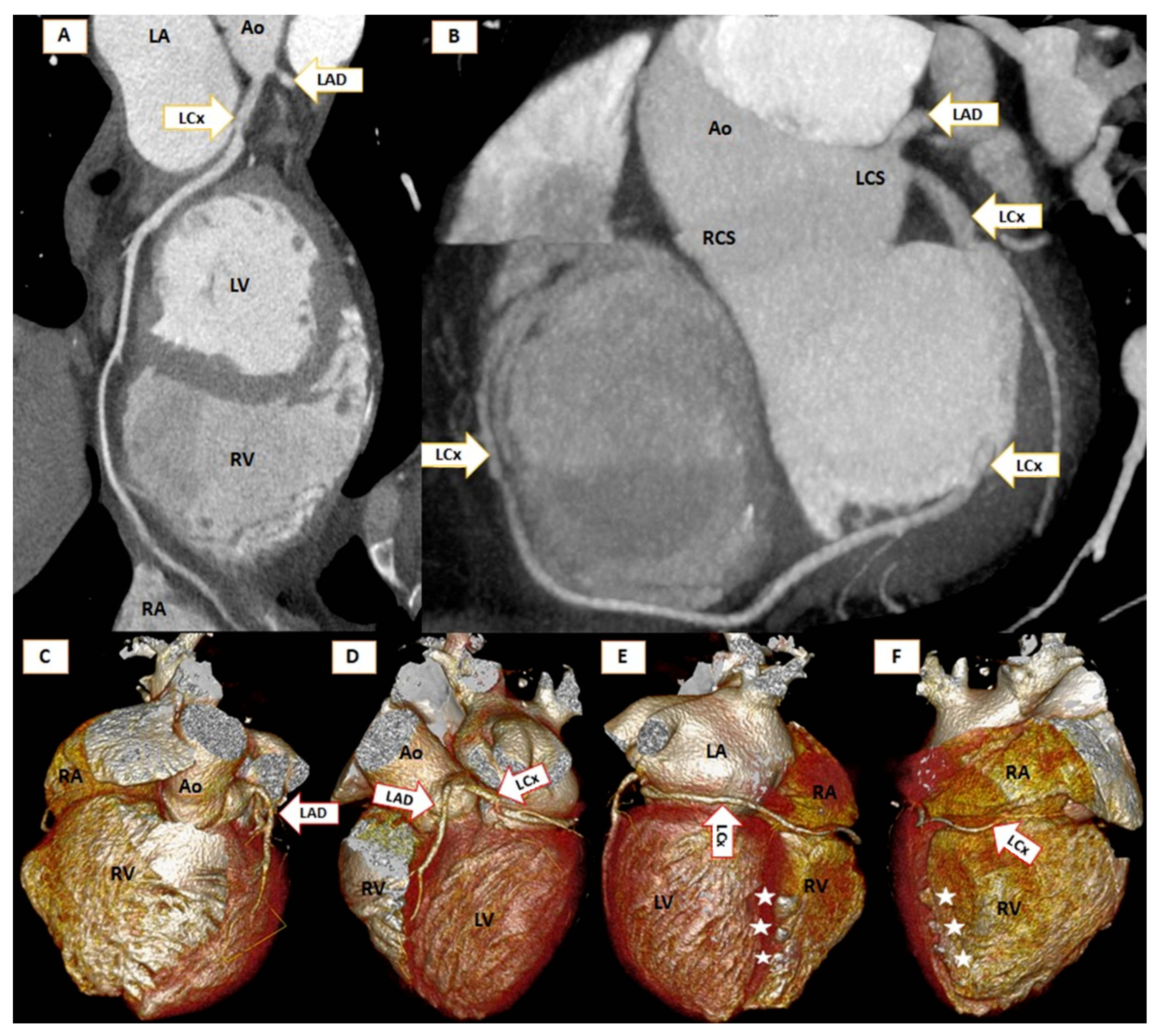
Absence of Both Right and Left Main Coronary in a COVID Survivor
Abstract

Author Contributions
Funding
Institutional Review Board Statement
Informed Consent Statement
Data Availability Statement
Conflicts of Interest
References
- Lipton, M.J.; Barry, W.H.; Obrez, I.; Silverman, J.F.; Wexler, L. Isolated Single Coronary Artery: Diagnosis, Angiographic Classification, and Clinical Significance. Diagn. Radiol. 1979, 130, 1. [Google Scholar] [CrossRef] [PubMed]
- Michalowska, A.M.; Tyczynski, P.; Pregowski, J.; Skowronski, J.; Mintz, G.S.; Kepka, C.; Kruk, M.; Witkowski, A.; Michalowska, I. Prevalence and Anatomic Characteristics of Single Coronary Artery Diagnosed by Computed Tomography Angiography. Am. J. Cardiol. 2019, 124, 939–946. [Google Scholar] [CrossRef] [PubMed]
- Kang, W.C.; Han, S.H.; Ahn, T.H.; Shin, E.K. Unusual dominant course of left circumflex coronary artery with absent right coronary artery. Heart 2006, 92, 657. [Google Scholar] [CrossRef] [PubMed][Green Version]
Publisher’s Note: MDPI stays neutral with regard to jurisdictional claims in published maps and institutional affiliations. |
© 2021 by the authors. Licensee MDPI, Basel, Switzerland. This article is an open access article distributed under the terms and conditions of the Creative Commons Attribution (CC BY) license (https://creativecommons.org/licenses/by/4.0/).
Share and Cite
Pop, M.; Pal, K.; Vaga, D. Absence of Both Right and Left Main Coronary in a COVID Survivor. Diagnostics 2021, 11, 1199. https://doi.org/10.3390/diagnostics11071199
Pop M, Pal K, Vaga D. Absence of Both Right and Left Main Coronary in a COVID Survivor. Diagnostics. 2021; 11(7):1199. https://doi.org/10.3390/diagnostics11071199
Chicago/Turabian StylePop, Marian, Krisztina Pal, and Diana Vaga. 2021. "Absence of Both Right and Left Main Coronary in a COVID Survivor" Diagnostics 11, no. 7: 1199. https://doi.org/10.3390/diagnostics11071199
APA StylePop, M., Pal, K., & Vaga, D. (2021). Absence of Both Right and Left Main Coronary in a COVID Survivor. Diagnostics, 11(7), 1199. https://doi.org/10.3390/diagnostics11071199


