Article

The Effect of Genomic DNA Contamination on the Detection of Circulating Long Non-Coding RNAs: The Paradigm of MALAT1

1
Analysis of Circulating Tumor Cells Lab, Lab of Analytical Chemistry, Department of Chemistry, National and Kapodistrian University of Athens, 15771 Athens, Greece
2
8th Department of Pulmonary Diseases, ‘Sotiria’ General Hospital for Chest Diseases, 11527 Athens, Greece
*
Author to whom correspondence should be addressed.
Diagnostics 2021, 11(7), 1160; https://doi.org/10.3390/diagnostics11071160
Submission received: 19 April 2021 / Revised: 24 May 2021 / Accepted: 21 June 2021 / Published: 25 June 2021

Abstract

:
The presence of contaminating gDNA in RNA preparations is a frequent cause of false positives in RT-PCR-based analysis. However, in some cases, this cannot be avoided, especially when there are no exons–intron junctions in the lncRNA sequences. Due to the lack of exons in few of long noncoding RNAs (lncRNAs) and the lack of DNAse treatment step in most studies reported so far, serious questions are raised about the specificity of lncRNA detection and the potential of reporting false-positive results. We hypothesized that minute amounts of gDNA usually co-extracted with RNA could give false-positive signals since primers would specifically bind to gDNA due to the lack of junction. In the current study, we evaluated the effect of gDNA and other forms of DNA like extrachromosomal circular DNAs (eccDNAs) contamination and the importance of including a DNAse treatment step on lncRNAsexpression.As a model, we have chosen as one of the most widely studied lncRNAs in cancer namely MALAT1, which lacks exons. When we tested this hypothesis in plasma and primary tissue samples from NSCLC patients, our findings clearly indicated that results on MALAT1 expression are highly affected by the presence of DNA contamination and that the DNAse treatment step is absolutely necessary to avoid false positive results.

1. Introduction

Non-coding RNAs (ncRNAs) are RNA molecules that are not translated into a protein but their functions are undoubtedly crucial in several mechanisms. They are divided into short ncRNAs and long ncRNAs(lncRNAs) based on their nucleotide length. The lncRNAs are non-protein-coding transcripts with a length over 200 nucleotides (nt), and consist of the broadest class of ncRNAs [1]. In a meta-analysis, Iyer et al. showed that from a consensus of around 91,000 human genes, over 68% of genes were classified as lncRNAs, of which 79% were previously unannotated [2]. During the last decades, lncRNAs were found to be involved in many biological processes [3] and have thus gained considerable interest as principal regulators of gene expression in several different ways [4,5]. In addition, numerous studies have shown that lncRNAs are abnormally expressed in many cancers, such as breast, lung and prostate [6,7].
Various total RNA and cfRNA isolation methods have been described for the extraction of non-long coding RNAs that are further detected by molecular techniques like RT-qPCR, RNA sequencing, and FISH analysis. Co-isolation of genomic DNA (gDNA), and other forms of DNA, like extrachromosomal circular DNAs (eccDNAs) during the extraction of total RNA from plasma and tissues, is inevitable, unless a DNAse treatment step is included prior to RT-PCR. The presence of contaminating DNAs in RNA preparations can cause false positives in RT-PCR in case those primers are fully overlapping with gDNA sequences. To avoid gDNA co-amplification, specific precautions must be taken in the primers’ design. However, in some cases, this cannot be avoided, especially when the target mRNA presents pseudogenes at the DNA level or when there are no exons–intron junctions.
Many of the well-studied lncRNAs, that are considered as regulatory molecules with various significant functions in cancer, have no junctions in their sequences such as MALAT1, NKILA, NEAT1 and NORAD [8,9,10,11] (Table 1).
Thus, RNA analysis in clinical samples could lead to false-positive results, due to gDNA contamination, and the lack of exons in few of the lncRNAs and the overlapping of all primers designed with gDNA. We noticed that in the vast majority of lncRNAs studies, the expression levels of lncRNAs were evaluated by RT-qPCR without taking into account the possibility of false-positive results due to gDNA contamination. For this reason, we aimed to examine this by analyzing clinical samples for Metastasis-Associated Lung Adenocarcinoma Transcript 1 (MALAT1) with and without DNAse treatment.
MALAT1 is one of the most widely studied lncRNAs in cancer. MALAT1, located on chromosome 11q13 and especially on nuclear speckles [12], was initially identified as a prognostic marker in non-small-cell lung cancer (NSCLC) [13]. Nowadays, its function as an oncogene has been evaluated in several types of cancer like colorectal [14], ovarian [15,16] and gastric cancer [17]. MALAT1 has also been proposed as a reliable biomarker, not only for diagnosis and prognosis, but also in targeted therapy for leukemia [18,19]. Interestingly, the most abundant transcript variant of MALAT1 is the long variant (NR_002819), which lacks exons. As expected, all studies published so far on MALAT1 in cancer use primers that co-hybridize to genomic DNA (Figure 1). However, in the majority of these studies, MALAT1 expression is evaluated by RT-qPCR without any prior DNAse treatment for the removal of contaminating gDNA (Table 2), thus giving rise to significant concerns on specificity, and the presence of false positives, which is highly crucial for the clinical significance of the results presented.
In the current study, we evaluated, for the first time, the effect of gDNA contamination and the importance of including a DNAse treatment step on the expression levels of one of the most widely studied lncRNAs, MALAT1. We tested this in plasma and primary tissue samples from NSCLC patients and healthy donors. Our findings clearly indicated that most results reported so far on MALAT1 expression are highly affected by gDNA contamination, and this could be also extrapolated to all lncRNAs without exons.

2. Materials and Methods

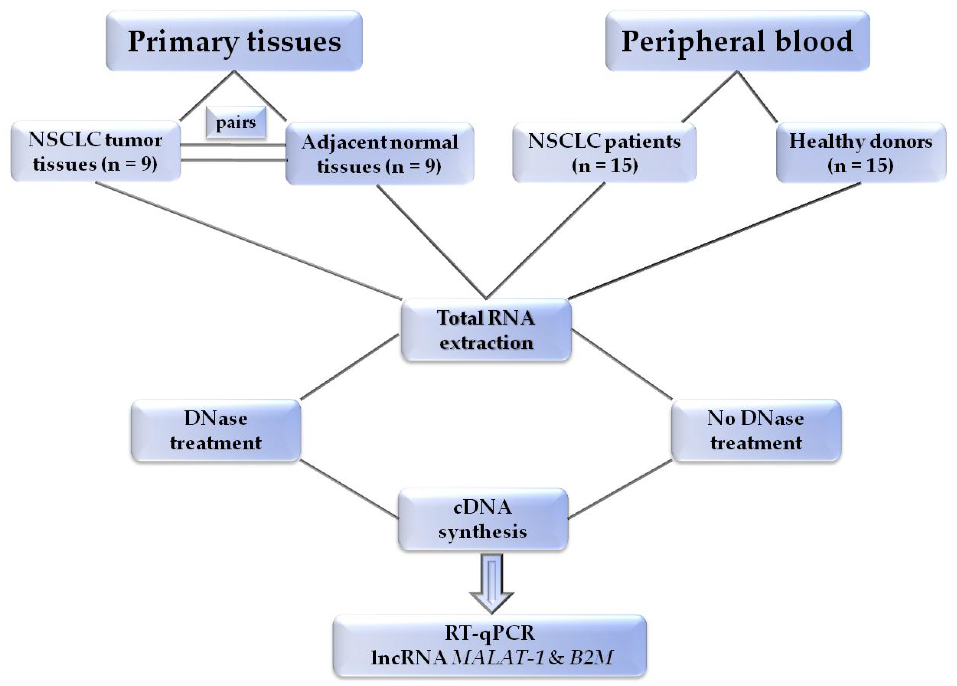
2.1. Patients and Samples

We analyzed a total of 48 clinical samples: (i) 15 peripheral blood samples from patients with early NSCLC, (ii) 15 peripheral blood samples from healthy donors, and (iii) 9 primary tissues of surgically resected NSCLC and their adjacent noncancerous tissue specimens. All patients gave their informed consent, and the Ethical and Scientific Committees of the participating institutions approved the study (28872/10-12-19). At the time of surgery, all tissue samples were immediately flashfrozen in liquid nitrogen and stored at −70 °C until use. We analyzed all samples histologically to assess the amount of tumor component (at least 70% tumor cells) and the quality of material (i.e., absence of necrosis).

2.2. Plasma Preparation

Peripheral blood samples (30 mL) isolated in K3-EDTA tubes were centrifuged at 530× g for 10 min at room temperature, without brakes, within 6 h after collection. Plasma was transferred to fresh tubes and centrifuged at 2000× g for 10 min. Finally, plasma was divided into 2 mL aliquots in fresh tubes, and stored at −80 °C. All samples were collected in the morning before surgery from early NSCLC patients.

2.3. RNA Extraction

Circulating cell-free RNA (ccfRNA) was extracted from 600 μL of plasma using miRNeasy Serum/Plasma Advanced Kit (Qiagen, Hilden, Germany) according to the manufacturer’s instructions, with an elution volume of 25 μL in RNase-free water. In tissue samples, total cellular RNA isolation was performed using the QiagenRNeasy Mini Reagent kit (Qiagen, Hilden, Germany)according to the manufacturer’s instructions [36]. All preparation and handling of RNA took place in a laminar flow hood, under RNase-free conditions, and the isolated RNA was stored at −70 °C until use. RNA concentration was determined with a NanoDrop ND-100 spectrophotometer (NanoDrop Technologies). To accurately assess sample quality, 260/280 and 260/230 ratios were analyzed in combination with overall spectral quality and the yield of 260/280 ratio was acceptable at ~2.0 for RNAs. RNA of each sample was spilt into two aliquots of 10 μL each.

2.4. DNAse Treatment

Initially, in each reaction tube were added: ≤200 ng/μL input RNA, 1 μL of TURBO DNAse Buffer (Ambion Life Technologies, Austin, TX, USA) and 0.4U DNAse I enzyme (Ambion Life Technologies, Austin, TX, USA), and the sample was incubated at 37 °C for 20min. One microliter of DNAse inactivation reagent was then added for 5min followed by centrifugation at 10,000× g for 1.5 min. The supernatant which contains the RNA was then carefully transferred into a fresh tube. The whole procedure was done under DNAse-free conditions to avoid DNA contamination (dedicated specific lab areas, labware, laminar-flow hood). In order to optimize the DNase treatment step, RT-PCR for MALAT1 and B2M in different concentrations of gDNA before and after treatment was performed. Due to the lack of exons in MALAT1, primers would specifically bind to gDNA but B2M primers were designed in the junction area; there was no influence of the presence of gDNA and DNase treatment. Complete degradation of DNA was considered when there was no detection of MALAT1 after DNase treatment in gDNA samples (Figure 2).

2.5. cDNA Synthesis

The high-capacity RNA-to-cDNA kit (Applied Biosystems, Foster City, CA, USA) was used for reverse transcription in 20 μL of total volume reaction. A negative control was included in each experiment to ensure that there was no contamination by genomic DNA (gDNA). All cDNA samples were stored at −20 °C until further molecular analysis.

2.6. RT-qPCR Assay

We first designed insilicohighly specific primers for MALAT1 based on its RNA sequence (NR_002819.4) using Primer Premier 5.0 software (Premier Biosoft, San Francisco, CA, USA). The designed primers for MALAT1 are the following: forward: 5′-CCCCACAAGCAACTTCTCTG-3′ and, reverse: 5′-TCCAAGCTACTGGCTGCATC-3′. The experimental conditions of RT-qPCR for MALAT1 expression were optimized (annealing temperature, time, primer MgCl2, dNTPs, and BSA concentrations). Each reaction was performed in the LightCycler® 2.0 System (IVD instrument, Roche Diagnostics, Mannheim, Germany) in a total volume of 10 μL, following the MIQE guidelines [37]. One microliter of cDNA was added to a 9 μL reaction mixture. The amplification reaction for MALAT1 contained 2 μL of the PCR Synthesis Buffer (5Χ), 1 μL of MgCl2 (25 mM), 0.2 μL dNTPs (10 mM), 0.5 μL BSA (10 μg/μL), 0.1 μL Hot Start DNA polymerase (HotStart, 5 U/μL, Promega, Dane County, WI, USA), 0.3 μL of forward and reverse primer (10 μM), 1 μL of 1X LC Green® (Idaho Technology, Salt Lake City, UT, USA). RT-qPCR protocol begins with one cycle at 95 °C for 2 min followed by 45 cycles of 95 °C for 10 s, 60 °C for 10 s, and 72 °C for 10 s. Immediately after amplification, a rapid cooling cycle to 40 °C for 30 s was introduced in order to prepare the melting curve acquisition step. Real-time fluorescence acquisition was set at the elongation step (72 °C). The following melting curve analysis included the steps of 55 °C for 20 s, 95 °C for 0 s with a ramp rate 0.19 °C/s (acquisition mode: continuous), and 40 °C for 10 s. Additionally, we used our previously developed and analytically validated RT-qPCR assays for beta-2-microglobulin (B2M), used as a reference gene [38]. In each RT-qPCR run, we used the same cDNA from MCF-7 cells as a positive control in order to evaluate the accuracy and reproducibility of the results.

2.7. Normalization of Data

Expression values of MALAT1 were normalized to B2M. ΔCq values were calculated by using Cq values for MALAT1 and the corresponding B2M for each sample. We calculated ΔΔCq values using ΔCq values for cancerous samples and the mean value of ΔCq for normal samples (ΔΔCq = ΔCqcancer − Δcqnormal). Relative quantification (RQ) was based on the ΔΔCq method as described [39]. For paired tissue samples, ΔCq values were calculated as the differences between ΔCq values for each cancerous sample and its corresponding adjacent normal tissue. MALAT1 expression data are presented as fold change relative to the reference gene based on the formula of RQ = 2−ΔΔCq.

2.8. Statistical Analysis

We performed statistical evaluation of data using SPSS (SPSS Statistics version 26.0). A level of p < 0.05 was considered statistically significant. Statistical analysis was performed in all cases by using paired sample t-test.

3. Results

The experimental flowchart of the study is outlined in Figure 3.

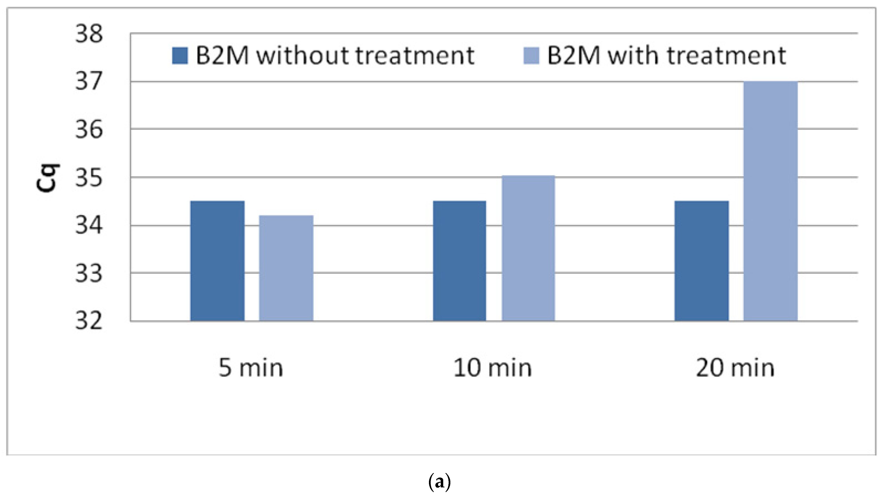
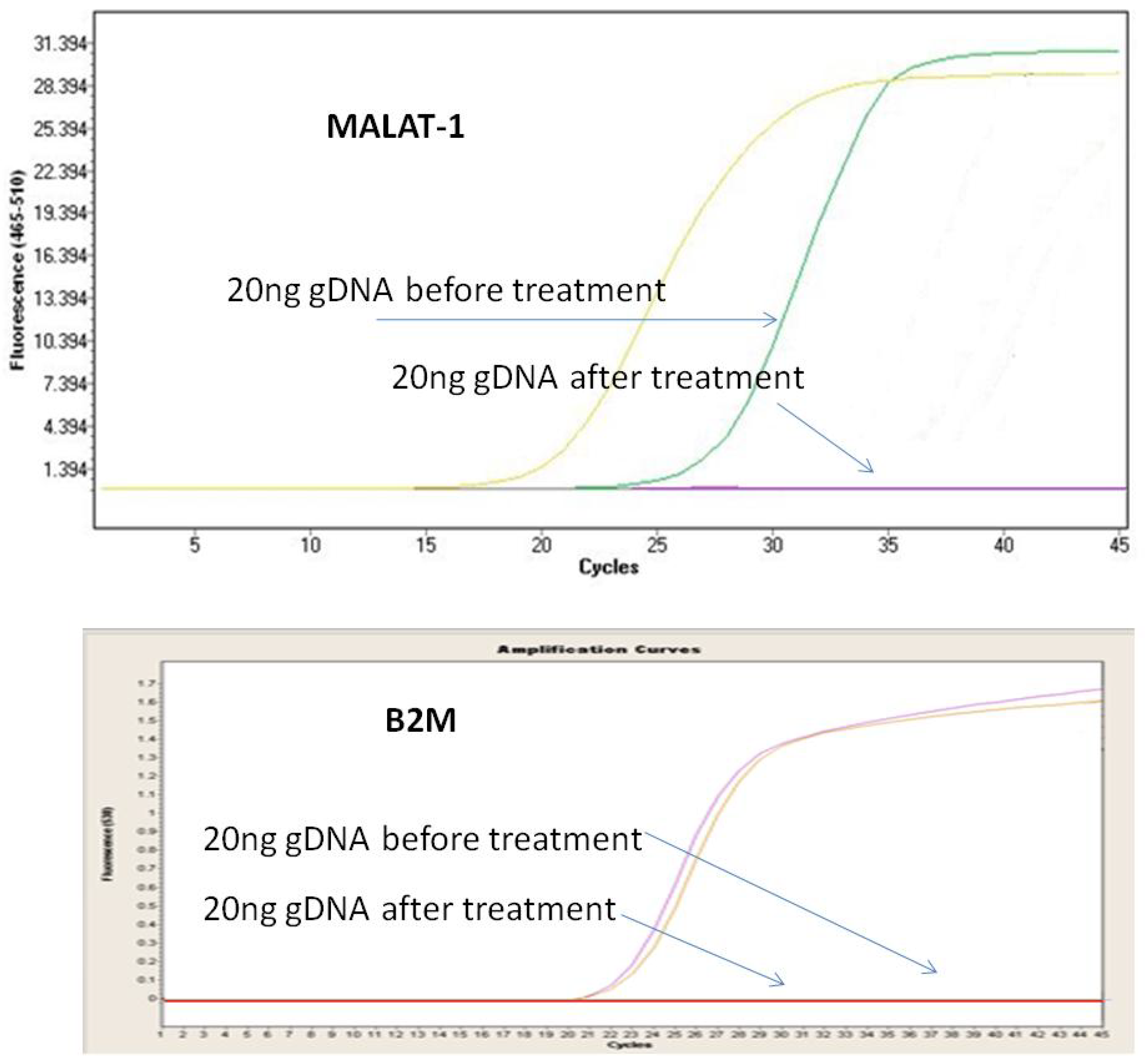
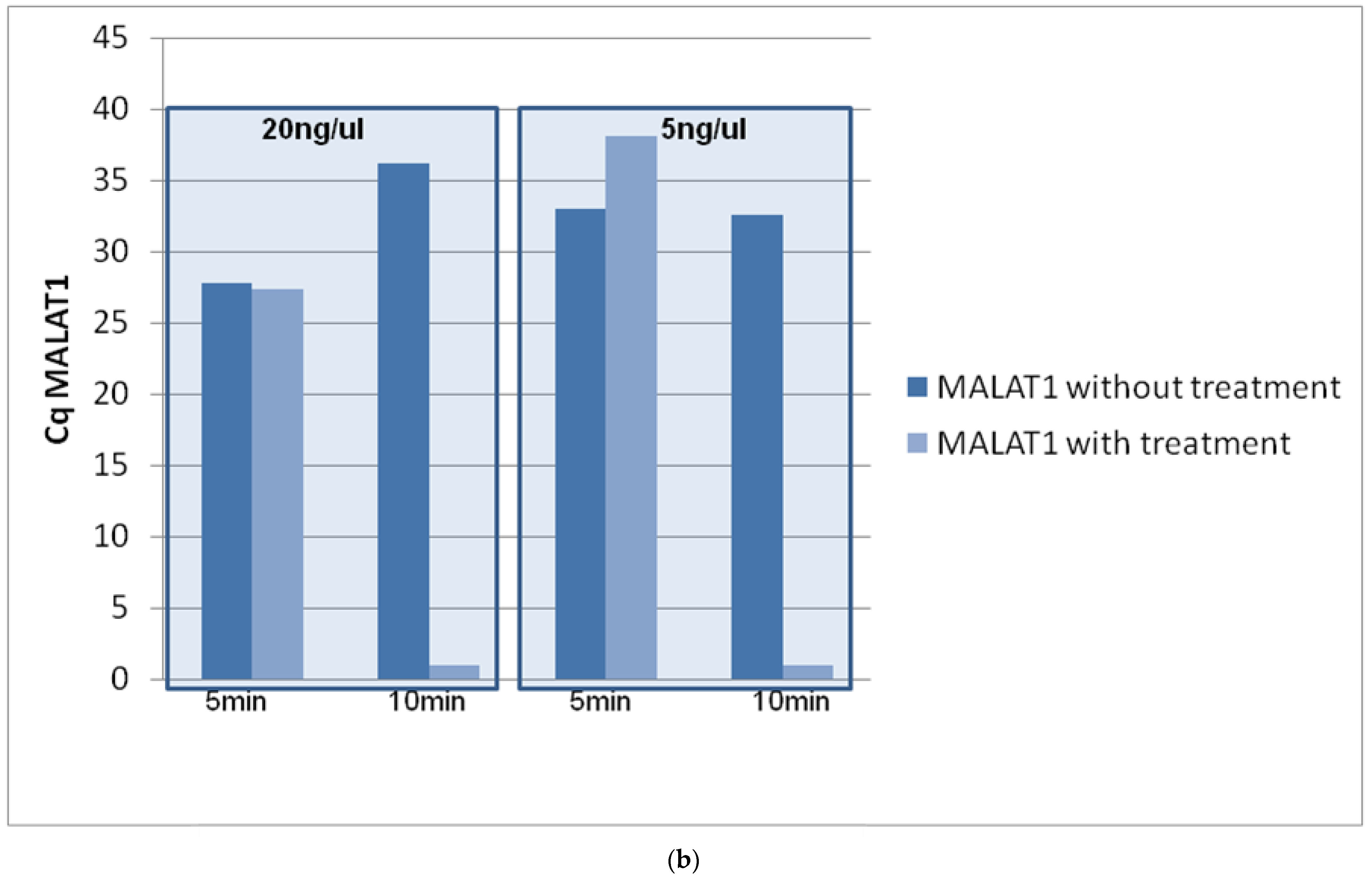
3.1. Optimization of DNase Treatment Conditions

3.1.1. Enzyme Incubation

Equal amounts of total RNA (200 ng) were either treated with TURBO DNA-free™ at different incubation times (5, 10, and, 20 min) at 37 °C, or were left untreated. Treated and untreated RNA samples were reverse-transcribed and cDNA were further analyzed by RT-PCR. RT-PCR was first performed for B2M as reference gene in order to ensure that treatment of RNA with TURBO DNA-free™ maintains target sensitivity in real-time RT-PCR. RNA quality as estimated through B2M expression is not affected when DNAse treatment was performed for 5 or 10 min in 37 °C (Figure 4a). All experiments were run in triplicate.

3.1.2. Concentration of gDNA

We evaluated the effectiveness of the DNAse treatment step by analyzing gDNA samples at concentrations of 20 ng/μL and 5 ng/μL, using 5 and 10 min as incubation time. gDNA was added in the same RNA samples that were split into two aliquots of 10 μL each. One aliquot was treated with TURBO DNA-free™ (for 5 and 10 min of enzyme incubation) and the other aliquots were left untreated. We found out that DNAse incubation at 37 °C for 5 min is not enough for the complete elimination of gDNA. However, when we increased the DNAse incubation time for 10 min, there was no signal for gDNA, while at the same time the effect of DNAse treatment in the quality of RNA was limited (Figure 4b).

3.1.3. Repeatability of the Procedure

We evaluated the repeatability of the whole procedure (within run imprecision) by analyzing the same RNA sample in 3 parallel determinations after DNAse treatment and without DNAse treatment. Intra-assay CVs were satisfactory in all cases.

3.2. False-Positive Results on MALAT1 Expression in Clinical Samples

3.2.1. NSCLC Primary Tissues

We compared the expression of MALAT1 in nine pairs of NSCLC tissues and their adjacent noncancerous tissues using RT-qPCR in samples with and without DNAse treatment. MALAT1 expression was normalized with respect to B2M gene expression based on the relative quantification approach [23]. We observed that in untreated samples, MALAT1 was found to be overexpressed in 6/9 (66.7%) NSCLC tissues, while only 3/6 (50%) remained positive for MALAT1 after DNAse treatment, showing that, in total, 3/9 (30%) tested samples were false positive before the DNAse treatment step. MALAT1 was found to be downregulated in 3/9 (33.3%) tested paired samples before treatment but only 1/9 (11.1%) remained downregulated after DNAse treatment. In 4/9 (44.4%) paired samples, there was no differentiation in the expression of MALAT1 (Supplementary Figure S1). More specifically, in 3/4 (75%) of the samples, we detected MALAT1 overexpression both with and without DNAse treatment, while in 1/4 (25%) of the samples we detected lower expression (Table 3).

3.2.2. ccfRNA in Plasma

MALAT1 and B2M expression were evaluated in 30 RNA samples directly isolated from plasma of early NSCLC patients (n = 15) and healthy donors (HD) (n = 15). Without DNAse treatment, MALAT1 was detected in all samples and overexpression was observed in 3/15 (20%) of NSCLC patients (Supplementary Figure S1). Interestingly, after DNAse treatment, MALAT1 was detected only in 4 of the tested samples and none of them was overexpressed in MALAT1 (Table 3), proving once again the effect of gDNA and the detection of false-positive results.

4. Discussion

lncRNAs have been evaluated as novel tumor biomarkers, not only in diagnosis and prognosis, but also in targeted therapy for different types of cancer [40,41]. RT-qPCR is extensively used for the quantification of lncRNAs transcripts. Contamination of gDNA and other forms of DNAs like extrachromosomal circular DNAs (eccDNAs)—which are the major form of extrachromosomal DNAs—is an inherent problem during RNA purification due to the similar physicochemical properties of RNA and DNA [42,43].
We report in this study that false-positive results could arise due to gDNA contamination and overestimate the abundance of lncRNAs transcript levels. RT-PCR assays can be designed to be gDNA insensitive only if primers can be designed. Such as those designed to target exons flanking a long intron or with primers that cross exon–exon junctions. It is expected that all RT-PCR assays for single-exon genes, like in the case of several lncRNAs, will readily amplify contaminating gDNA.
NEAT1overexpression was associated with poor prognosis in several types of cancer, like breast [44] and digestive system tumors [9], and it was suggested that it could be used as a promising biomarker for diagnosis [45]. NORAD is another lncRNA, which is reported to be overexpressed in many cancers and several studies have explored its involvement in numerous processes associated with carcinogenesis [10]. Moreover, NKILA underexpression was shown to be an effective prognostic and diagnostic biomarker in human cancer [8,46]. The expression of MALAT1 has been evaluated in numerous studies and its increased expression has been correlated with poor overall survival in patients with solid malignancies [47,48]. A common characteristic of all these lncRNAs is the lack of exons in their sequences. Intriguingly almost all studies that have evaluated the expression of these lncRNAs do not include any DNAse treatment step, and thus there is a high probability of reporting falsepositive results.
In this study, we evaluated for the first time the effect of gDNA on the expression levels of MALAT1, a well-studied lncRNA that has a single exon, using RT-qPCR. We initially optimized the protocol to achieve specific DNAse treatment with the lowest effect on RNA and further compared MALAT1 expression in clinical samples before and after DNAse treatment. Our findings clearly indicate that the expression levels of MALAT1 were significantly affected by the presence of gDNA. In paired NSCLC tissue samples, we observed a significant difference in the expression of MALAT1 before and after DNAse treatment in the majority of samples (56.6%). It is highly important that MALAT1 expression was not detected in 73.4% of plasma samples after DNAse treatment. Τhis observation, combined with the lack of DNAse treatment, may explain the ambiguous results of various studies that characterized MALAT1 either as oncogene or as tumor suppressor and consequently report that its expression is upregulated or downregulated, respectively [21,30,49,50]. One of the few studies that performed DNAse treatment before quantification of MALAT1 expression demonstrated that MALAT1 is a metastasis-suppressing lncRNA rather than a metastasis promoter lncRNA in breast cancer [24].
In conclusion, we report for the first time that contamination of gDNA can seriously affect lncRNAs expression results and cause false positives. Our findings need to be further evaluated and validated in a large and well-defined patient cohort. Taking into account that lncRNAs have gained widespread attention in recent years as potentially new and crucial candidates for tumor biomarkers, we conclude that DNAse treatment is a mandatory step in cases where there are no exons in lncRNAs sequence in order to ensure specificity. It is only under these conditions that the clinical significance of lncRNAs will be reliably revealed.

Supplementary Materials

The following are available online at https://www.mdpi.com/article/10.3390/diagnostics11071160/s1, Figure S1: Direct comparison study of MALAT1 expression before and after DNase treatment from (a) NSCLC pairs tissue samples and (b) NSCLC plasma samples.

Author Contributions

Conceptualization, A.N.M.; methodology, A.N.M.; validation, A.N.M., S.S.; formal analysis, A.N.M.; data curation, A.N.M.; writing—original draft preparation, A.N.M., E.L., E.T.; writing—review and editing, A.N.M., E.L.; supervision, A.N.M.; project administration, A.N.M.; funding acquisition, A.N.M. All authors have read and agreed to the published version of the manuscript.

Funding

This research was funded by the Hellenic Foundation for Research and Innovation (HFRI) and the General Secretariat for Research and Technology (GSRT), grant number 1964.

Institutional Review Board Statement

The study was conducted according to the guidelines of the Declaration of Helsinki, and approved by Ethics Committee of Sotiria General Hospital; (28872/10.12.19).

Informed Consent Statement

Informed consent was obtained from all subjects involved in the study.

Data Availability Statement

The data presented in this study are available on request from the corresponding author.

Acknowledgments

This project has received funding from the Hellenic Foundation for Research and Innovation (HFRI) and the General Secretariat for Research and Technology (GSRT), under Grant Agreement No. 1964 and from the European Union and Greek national funds through the Operational Program Competitiveness, Entrepreneurship and Innovation, under the call RESEARCH—CREATE—INNOVATE (project code: T1RCI-02935). We would also like to thank L. Kaklamanis for evaluating the tumor content in all tissue samples analyzed.

Conflicts of Interest

The authors declare no conflict of interest.

References

  1. Kopp, F.; Mendell, J.T. Functional Classification and Experimental Dissection of Long Noncoding RNAs. Cell 2018, 172, 393–407. [Google Scholar] [CrossRef] [Green Version]
  2. Iyer, M.K.; Niknafs, Y.S.; Malik, R.; Singhal, U.; Sahu, A.; Hosono, Y.; Barrette, T.R.; Prensner, J.R.; Evans, J.R.; Zhao, S.; et al. The landscape of longnoncoding RNAs in the human transcriptome. Nat. Genet. 2015, 47, 199–208. [Google Scholar] [CrossRef] [PubMed]
  3. Jarroux, J.; Morillon, A.; Pinskaya, M. History, Discovery, and Classification of lncRNAs. Adv. Exp. Med. Biol. 2017, 1008, 1–46. [Google Scholar] [CrossRef] [PubMed]
  4. Gupta, S.C.; Awasthee, N.; Rai, V.; Chava, S.; Gunda, V.; Challagundla, K.B. Long non-coding RNAs and nuclear factor-κB crosstalk in cancer and other human diseases. Biochim. Biophys. Acta Rev. Cancer 2020, 1873, 188316. [Google Scholar] [CrossRef] [PubMed]
  5. Bassett, A.R.; Akhtar, A.; Barlow, D.P.; Bird, A.P.; Brockdorff, N.; Duboule, D.; Ephrussi, A.; Ferguson-Smith, A.C.; Gingeras, T.R.; Haerty, W.; et al. Considerations when investigating lncRNA function in vivo. Elife 2014, 3, e03058. [Google Scholar] [CrossRef]
  6. Zhang, H.; Wang, J.; Yin, Y.; Meng, Q.; Lyu, Y. The role of EMT-related lncRNA in the process of triple-negative breast cancer metastasis. Biosci. Rep. 2021, 41. [Google Scholar] [CrossRef]
  7. Acha-Sagredo, A.; Uko, B.; Pantazi, P.; Bediaga, N.G.; Moschandrea, C.; Rainbow, L.; Marcus, M.W.; Davies, M.P.A.; Field, J.K.; Liloglou, T. Long non-coding RNA dysregulation is a frequent event in non-small cell lung carcinoma pathogenesis. Br. J. Cancer 2020, 122, 1050–1058. [Google Scholar] [CrossRef] [PubMed]
  8. Tian, S.; Yu, Y.; Huang, H.; Xu, A.; Xu, H.; Zhou, Y. Expression Level and Clinical Significance of NKILA in Human Cancers: A Systematic Review and Meta-Analysis. BioMed. Res. Int. 2020, 2020, 1–9. [Google Scholar] [CrossRef]
  9. Guo, L.; Tang, Y.; Wang, Y.; Xu, H. Prognostic Value of lncRNA NEAT1 as a New Biomarker in Digestive System Tumors: A Systematic Study and Meta-analysis. Expert Rev. Mol. Diagn. 2021, 21, 91–99. [Google Scholar] [CrossRef]
  10. Soghli, N.; Yousefi, T.; Abolghasemi, M.; Qujeq, D. NORAD, a critical long non-coding RNA in human cancers. Life Sci. 2021, 264, 118665. [Google Scholar] [CrossRef] [PubMed]
  11. Amirinejad, R.; Rezaei, M.; Shirvani-Farsani, Z. An update on long intergenic noncoding RNA p21: A regulatory molecule with various significant functions in cancer. Cell Biosci. 2020, 10, 82. [Google Scholar] [CrossRef] [PubMed]
  12. Tripathi, V.; Ellis, J.D.; Shen, Z.; Song, D.Y.; Pan, Q.; Watt, A.T.; Freier, S.M.; Bennett, C.F.; Sharma, A.; Bubulya, P.A.; et al. The nuclear-retained noncoding RNA MALAT1 regulates alternative splicing by modulating SRsplicing factor phosphorylation. Mol. Cell. 2010, 39, 925–938. [Google Scholar] [CrossRef] [PubMed] [Green Version]
  13. Ji, P.; Diederichs, S.; Wang, W.; Böing, S.; Metzger, R.; Schneider, P.M.; Tidow, N.; Brandt, B.; Buerger, H.; Bulk, E.; et al. MALAT-1, a novel noncoding RNA, and thymosin beta4 predict metastasis and survival in early-stage non-small cell lung cancer. Oncogene 2003, 22, 8031–8041. [Google Scholar] [CrossRef] [Green Version]
  14. Wu, S.; Sun, H.; Wang, Y.; Yang, X.; Meng, Q.; Yang, H.; Zhu, H.; Tang, W.; Li, X.; Aschner, M.; et al. MALAT1 rs664589 Polymorphism Inhibits Binding to miR-194-5p, Contributing to Colorectal Cancer Risk, Growth, and Metastasis. Cancer Res. 2019, 79, 5432–5441. [Google Scholar] [CrossRef] [Green Version]
  15. Pei, C.; Gong, X.; Zhang, Y. LncRNA MALAT-1 promotes growth and metastasis of epithelial ovarian cancer via sponging microrna-22. Am. J. Transl. Res. 2020, 12, 6977–6987. [Google Scholar]
  16. Wang, K.; Zhao, Y.; Wang, Y.-M. LncRNA MALAT1 Promotes Survival of Epithelial Ovarian Cancer Cells by Downregulating miR-145-5p. Cancer Manag. Res. 2020, 12, 11359–11369. [Google Scholar] [CrossRef]
  17. Zhang, Z.; Li, M.; Zhang, Z. Lncrna malat1 modulates oxaliplatin resistance of gastric cancer via sponging mir-22-3p. Onco Targets Ther. 2020, 13, 1343–1354. [Google Scholar] [CrossRef] [PubMed] [Green Version]
  18. Amodio, N.; Stamato, M.A.; Juli, G.; Morelli, E.; Fulciniti, M.; Manzoni, M.; Taiana, E.; Agnelli, L.; Cantafio, M.E.G.; Romeo, E.; et al. Drugging the lncRNA MALAT1 via LNA gapmeR ASO inhibits gene expression of proteasome subunits and triggers anti-multiple myeloma activity. Leukemia 2018, 32, 1948–1957. [Google Scholar] [CrossRef] [Green Version]
  19. Gong, N.; Teng, X.; Li, J.; Liang, X.-J. Antisense Oligonucleotide-Conjugated Nanostructure-Targeting lncRNA MALAT1 Inhibits Cancer Metastasis. ACS Appl. Mater. Interfaces 2018, 11, 37–42. [Google Scholar] [CrossRef] [PubMed]
  20. Tiansheng, G.; Junming, H.; Xiaoyun, W.; Peixi, C.; Shaoshan, D.; Qianping, C. lncRNA Metastasis-Associated Lung Adenocarcinoma Transcript 1 Promotes Proliferation and Invasion of Non-Small Cell Lung Cancer Cells via Down-Regulating miR-202 Expression. Cell J. 2019, 22, 375–385. [Google Scholar]
  21. Guo, F.; Yu, F.; Wang, J.; Li, Y.; Li, Y.; Li, Z.; Zhou, Q. Expression of MALAT1 in the peripheral whole blood of patients with lung cancer. Biomed. Rep. 2015, 3, 309–312. [Google Scholar] [CrossRef] [PubMed] [Green Version]
  22. Wu, J.; Weng, Y.; He, F.; Liang, D.; Cai, L. LncRNA MALAT-1 competitively regulates miR-124 to promote EMT and development of non-small-cell lung cancer. Anti-Cancer Drugs 2018, 29, 628–636. [Google Scholar] [CrossRef]
  23. Zhang, R.; Xia, Y.; Wang, Z.; Zheng, J.; Chen, Y.; Li, X.; Wang, Y.; Ming, H. Serum long non coding RNA MALAT-1 protected by exosomes is up-regulated and promotes cell proliferation and migration in non-small cell lung cancer. Biochem. Biophys. Res. Commun. 2017, 490, 406–414. [Google Scholar] [CrossRef]
  24. Lv, P.; Yang, S.; Liu, W.; Qin, H.; Tang, X.; Wu, F.; Liu, Z.; Gao, H.; Liu, X. Circulating plasma lncRNAs as novel markers of EGFR mutation status and monitors of epidermal growth factor receptor-tyrosine kinase inhibitor therapy. Thorac. Cancer 2020, 11, 29–40. [Google Scholar] [CrossRef] [PubMed]
  25. Chen, W.; Zhao, W.; Zhang, L.; Wang, L.; Wang, J.; Wan, Z.; Hong, Y.; Yu, L. MALAT1-miR-101-SOX9 feedback loop modulates the chemo-resistance of lung cancer cell to DDP via Wnt signaling pathway. Oncotarget 2017, 8, 94317–94329. [Google Scholar] [CrossRef] [Green Version]
  26. Song, J.; Su, Z.-Z.; Shen, Q.-M. Long non-coding RNA MALAT1 regulates proliferation, apoptosis, migration and invasion via miR-374b-5p/SRSF7 axis in non-small cell lung cancer. Eur. Rev. Med. Pharmacol. Sci. 2020, 24, 1853–1862. [Google Scholar]
  27. Liu, J.; Tian, W.; Zhang, W.; Jia, Y.; Yang, X.; Wang, Y.; Zhang, J. MicroRNA-142-3p/MALAT1 inhibits lung cancer progression through repressing β-catenin expression. Biomed. Pharmacother. 2019, 114, 108847. [Google Scholar] [CrossRef]
  28. Ren, S.; Wang, F.; Shen, J.; Sun, Y.; Xu, W.; Lu, J.; Wei, M.; Xu, C.; Wu, C.; Zhang, Z.; et al. Long non-coding RNA metastasis associated in lung adenocarcinoma transcript 1 derived miniRNA as a novel plasma-based biomarker for diagnosing prostate cancer. Eur. J. Cancer 2013, 49, 2949–2959. [Google Scholar] [CrossRef]
  29. Hao, T.; Wang, Z.; Yang, J.; Zhang, Y.; Shang, Y.; Sun, J. MALAT1 knockdown inhibits prostate cancer progression by regulating miR-140/BIRC6 axis. Biomed. Pharmacother. 2020, 123, 109666. [Google Scholar] [CrossRef] [PubMed]
  30. Kim, J.; Piao, H.-L.; Kim, B.-J.; Yao, F.; Han, Z.; Wang, Y.; Xiao, Z.; Siverly, A.N.; Lawhon, S.E.; Ton, B.N.; et al. Long noncoding RNA MALAT1 suppresses breast cancer metastasis. Nat. Genet. 2018, 50, 1705–1715. [Google Scholar] [CrossRef]
  31. Stone, J.K.; Kim, J.-H.; Vukadin, L.; Richard, A.; Giannini, H.K.; Lim, S.-T.S.; Tan, M.; Ahn, E.-Y.E. Hypoxia induces cancer cell-specific chromatin interactions and increases MALAT1 expression in breast cancer cells. J. Biol. Chem. 2019, 294, 11213–11224. [Google Scholar] [CrossRef] [PubMed]
  32. Zheng, L.; Zhang, Y.; Fu, Y.; Gong, H.; Guo, J.; Wu, K.; Jia, Q.; Ding, X. Long non-coding RNA MALAT1 regulates BLCAP mRNA expression through binding to miR-339-5p and promotes poor prognosis in breast cancer. Biosci. Rep. 2019, 39. [Google Scholar] [CrossRef] [Green Version]
  33. Zhu, K.; Ren, Q.; Zhao, Y. lncRNA MALAT1 overexpression promotes proliferation, migration and invasion of gastric cancer by activating the PI3K/AKT pathway. Oncol. Lett. 2019, 17, 5335–5342. [Google Scholar] [CrossRef] [Green Version]
  34. Shao, G.; Zhao, Z.; Zhao, W.; Hu, G.; Zhang, L.; Li, W.; Xing, C.; Zhang, X. Long non-coding RNA MALAT1 activates autophagy and promotes cell proliferation by downregulating microRNA-204 expression in gastric cancer. Oncol. Lett. 2019, 19, 805–812. [Google Scholar] [CrossRef]
  35. Yiren, H.; Yingcong, Y.; Sunwu, Y.; Keqin, L.; XiaoChun, T.; Senrui, C.; Ende, C.; Xizhou, L.; Yanfan, C. Long noncoding RNA MALAT1 regulates autophagy associated chemoresistance via miR-23b-3p sequestration in gastric cancer. Mol. Cancer 2017, 16, 174. [Google Scholar] [CrossRef] [PubMed] [Green Version]
  36. Zygalaki, E.; Tsaroucha, E.G.; Kaklamanis, L.; Lianidou, E.S. Quantitative Real-Time Reverse Transcription–PCR Study of the Expression of Vascular Endothelial Growth Factor (VEGF) Splice Variants and VEGF Receptors (VEGFR-1 and VEGFR-2) in Non–Small Cell Lung Cancer. Clin. Chem. 2007, 53, 1433–1439. [Google Scholar] [CrossRef] [Green Version]
  37. Bustin, S.A.; Benes, V.; Garson, J.A.; Hellemans, J.; Huggett, J.; Kubista, M.; Mueller, R.; Nolan, T.; Pfaffl, M.W.; Shipley, G.L.; et al. The MIQE Guidelines: Minimum Information for Publication of Quantitative Real-Time PCR Experiments. Clin. Chem. 2009, 55, 611–622. [Google Scholar] [CrossRef] [PubMed] [Green Version]
  38. Zavridou, M.; Mastoraki, S.; Strati, A.; Tzanikou, E.; Chimonidou, M.; Lianidou, E. Evaluation of Preanalytical Conditions and Implementation of Quality Control Steps for Reliable Gene Expression and DNA Methylation Analyses in Liquid Biopsies. Clin. Chem. 2018, 64, 1522–1533. [Google Scholar] [CrossRef]
  39. Livak, K.J.; Schmittgen, T.D. Analysis of Relative Gene Expression Data Using Real-Time Quantitative PCR and the 2-Delta Delta C(T) Method. Methods 2001, 25, 402–408. [Google Scholar] [CrossRef]
  40. Nandwani, A.; Rathore, S.; Datta, M. LncRNAs in cancer: Regulatory and therapeutic implications. Cancer Lett. 2021, 501, 162–171. [Google Scholar] [CrossRef] [PubMed]
  41. Taniue, K.; Akimitsu, N. The Functions and Unique Features of LncRNAs in Cancer Development and Tumorigenesis. Int. J. Mol. Sci. 2021, 22, 632. [Google Scholar] [CrossRef] [PubMed]
  42. Laurell, H.; Iacovoni, J.S.; Abot, A.; Svec, D.; Maoret, J.J.; Arnal, J.F.; Kubista, M. Correction of RT-qPCR data for genomic DNA-derived signals with ValidPrime. Nucleic Acids Res. 2012, 40, e51. [Google Scholar] [CrossRef]
  43. Wang, M.; Chen, X.; Yu, F.; Ding, H.; Zhang, Y.; Wang, K. Extrachromosomal Circular DNAs: Origin, formation and emerging function in Cancer. Int. J. Biol. Sci. 2021, 17, 1010–1025. [Google Scholar] [CrossRef]
  44. Swellam, M.; EL Magdoub, H.M.; Shawki, M.A.; Adel, M.; Hefny, M.M.; El-Shazly, S.S. Clinical impact of LncRNA XIST and LncRNA NEAT1 for diagnosis of high-risk group breast cancer patients. Curr. Probl. Cancer 2021, 2021, 100709. [Google Scholar] [CrossRef] [PubMed]
  45. Yuan, S.; Xiang, Y.; Guo, X.; Zhang, Y.; Li, C.; Xie, W.; Wu, N.; Wu, L.; Cai, T.; Ma, X.; et al. Circulating Long Noncoding RNAs Act as Diagnostic Biomarkers in Non-Small Cell Lung Cancer. Front. Oncol. 2020, 10. [Google Scholar] [CrossRef] [PubMed]
  46. Jiang, P.; Han, X.; Zheng, Y.; Sui, J.; Bi, W. Long non-coding RNA NKILA serves as a biomarker in the early diagnosis and prognosis of patients with colorectal cancer. Oncol. Lett. 2019, 18, 2109–2117. [Google Scholar] [CrossRef] [Green Version]
  47. Gutschner, T.; Hämmerle, M.; Diederichs, S. MALAT1—A paradigm for long noncoding RNA function in cancer. J. Mol. Med. 2013, 91, 791–801. [Google Scholar] [CrossRef]
  48. Goyal, B.; Yadav, S.R.M.; Awasthee, N.; Gupta, S.; Kunnumakkara, A.B.; Gupta, S.C. Diagnostic, prognostic, and therapeutic significance of long non-coding RNA MALAT1 in cancer. Biochim. Biophys. ActaBioenerg. 2021, 1875, 188502. [Google Scholar] [CrossRef]
  49. Huang, Z.; Qin, Q.; Xia, L.; Lian, B.; Tan, Q.; Yu, Y.; Mo, Q. Significance of Oncotype DX 21-Gene Test and Expression of Long Non-Coding RNA MALAT1 in Early and Estrogen Receptor-Positive Breast Cancer Patients. Cancer Manag. Res. 2021, 13, 587–593. [Google Scholar] [CrossRef]
  50. Liu, X.; Huang, G.; Zhang, J.; Zhang, L.; Liang, Z. Prognostic and clinicopathological significance of long noncoding RNA MALAT-1 expression in patients with non-small cell lung cancer: A meta-analysis. PLoS ONE 2020, 15, e0240321. [Google Scholar] [CrossRef]
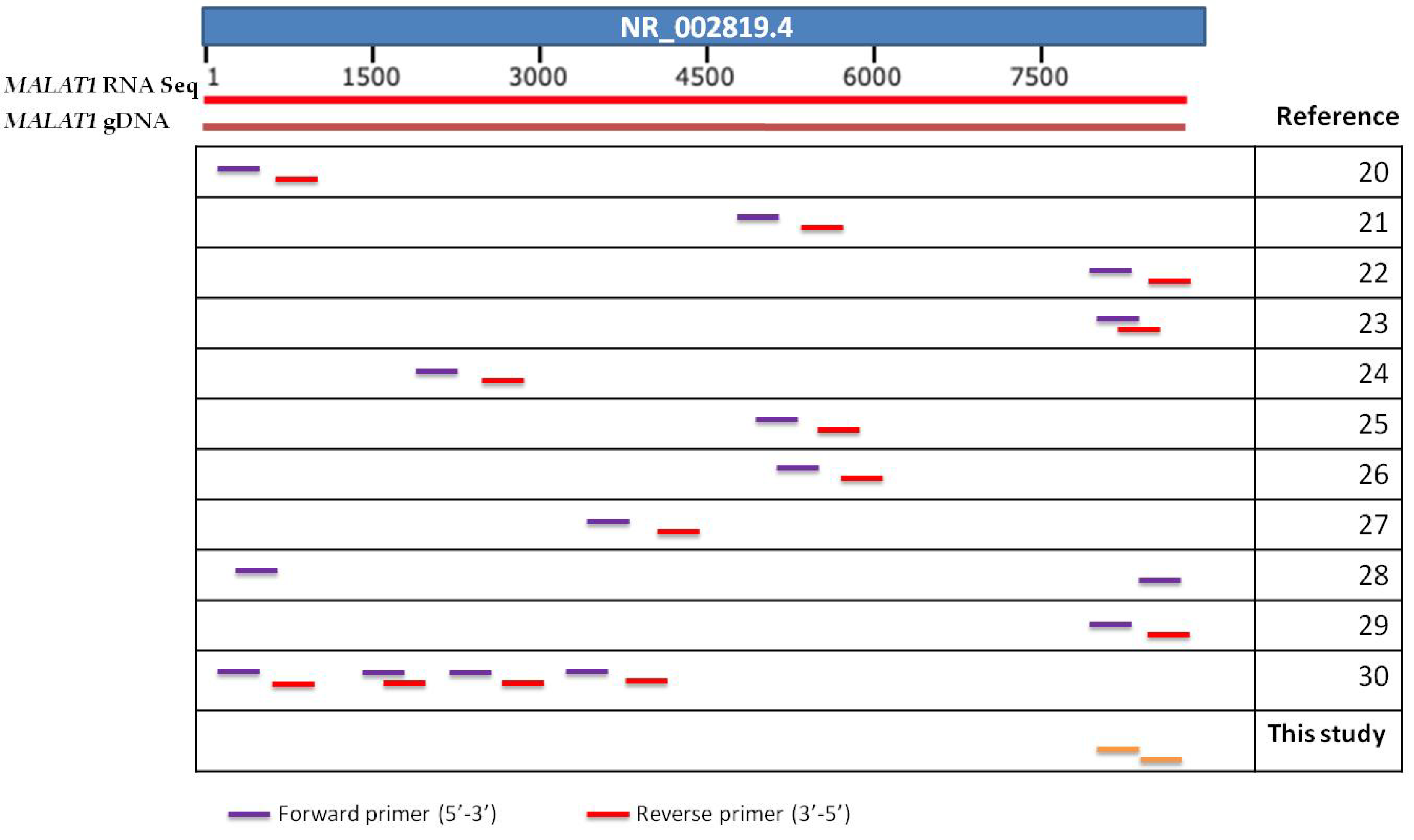
Figure 1. Graphic summary of lncRNA MALAT1 reference sequence, MALAT1 gDNA and position of different pairs of primers designed used in various studies. The last pair (green) was the one designed in the present study.
Diagnostics 11 01160 g001
Figure 2. B2M and MALAT1 expression before and after DNase treatment.
Diagnostics 11 01160 g002
Figure 3. Outline of the experimental procedure.
Diagnostics 11 01160 g003
Figure 4. (a): Effect of enzyme incubation time. (b):Effect of gDNA concentration.
Diagnostics 11 01160 g004aDiagnostics 11 01160 g004b
Table 1. Long non-coding RNAs which lack exons.
Long Non-Coding RNACancer TypesRef. Sequence
MALAT1Lung cancer,
Esophageal carcinoma,
Acute myeloid leukemia,
Ovarian cancer, thyroid, nerve—tibial, skin, uterus, prostate
NR_002819.4
NEAT1Breast, lung,
Prostate cancer,
Head and neck squamous cell carcinoma, colon cancer, thyroid cancer
NR_028272.1
DLEUAcute myeloid leukemia, spleen, lung cancer, esophageal carcinoma, pancreatic, laryngeal, renal, cervical cancerNR_002612.1
ANRASSF1Breast, prostate, astric cancerNR_109831.1
NKILAPancreatic adenocarcinoma, prostate, breast cancer, uterine carcinosarcoma, lung cancerNR_131157.1
NORADLymph node metastasis, pancreatic, bladder, gastric cancer; esophageal squamous cell carcinoma, epatocellularcarcinoma, colorectal cancerNR_027451.1
KCNQ1OT1Esophageal carcinoma,
Acute myeloid leukemia,
Ovarian,
Stomach adenocarcinoma
NR_002728.3
CCAT2Colon cancer, breast cancer, hepatocellular carcinomaNR_109834.1
LincRNA-p21Prostate, gastric, colorectal cancer, head and neck squamous cell carcinoma, lung cancerCD515754.1
Table 2. MALAT1 expression in cancer.
Cancer TypeSample OriginNumber of SamplesReference GeneDNase TreatmentExpression of MALAT-1SignificanceReference
NSCLCTissues (tumor and adjacent) Cell Lines40GAPDHYesOver-expressedTherapeutic target[20]
Plasma Tissues105
65
GAPDHNoUnder-expressedDiagnostic[21]
Tissues (tumor and adjacent)86GAPDHNoOver-expressedTumor progression and development[22]
Serum (exosomes)77GAPDHNoOver-expressedDiagnostic, prognostic, therapeutic target[23]
Plasma14218S rRNANoOver-expressedDiagnosis of EGFR-mutant patients[24]
Cell Lines
Tissues (tumor and adjacent)
42RNU6BNoOver-expressedTherapeutic target[25]
Cell Lines
Tissues (tumor and adjacent)
30GAPDH or U6NoOver-expressedDiagnostic[26]
Cell Lines
Tissues (tumor and adjacent)
36GAPDHNoOver-expressedTherapeutic target[27]
Prostate cancerPlasma Tissues (tumor and adjacent)169
14
β-actinYesOver-expressedDiagnostic[28]
Cell Lines Tissues (tumor and adjacent) Mice52β-actinNoOver-expressedTherapeutic target[29]
Breast cancerCell Lines Mice-GAPDHYesUnder-expressedPrognostic, Therapeutic target[30]
Cell Lines Mice-YWHAZNoUnder-expressedTherapeutic target[31]
Cell Lines Clinical samples-GAPDHNoOver-expressedPrognostic[32]
Gastric cancerTissues Plasma64β-actinNoOver-expressedPrognostic, diagnostic[33]
Cell Lines Tissues (tumor and adjacent)57GAPDHNoOver-expressedTherapeutic target[34]
Cell Lines Tissues Mice153GAPDHNoOver-expressedTherapeutic[35]
The bold “Yes” means that in this study DNase treatment was performed.
Table 3. MALAT1 expression in plasma (n = 15) and tissue samples (n = 9 pairs) of NSCLC patients before and after DNase treatment.
Plasma
After DNAse treatment Before DNAse treatment
OverexpressionUnderexpression
Overexpression00
Underexpression3 (False positive)12
Paired t-test: 0.082
False positives: 3/15 (20%)
Primary Tissues
After DNAse treatment Before DNAse treatment
OverexpressionUnderexpression
Overexpression32
Underexpression3 (False positive)1
Paired t-test: 0.681
False positives: 3/9 (30%)
Publisher’s Note: MDPI stays neutral with regard to jurisdictional claims in published maps and institutional affiliations.

