Article

Elastic Modulus and Elasticity Ratio of Malignant Breast Lesions with Shear Wave Ultrasound Elastography: Variations with Different Region of Interest and Lesion Size

by
Antonio Bulum
1,*,
Gordana Ivanac
1,2,
Eugen Divjak
1,
Iva Biondić Špoljar
1,
Martina Džoić Dominković
3,
Kristina Bojanić
4,5,6,
Marko Lucijanić
2,7 and
Boris Brkljačić
1,2
1
Department of Diagnostic and Interventional Radiology, University Hospital Dubrava, Avenija Gojka Šuška 6, 10000 Zagreb, Croatia
2
Department of Radiology, School of Medicine, University of Zagreb, Šalata 3, 10000 Zagreb, Croatia
3
Department of Radiology, General Hospital Orašje, 3rd Street, 76270 Orašje, Bosnia and Herzegovina
4
Department of Otorhinolaryngology, Neurosurgery and Radiology, Faculty of Dental Medicine and Health Osijek, J.J. Strossmayer University of Osijek, Crkvena ul. 21, 31000 Osijek, Croatia
5
Department of Biophysics and Radiology, Faculty of Medicine Osijek, J.J. Strossmayer University of Osijek, Ul. Josipa Huttlera 4, 31000 Osijek, Croatia
6
Department of Radiology, Health Center Osječko-Baranjska County, Ul. Josipa Huttlera 4, 31000 Osijek, Croatia
7
Hematology Department, University Hospital Dubrava, Avenija Gojka Šuška 6, 10000 Zagreb, Croatia
*
Author to whom correspondence should be addressed.
Diagnostics 2021, 11(6), 1015; https://doi.org/10.3390/diagnostics11061015
Submission received: 30 March 2021 / Revised: 22 May 2021 / Accepted: 27 May 2021 / Published: 1 June 2021
(This article belongs to the Special Issue Elastography 2.0)

Abstract

:
Shear wave elastography (SWE) is a type of ultrasound elastography with which the elastic properties of breast tissues can be quantitatively assessed. The purpose of this study was to determine the impact of different regions of interest (ROI) and lesion size on the performance of SWE in differentiating malignant breast lesions. The study included 150 female patients with histopathologically confirmed malignant breast lesions. Minimal (Emin), mean (Emean), maximal (Emax) elastic modulus and elasticity ratio (e-ratio) values were measured using a circular ROI size of 2, 4 and 6 mm diameters and the lesions were divided into large (diameter ≥ 15 mm) and small (diameter < 15 mm). Highest Emin, Emean and e-ratio values and lowest variability were observed when using the 2 mm ROI. Emax values did not differ between different ROI sizes. Larger lesions had significantly higher Emean and Emax values, but there was no difference in e-ratio values between lesions of different sizes. In conclusion, when measuring the Emin, Emean and e-ratio of malignant breast lesions using SWE the smallest possible ROI size should be used regardless of lesion size. ROI size has no impact on Emax values while lesion size has no impact on e-ratio values.

1. Introduction

Ultrasound elastography is an ultrasound technology which detects malignant breast lesions by measuring the changes of the elastic properties of breast tissue. There are two types of ultrasound elastography, strain and shear wave elastography (SWE) [1,2,3,4].
Strain elastography is performed by manual compression using the transducer which then produces an image based on the resulting displacement of the breast tissue caused by the compression. However, it is difficult to measure the exact amount of the applied force during compression resulting in the method being difficult to standardize. Additionally, the absolute elasticity values cannot be calculated, only qualitative results can be obtained [5]. Unlike strain elastography, SWE is a type of ultrasound elastography where the elastic properties of breast tissue can be both qualitatively and quantitatively assessed. The quantitative results are obtained by measuring the Young’s modulus of tissue elasticity in kilopascals (kPa), based on the focused ultrasound beam inducing mechanical vibrations and tissue displacement resulting in shear waves perpendicular to ultrasound beam direction. The velocity of shear waves can be measured and is proportional to tissue elasticity, allowing elastic modulus to be calculated. These values can be color-coded in various colors ranging between red and dark blue, which are superimposed over the gray-scale image in real time (so-called elastogram) [6]. In addition to the elastic modulus values, the elasticity ratio (e-ratio) can also be measured and provides another quantitative tool for the assessment of the tissue in focus [7]. The SWE acquisition box can be enlarged and positioned over the examined parts of the breast, encompassing focal lesions and surrounding fatty tissues. The elastic modulus is measured using a region of interest (ROI) circle that is positioned over the examined lesion in the breast, and a second ROI is positioned over the surrounding fatty tissue to calculate the e-ratio value between the two. The influence of the size of the ROI on the measured values of the elasticity modulus was addressed in only a few other publications assessing the utility of SWE in diagnosing breast cancer. In addition to that, the studies differed in methodology in regard to the ROI size, from using a ROI size of 1 mm to encompassing the entire lesion [8,9,10].
The aim of this study was to investigate the relation of the ROI area and the size of the breast lesion and their influence on the measured values of minimal (Emin), mean (Emean), maximal elastic modulus (Emax) and e-ratio of malignant breast lesions.

2. Materials and Methods

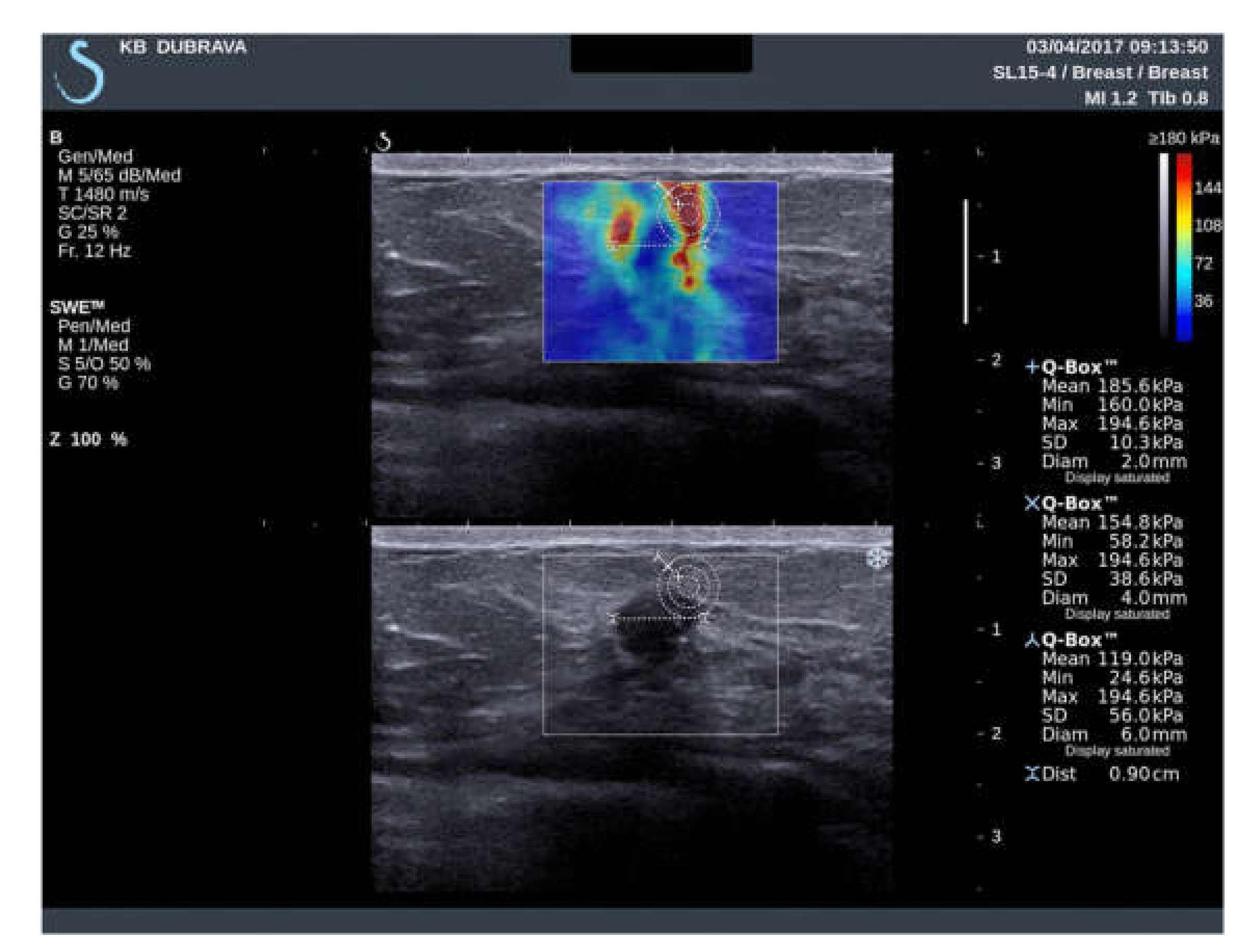
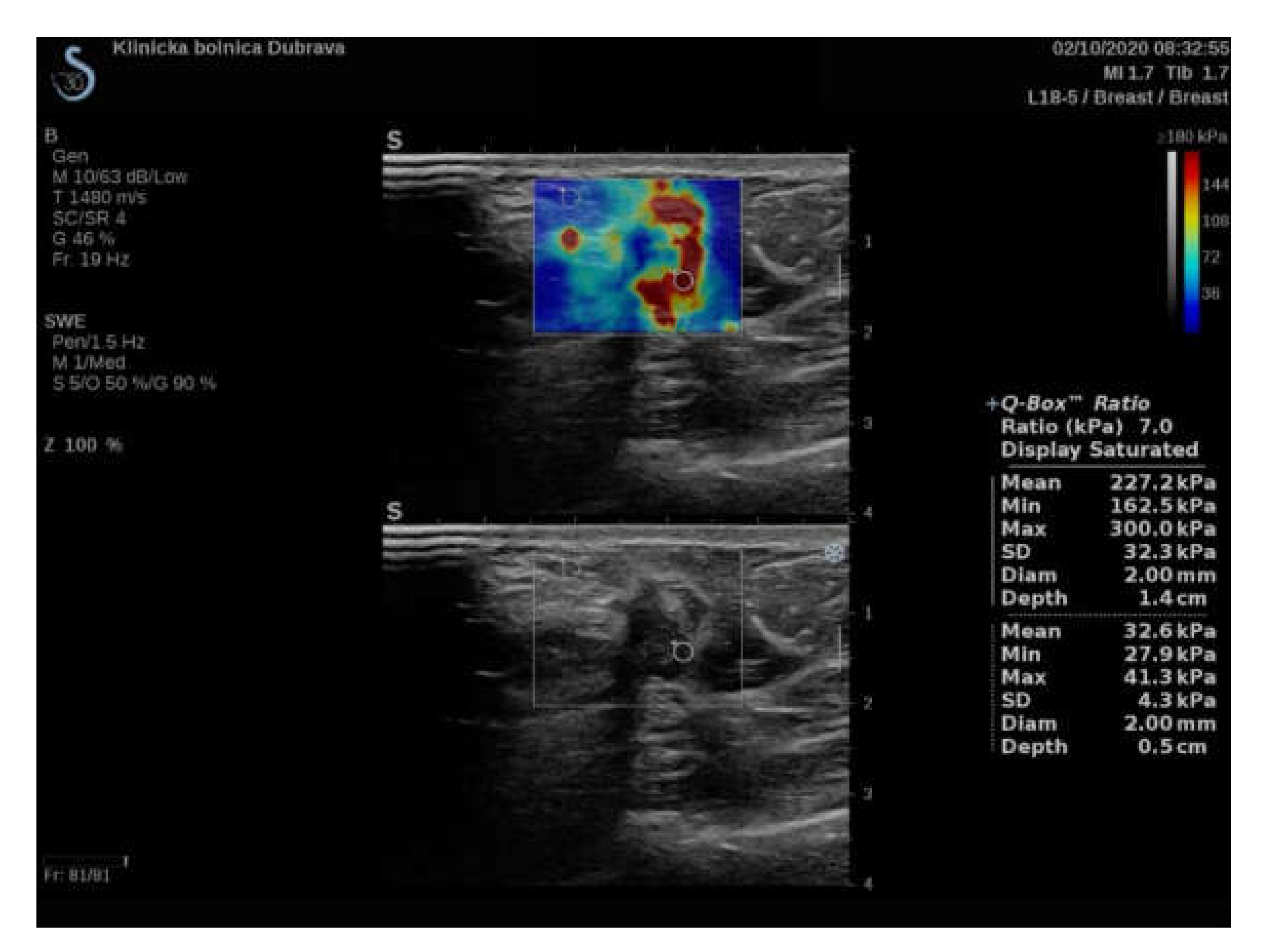
This retrospective single-center study conducted at our institution included 150 female patients (ages 37–92, median age 59) with histopathologically confirmed malignant breast lesions (PHD: invasive ductal carcinoma) between September 2017 and February 2019. Patients were excluded from the study if they had previous breast cancer, underwent surgery of the breast, chemotherapy, hormonal or radiation therapy. All patients were examined using a 4–15 MHz frequency 40 mm long linear probe, on the same ultrasound scanner (Aixplorer®, SuperSonic Imagine, Aix, France, product version 6.2.0, software 6.2.23751). Grey scale and shear-wave sonoelastography examinations were performed by an experienced breast radiologist with over 15 years of practice in breast ultrasound. The regions of interest used for the elastography measurements were set at diameters of 2 mm, 4 mm, and 6 mm. The stiffest areas of the lesion in the SWE acquisition box, including the immediately adjacent stiff tissue, were measured at the center of the ROI while the corresponding e-ratio value was obtained by placing a second ROI over the surrounding fatty tissue (Figure 1, Figure 2 and Figure 3) [11,12,13].
Additionally, all lesions were divided into small (diameter < 15 mm, Nm = 95) and large lesions (diameter ≥ 15 mm, Nm = 55) [8]. Normality of distribution of numerical variables was assessed using the Shapiro–Wilk test. Since most measurements were non-normally distributed, we presented all numerical variables as median and interquartile range (IQR) and used non-parametrical statistical tests. Coefficient of variation was obtained by dividing standard deviation of multiple assessments with the mean value. Measurements were mutually compared using the Friedman ANOVA for repeated measures and between different lesion sizes using the Mann Whitney U test. Correlation between different measurements was assessed using the Spearman rank correlation. p values < 0.05 were considered to be statistically significant. Bonferroni correction for multiple comparisons was used where appropriate (<0.017 considered to be statistically significant). All analyses were done using the MedCalc statistical software ver. 19.7.

3. Results

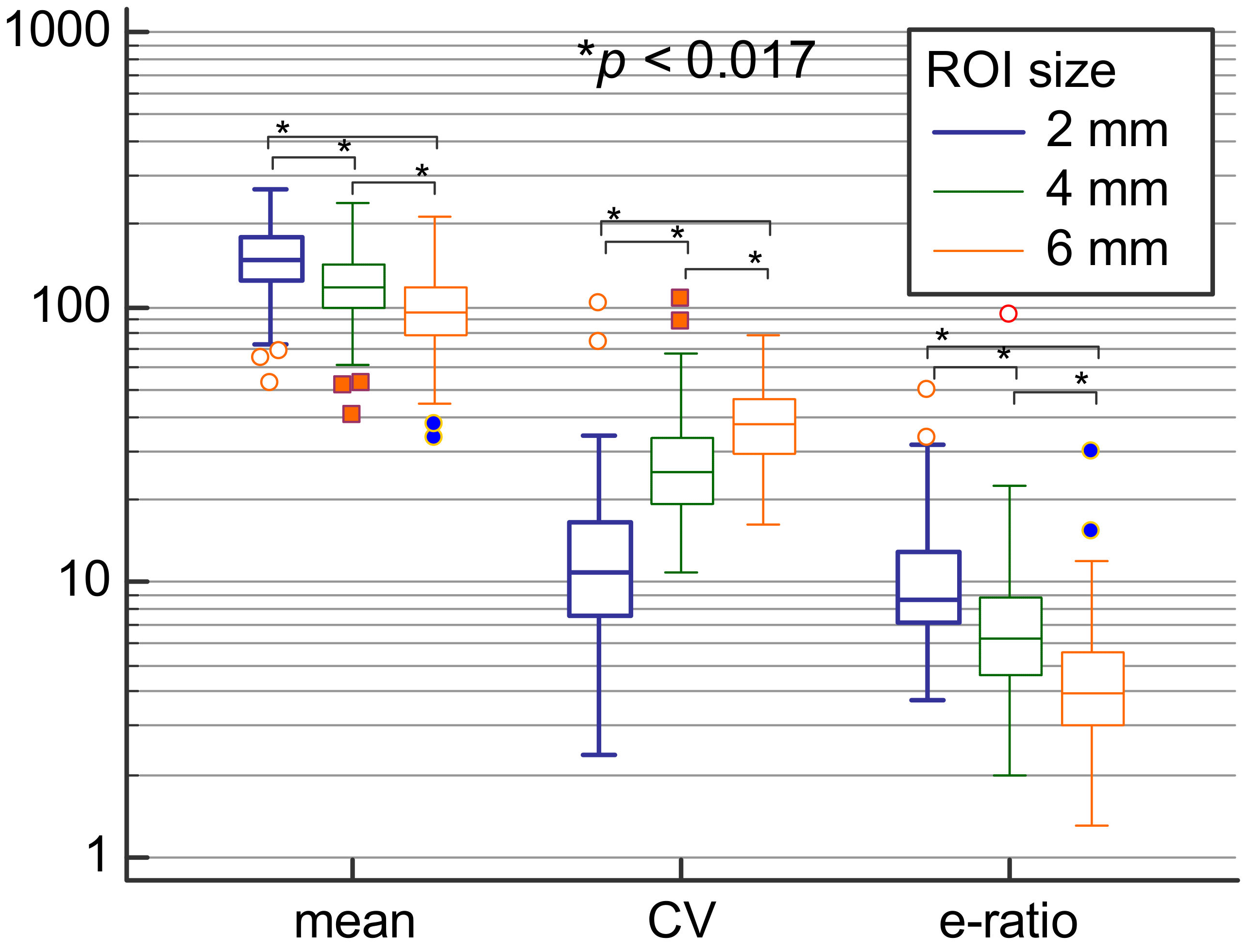
SWE measures in a total of 150 patients were analyzed. There was a significant difference between SWE parameters, with Emean, standard deviation, coefficient of variation, Emin and e-ratio values all being statistically significantly different between different ROI sizes (p < 0.017; Table 1; Figure 4).
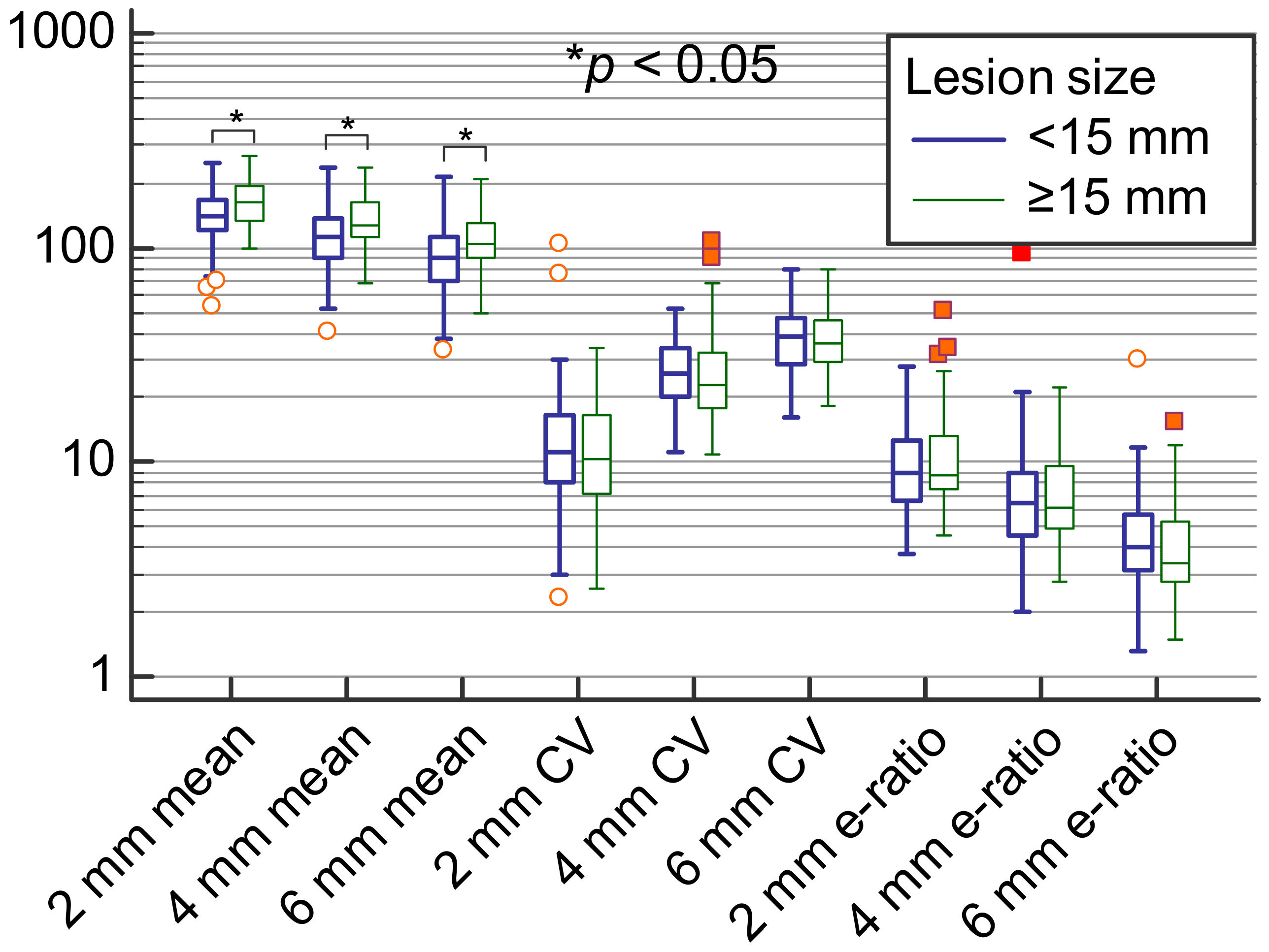
Highest Emin, Emean and e-ratio values and lowest variability were observed when using 2 mm in comparison to other ROI sizes. It should be noted that the lowest detected e-ratio values were 3.7, 2 and 1.3 for 2 mm, 4 mm, and 6 mm ROI sizes, respectively, showing that using higher ROI values results in substantially lower e-ratio measurements. However, Emax values did not differ according to different ROI sizes. Regarding lesion size, larger lesions had significantly higher Emean and Emax values as assessed using all three ROI sizes (p < 0.05; Table 2; Figure 5).
However, significant differences between lesion size and other SWE parameters like e-ratio (significant difference for 2 mm ROI size) and Emin values (significant differences for 2 mm and 4 mm ROI sizes) were present only when lower ROI sizes were used whereas variability of measurement did not differ according to the lesion size (Table 2). Median absolute differences between ROI measurements were 26 kPa for 2 mm vs. 4 mm, 23 kPa for 4 mm vs. 6 mm, and 50 kPa for 2 mm vs. 6 mm. Differences between 2 and 4 mm and 2 and 6 mm ROI were statistically significantly more pronounced with larger lesion size (p < 0.05; Table 2) and with higher e-ratio values (p < 0.05, Table 3) suggesting that with larger lesions it is more likely to obtain different measurements when using different ROI sizes.

4. Discussion

The influence of the ROI size on the measured values of the Emin, Emean and e-ratio of malignant breast lesions is significant. The highest values and lowest variability were observed when using the 2 mm diameter in comparison to other ROI sizes. The Emean value is calculated as a sum of elasticities of all pixels within the ROI, divided by the number of pixels. Therefore, the value of Emean is influenced if the lower elasticity values from the tissues surrounding the lesion are included into the measurements [7]. When a larger ROI is used to measure the lower elasticity values of the surrounding tissues that are not influenced by the malignant lesion, the effect on the measured Emin and e-ratio is the same as on the measured Emean values. Emax values, on the other hand, are not influenced by the change in the ROI size, and it was demonstrated in multiple studies that significant differences in its values were present between benign and malignant breast lesions versus normal breast tissues [8,14,15,16,17]. Regarding the lesion size, larger lesions had significantly higher Emean and Emax values as assessed using all three ROI sizes which is in line with the results from previous studies where some of these values were assessed in malignant breast lesions [8,18]. However, there was no significant change in e-ratio values between small and large lesions when a 2 mm ROI was used.
In conclusion, when measuring the Emin, Emean and e-ratio of malignant breast lesions using SWE the smallest possible ROI size should be used regardless of lesion size. The effect of the ROI size is most pronounced on the Emean values and can influence the measured elasticity values and, subsequently, sensitivity and specificity of the examination. If different ROI sizes are used, Emax values can be measured but different values will be obtained depending on the size of the lesion. In that case, the use of e-ratio with the smallest possible ROI is recommended to achieve uniform results regardless of lesion size. There are some limitations to this retrospective, single-center study. All lesions in this study were histologically confirmed as malignant, therefore the performance of SWE to differentiate between benign and malignant breast lesions was not assessed. The lesions were also divided into groups of small and large lesions by consideration of the mean value of their diameter (15 mm). Hence, further research defining the optimal cut-off thresholds of the lesion diameters is needed. Additionally, the influence of lesion depth and breast thickness was not considered in this study and it is suggested that both might influence the performance of SWE [19]. It is also known that soft tissues are non-linear and that any pressure applied by the probe may influence the results. In this study, minimal manual compression of the examined region was applied to avoid false positive results, as suggested by previous research [20]. A recent study also suggested that some amount of compression is essential to avoid false negative results [21]. However, these are also areas that also require further research.

Author Contributions

Conceptualization, A.B. and G.I.; methodology, A.B.; validation, A.B., G.I.; formal analysis, G.I.; investigation, A.B.; resources, A.B.; data curation, M.L.; writing—original draft preparation, A.B.; writing—review and editing, A.B., G.I., E.D., I.B.Š., M.D.D., K.B., B.B.; visualization, A.B.; supervision, B.B. All authors have read and agreed to the published version of the manuscript.

Funding

This research has been supported by the Croatian Science Foundation under project IP-2016-06-2997 “Sonoelastography and MRI in Diagnosis and Treatment of Breast Cancer”, University of Zagreb School of Medicine, Department of Diagnostic and Interventional Radiology, University Hospital Dubrava, Zagreb (to B.B.).

Institutional Review Board Statement

The study was reviewed and approved by the University of Zagreb, School of Medicine Ethical Committee on 23 February 2017 (Approval No. 641-01/02-01).

Informed Consent Statement

Informed consent was obtained from all subjects involved in the study.

Data Availability Statement

Data available on request due to restrictions. The data presented in this study is available on request from the corresponding author. The data is not publicly available due to patient privacy reasons.

Conflicts of Interest

The authors declare no conflict of interest.

References

  1. Barr, R. Sonographic breast elastography: A primer. J. Ultrasound Med. 2012, 31, 773–783. [Google Scholar] [CrossRef] [PubMed]
  2. Goddi, A.; Bonardi, M.; Alessi, S. Breast elastography: A literature review. J. Ultrasound 2012, 15, 192–198. [Google Scholar] [CrossRef] [PubMed] [Green Version]
  3. Gennisson, J.-L.; Deffieux, T.; Fink, M.; Tanter, M. Ultrasound elastography: Principles and techniques. Diagn. Interv. Imaging 2013, 94, 487–495. [Google Scholar] [CrossRef] [PubMed]
  4. Youk, J.H.; Son, E.J.; Gweon, H.M.; Kim, H.; Park, Y.J.; Kim, J.A. Comparison of strain and shear wave elastography for the differentiation of benign from malignant breast lesions, combined with B-mode ultrasonography: Qualitative and quantitative assessments. Ultrasound Med. Biol. 2014, 40, 2336–2344. [Google Scholar] [CrossRef] [PubMed]
  5. Athanasiou, A.; Tardivon, A.; Tanter, M.; Sigal-Zafrani, B.; Bercoff, J.; Deffieux, T.; Gennisson, J.-L.; Fink, M.; Neuenschwander, S. Breast lesions: Quantitative elastography with supersonic shear imaging: Preliminary results. Radiology 2010, 256, 297–303. [Google Scholar] [CrossRef] [PubMed]
  6. Nowicki, A.; Dobruch-Sobczak, K. Introduction to ultrasound elastography. J. Ultrason. 2016, 16, 113–124. [Google Scholar] [CrossRef] [PubMed]
  7. Youk, J.H.; Gweon, H.M.; Son, E.J. Shear-wave elastography in breast ultrasonography: The state of the art. Ultrasonography 2017, 36, 300–309. [Google Scholar] [CrossRef] [PubMed] [Green Version]
  8. Skerl, K.; Vinnicombe, S.; Giannotti, E. Influence of region of interest size and ultrasound lesion size on the performance of 2D shear wave elastography (SWE) in solid breast masses. Clin. Radiol. 2015, 70, 1421–1427. [Google Scholar] [CrossRef] [PubMed] [Green Version]
  9. Moon, J.H.; Hwang, J.Y.; Park, J.S.; Koh, S.H.; Park, S.Y. Impact of region of interest (ROI) size on the diagnostic performance of shear wave elastography in differentiating solid breast lesions. Acta Radiol. 2017, 59, 657–663. [Google Scholar] [CrossRef] [PubMed] [Green Version]
  10. Youk, J.H.; Son, E.J.; Han, K.; Gweon, H.M.; Kim, J.A. Performance of shear-wave elastography for breast masses using different region-of-interest (ROI) settings. Acta. Radiol. 2018, 59, 789–797. [Google Scholar] [CrossRef] [PubMed]
  11. Olgun, D.Ç.; Korkmazer, B.; Kılıç, F. Use of shear wave elastography to differentiate benign and malignant breast lesions. Diagn. Interv. Radiol. 2014, 20, 239–244. [Google Scholar] [CrossRef] [PubMed]
  12. Au, F.W.-F.; Ghai, S.; Moshonov, H. Diagnostic performance of quantitative shear wave elastography in the evaluation of solid breast masses: Determination of the most discriminatory parameter. Am. J. Roentgenol. 2014, 203, 328–336. [Google Scholar] [CrossRef] [PubMed]
  13. Kim, S.; Choi, S.; Choi, Y. Diagnostic performance of shear wave elastography of the breast according to scanning orientation. J. Ultrasound Med. 2014, 33, 1797–1804. [Google Scholar] [CrossRef] [PubMed]
  14. Berg, W.A.; Cosgrove, D.O.; Doré, C.J.; Schäfer, F.K.; Svensson, W.E.; Hooley, R.J.; Ohlinger, R.; Mendelson, E.B.; Balu-Maestro, K.; Locatelli, M.; et al. Shear-wave elastography improves the specificity of breast US: The BE1 multinational study of 939 masses. Radiology 2012, 262, 435–449. [Google Scholar] [CrossRef] [PubMed]
  15. Lee, E.J.; Jung, H.K.; Ko, K.H.; Lee, J.T.; Yoon, J.H. Diagnostic performances of shear wave elastography: Which parameter to use in differential diagnosis of solid breast masses? Eur. Radiol. 2013, 23, 1803–1811. [Google Scholar] [CrossRef] [PubMed]
  16. Evans, A.; Whelehan, P.; Thomson, K.; Brauer, K.; Jordan, L.; Purdie, C.; McLean, D.; Baker, L.; Vinnicombe, S.; Thompson, A. Differentiating benign from malignant solid breast masses: Value of shear wave elastography according to lesion stiffness combined with greyscale ultrasound according to BI-RADS classification. Br. J. Cancer 2012, 107, 224–229. [Google Scholar] [CrossRef] [PubMed]
  17. Zhou, J.; Zhan, W.; Chang, C.; Zhang, X.; Jia, Y.; Dong, Y.; Zhou, C.; Sun, J.; Grant, E.G. Breast lesions: Evaluation with shear wave elastography, with special emphasis on the “stiff rim” sign. Radiology 2014, 272, 63–72. [Google Scholar] [CrossRef] [PubMed]
  18. Chang, J.M.; Park, I.A.; Lee, S.H.; Kim, W.H.; Bae, M.S.; Koo, H.R.; Yi, A.; Kim, S.J.; Cho, N.; Moon, W.K. Stiffness of tumours measured by shear-wave elastography correlated with subtypes of breast cancer. Eur. Radiol. 2013, 23, 2450–2458. [Google Scholar] [CrossRef] [PubMed]
  19. Yoon, J.H.; Jung, H.K.; Lee, J.T.; Ko, K.H. Shear-wave elastography in the diagnosis of solid breast masses: What leads to false-negative or false-positive results? Eur. Radiol. 2013, 23, 2432–2440. [Google Scholar] [CrossRef] [PubMed]
  20. Barr, R. Elastography: A Practical Approach; Thieme: New York, NY, USA, 2016. [Google Scholar]
  21. Chamming’s, F.; Hangard, C.; Gennisson, J.L.; Reinhold, C.; Fournier, L.S. Diagnostic Accuracy of Four Levels of Manual Compression Applied in Supersonic Shear Wave Elastography of the Breast. Acad. Radiol. 2021, 28, 481–486. [Google Scholar] [CrossRef] [PubMed]
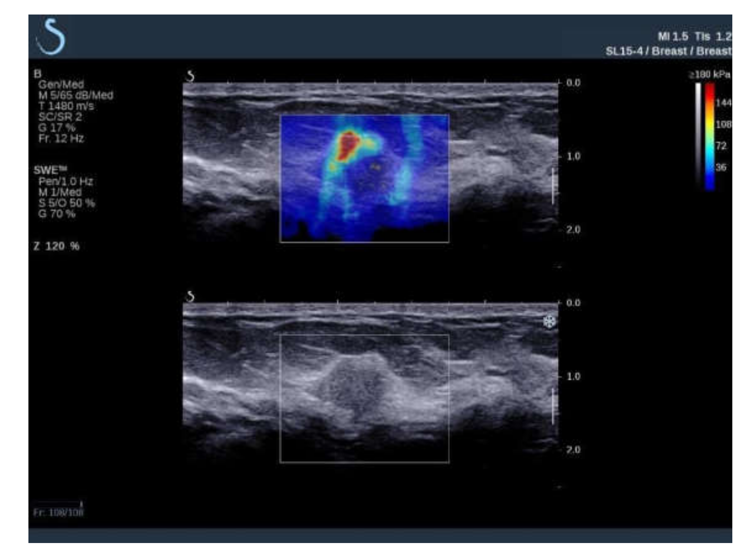
Figure 1. Ultrasound examination of a malignant breast lesion with B-mode (bottom) and shear wave elastography (top) where elastic properties of breast tissues are displayed qualitatively, color-coded, and superimposed on the B-mode image. Red color denotes areas with the highest elastic modulus values.
Diagnostics 11 01015 g001
Figure 2. Ultrasound examination with shear wave elastography of a hypoechoic breast lesion, later pathohistologically confirmed after core-biopsy as an invasive ductal carcinoma. Elastic modulus values measurements with ROI size of 2 mm, 4 mm and 6 mm were performed and the measured values are displayed.
Diagnostics 11 01015 g002
Figure 3. Ultrasound examination with shear wave elastography of a hypoechoic breast lesion, later pathohistologically confirmed after core-biopsy as an invasive ductal carcinoma. An e-ratio value measurement with ROI size of 2 mm with the measured values is displayed (measured mean values are used to calculate the ratio). Additional ROI with a size of 4 mm and 6 mm were placed at the same points as the 2 mm ROI to obtain additional values (not displayed in this image).
Diagnostics 11 01015 g003
Figure 4. Comparison of means, coefficients of variation and e-ratio values between 2 mm, 4 mm, and 6 mm ROI sizes.
Diagnostics 11 01015 g004
Figure 5. Means, coefficients of variation and e-ratio measurements for ROI 2 mm, 4 mm and 6 mm sizes stratified according to the lesion size.
Diagnostics 11 01015 g005
Table 1. Pairwise comparisons of different SWE measures according to the ROI size.
VariableROI 2 mm (1)ROI 4 mm (2)ROI 6 mm (3)p Value
Emean (kPa)148.85117.5595.65<0.001 * (1 vs. 2; 1 vs. 3; 2 vs. 3) *
Sd (kPa)16.631.1536.65<0.001 * (1 vs. 2; 1 vs. 3; 2 vs. 3) *
Coeff. var. (%)10.925.2237.77<0.001 * (1 vs. 2; 1 vs. 3; 2 vs. 3) *
Emin (kPa)108.3550.823.85<0.001 * (1 vs. 2; 1 vs. 3; 2 vs. 3) *
Emax (kPa)184.45184.45184.450.236
e-ratio8.76.33.95<0.001 * (1 vs. 2; 1 vs. 3; 2 vs. 3) *
* Statistically significant at level p < 0.05 (Bonferroni corrected p < 0.017 for three simultaneous pairwise comparisons). Results are presented as median values. Friedman ANOVA for repeated measures was used. Abbreviations: ROI—region of interest; sd—standard deviation; coeff. var.—coefficient of variation; min—minimum; max—maximum.
Table 2. Different SWE measurements stratified according to the lesion size.
VariableLesion Size < 15 mmLesion Size ≥ 15 mmp ValueCorrelation with Lesion Size as a Cont. Variable
Nm of patients9555--
Emean ROI 2 mm (kPa)141.8 IQR (119.6–165.9)161.7 IQR (134.95–190.6)0.003 *Rho = 0.4; p < 0.001 *
Sd ROI 2 mm (kPa)16.6 IQR (11–23)16.6 IQR (11.35–23.3)0.729Rho = 0.18; p = 0.030 *
Coeff. var. ROI 2 mm (%)11.1 IQR (8.08–16.42)10.1 IQR (6.7–16.32)0.245Rho = −0.01; p = 0.894
Emin ROI 2 mm (kPa)104.9 IQR (79.4–127.4)123.8 IQR (91.3–154.6)0.015 *Rho = 0.3; p < 0.001 *
Emax ROI 2 mm (kPa)171.8 IQR (137.9–192)192 IQR (171.85–221.6)0.001 *Rho = 0.44; p < 0.001 *
e-ratio ROI 2 mm8.8 IQR (6.6–12.75)8.6 IQR (7.5–12.95)0.201Rho = 0.19; p = 0.023 *
Emean ROI 4 mm (kPa)111.3 IQR (89.15–135.55)128.6 IQR (111.9–161.8)0.001 *Rho = 0.42; p < 0.001 *
Sd ROI 4 mm (kPa)31 IQR (21.9–37.65)32.8 IQR (24.15–40.1)0.256Rho = 0.21; p = 0.009 *
Coeff. var. ROI 4 mm (%)25.7 IQR (20.72–34.14)22.7 IQR (17.93–32.1)0.154Rho = −0.1; p = 0.219
Emin ROI 4 mm (kPa)49.3 IQR (32.3–66.9)57.2 IQR (40.05–82)0.063Rho = 0.26; p = 0.002 *
Emax ROI 4 mm (kPa)171.8 IQR (137.9–192)192 IQR (171.85–214.6)0.001 *Rho = 0.43; p < 0.001 *
e-ratio ROI 4 mm6.3 IQR (4.55–8.8)6.3 IQR (4.95–10.05)0.638Rho = 0.14; p = 0.092
Emean ROI 6 mm (kPa)89.4 IQR (69.8–110.1)105.1 IQR (89.65–129.8)0.001 *Rho = 0.4; p < 0.001 *
Sd ROI6 mm (kPa)34.5 IQR (24.75–43.45)39.7 IQR (32.95–46.55)0.005 *Rho = 0.39; p < 0.001 *
Coeff. var. ROI6 (kPa) 38.4 IQR (28.82–46.59)35.7 IQR (29.24–44.9)0.785Rho = 0.01; p = 0.942
Emin ROI 6 mm (kPa)23.1 IQR (15.1–38.45)25.4 IQR (10.85–41.1)0.685Rho = 0.09; p = 0.295
Emax ROI 6 mm (kPa)171.8 IQR (137.9–192)192 IQR (171.85–221.6)0.001 *Rho = 0.43; p < 0.001 *
e-ratio ROI 6 mm4 IQR (3.15–5.7)3.4 IQR (2.85–5.35)0.335Rho = −0.05; p = 0.527
ROI 2 mm vs. 4 mm mean difference26.3 IQR (18.55–37.95)26.2 IQR (21.4–38.75)0.408Rho = 0.13; p = 0.116
ROI 2 mm vs. 6 mm mean difference47.4 IQR (34.05–64.6)52.7 IQR (41.45–69)0.116Rho = 0.22; p = 0.006 *
ROI 4 mm vs. 6 mm mean difference22.2 IQR (15.65–26)26.1 IQR (18.25–32.2)0.008 *Rho = 0.32; p < 0.001 *
* Statistically significant at level p < 0.05. Results are presented as median values and interquartile range. Mann–Whitney U test and Spearman rank correlation were used. Abbreviations: ROI—region of interest; cont.—continuous; sd—standard deviation; coeff. var.—coefficient of variation; min—minimum; max—maximum.
Table 3. Correlation between e-ratio assessed by different ROI sizes and differences in SWE measurements.
Variablee-Ratio ROI 2 mm e-Ratio ROI 4 mme-Ratio ROI 6 mm
ROI 2 mm vs. 4 mm mean differenceRho = 0.14; p = 0.088Rho = 0.07; p = 0.388Rho = −0.01; p = 0.952
ROI 2 mm vs. 6 mm mean differenceRho = 0.25; p = 0.002 *Rho = 0.2; p = 0.017 *Rho = 0.08; p = 0.362
ROI 4 mm vs. 6 mm mean differenceRho = 0.39; p < 0.001 *Rho = 0.39; p < 0.001 *Rho = 0.22; p = 0.007 *
* Statistically significant at level p < 0.05/Spearman rank correlation was used. Abbreviations: ROI—region of interest.
Publisher’s Note: MDPI stays neutral with regard to jurisdictional claims in published maps and institutional affiliations.