Case Report

Kingella kingae Intrauterine Infection: An Unusual Cause of Chorioamnionitis and Miscarriage in a Patient with Undifferentiated Connective Tissue Disease

1
Pathology Unit, Azienda Unità Sanitaria Locale—IRCCS di Reggio Emilia, 42122 Reggio Emilia, Italy
2
Pathology Unit, “Maggiore della Carità” Hospital, 28100 Novara, Italy
3
Department of Obstetrics & Gynaecology, Azienda Unità Sanitaria Locale—IRCCS di Reggio Emilia, 42122 Reggio Emilia, Italy
4
Rheumatology Unit, Department of Internal Medicine, Azienda USL-IRCCS di Reggio Emilia e Università di Modena e Reggio Emilia, 42122 Reggio Emilia, Italy
5
Clinical Microbiology Laboratory, Azienda Unità Sanitaria Locale—IRCCS di Reggio Emilia, 42122 Reggio Emilia, Italy
*
Author to whom correspondence should be addressed.
Diagnostics 2021, 11(2), 243; https://doi.org/10.3390/diagnostics11020243
Submission received: 12 January 2021 / Revised: 25 January 2021 / Accepted: 1 February 2021 / Published: 4 February 2021
(This article belongs to the Section Diagnostic Microbiology and Infectious Disease)

Abstract

:
Kingella kingae is a Gram-negative coccobacillus belonging to the Neisseriaceae family. In children less than 4 years old, K. kingae invasive infection can induce septic arthritis and osteomyelitis, and more rarely endocarditis, meningitis, ocular infections, and pneumonia. In adults, it may be a cause of endocarditis. To date, K. kingae acute chorioamnionitis (AC) leading to preterm rupture of membranes (PPROM) and miscarriage has never been reported. Herein, we describe a case of intrauterine fetal death (IUFD) at 22 weeks’ gestation due to K. kingae infection occurred in a patient affected by undifferentiated connective tissue disease (UCTD) in lupus erythematosus systemic (LES) evolution with severe neutropenia. K. kingae was isolated in placental subamnionic swab and tissue cultures as well as fetal ear, nose, and pharyngeal swabs. Placental histological examination showed necrotizing AC and funisitis. In the fetus, neutrophils were observed within the alveoli and in the gastrointestinal lumen. Maternal medical treatment for UCTD was modified according to the K. kingae invasive infection. In the event of IUFD due to AC, microbiological cultures on placenta and fetal tissues should always be carried out in order to isolate the etiologic agent and target the correct medical treatment.

1. Introduction

Kingella kingae is a Gram-negative, facultatively anaerobic coccobacillus of the Neisseriaceae family. It is a slowly growing bacterium also a member of the HACEK group (Haemophilus spp., Aggregatibacter actinomycetemcomitans, Cardiobacterium hominis, Eikenella corrodens, and Kingella kingae) [1,2]. K. kingae is currently recognized as the most common etiology of septic arthritis and osteomyelitis in children between the ages of 6 and 48 months. More rarely, it can cause complicated endocarditis, meningitis, ocular infections, pericarditis, peritonitis, and pneumonia [3,4].
To the best of our knowledge, K. kingae infection has never been reported in pregnancy. Only a case of early onset sepsis (EOS) in a premature infant has been described [5]. Herein, we present an unusual case of preterm premature rupture of membranes (PPROM) at 22 weeks’ gestation with subsequent intrauterine fetal death (IUFD) due to severe acute chorioamnionitis (AC). Placental subamnionic swab and tissue microbiological cultures isolated K. kingae, including fetal ear, nose, and pharyngeal swabs. Moreover, the 31-year-old mother was affected by undifferentiated connective tissue disease (UCTD) in evolution toward lupus erythematosus systemic (LES); severe neutropenia, antinuclear antibodies (ANA), and low titer of anti-neutrophil cytoplasmic antibodies (ANCA) were also present.

2. Case Description

2.1. Mother Clinical Presentation and Treatment

A 31-year-old mother, gravida 2 para 1, presented at 22 weeks’ gestation to the Emergency Department of our Institution for miscarriage. The patient had been suffering from shaking chills, fever (38 °C), stomatitis, sore throat, abdominal pain, and minimal vaginal bleeding for the previous three days. Gynaecological examination revealed on speculum mild fresh blood in vagina and abdominal ultrasound (US) observed IUFD. She was admitted to the hospital, labour was induced and a stillborn female fetus was delivered. Placental microbiological samples and fetal swabs were sent to the Clinical Microbiology laboratory where cultures on different media were immediately performed.
Overall, the patient had a complex clinical history. She had suffered from monoarticular juvenile idiopathic arthritis, completely resolved at 15 years of age.
Six years before the current miscarriage, she had complained of bilateral gonalgia. Laboratory findings had shown severe neutropenia (250 neutrophils) and low titer of ANCA. Antinuclear antibodies (ANA) titer had been 1:160. Anti extractable nuclear antigens (ENA), anti-DNA antibodies, and antiphospholipid antibodies (lupus anticoagulant—LAC; anticardiolipin antibodies—aCL; anti-β2-glycoprotein-1—anti-β2GP-1) had been negative. Bone marrow biopsy had been reported within normal limits.
In the previous six years, although the patient had always been presenting with low neutrophils, she had never contracted opportunistic infections.
One year before the miscarriage, she had had her first pregnancy with a vaginally delivered male infant of 3200 g at 40 weeks + 6 days of gestational age. One month after delivery, she had severe mastitis, which required hospital admission and surgical treatment. After that, she presented with two episodes of bilateral knee arthritis, and then she was put on hydroxychloroquine 200 mg twice a day (tablets).
Regarding the current miscarriage, at hospital admission, maternal laboratory findings were as follows: WBC 2.6 x 1.000/μL and C-reactive protein (CRP) 13.45 mg/dL. During the first day after IUFD, CRP worsened to 18.02 mg/dL, then progressively decreased from 16.46 (2 days), 2.7 mg/dL (3 days), 1.43 mg/dL (5 days) to 0.11 mg/dL (10 days). Blood cultures performed at admission resulted negative.
Maternal medical treatment, started soon after IUFD, consisted of 2 days of meropenem, subsequently switched to piperacillin/tazobactam for 6 days. She fully recovered and was discharged after 10 days.
After hematologic and rheumatologic consult, due to low neutrophil count and recent intrauterine infection, Granulocyte-Colony Stimulating Factor (G-CSF) and cyclosporine (200 mg a day) were added to hydroxychloroquine.

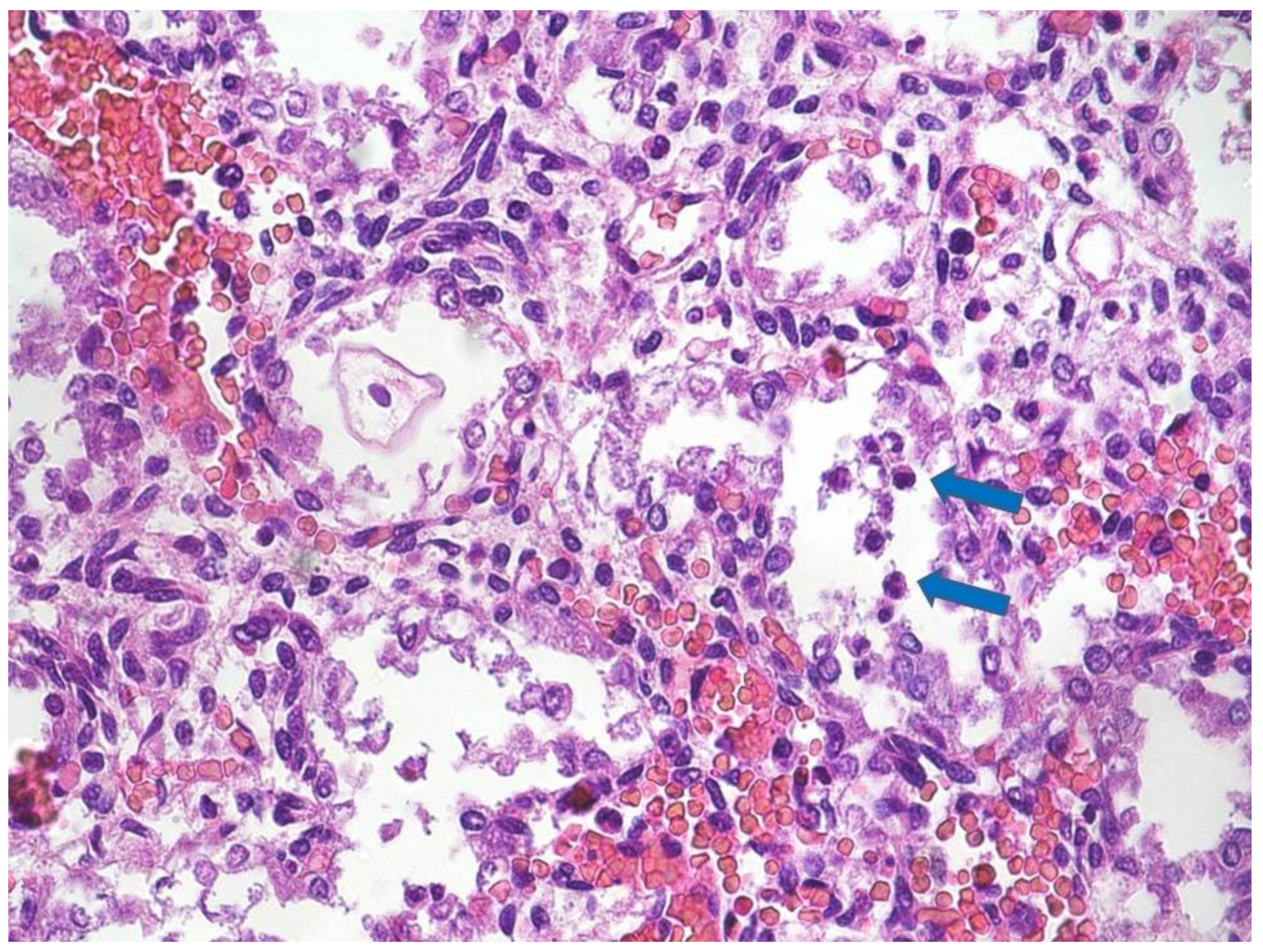
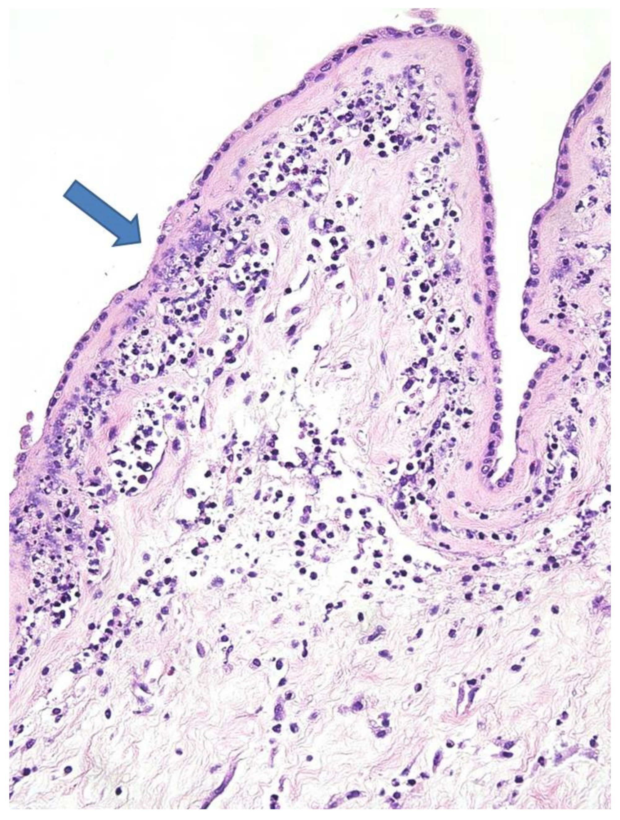
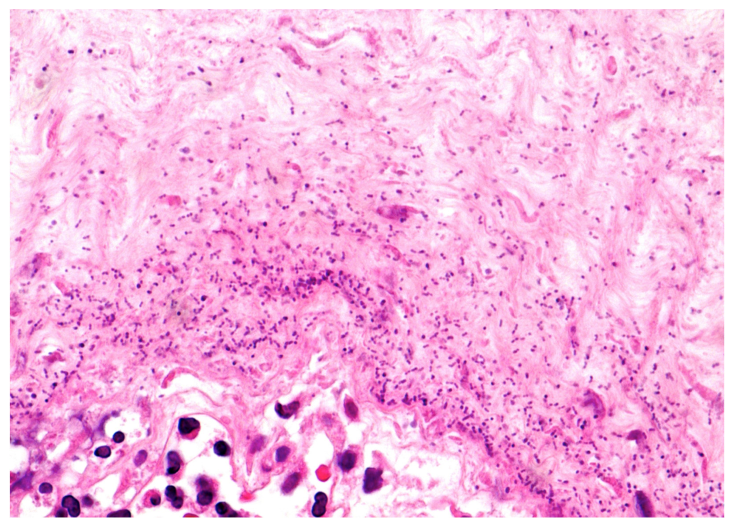
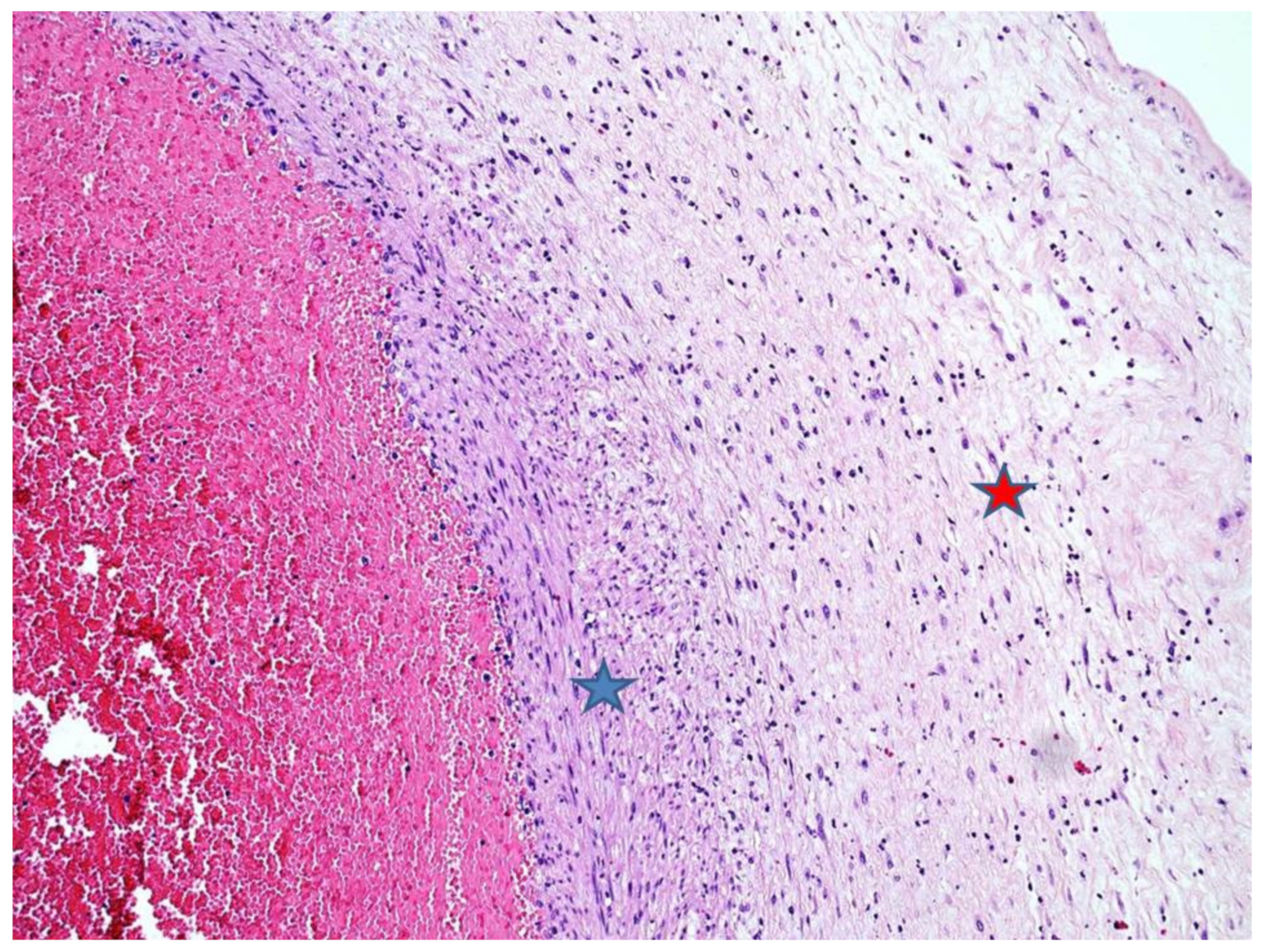
2.2. Fetal Autopsy and Microbiological Results

Postmortem examination revealed a nonmacerated female fetus weighing 420 g and measuring 29 cm in crown-heel length. The other measurements were as follows: Crown-rump length, 19.5 cm; foot length, 3.7 cm; head, chest and abdominal circumference, 18, 16, and 15 cm, respectively. Overall, anthropometric measurements were consistent with 22 weeks’ gestation [6]. External examination showed a normal fetus with mild eyelid and nuchal oedema. On internal examination, intestinal rotation was incomplete (malrotation) with short midgut mesenteric attachment and mobile intestine. No volvulus was observed. Microscopic analysis revealed the presence of intra-alveolar, gastric and intestinal neutrophils (Figure 1); oedema, microhemorrhages and acute tubular necrosis (NTA) of the renal parenchyma were also present.
The placenta was received complete, weighed 137 g, and measured 10 × 10 × 2 cm. The membranes were yellowish and opaque. Microscopically, there was necrotizing AC (Figure 2) corresponding to a maternal inflammatory response stage 3/3 and grade 2/2 [7]. Coccoid bacteria were noted at high magnification within the chorion (Figure 3). Funisitis was also observed with neutrophilic infiltrate of the umbilical vein, and the two arteries with extension to Wharton’s jelly (Figure 4). These findings were consistent with fetal inflammatory response stage 2/3 and grade 2/2 [7]. In addition, in the placental parenchyma some recent infarcts were detected, and in the decidua few spiral arteries presented initial thrombosis.
Autoptical samples were cultured on Columbia blood agar (CBA), McConkey agar (MCA), mannitol salt agar (MSA) and Sabouraud agar (SAB; all these media were incubated at 36 °C, ambient air), chocolate agar (CA, incubated at 36 °C, 5% CO2), and Schaedler agar (SA, incubated at 36 °C, anaerobic atmosphere). After 48 hours from the fetus’s pharyngeal, nose, and ear swabs, small colonies grew on CBA and CA (better on the latter). No growth was observed on MCA. These smooth colonies were catalase negative and oxidase positive. They were identified as K. kingae using the MALDI-ToF technology (Bruker Daltonics, Germany), Biotyper OC software version 3.1. Microbiological cultures on fetal blood and tissues (lung and liver) showed no growth after 72 hours and were discarded as negative. Instead, K. kingae also grew on subamniotic swab and placental tissue cultures. If present, the microorganism grew as a pure culture in all the samples. Antimicrobial susceptibility testing was not performed.

3. Materials and Methods

We searched for (Kingella kingae AND (chorioamnionitis OR genital OR amniotic OR amnios OR placenta OR placental OR funisitis OR fetus OR fetal OR pregnancy OR pregnant OR (membrane AND rupture) OR uterus OR uterine OR intrauterine)) in Pubmed (all fields, 2 results), Scopus (Title/Abstract/Keyword, 9 results) and Web of Science (Topic/Title, 2 results). No limitations were set. The bibliographic research ended on 25 December 2020. Globally, a total of 10 articles resulted from our search. Titles and Abstract of all the articles were screened. All articles were excluded as non-relevant.

4. Discussion

K. kingae is a Gram-negative coccobacillus belonging to the Neisseriaceae family. It is a normal component of the oropharyngeal flora of children less than 4 years old. However, the bacterium, after having colonized and breached the epithelial surface, may disseminate in the bloodstream causing, especially in children, arthritis, osteomyelitis, and endocarditis. This latter complication may also occur in adults [1,8].
Mechanisms of virulence include biofilm formation, pili, RTX toxin production, polysaccharide capsule, and secretion of outer membrane vesicles (OMV).
K. kingae is able to form biofilms, which consist of large quantities of bacteria clustered together in a polysaccharide “slime”, that tightly attaches to the mucosal surface. This kind of colonization allows bacteria to survive in a protected environment preserving them from dehydratation, immune response, and antibiotics [9,10].
K. kingae anchors to the epithelia thanks to type 4 pili, which are proteinaceous fimbriae necessary for efficient adherence to respiratory mucosa and synovia [11].
Moreover, RTX toxin expression exerts a wide-spectrum cytotoxic effect on macrophages, leukocytes, synoviocytes, and respiratory epithelial cells. This toxin plays a key role in infection spreading favouring epithelial disruption and bloodstream dissemination [12,13]. Of note, type a and b polysaccharide capsule predominate in invasive isolates [14]. A further virulence factor possessed by K. kingae is the secretion of OMV, which are small parts of the outer membrane encasing periplasm proteins that bulge away from the bacterium and then are released in the extracellular space. These OMV’s are hemolytic and leukotoxic in an in vitro model and internalized by synoviocytes inducing the synthesis of granulocyte-macrophage colony stimulating factor (GM-CSF) and interleukin 6 (IL-6), likely representing the in vivo immunitary response in joint infections [15].
K. kingae has been recognized as a leading cause of septic arthritis and osteomyelitis in children less than 4 years old, usually with no underlying medical conditions [3,4]. Other rare primary manifestations in pediatric population are: Pneumonia, endocarditis, soft tissue infection, endophtalmitis, orbital cellulitis, and meningitis [1,2,16,17,18,19,20].
Being part of the HACEK group (Haemophilus spp, Aggregatibacter actinomycetemcomitans, Cardiobacterium hominis, Eikenella corrodens, Kingella kingae), K. kingae can cause endocarditis, affecting more frequently children and young adults [21].
Adult patients usually have predisposing conditions in order to develop invasive K. kingae disease, such as malignancies, liver cirrhosis, diabetes, sickle cell anemia, and renal transplant [1,22,23,24,25].
However, to the best of our knowledge, K. kingae infection has never been reported in pregnancy and besides as a cause of PPROM and miscarriage. Moreover, the patient was affected by UCTD in evolution toward LES with severe neutropenia, ANA, and ANCA.
The 31-year-old mother presented to the hospital after having suffered shaking chills, fever (38 °C), stomatitis, sore throat, abdominal pain, and minimal vaginal bleeding for the previous three days. US revealed IUFD. Microbiological cultures on placental subamnionic swab and parenchymal tissue isolated K. kingae. The same agent grew on fetal swabs (ear, nose, and pharyngeal). Fetal tissue cultures (blood, lung and liver) resulted negative. Placental histological examination confirmed severe necrotizing chorioamnionitis and funisitis. Within the chorion, coccoid bacteria were also observed. Fetal histology showed few neutrophils within the alveoli, and gastro-intestinal lumen, respectively.
K. kingae was then considered the etiologic agent responsible for the miscarriage. While other members of the genus Kingella such as K. negevensis and K. denitrificans are known to cause bacterial vaginosis, chorioamnionitis, and pediatric vaginitis [26,27,28]; K. kingae infections of the urogenital tract are extremely rare [29].
However, AC due to K. kingae has never been described. The main microorganisms responsible for AC usually are group B Streptococcus, Fusobacterium nucleatum, Peptostreptococcus, Escherichia coli, Bacteroides species, Ureaplasma urealyticum, and Listeria monocytogenes [30,31]. As K. kingae is not part of the lower genital tract flora, in the case described, an ascending infection was unlikely. The bacterium probably reached the amniotic cavity through the hematogenous pathway from the oropharynx. In fact, the mother presented with sore throat and stomatitis. Although reported in children, K. kingae invasive disease has been associated with recent or concomitant virus infections like coxsackievirus, herpes simplex, varicella zoster, and rhinovirus in the oropharynx and upper respiratory tract [32,33,34,35]. Therefore, a concomitant viral infection in those locations may represent a cofactor in the pathophysiology of invasive K. kingae, probably favoring epithelial breaching and altering the local immune response [36].
In the case we described, it is impossible to differentiate a condition of maternal oropharyngeal carrier exacerbated by a concomitant viral infection or a K. kingae primary oropharyngeal infection turned out to be invasive. Notwithstanding, the mother was affected by UCTD in evolution toward LES with severe neutropenia. To date, K. kingae infection has never been described in this kind of peculiar setting. Only few cases have been observed in a granulocytopenic host [22], LES [37,38,39,40], rheumatoid arthritis [41], and acquired immunodeficiency syndrome (AIDS) [42,43,44,45,46].
Besides, recognition of K. kingae intrauterine infection allowed modifying maternal therapeutic plan, as G-CSF and cyclosporine were added to hydroxychloroquine.
In our specific case, placental cultures and fetal swabs revealed K. kingae infection, but the microorganism was not retrieved from fetal blood and tissues. This may be explained as a low bacterial load or a possible specimen type inhibition. In fact, it is known that recovery of K. kingae from bacteriological solid media may be difficult and nucleic acid amplification is advisable [1]. In our case, polymerase chain reaction (PCR) on the fetal tissues was not performed as infection was clearly demonstrated by neutrophils within the alveoli and in the lumen of gastrointestinal tract.
Usually, in the event of miscarriage due to chorioamnionitis, identification of the correct etiologic agent is of paramount importance as maternal medical treatment can be adjusted accordingly. Placental subamniotic swab and parenchymal cultures as well as fetal blood and tissues microbiological studies should always be carried out as recommended by perinatal autopsy protocols [47,48].

Author Contributions

M.P.B.; carried out the pathological diagnosis and written the manuscript—original draft preparation, review and editing, A.P.; provided the histological pictures and searched for the bibliograhy, G.D.D.; provided the draft preparation and writing, G.C. and G.P.; clinically managed the patient, G.R., G.B., M.B., C.Z., and E.C.; carried out the microbiological diagnosis. All authors have read and agreed to the published version of the manuscript.

Funding

This research received no external funding.

Institutional Review Board Statement

Our investigations were carried out following the rules of the Declaration of Helsinki of 1975, revised in 2013. According to Italian legislation, Ethical Approval for a single case is not required, as long as the data are kept anonymous and the investigations performed do not imply genetic results.

Informed Consent Statement

The current Italian legislation neither requires the family’s consent or ethical approval for a single case, as long as the data are strictly kept anonymous. As summoning the mother was not possible because it would interfere with the grieving process, patient’s consent was completely waived, according to the Italian Authority of Privacy and Data Protection (“Garante della Privacy”: GDPR nr 679/2016; 9/2016 and recent law addition number 424/ 19th of July 2018; http://www.garanteprivacy.it).

Data Availability Statement

The data presented in this study are available on request from the corresponding author.

Conflicts of Interest

The authors declare no conflict of interest.

References

  1. Yagupsky, P. Kingella kingae: Carriage, Transmission, and Disease. Clin. Microbiol. Rev. 2015, 28, 54–79. [Google Scholar] [CrossRef] [Green Version]
  2. Syridou, G.; Giannopoulou, P.; Charalampaki, N.; Papaparaskevas, J.; Korovessi, P.; Papagianni, S.; Tsakris, A.; Trikka-Grafakou, E. Invasive infection from Kingella kingae: Not only arthritis. IDCases 2020, 20, e00732. [Google Scholar] [CrossRef]
  3. Wong, M.; Williams, N.; Cooper, C. Systematic Review of Kingella kingae Musculoskeletal Infection in Children: Epidemiology, Impact and Management Strategies. Pediatr. Heal. Med. Ther. 2020, 11, 73–84. [Google Scholar] [CrossRef] [Green Version]
  4. Principi, N.; Esposito, S. Kingella kingae infections in children. BMC Infect. Dis. 2015, 15, 260. [Google Scholar] [CrossRef] [Green Version]
  5. Xia, Y.F.; Ma, L.; Qiu, X.L.; Li, G.X.; Guo, Y.H.; Liu, S.H.; Pu, W.C.; Wang, L. Early-onset septic shock in a preterm infant caused by Kingella Kingae. Zhongguo Dang Dai Er Ke Za Zhi. 2020, 22, 804. [Google Scholar] [PubMed]
  6. Archie, J.G.; Collins, J.S.; Lebel, R.R. Quantitative Standards for Fetal and Neonatal Autopsy. Am. J. Clin. Pathol 2006, 126, 256–265. [Google Scholar] [CrossRef] [PubMed]
  7. Kraus, F.T.; Redline, R.W.; Gersell, D.J.; Nelson, D.M.; Dicke, J.M. Chapter 5 Inflammation and Infection. In Placental Pathology; Armed Forces Institue of Pathology (AFIP): Washington, DC, USA, 2004; pp. 75–86. [Google Scholar]
  8. Kaplan, J.B.; Sampathkumar, V.; Bendaoud, M.; Giannakakis, A.K.; Lally, E.T.; Balashova, N.V. In vitro characterization of biofilms formed by Kingella kingae. Mol. Oral Microbiol. 2016, 32, 341–353. [Google Scholar] [CrossRef]
  9. Bendaoud, M.; Vinogradov, E.; Balashova, N.V.; Kadouri, D.E.; Kachlany, S.C.; Kaplan, J.B. Broad-Spectrum Biofilm Inhibition by Kingella kingae Exopolysaccharide. J. Bacteriol. 2011, 193, 3879–3886. [Google Scholar] [CrossRef] [Green Version]
  10. Kaplan, J.B. Biofilm Dispersal: Mechanisms, Clinical Implications, and Potential Therapeutic Uses. J. Dent. Res. 2010, 89, 205–218. [Google Scholar] [CrossRef] [PubMed] [Green Version]
  11. Kehl-Fie, T.E.; Miller, S.E.; Geme, J.W.S. Kingella kingae Expresses Type IV Pili That Mediate Adherence to Respiratory Epithelial and Synovial Cells. J. Bacteriol. 2008, 190, 7157–7163. [Google Scholar] [CrossRef] [Green Version]
  12. Kehl-Fie, T.E.; Geme, J.W.S. Identification and Characterization of an RTX Toxin in the Emerging Pathogen Kingella kingae. J. Bacteriol. 2006, 189, 430–436. [Google Scholar] [CrossRef] [Green Version]
  13. Chang, D.W.; Nudell, Y.A.; Lau, J.; Zakharian, E.; Balashova, N.V. RTX Toxin Plays a Key Role in Kingella kingae Virulence in an Infant Rat Model. Infect. Immun. 2014, 82, 2318–2328. [Google Scholar] [CrossRef] [Green Version]
  14. Porsch, E.A.; Starr, K.F.; Yagupsky, P.; Geme, J.W.S. The Type a and Type b Polysaccharide Capsules Predominate in an International Collection of Invasive Kingella kingae Isolates. mSphere 2017, 2, e00060-17. [Google Scholar] [CrossRef] [Green Version]
  15. Maldonado, R.; Wei, R.; Kachlany, S.; Kazi, M.; Balashova, N. Cytotoxic effects of Kingella kingae outer membrane vesicles on human cells. Microb. Pathog. 2011, 51, 22–30. [Google Scholar] [CrossRef] [Green Version]
  16. Claesson, B.; Falsen, E.; Kjellman, B. Kingella kingae Infections: A Review and a Presentation of Data from 10 Swedish Cases. Scand. J. Infect. Dis. 1985, 17, 233–243. [Google Scholar] [CrossRef]
  17. Rolle, U.; Schille, R.; Hörmann, D.; Friedrich, T.; Handrick, W. Soft tissue infection caused by kingella kingae in a child. J. Pediatr. Surg. 2001, 36, 946–947. [Google Scholar] [CrossRef]
  18. Carden, S.M.; Colville, D.J.; Gonis, G.; Gilbert, G.L. Kingella kingae endophthalmitis in an infant. Aust. New Zealand J. Ophthalmol. 1991, 19, 217–220. [Google Scholar] [CrossRef]
  19. Connell, P.P.; Carey, B.; Kollpiara, D.; Fenton, S. Kingella kingae orbital cellulitis in a 3-year-old. Eye 2005, 20, 1086–1088. [Google Scholar] [CrossRef] [Green Version]
  20. Van Erps, J.; Schmedding, E.; Naessens, A.; Keymeulen, B. Kingella kingae, a rare cause of bacterial meningitis. Clin. Neurol. Neurosurg. 1992, 94, 173–175. [Google Scholar] [CrossRef]
  21. Sharara, S.L.; Tayyar, R.; Kanafani, Z.A.; Kanj, S.S. HACEK endocarditis: A review. Expert Rev. Anti-infective Ther. 2016, 14, 539–545. [Google Scholar] [CrossRef]
  22. Kennedy, C.A.; Rosen, H. Kingella kingae bacteremia and adult epiglottitis in a granulocytopenic host. Am. J. Med. 1988, 85, 701–702. [Google Scholar] [CrossRef]
  23. Toshniwal, R.; Draghi, T.C.; Kocka, F.E.; Kallick, C.A. Manifestations of Kingella kingae infections in adults: Resemblance to neisserial infections. Diagn. Microbiol. Infect. Dis. 1986, 5, 81–85. [Google Scholar] [CrossRef]
  24. Elyès, B.; Mehdi, G.; Kamel, B.H.S.; Hela, Z.; Imen, B.S. Kingella kingae septic arthritis with endocarditis in an adult. Jt. Bone Spine 2006, 73, 472–473. [Google Scholar] [CrossRef]
  25. Verbruggen, A.-M.; Hauglustaine, D.; Schildermans, F.; Van Der Hauwaert, L.; Rombouts, J.-J.; Wauters, G.; Vandepitte, J. Infections caused by Kingella kingae: Report of four cases and review. J. Infect. 1986, 13, 133–142. [Google Scholar] [CrossRef]
  26. Opota, O.; Laurent, S.; Pillonel, T.; Léger, M.; Trachsel, S.; Prod’Hom, G.; Jaton, K.; Greub, G. Genomics of the new species Kingella negevensis: Diagnostic issues and identification of a locus encoding a RTX toxin. Microbes Infect. 2017, 19, 546–552. [Google Scholar] [CrossRef]
  27. Maccato, M.; McLean, W.; Riddle, G.; Faro, S. Isolation of Kingella denitrificans from amniotic fluid in a woman with chorioam-nionitis. A case report. J Reprod Med. 1991, 36, 685–687. [Google Scholar]
  28. Salvo, S.; Mazón, A.; Kutz, M.; Inza, E. Vaginitis por Kingella denitrificans en una paciente de 3 años [Vaginitis caused by Kingella denitrificans in a 3-year-old female patient]. Enferm. Infecc. Microbiol. Clin. 1993, 11, 395–396. [Google Scholar]
  29. Mohanty, S. An adult case of urinary tract infection with Kingella kingae: A case report. J. Med. Case Rep. 2009, 3, 7236. [Google Scholar]
  30. Baergen, R.N. Manual of Pathology of the Human Placenta. In Manual of Pathology of the Human Placenta; Springer Science+Business Media, LLC: New York, NY, USA, 2011; pp. 281–297. [Google Scholar]
  31. Khong, T.Y.M.E.; Nikkels, P.G.J.; Morgan, T.K.; Gordijn, J.G. Chapter 12 Acute Chorioamnionitis. In Pathology of the Placenta—A Pratical Guide; Springer: Berlin/Heidelberg, Germany, 2019; pp. 103–108. [Google Scholar]
  32. Amir, J.; Yagupsky, P. Invasive Kingella Kingae Infection Associated with Stomatitis in Children. Pediatr. Infect. Dis. J. 1998, 17, 757–758. [Google Scholar] [CrossRef] [PubMed]
  33. El Houmami, N.; Mirand, A.; Dubourg, G.; Hung, D.; Minodier, P.; Jouve, J.-L.; Charrel, R.; Raoult, D.; Fournier, P.-E. Hand, Foot and Mouth Disease and Kingella kingae Infections. Pediatr. Infect. Dis. J. 2015, 34, 547–548. [Google Scholar] [CrossRef] [PubMed]
  34. Yagupsky, P.; Press, J. Arthritis following stomatitis in a sixteen-month-old child. Pediatr. Infect. Dis. J. 2003, 22, 573–574. [Google Scholar] [CrossRef]
  35. Basmaci, R.; Ilharreborde, B.; Doit, C.; Presedo, A.; Lorrot, M.; Alison, M.; Mazda, K.; Bidet, P.; Bonacorsi, S. Two Atypical Cases of Kingella kingae Invasive Infection with Concomitant Human Rhinovirus Infection. J. Clin. Microbiol. 2013, 51, 3137–3139. [Google Scholar] [CrossRef] [Green Version]
  36. Basmaci, R.; Bonacorsi, S. Kingella kingae: From carriage to infection. Can. Med Assoc. J. 2017, 189, E1105–E1106. [Google Scholar] [CrossRef] [Green Version]
  37. Huhn, P. Moraxella endocarditis in a patient with systemic lupus erythematosus. West Va. Med. J. 1973, 69, 35–36. [Google Scholar]
  38. Ødum, L.; Jensen, K.T.; Slotsbjerg, T.D. Endocarditis due toKingella kingae. Eur. J. Clin. Microbiol. Infect. Dis. 1984, 3, 263–266. [Google Scholar] [CrossRef]
  39. Shinagawa, T.; Kato, Y.; Matsuo, H.; Takii, M.; Fujino, M. Infective Endocarditis due to Kingella kingae in a Patient with Systemic Lupus Erythematos. Kansenshogaku Zasshi 1987, 61, 1285–1291. [Google Scholar] [CrossRef] [PubMed] [Green Version]
  40. Wolak, T.; Abu-Shakra, M.; Flusser, D.; Liel-Cohen, N.; Buskila, D.; Sukenik, S. Kingella endocarditis and meningitis in a patient with SLE and associated antiphospholipid syndrome. Lupus 2000, 9, 393–396. [Google Scholar] [CrossRef] [PubMed]
  41. Lewis, D.A.; Settas, L. Kingella kingae causing septic arthritis in Felty’s syndrome. Postgrad. Med. J. 1983, 59, 525–526. [Google Scholar] [CrossRef] [PubMed] [Green Version]
  42. Kerlikowske, K.; Chambers, H.F. Kingella kingae endocarditis in a patient with the acquired immunodeficiency syndrome. West. J. Med. 1989, 151, 558–560. [Google Scholar]
  43. Sordillo, E.M.; Rendel, M.; Sood, R.; Belinfanti, J.; Murray, O.; Brook, D. Septicemia Due to -Lactamase-Positive Kingella kingae. Clin. Infect. Dis. 1993, 17, 818–819. [Google Scholar] [CrossRef]
  44. Urs, S.; D’Silva, B.S.V.; Jeena, C.P.; Sona, C.P.; Beena, K.; Shetty, K.R. Kingella Kingae Septicaemia in Association with HIV Disease. Trop. Dr. 1994, 24, 127. [Google Scholar] [CrossRef]
  45. Muñoz-Egea, M.-C.; García-Pedrazuela, M.; González-Pallarés, I.; Martínez-Pérez, M.; Fernández-Roblas, R.; Esteban, J. Kingella kingae Keratitis. J. Clin. Microbiol. 2013, 51, 1627–1628. [Google Scholar] [CrossRef] [PubMed] [Green Version]
  46. Yip, H.; Whiting, M. Kingella kingae microbial keratitis in a human immunodeficiency virus patient with orthokeratology lens wear. Clin. Exp. Ophthalmol. 2017, 45, 420–421. [Google Scholar] [CrossRef]
  47. Ernst, L. A pathologist’s perspective on the perinatal autopsy. Semin. Perinatol. 2015, 39, 55–63. [Google Scholar] [CrossRef] [PubMed]
  48. Aquino, T.I.; Zhang, J.; Kraus, F.T.; Knefel, R.; Taff, T. Subchorionic Fibrin Cultures for Bacteriologic Study of the Placenta. Am. J. Clin. Pathol. 1984, 81, 482–486. [Google Scholar] [CrossRef] [PubMed]
Figure 1. Fetal lung: A few neutrophils within the alveoli (blue arrows; Hematoxylin Eosin (HE) staining 20×).
Diagnostics 11 00243 g001
Figure 2. Amniochorial membranes: Severe chorioamnionitis with amnion necrosis (blue arrow; HE staining 10×).
Diagnostics 11 00243 g002
Figure 3. Chorion: Abundant coccoid bacteria within the chorionic stroma (HE staining 40×).
Diagnostics 11 00243 g003
Figure 4. Umbilical artery acute vasculitis: Neutrophils within the arterial wall (blue star) with extension to Warthon’s jelly (red star) (HE staining, 4×).
Diagnostics 11 00243 g004
Publisher’s Note: MDPI stays neutral with regard to jurisdictional claims in published maps and institutional affiliations.

