
Detectability of Head and Neck Cancer via New Computed Tomography Reconstruction Tools including Iterative Reconstruction and Metal Artifact Reduction

 ,
,  , ,
, ,
Abstract
1. Introduction
2. Materials and Methods
2.1. Study Population
2.2. CT Protocol
2.3. Data Assessment
2.4. Statistical Analysis
3. Results
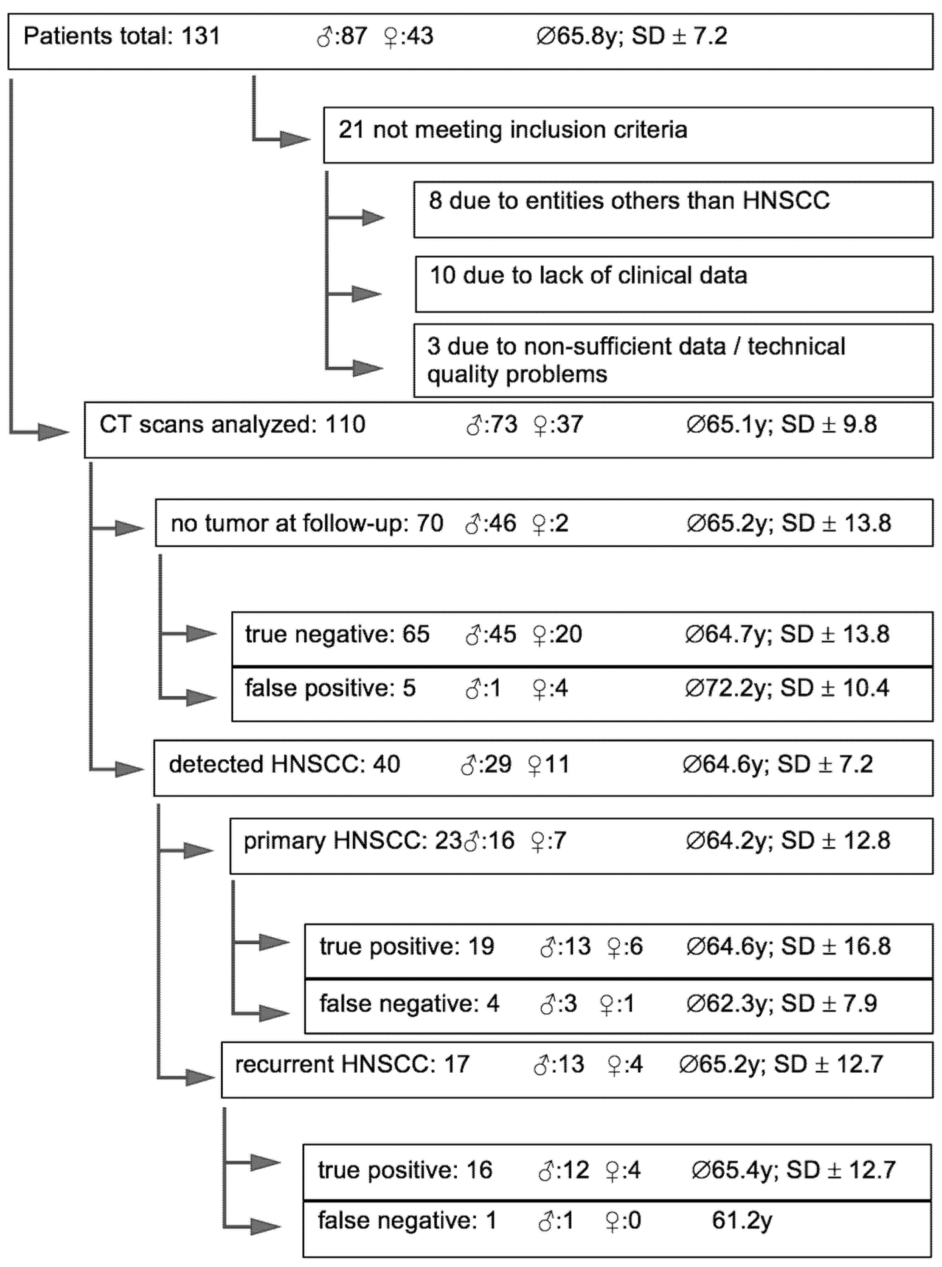
3.1. Patients
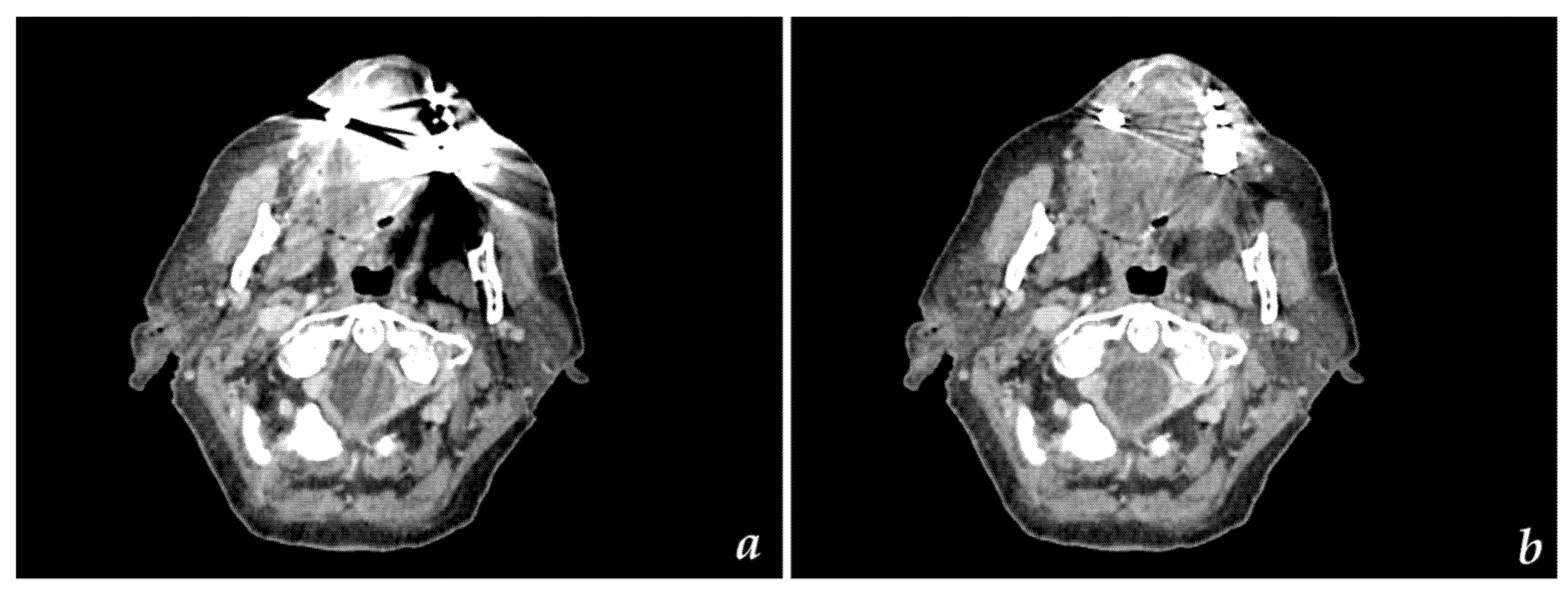
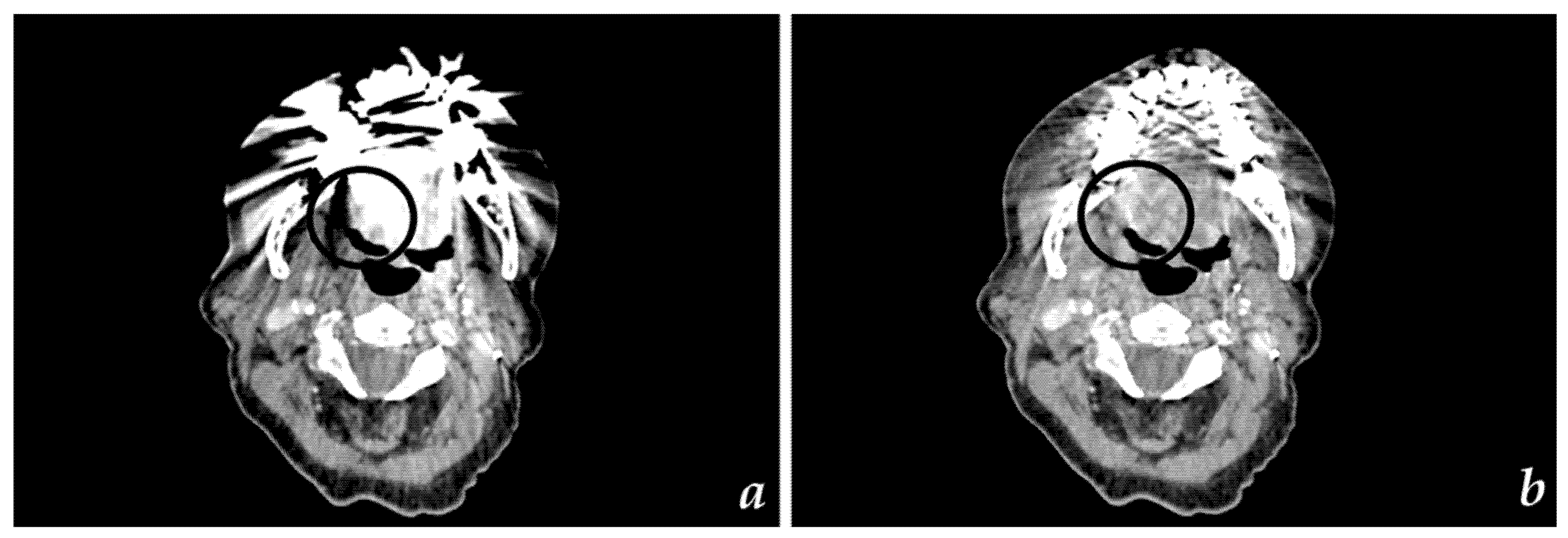
3.2. CT Findings
3.3. Technical Results
3.4. Detectability of Primary (I) and Recurrent HNSCC (II) via CT
4. Discussion
Author Contributions
Funding
Institutional Review Board Statement
Informed Consent Statement
Data Availability Statement
Conflicts of Interest
References
- Bray-Wiley, S.; Cooper-Pellegrini, W. World Cancer Report 2014; World Health Organization, International Agency for Research on Cancer: Geneva, Switzerland; ISBN 978-92-832-0429-9.
- Bray, F.; Ferlay, J.; Soerjomataram, I.; Siegel, R.L.; Torre, L.A.; Jemal, A. Global Cancer Statistics 2018: GLOBOCAN Estimates of Incidence and Mortality Worldwide for 36 Cancers in 185 Countries. CA Cancer J. Clin. 2018, 68, 394–424. [Google Scholar] [CrossRef] [PubMed]
- Johnson, N.W.; Jayasekara, P.; Amarasinghe, A.A.H.K. Squamous Cell Carcinoma and Precursor Lesions of the Oral Cavity: Epidemiology and Aetiology: Oral Cancer Epidemiology/Aetiology. Periodontology 2000 2011, 57, 19–37. [Google Scholar] [CrossRef] [PubMed]
- Ferlay, J.; Soerjomataram, I.; Dikshit, R.; Eser, S.; Mathers, C.; Rebelo, M.; Parkin, D.M.; Forman, D.; Bray, F. Cancer Incidence and Mortality Worldwide: Sources, Methods and Major Patterns in GLOBOCAN 2012: Globocan 2012. Int. J. Cancer 2015, 136, E359–E386. [Google Scholar] [CrossRef] [PubMed]
- Arya, S.; Rane, P.; Deshmukh, A. Oral Cavity Squamous Cell Carcinoma: Role of Pretreatment Imaging and Its Influence on Management. Clin. Radiol. 2014, 69, 916–930. [Google Scholar] [CrossRef] [PubMed]
- Blatt, S.; Ziebart, T.; Krüger, M.; Pabst, A.M. Diagnosing Oral Squamous Cell Carcinoma: How Much Imaging Do We Really Need? A Review of the Current Literature. J. Craniomaxillofac. Surg. 2016, 44, 538–549. [Google Scholar] [CrossRef]
- Gong, X.-Y.; Meyer, E.; Yu, X.-J.; Sun, J.-H.; Sheng, L.-P.; Huang, K.-H.; Wu, R.-Z. Clinical Evaluation of the Normalized Metal Artefact Reduction Algorithm Caused by Dental Fillings in CT. Dentomaxillofac. Radiol. 2013, 42, 105. [Google Scholar] [CrossRef]
- Fazel, R.; Krumholz, H.M.; Wang, Y.; Ross, J.S.; Chen, J.; Ting, H.H.; Shah, N.D.; Nasir, K.; Einstein, A.J.; Nallamothu, B.K. Exposure to Low-Dose Ionizing Radiation from Medical Imaging Procedures. N. Engl. J. Med. 2009, 361, 849–857. [Google Scholar] [CrossRef]
- Bundesamt für Strahlenschutz Diagnostische Referenzwerte für diagnostische und interventionelle Röntgenanwendungen 2016. Available online: https://www.tuvsud.com/de-de/-/media/de/aerztliche-stelle-hessen/pdf/newsletter/schegerer-diagnostische-referenzwerte.pdf (accessed on 18 June 2021).
- Schmid, A.I.; Uder, M.; Lell, M.M. Reaching for Better Image Quality and Lower Radiation Dose in Head and Neck CT: Advanced Modeled and Sinogram-Affirmed Iterative Reconstruction in Combination with Tube Voltage Adaptation. Dentomaxillofac. Radiol. 2017, 46, 131. [Google Scholar] [CrossRef]
- Vachha, B.; Brodoefel, H.; Wilcox, C.; Hackney, D.B.; Moonis, G. Radiation Dose Reduction in Soft Tissue Neck CT Using Adaptive Statistical Iterative Reconstruction (ASIR). Eur. J. Radiol. 2013, 82, 2222–2226. [Google Scholar] [CrossRef]
- Hounsfield, G.N. Computerized Transverse Axial Scanning (Tomography). 1. Description of System. Br. J. Radiol. 1973, 46, 1016–1022. [Google Scholar] [CrossRef]
- Kano, T.; Koseki, M. Metal Artifact Reduction Using Iterative Reconstruction Algorithm. SICE 2015, 51, 836–844. [Google Scholar] [CrossRef]
- Seibert, J.A. Iterative Reconstruction: How It Works, How to Apply It. Pediatr. Radiol. 2014, 44, 431–439. [Google Scholar] [CrossRef] [PubMed]
- Weiß, J.; Schabel, C.; Bongers, M.; Raupach, R.; Clasen, S.; Notohamiprodjo, M.; Nikolaou, K.; Bamberg, F. Impact of Iterative Metal Artifact Reduction on Diagnostic Image Quality in Patients with Dental Hardware. Acta Radiol. 2017, 58, 279–285. [Google Scholar] [CrossRef] [PubMed]
- Leslie, A.; Fyfe, E.; Guest, P.; Goddard, P.; Kabala, J.E. Staging of Squamous Cell Carcinoma of the Oral Cavity and Oropharynx: A Comparison of MRI and CT in T- and N-Staging. J. Comput. Assist. Tomogr. 1999, 23, 43–49. [Google Scholar] [CrossRef]
- Huang, S.-H.; Chien, C.-Y.; Lin, W.-C.; Fang, F.-M.; Wang, P.-W.; Lui, C.-C.; Huang, Y.-C.; Hung, B.-T.; Tu, M.-C.; Chang, C.-C. A Comparative Study of Fused FDG PET/MRI, PET/CT, MRI, and CT Imaging for Assessing Surrounding Tissue Invasion of Advanced Buccal Squamous Cell Carcinoma. Clin. Nucl. Med. 2011, 36, 518–525. [Google Scholar] [CrossRef] [PubMed]
- Di Martino, E.; Nowak, B.; Hassan, H.A.; Hausmann, R.; Adam, G.; Buell, U.; Westhofen, M. Diagnosis and Staging of Head and Neck Cancer: A Comparison of Modern Imaging Modalities (Positron Emission Tomography, Computed Tomography, Color-Coded Duplex Sonography) with Panendoscopic and Histopathologic Findings. Arch. Otolaryngol. Head Neck Surg. 2000, 126, 1457–1461. [Google Scholar] [CrossRef]
- Pałasz, P.; Adamski, Ł.; Górska-Chrząstek, M.; Starzyńska, A.; Studniarek, M. Contemporary Diagnostic Imaging of Oral Squamous Cell Carcinoma—A Review of Literature. Pol. J. Radiol. 2017, 82, 193–202. [Google Scholar] [CrossRef]
- Hao, S.P.; Ng, S.H. Magnetic Resonance Imaging versus Clinical Palpation in Evaluating Cervical Metastasis from Head and Neck Cancer. Otolaryngol. Head Neck Surg. 2000, 123, 324–327. [Google Scholar] [CrossRef]
- Bolzoni, A.; Cappiello, J.; Piazza, C.; Peretti, G.; Maroldi, R.; Farina, D.; Nicolai, P. Diagnostic Accuracy of Magnetic Resonance Imaging in the Assessment of Mandibular Involvement in Oral-Oropharyngeal Squamous Cell Carcinoma: A Prospective Study. Arch. Otolaryngol. Head Neck Surg. 2004, 130, 837–843. [Google Scholar] [CrossRef]
- Imaizumi, A.; Yoshino, N.; Yamada, I.; Nagumo, K.; Amagasa, T.; Omura, K.; Okada, N.; Kurabayashi, T. A Potential Pitfall of MR Imaging for Assessing Mandibular Invasion of Squamous Cell Carcinoma in the Oral Cavity. Am. J. Neuroradiol. 2006, 27, 114–122. [Google Scholar] [PubMed]
- Pinter, N.K.; Klein, J.P.; Mechtler, L.L. Potential Safety Issues Related to the Use of Gadolinium-Based Contrast Agents. Continuum 2016, 22, 1678–1684. [Google Scholar] [CrossRef] [PubMed]
- Gulani, V.; Calamante, F.; Shellock, F.G.; Kanal, E.; Reeder, S.B. International Society for Magnetic Resonance in Medicine Gadolinium Deposition in the Brain: Summary of Evidence and Recommendations. Lancet Neurol. 2017, 16, 564–570. [Google Scholar] [CrossRef]
- Sheikhbahaei, S.; Taghipour, M.; Ahmad, R.; Fakhry, C.; Kiess, A.P.; Chung, C.H.; Subramaniam, R.M. Diagnostic Accuracy of Follow-Up FDG PET or PET/CT in Patients With Head and Neck Cancer After Definitive Treatment: A Systematic Review and Meta-Analysis. Am. J. Roentgenol. 2015, 205, 629–639. [Google Scholar] [CrossRef]
- Jeong, H.-S.; Baek, C.-H.; Son, Y.-I.; Ki Chung, M.; Kyung Lee, D.; Young Choi, J.; Kim, B.-T.; Kim, H.-J. Use of Integrated 18F-FDG PET/CT to Improve the Accuracy of Initial Cervical Nodal Evaluation in Patients with Head and Neck Squamous Cell Carcinoma. Head Neck 2007, 29, 203–210. [Google Scholar] [CrossRef] [PubMed]
- Yamashiro, T.; Miyara, T.; Honda, O.; Kamiya, H.; Murata, K.; Ohno, Y.; Tomiyama, N.; Moriya, H.; Koyama, M.; Noma, S.; et al. Adaptive Iterative Dose Reduction Using Three Dimensional Processing (AIDR3D) Improves Chest CT Image Quality and Reduces Radiation Exposure. PLoS ONE 2014, 9, e0105735. [Google Scholar] [CrossRef] [PubMed]


| Tumor Localization at Initial Diagnosis | Frequency in Total (n) | Percentage | Frequency (n) and Percentage of Locations in Primary (I), Recurrent (II) and no Tumor (III) |
|---|---|---|---|
| Mouth floor | 35 | 31.8% | I: 9 (25.7%) II: 5 (14.3%) III: 21 (60.0%) |
| Oropharynx | 22 | 20.0% | I: 4 (18.2%) II: 3 (13.6%) III: 15 (68.2%) |
| (Alveolar ridge of upper/lower jaw/buccal mucosa | 23 | 20.9% | I: 6 (26.1%) II: 2 (8.7%) III: 15 (65.2%) |
| Edge of tongue | 20 | 18.2% | I: 3 (15.0%) II: 3 (15.0%) III: 14 (70.0%) |
| Laryngopharynx | 5 | 4.6% | I: 0 (0.0%) II: 3 (60.0%) III: 2 (40.0%) |
| Nasopharynx | 2 | 1.8% | I: 1 (50.0%) II: 0 (0.0%) III: 1 (50.0%) |
| Others | 3 | 2.7% | I: 0 (0.0%) II: 1 (33.3%) III: 2 (66.7%) |
| T-Stage | Number n | Percentage % |
|---|---|---|
| T1 | 36 | 32.7 |
| T2 | 33 | 30.0 |
| T3 | 11 | 10.0 |
| T4 | 24 | 21.8 |
| Tx | 6 | 5.5 |
| Patient Group | Number of Patients | True Positive | False Negative | True Negative | False Positive |
|---|---|---|---|---|---|
| Primary HNSCC (I) | 23 | 19 | 4 | 0 | 0 |
| Recurrent HNSCC (II) | 17 | 16 | 1 | 0 | 0 |
| No HNSCC (III) | 70 | 0 | 0 | 65 | 5 |
| Statistical Results | Primary Data Primary HNSCC Recurrent HNSCC | Comparative Range from Literature Primary HNSCC Recurrent HNSCC | ||
|---|---|---|---|---|
| Sensitivity | 0.83 (95% CI; 0.61–0.95) | 0.94 (95% CI; 0.71–0.99) | 0.42–0.82 [16,17,18] | 0.50 [19]–0.68 [16,17,18] |
| Specificity | 0.93 (95% CI; 0.84–0.98) | 0.93 (95% CI; 0.84–0.98) | 0.69–1 [18,19] | 0.88 [19] |
| PPV | 0.79 (95% CI; 0.62–0.90) | 0.76 (95% CI; 0.58–0.88) | ||
| NPV | 0.94 (95% CI; 0.87–0.98) | 0.98 (95% CI; 0.91–0.99) | ||
| Accuracy | 0.90 (95% CI; 0.82–0.95) | 0.93 (95% CI; 0.86–0.97) | ||
Publisher’s Note: MDPI stays neutral with regard to jurisdictional claims in published maps and institutional affiliations. |
© 2021 by the authors. Licensee MDPI, Basel, Switzerland. This article is an open access article distributed under the terms and conditions of the Creative Commons Attribution (CC BY) license (https://creativecommons.org/licenses/by/4.0/).
Share and Cite
Troeltzsch, D.; Shnayien, S.; Heiland, M.; Kreutzer, K.; Raguse, J.-D.; Hamm, B.; Niehues, S.M. Detectability of Head and Neck Cancer via New Computed Tomography Reconstruction Tools including Iterative Reconstruction and Metal Artifact Reduction. Diagnostics 2021, 11, 2154. https://doi.org/10.3390/diagnostics11112154
Troeltzsch D, Shnayien S, Heiland M, Kreutzer K, Raguse J-D, Hamm B, Niehues SM. Detectability of Head and Neck Cancer via New Computed Tomography Reconstruction Tools including Iterative Reconstruction and Metal Artifact Reduction. Diagnostics. 2021; 11(11):2154. https://doi.org/10.3390/diagnostics11112154
Chicago/Turabian StyleTroeltzsch, Daniel, Seyd Shnayien, Max Heiland, Kilian Kreutzer, Jan-Dirk Raguse, Bernd Hamm, and Stefan M. Niehues. 2021. "Detectability of Head and Neck Cancer via New Computed Tomography Reconstruction Tools including Iterative Reconstruction and Metal Artifact Reduction" Diagnostics 11, no. 11: 2154. https://doi.org/10.3390/diagnostics11112154
APA StyleTroeltzsch, D., Shnayien, S., Heiland, M., Kreutzer, K., Raguse, J.-D., Hamm, B., & Niehues, S. M. (2021). Detectability of Head and Neck Cancer via New Computed Tomography Reconstruction Tools including Iterative Reconstruction and Metal Artifact Reduction. Diagnostics, 11(11), 2154. https://doi.org/10.3390/diagnostics11112154