Article

Comparative Analysis of BIOCHIP Mosaic-Based Indirect Immunofluorescence with Enzyme-Linked Immunosorbent Assay for Diagnosing Myasthenia Gravis

by
Caterina Maria Gambino
1,
Luisa Agnello
1,
Bruna Lo Sasso
1,
Concetta Scazzone
1,
Rosaria Vincenza Giglio
1,
Giuseppina Candore
1,
Anna Maria Ciaccio
2,
Vincenzo Di Stefano
3,
Filippo Brighina
3,
Matteo Vidali
4 and
Marcello Ciaccio
1,5,*
1
Clinical Molecular Medicine and Laboratory Medicine, Institute of Clinical Biochemistry, Department of Biomedicine, Neurosciences and Advanced Diagnostics, University of Palermo, 90127 Palermo, Italy
2
Unit of Clinical Biochemistry, University of Palermo, 90127 Palermo, Italy
3
Unit of Neurology, Department of Biomedicine, Neurosciences and Advanced Diagnostics, 90127 Palermo, Italy
4
Foundation IRCCS Ca’ Granda Ospedale Maggiore Policlinico, 20122 Milan, Italy
5
Department of Laboratory Medicine, Azienda Ospedaliera Universitaria Policlinico “P. Giaccone”, 90127 Palermo, Italy
*
Author to whom correspondence should be addressed.
Diagnostics 2021, 11(11), 2098; https://doi.org/10.3390/diagnostics11112098
Submission received: 28 September 2021 / Revised: 4 November 2021 / Accepted: 10 November 2021 / Published: 13 November 2021
(This article belongs to the Section Medical Imaging and Theranostics)

Abstract

:
Background: The detection of anti-acetylcholine receptor (AChR) and anti-muscle-specific tyrosine kinase (MuSK) antibodies is useful in myasthenia gravis (MG) diagnosis and management. BIOCHIP mosaic-based indirect immunofluorescence is a novel analytical method, which employs the simultaneous detection of anti-AChR and anti-MuSK antibodies in a single miniature incubation field. In this study, we compare, for the first time, the BIOCHIP MG mosaic with conventional enzyme-linked immunosorbent assay (ELISA) in the diagnosis of MG. Methods: A total of 71 patients with MG diagnosis were included in the study. Anti-AChR and anti-MuSK antibodies were measured separately by two different ELISA and simultaneously by BIOCHIP. The results were then compared. Results: The overall concordance between ELISA and BIOCHIP for anti-AChR reactivity was 74%. Cohen’s kappa was 0.51 (95% CI 0.32–0.71), which corresponds to 90% of the maximum possible kappa (0.57), given the observed marginal frequencies. The overall concordance for anti-MuSK reactivity was 84%. Cohen’s kappa was 0.11 (95% CI 0.00–0.36), which corresponds to 41% of the maximum possible kappa (0.27). Conclusion: The overall concordance among assays is not optimal.

1. Introduction

Myasthenia gravis (MG) is a chronic autoimmune disease characterized by antibody-mediated reduction of neuromuscular transmission resulting in skeletal muscle weakness and fatigue [1,2,3]. The disease onset is characterized by the weakness of eye muscles, which leads to diplopia and/or ptosis. About 20% of patients present only eye muscle symptoms, commonly referred to as ocular MG (OMG), while most patients develop other skeletal muscles symptoms within a few years, leading to generalized MG (GMG). The disease onset is characterized by 2 peaks of incidence: early-onset and late-onset, which commonly occurs in women of up to 40 years of age and in men of around 50 years of age, respectively [4].
The detection of autoantibodies is crucial for MG diagnosis, prognosis, and monitor response to treatment [5].
Autoantibodies against the nicotinic acetylcholine receptor (AChR) localized on the post-synaptic membrane of the neuromuscular junction are highly specific to MG. They are pathogenic and are detectable in approximately 85% of MG patients [6]. In approximately 6% of MG patients, the autoimmune response is against the muscle-specific tyrosine kinase (MuSK), a protein belonging to the clustering of AChR and other postsynaptic components of the neuromuscular junction [7]. Anti-MuSK autoantibodies are also pathogenic. They are mainly detectable in younger women and are associated with a severe disease course [8]. Recently, the low-density lipoprotein receptor-related protein 4 (LRP4), a molecule that forms a complex with MuSK, has been described as a novel auto-antigenic target in about 2% of seronegative MG patients [6], but its clinical significance is still unknown.
Since their discovery, several assays have been developed and commercialized for the detection of AChR and MuSK autoantibodies, including the radioimmunoprecipitation assay (RIPA) and enzyme-linked immunosorbent assay (ELISA) [9,10,11,12,13]. Although such methods are characterized by their high diagnostic accuracy, they are laborious and, especially for RIPA, the use of isotopes containing radioactivity has discouraged many laboratories. Moreover, ELISA methods have the disadvantage of being impractical when only a few samples must be analyzed. Different ELISA assays are commercially available and are based either on direct or on competitive methods, leading to discordant results among laboratories. Indeed, incongruous results have often been reported for identical samples tested with different assays. In addition, there is always the need to run two different antibody assays (e.g., anti-AChR and MuSK) to test a complete antibody panel. Finally, the sensitivity of antibody detection is limited because some antibodies may bind more effectively to the antigen when expressed on the cell surface rather than recombinant or soluble antigens.
The present study aims to compare a new commercially available multiparametric indirect immunofluorescence (IIF) assay, noted as the BIOCHIP mosaic, with conventional ELISA for the detection of anti-AChR and anti-MuSK autoantibodies for the diagnosis of MG. The BIOCHIP mosaic employs the simultaneous detection of anti-AChR and anti-MuSK autoantibodies in a single miniature incubation field. Thus, it is fast to perform and highly reproducible, with great potential in clinical practice by avoiding the use of radioactive materials and reducing the turn-around time (TAT).

2. Materials and Methods

2.1. Study Population

We performed an observational retrospective study on consecutive patients with MG enrolled at the Unit of Neurology at the University Hospital Policlinico “P. Giaccone” of Palermo, Italy.
The demographic and clinical data of the patients, including sex, age at onset, initial symptoms, presence of MG crisis, the severity of MG based on the Myasthenia Gravis Foundation of America (MGFA) clinical classification, and the state of immunosuppressive treatment, were recorded by reviewing medical records.
The diagnosis of MG was made according to the International Consensus Guidance for Management of MG [14], including the following criteria: a diffuse weakness with or without ocular and respiratory involvement; decremented U-shaped response at 3 Hz repetitive nerve stimulation and/or increased jitter at single-fiber electromyography; and exclusion of any other neurologic or inflammatory condition. All patients were screened for the presence of thymoma with computed tomography (CT) or/and magnetic resonance (MR) imaging scanning of the mediastinum.
The study was conducted in accordance with the ethical standards as formulated in the Helsinki Declaration. All patients remained anonymous and gave informed consent to be included in the study.

2.2. Biochemical Analysis

The blood samples of the patients were collected in dry tubes and the serum obtained after centrifugation was stored at −80 °C until analysis.
Anti AChR and MuSK autoantibodies were measured by two different assays: a new commercially available BIOCHIP method (MG mosaics, Euroimmun, Lübeck, Germany), which measures antibodies against AChR and MuSK simultaneously, and two different commercially available ELISA methods for detecting AChR (RSR Ltd., Cardiff, UK) and MuSK autoantibodies (IBL International GmbH, Männedorf, Germany).
All analyses were performed at the Institute of Clinical Biochemistry, Clinical Molecular Medicine and Clinical Laboratory Medicine at the University of Palermo.

2.2.1. BIOCHIP Mosaic Indirect Immunofluorescence Assay

The BIOCHIP mosaic-based IIF method for detecting autoantibodies against AChR and MuSK relies on the combination of different substrates put on each field that enables the simultaneous detection of different antibodies. In detail, each field of the slide is a mosaic of 4 substrates: (1) EU90 recombinant cells transfected with the adult acetylcholine receptor (AChR-E); (2) EU90 recombinant cells transfected with fetal acetylcholine receptor (AChR-G); (3) EU90 recombinant cells transfected with MuSK; and (4) EU90 recombinant cells not transfected used as a negative control. This test is designed exclusively for the in vitro determination of human antibodies in serum or plasma.
The assays were performed at a serum dilution of 1:10 in a sample buffer. If the reaction was positive, specific antibodies of classes IgA, IgG, and IgM bound the antigens. In the second and third steps, the attached antibodies were stained with biotin-labeled anti-human antibodies, followed by fluorescein isothiocyanate-labeled avidin and made visible with the fluorescence microscope. Positive and negative control sera provided by the manufacturer were tested in each working session to evaluate run validity, in order to generate a valid result.
Slides were analyzed under a DMIRE2 Leica fluorescence microscope (Leica, Milan, Italy) with a 10× lens. Pictures were acquired with a digital camera model DC250 Leica, using the acquisition software Qfluor550 Leica. Two expert operators, who worked independently and were blinded to the clinical data, interpreted the results. Samples were scored positive if a fluorescence reaction was observed at 1:10 sample dilution.
We also assessed intra-laboratory reproducibility by running positive and negative control samples 10 times in separate runs. Moreover, each serum sample was tested in our laboratory twice by two different operators in a blinded fashion (CMG and GC).

2.2.2. ELISA Methods

The RSR AChR Autoantibody (AChRAb) ELISA kit was used for measuring anti-AChR antibodies. This assay depended on the ability of patient serum AChRAb to compete with 3 different AChR monoclonal antibodies (MAbs 1–3) for binding sites on fetal and adult-type AChR. One MAb (MAb1) was coated onto ELISA plate wells and the other two were labeled with biotin and used in the assay in the liquid phase. In the absence of AChRAb, a sandwich was formed among MAb1, the AChR, and the two biotinylated MAbs. The bind of Mab2- and Mab3-biotin were then detected by the addition of streptavidin peroxidase, which bound specifically to biotin. In the presence of serum AChRAb, the formation of the sandwich was inhibited, and the amount of biotinylated MAbs bound to the plate wells was reduced. A higher concentration of serum AChRAb was associated with a greater inhibition of MAb-biotin binding.
Anti-MuSK antibodies were measured according to the manufacturer’s instructions. Briefly, serum patient antibodies bound to the antigen-coated wells and were detected by a signal amplification system. The substrate reaction was catalyzed via the alkaline phosphate coupled detection antibody. The intensity of the color developed was proportional to the number of patient antibodies detected. All assays were performed in duplicate, with the final concentration calculated by averaging the results. The results were reported as negative (<0.4 nmol/L), borderline (≥0.40 and <0.50 nmol/L), or positive (≥0.50 nmol/L).

2.3. Statistical Analysis

Statistical analyses were performed using SPSS statistical software v.17.0 (SPSS Inc., Chicago, IL, USA) and R Language v.4.0.3 (R Foundation for Statistical Computing, Vienna, Austria). The normality distribution was assessed preliminarily by the q–q plot and Shapiro–Wilk test. Quantitative variables were expressed by the median and interquartile range (IQR), while categorical variables were expressed as absolute frequencies and percentages. The concordance was evaluated by concordant pairs and unweighted Cohen’s kappa with 95% CI. Kappa was also reported as the proportion of maximum possible, given the observed marginal frequencies.

3. Results

In this study, we included a total of 71 Caucasian patients with confirmed MG, 40 males, ages ranging from 31 to 83 years, and 31 females, ages ranging from 18 to 78 years.
At the time of enrolment, among all MG patients, 10 (14%) had thymoma, 9 (13%) used symptomatic monotherapy with cholinesterase inhibitors, and the majority (55%) additionally used oral corticosteroids and/or steroid-sparing immunosuppressive therapy (29%).
The BIOCHIP mosaic method provided a qualitative evaluation of circulating antibodies. All samples were tested at 1:10 dilution, in accordance with the manufacturer’s recommendations. Negative samples were defined by the complete absence of staining in all four substrates (Figure 1, row C). However, as the use of only a single dilution may lead to blocking or masking effects in high-titered sera, causing false–negative results, sera that were negative at 1:10 were retested at 1:100 dilutions. At this dilution, no pro-zone effects (defined as the lack of detection of antibodies in low dilutions, but their presence in high dilution) were observed, confirming that those sera were true negatives.
Serum reactivity was evaluated as follows:
-
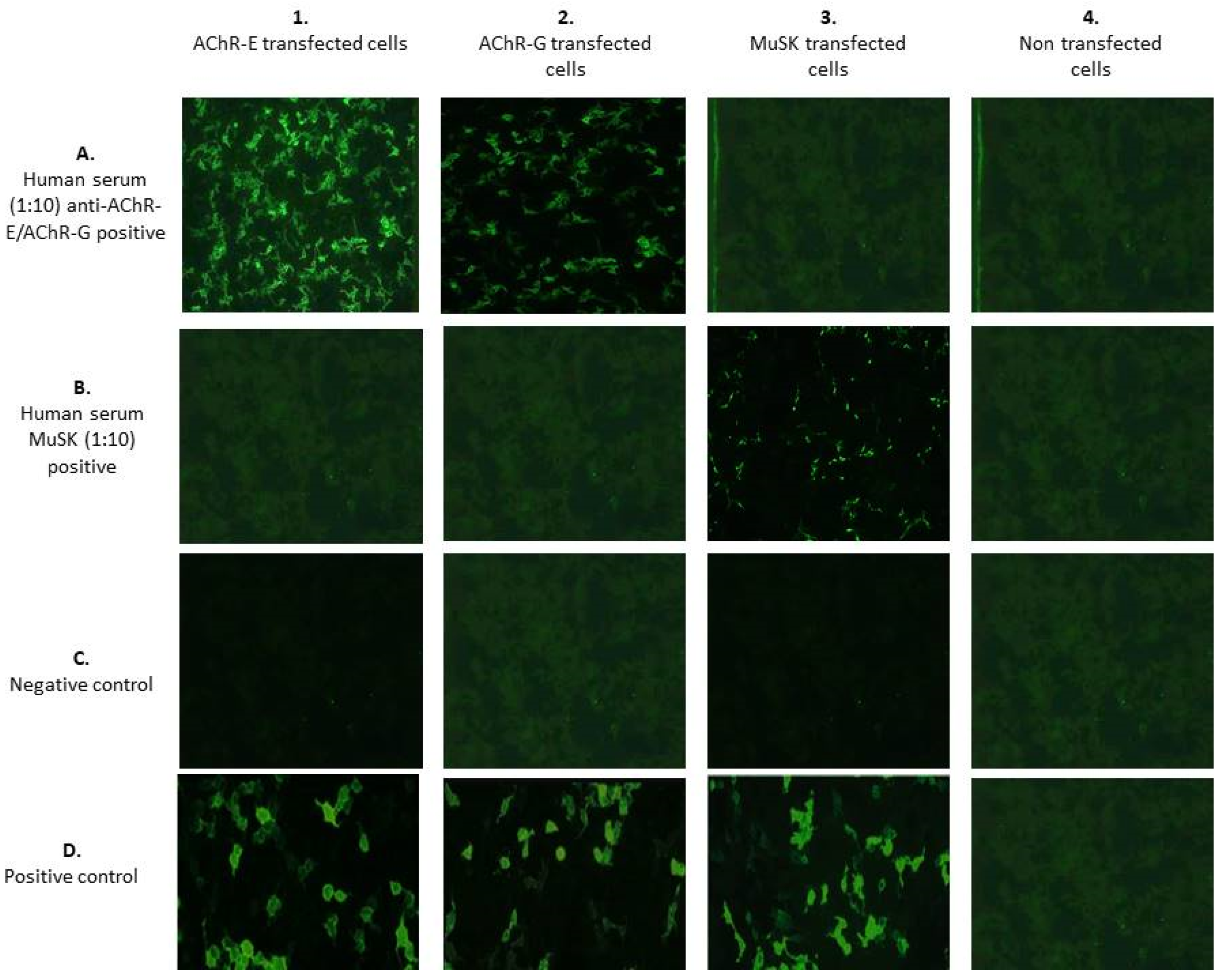
Antibodies against AChR-E showed a flat or fine-to-coarse granular fluorescence of the cells, and also a smooth to very fine granular fluorescence of the cells in the absence of the staining of the membrane of the non-transfected cells. The area of the cell nucleus was only slightly stained (Figure 1(1A)).
-
Antibodies against AChR-G showed a spotted, and also a smooth-to-very fine granular fluorescence of the cells in the absence of the staining of the membrane of the non-transfected cells. The area of the cell nucleus was only slightly stained (Figure 1(2A)).
-
Antibodies against MuSK produced a smooth-to-very fine granular fluorescence of the cells in the absence of the staining of the membrane of the non-transfected cells. The area of the cell nucleus was only slightly stained (Figure 1(3B)). To evaluate the BIOCHIP mosaic intra-laboratory reproducibility, positive and negative control samples were tested 10 times in separate runs by 2 blinded operators. The overall agreement within the intra-laboratory runs was close to 100%.
The Anti-AChR autoantibodies were positive (≥0.50 nmol/L) in 44 out of 71 (62%) by ELISA and in 27 out of 71 (38%) by the BIOCHIP mosaic (combining the results of fetal and adult AChR assays).
The median (IQR) levels of anti-AChR autoantibodies in ELISA were 1.25 (0.25–12.88), with 13 subjects displaying levels higher than 20 (Figure 2). Of note, 15 out of 17 patients, whose diagnoses of MG were not identified by the BIOCHIP mosaic, with a lack of AChR positivity, had a low autoantibodies titer on the ELISA method, whilst the other 2 patients had a higher median anti-AChR titer.
Anti-MuSK antibodies were considered as positive (≥0.50 nmol/L) in 11 out of 71 (15%) by ELISA and in 2 out of 71 (4%) by the BIOCHIP mosaic. Similarly, anti-MuSK negative patients on the BIOCHIP mosaic had a low antibodies titer, whilst anti-MuSK positive patients on the BIOCHIP mosaic had higher antibodies levels (Figure 2).
Patients with anti-AChR and anti-MuSK double positivity in the ELISA and BIOCHIP mosaic were respectively 5 (7%) and 0 (0%). A total of 20 MG patients (28.2%) were seronegative tested by ELISA, while 41 (57.7%) tested by BIOCHIP. We also associated either the ELISA positivity or the fluorescence patterns observed by BIOCHIP with the clinical manifestations (OMG or GMG) of each patient. This clinical information was available only in 58 (41 GMG and 17 OMG) patients. Positivity to anti-AChR autoantibodies by ELISA was found in 26/41 (63%) patients with GMG and in 8/17 (47%) patients with OMG; no significant association was evident (Fisher’s exact test p = 0.380). The AChRE positive pattern was found in 18/41 (44%) patients with GMG and in 2/17 (12%) patients with OMG. Similarly, the AChRG positive pattern was present in 20/41 (49%) of GMG patients and in only 2/17 (12%) with OMG. A statistically significant association was found between the clinical manifestation and both the AchRE or the AchRG pattern (Fisher’s exact test, respectively, p = 0.032 and p = 0.009). Anti-MuSK positivity by ELISA was present in 5/41 (12%) patients with GMG and in 3/17 (18%) patients with OMG; no significant association was found (Fisher’s exact test p = 0.681). A MuSK positive pattern was observed in only 2/41 (5%) of GMG patients.
The cross-tabulation of anti-AChR positivity in ELISA and IIF (fetal and/or adult AChR) is shown in Table 1. The overall concordance between ELISA and IIF for anti-AChR reactivity, calculated as concordant pairs, was (26 + 26)/70 = 74%. Cohen’s kappa was 0.51 (95% CI 0.32–0.71), which corresponds to 90% of the maximum possible kappa (0.57), given the observed marginal frequencies.
The cross-tabulation of anti-MuSK positivity in ELISA and IIF is shown in Table 2. The overall concordance between ELISA and IFI for anti-MuSK reactivity, calculated as concordant pairs, was (58 + 1)/70 = 84%. Cohen’s kappa was 0.11 (95% CI 0.00–0.36), which corresponds to 41% of the maximum possible kappa (0.27), given the observed marginal frequencies.

4. Discussion

The diagnosis of MG is based on a multistep approach, which combines clinical features and laboratory criteria (Figure 3). The sensitive and specific detection of anti-AChR and anti-MuSK autoantibodies has become an essential laboratory investigation in evaluating patients with MG, because seropositivity has diagnostic, prognostic, and therapeutic implications [15,16,17,18,19].
Recent studies report that anti-MuSK positive patients have a worse prognosis with predominant bulbar and respiratory muscle involvement and frequent respiratory crises [3,20,21,22]. Moreover, they respond poorly to acetylcholinesterase inhibitors, and conventional pyridostigmine doses frequently induce side effects, while they respond well to corticosteroids and to many steroid-sparing agents [23,24]. In addition, Rituximab should be considered as an early therapeutic option in patients who have an unsatisfactory response to initial immunotherapy [25,26]. Anti-MuSK autoantibodies primarily belong to the IgG4 subclass, which does not actives complement but rather directly inhibits protein function [27,28]. Interestingly, their titers positively correlate with symptom severity and a decrease in their response to plasma exchange [29]. Unlike anti-MuSK antibodies, anti-AChR autoantibodies primarily belong to the IgG1 and IgG3 subclasses and active complement cascades, causing the destruction of the post-synaptic membrane [30,31]. Their titers, generally, do not correlate with disease severity; however, the temporal variation of anti-AChR autoantibodies levels in a patient can be associated with symptom severity and the response to treatment as well. Thus, anti-AChR and anti-MuSK antibodies are associated to distinct disease entities with unique immunopathologic mechanisms.
Over the past decades, several analytical methods have been developed to measure anti-AChR and anti-MuSK autoantibodies, such as RIPA and ELISA [32,33,34]. However, these methods have some important analytical drawbacks, such as the use of radioactive isotopes for RIPA or a long TAT, the lack of standardization, and excessive cost for ELISA, resulting in an inconvenience for small laboratories. Different types of ELISA have been developed based either on direct or competitive methods, as shown in Table 3. Recently, a new method, the BIOCHIP mosaic, which simultaneously detects anti-AChR and anti-MuSK autoantibodies, has been developed. Although the BIOCHIP mosaic does not provide quantitative autoantibody serum levels, the simultaneous and multi-parametric analysis of all relevant antibodies provides faster results and a more cost-effective approach. Moreover, the BIOCHIP mosaic allows the discrimination between antibodies directed against fetal or adult AChR, which is important for the diagnosis of transient neonatal MG [35]. This condition is characterized by the presence in the mother of antibodies against the fetal AChR form only, which could not cause symptoms of MG in the mother, but could be harmful for the newborn [36,37].
In this study, for the first time, we compared the performance of a new multiparametric BIOCHIP mosaic-based IIF technique with ELISA for anti-AChR and anti-MuSK autoantibodies evaluation. Our results showed a concordance between ELISA and the BIOCHIP mosaic of 74% and 84% for anti-AChR reactivity and anti-MuSK reactivity, respectively. The Cohen’s kappa was 0.51 (95% CI 0.32–0.71) and 0.11 (95% CI 0.00–0.36) for anti-AChR and anti-MuSK reactivity, respectively. Thus, the overall concordance between ELISA and the BIOCHIP mosaic is not optimal. Interestingly, we observed a significant association between the clinical manifestation and both the AChRE or the AChRG pattern, suggesting a possible discriminatory power between ocular and generalized MG by using the BIOCHIP method.
A number of studies assessed the validity of the BIOCHIP mosaic method for detecting anti-desmoglein 1 and 3 antibodies in the diagnosis of autoimmune bullous diseases [38,39,40,41,42,43], presenting encouraging results. In addition, only one study reported the BIOCHIP mosaic assay as a powerful tool for the diagnosis of neuromyelitis optica [44].
Unfortunately, our findings showed that the BIOCHIP mosaic for MG diagnosis did not accurately identify specific autoantibodies. In particular, we observed that low autoantibody titrrs, detected by ELISA, were undetectable by the BIOCHIP method and, thus, the patients were identified as “seronegative”.
In conclusion, although BIOCHIP has great potential, since it offers different advantages, being easy to perform and less-time consuming, eliminating the need for multiple runs, more efforts are required to improve the antibodies detection in order to use it as an alternative test to ELISA and RIPA in MG diagnosis.

Author Contributions

Conceptualization, C.M.G. and M.C.; formal analysis, C.S. and R.V.G.; data curation, V.D.S. and F.B.; writing—original draft preparation, C.M.G. and A.M.C.; statistical analysis, M.V.; writing—review and editing, G.C., L.A. and B.L.S.; and supervision, M.C. All authors have read and agreed to the published version of the manuscript.

Funding

This research received no external funding.

Institutional Review Board Statement

The study was conducted according to the guidelines of the Declaration of Helsinki and approved by the Institutional Review Board of the University Hospital of Palermo (nr 05/2021) on 19 May 2021.

Informed Consent Statement

Informed consent was obtained from all subjects involved in the study.

Data Availability Statement

Derived data supporting the findings of this study are available from the corresponding author on request.

Conflicts of Interest

The authors declare no conflict of interest.

References

  1. Berrih-Aknin, S.; Le Panse, R. Myasthenia gravis: A comprehensive review of immune dysregulation and etiological mechanisms. J. Autoimmun. 2014, 52, 90–100. [Google Scholar] [CrossRef]
  2. Gilhus, N.E.; Verschuuren, J.J. Myasthenia gravis: Subgroup classification and therapeutic strategies. Lancet Neurol. 2015, 14, 1023–1036. [Google Scholar] [CrossRef]
  3. Gilhus, N.E.; Tzartos, S.; Evoli, A.; Palace, J.; Burns, T.M.; Verschuuren, J. Myasthenia gravis. Nat. Rev. Dis. Primers 2019, 5, 30. [Google Scholar] [CrossRef]
  4. Gilhus, N.E. Myasthenia Gravis. N. Engl. J. Med. 2016, 375, 2570–2581. [Google Scholar] [CrossRef] [PubMed]
  5. Gastaldi, M.; Scaranzin, S.; Businaro, P.; Mobilia, E.; Benedetti, L.; Pesce, G.; Franciotta, D. Improving laboratory diagnostics in myasthenia gravis. Expert Rev. Mol. Diagn. 2021, 21, 579–590. [Google Scholar] [CrossRef] [PubMed]
  6. Verschuuren, J.J.; Huijbers, M.G.; Plomp, J.J.; Niks, E.H.; Molenaar, P.C.; Martinez-Martinez, P.; Gomez, A.M.; De Baets, M.H.; Losen, M. Pathophysiology of myasthenia gravis with antibodies to the acetylcholine receptor, muscle-specific kinase and low-density lipoprotein receptor-related protein 4. Autoimmun. Rev. 2013, 12, 918–923. [Google Scholar] [CrossRef]
  7. Pevzner, A.; Schoser, B.; Peters, K.; Cosma, N.C.; Karakatsani, A.; Schalke, B.; Melms, A.; Kröger, S. Anti-LRP4 autoantibodies in AChR- and MuSK-antibody-negative myasthenia gravis. J. Neurol. 2012, 259, 427–435. [Google Scholar] [CrossRef] [PubMed]
  8. Borges, L.S.; Richman, D.P. Muscle-Specific Kinase Myasthenia Gravis. Front. Immunol. 2020, 11, 707. [Google Scholar] [CrossRef] [PubMed]
  9. Lindstrom, J. An assay for antibodies to human acetylcholine receptor in serum from patients with myasthenia gravis. Clin. Immunol. Immunopathol. 1977, 7, 36–43. [Google Scholar] [CrossRef]
  10. Matthews, I.; Chen, S.; Hewer, R.; McGrath, V.; Furmaniak, J.; Rees Smith, B. Muscle-specific receptor tyrosine kinase autoantibodies--A new immunoprecipitation assay. Clin. Chim. Acta 2004, 348, 95–99. [Google Scholar] [CrossRef]
  11. Hewer, R.; Matthews, I.; Chen, S.; McGrath, V.; Evans, M.; Roberts, E.; Nute, S.; Sanders, J.; Furmaniak, J.; Smith, B.R. A sensitive non-isotopic assay for acetylcholine receptor autoantibodies. Clin. Chim. Acta 2006, 364, 159–166. [Google Scholar] [CrossRef] [PubMed]
  12. Leite, M.I.; Jacob, S.; Viegas, S.; Cossins, J.; Clover, L.; Morgan, B.P.; Beeson, D.; Willcox, N.; Vincent, A. IgG1 antibodies to acetylcholine receptors in ‘seronegative’ myasthenia gravis. Brain 2008, 131, 1940–1952. [Google Scholar] [CrossRef] [PubMed] [Green Version]
  13. Zisimopoulou, P.; Lagoumintzis, G.; Kostelidou, K.; Bitzopoulou, K.; Kordas, G.; Trakas, N.; Poulas, K.; Tzartos, S.J. Towards antigen-specific apheresis of pathogenic autoantibodies as a further step in the treatment of myasthenia gravis by plasmapheresis. J. Neuroimmunol. 2008, 201–202, 95–103. [Google Scholar] [CrossRef] [PubMed]
  14. Narayanaswami, P.; Sanders, D.B.; Wolfe, G.; Benatar, M.; Cea, G.; Evoli, A.; Gilhus, N.E.; Illa, I.; Kuntz, N.L.; Massey, J.; et al. International Consensus Guidance for Management of Myasthenia Gravis: 2020 Update. Neurology 2021, 96, 114–122. [Google Scholar] [CrossRef]
  15. Leite, M.I.; Waters, P.; Vincent, A. Diagnostic use of autoantibodies in myasthenia gravis. Autoimmunity 2010, 43, 371–379. [Google Scholar] [CrossRef]
  16. Frykman, H.; Kumar, P.; Oger, J. Immunopathology of Autoimmune Myasthenia Gravis: Implications for Improved Testing Algorithms and Treatment Strategies. Front. Neurol. 2020, 11, 596621. [Google Scholar] [CrossRef]
  17. Lazaridis, K.; Tzartos, S.J. Myasthenia Gravis: Autoantibody Specificities and Their Role in MG Management. Front. Neurol. 2020, 11, 596981. [Google Scholar] [CrossRef]
  18. Lindstrom, J. Acetylcholine receptors and myasthenia. Muscle Nerve 2000, 23, 453–477. [Google Scholar] [CrossRef]
  19. Vincent, A.; Newsom-Davis, J. Acetylcholine receptor antibody as a diagnostic test for myasthenia gravis: Results in 153 validated cases and 2967 diagnostic assays. J. Neurol. Neurosurg. Psychiatry 1985, 48, 1246–1452. [Google Scholar] [CrossRef] [Green Version]
  20. Tomschik, M.; Hilger, E.; Rath, J.; Mayer, E.M.; Fahrner, M.; Cetin, H.; Löscher, W.N.; Zimprich, F. Subgroup stratification and outcome in recently diagnosed generalized myasthenia gravis. Neurology 2020, 95, e1426–e1436. [Google Scholar] [CrossRef]
  21. Kohler, S.; Losen, M.; Alexander, T.; Hiepe, F.; Meisel, A. Myasthenia gravis: Subgroup classifications. Lancet Neurol. 2016, 15, 356–357. [Google Scholar] [CrossRef] [Green Version]
  22. Mantegazza, R.; Antozzi, C. When myasthenia gravis is deemed refractory: Clinical signposts and treatment strategies. Ther Adv Neurol Disord. 2018, 11, 1756285617749134. [Google Scholar] [CrossRef] [Green Version]
  23. Mantegazza, R.; Antozzi, C. From Traditional to Targeted Immunotherapy in Myasthenia Gravis: Prospects for Research. Front. Neurol. 2020, 11, 981. [Google Scholar] [CrossRef] [PubMed]
  24. Dalakas, M.C. Progress in the therapy of myasthenia gravis: Getting closer to effective targeted immunotherapies. Curr. Opin. Neurol. 2020, 33, 545–552. [Google Scholar] [CrossRef] [PubMed]
  25. Litchman, T.; Roy, B.; Kumar, A.; Sharma, A.; Njike, V.; Nowak, R.J. Differential response to rituximab in anti-AChR and anti-MuSK positive myasthenia gravis patients: A single-center retrospective study. J. Neurol. Sci. 2020, 411, 116690. [Google Scholar] [CrossRef] [PubMed]
  26. Lazaridis, K.; Tzartos, S.J. Autoantibody Specificities in Myasthenia Gravis; Implications for Improved Diagnostics and Therapeutics. Front. Immunol. 2020, 11, 212. [Google Scholar] [CrossRef]
  27. Niks, E.H.; van Leeuwen, Y.; Leite, M.I.; Dekker, F.W.; Wintzen, A.R.; Wirtz, P.W.; Vincent, A.; van Tol, M.J.; Jol-van der Zijde, C.M.; Verschuuren, J.J. Clinical fluctuations in MuSK myasthenia gravis are related to antigen-specific IgG4 instead of IgG1. J. Neuroimmunol. 2008, 195, 151–156. [Google Scholar] [CrossRef]
  28. Huijbers, M.G.; Plomp, J.J.; van der Maarel, S.M.; Verschuuren, J.J. IgG4-mediated autoimmune diseases: A niche of antibody-mediated disorders. Ann. NY Acad. Sci. 2018, 1413, 92–103. [Google Scholar] [CrossRef]
  29. Rivner, M.H.; Pasnoor, M.; Dimachkie, M.M.; Barohn, R.J.; Mei, L. Muscle-Specific Tyrosine Kinase and Myasthenia Gravis Owing to Other Antibodies. Neurol. Clin. 2018, 36, 293–310. [Google Scholar] [CrossRef]
  30. Engel, A.G.; Arahata, K. The membrane attack complex of complement at the endplate in myasthenia gravis. Ann. NY Acad. Sci. 1987, 505, 326–332. [Google Scholar] [CrossRef]
  31. Rødgaard, A.; Nielsen, F.C.; Djurup, R.; Somnier, F.; Gammeltoft, S. Acetylcholine receptor antibody in myasthenia gravis: Predominance of IgG subclasses 1 and 3. Clin. Exp. Immunol. 1987, 67, 82–88. [Google Scholar]
  32. Bokoliya, S.; Patil, S.; Nagappa, M.; Taly, A. A Simple, Rapid and Non-Radiolabeled Immune Assay to Detect Anti-AChR Antibodies in Myasthenia Gravis. Lab. Med. 2019, 50, 229–235. [Google Scholar] [CrossRef] [PubMed]
  33. Trakas, N.; Tzartos, S.J. Immunostick ELISA for rapid and easy diagnosis of myasthenia gravis. J. Immunol. Methods 2018, 460, 107–112. [Google Scholar] [CrossRef] [PubMed]
  34. Zisimopoulou, P.; Brenner, T.; Trakas, N.; Tzartos, S.J. Serological diagnostics in myasthenia gravis based on novel assays and recently identified antigens. Autoimmun. Rev. 2013, 12, 924–930. [Google Scholar] [CrossRef] [PubMed]
  35. Shi, Q.G.; Wang, Z.H.; Ma, X.W.; Zhang, D.Q.; Yang, C.S.; Shi, F.D.; Yang, L. Clinical significance of detection of antibodies to fetal and adult acetylcholine receptors in myasthenia gravis. Neurosci. Bull. 2012, 28, 469–474. [Google Scholar] [CrossRef] [PubMed]
  36. Riemersma, S.; Vincent, A.; Beeson, D.; Newland, C.; Hawke, S.; Vernet-der Garabedian, B.; Eymard, B.; Newsom-Davis, J. Association of arthrogryposis multiplex congenita with maternal antibodies inhibiting fetal acetylcholine receptor function. J. Clin. Investig. 1966, 98, 2358–2363. [Google Scholar] [CrossRef]
  37. Saxena, A.; Stevens, J.; Cetin, H.; Koneczny, I.; Webster, R.; Lazaridis, K.; Tzartos, S.; Vrolix, K.; Nogales-Gadea, G.; Machiels, B.; et al. Characterization of an anti-fetal AChR monoclonal antibody isolated from a myasthenia gravis patient. Sci. Rep. 2017, 7, 14426. [Google Scholar] [CrossRef] [Green Version]
  38. Adaszewska, A.; Kalinska-Bienias, A.; Jagielski, P.; Wozniak, K.; Kowalewski, C. The use of BIOCHIP technique in diagnosis of different types of pemphigus: Vulgaris and foliaceus. J. Immunol. Methods 2019, 468, 35–39. [Google Scholar] [CrossRef]
  39. Tampoia, M.; Zucano, A.; Villalta, D.; Antico, A.; Bizzaro, N. Anti-skin specific autoantibodies detected by a new immunofluorescence multiplex biochip method in patients with autoimmune bullous diseases. Dermatology 2012, 225, 37–44. [Google Scholar] [CrossRef]
  40. Arunprasath, P.; Rai, R.; Venkataswamy, C. Comparative Analysis of BIOCHIP Mosaic-based Indirect Immunofluorescence with Direct Immunofluorescence in Diagnosis of Autoimmune Bullous Diseases: A Cross-Sectional Study. Indian Dermatol. Online J. 2021, 12, 105–109. [Google Scholar]
  41. van Beek, N.; Rentzsch, K.; Probst, C.; Komorowski, L.; Kasperkiewicz, M.; Fechner, K.; Bloecker, I.M.; Zillikens, D.; Stöcker, W.; Schmidt, E. Serological diagnosis of autoimmune bullous skin diseases: Prospective comparison of the BIOCHIP mosaic-based indirect immunofluorescence technique with the conventional multi-step single test strategy. Orphanet J. Rare Dis. 2019, 7, 49. [Google Scholar] [CrossRef] [PubMed] [Green Version]
  42. Xuan, R.R.; Yang, A.; Murrell, D.F. New biochip immunofluorescence test for the serological diagnosis of pemphigus vulgaris and foliaceus: A review of the literature. Int. J. Womens Dermatol. 2018, 4, 102–108. [Google Scholar] [CrossRef] [PubMed]
  43. Yang, A.; Xuan, R.; Melbourne, W.; Tran, K.; Murrell, D.F. Validation of the BIOCHIP test for the diagnosis of bullous pemphigoid, pemphigus vulgaris and pemphigus foliaceus. J. Eur. Acad. Dermatol. Venereol. 2020, 34, 153–160. [Google Scholar] [CrossRef] [PubMed]
  44. Granieri, L.; Marnetto, F.; Valentino, P.; Frau, J.; Patanella, A.K.; Nytrova, P.; Sola, P.; Capobianco, M.; Jarius, S.; Bertolotto, A. Evaluation of a multiparametric immunofluorescence assay for standardization of neuromyelitis optica serology. PLoS ONE 2012, 7, e38896. [Google Scholar] [CrossRef] [PubMed] [Green Version]
Figure 1. BIOCHIP mosaic for MG diagnosis. On a standard-sized slide, there are five incubation fields, each with four different substrates. (1) Anti-adult acetylcholine receptor (AChR-E) positive transfected cells; (2) anti-fetal acetylcholine receptor (AChR-G) positive transfected cells; (3) anti-MuSK positive transfected cells; and (4) no transfected cells.
Diagnostics 11 02098 g001
Figure 2. (a) Histogram of anti-AChR antibodies. Levels higher than 20 were represented here as having levels equal to 20; (b) histogram of anti-MuSK antibodies.
Diagnostics 11 02098 g002
Figure 3. The algorithm for the diagnosis of MG. Serological antibody testing is the gold standard for the diagnosis of MG. Autoantibodies identification is important for designing better diagnosis.
Diagnostics 11 02098 g003
Table 1. The analysis of the agreement between BIOCHIP and ELISA assays for the detection of anti-AChR antibodies.
ELISA Anti-AChR
BIOCHIP IIFPositiveNegativeTotalBIOCHIP IIF
Positive26127Positive
Negative172643Negative
Total432770Total
Table 2. The analysis of the agreement between BIOCHIP IIF and ELISA assays for the detection of anti-MuSK antibodies.
ELISA anti-MuSK
BIOCHIP IIFPositiveNegativeTotalKappa
Positive112
Negative1058680.11
Total115970
Table 3. Summary of commercially ELISA assays for anti-AChR detection.
NameAssay TypeSensitivityDetection RangeCompanyRegulatory Status
Human AChR-AbQuantitative Sandwich0.04 pmol/mL0.06–4 pmol/mLMyBioSourceRUO
Anti-AChR AbCompetitive0.23 nmol/L0.2–20 IU/mLEagle BioscienceRUO
Human Anti-AChR AbIndirect0.938 ng/mL1.563–100 ng/mLBiomatikRUO
Medizym® Anti-AChR AbCompetitive0.23 U/mL0.2–20 U/mLMedipanIVD
Anti-AChR AbCompetitive0.25 nmol/L0.2–20 nmol/LRSR LimitedIVD
Anti-AChR AbIndirect0.11 nmol/L0–8 nmol/LEuroimmunIVD
AChR-Ab, Acetylcholine Receptor Antibody; AChR, Acetylcholine Receptor; RUO, Research Use Only; and IVD, In Vitro Diagnostic.
Publisher’s Note: MDPI stays neutral with regard to jurisdictional claims in published maps and institutional affiliations.

