
Atrial Fibrillation and Peri-Atrial Inflammation Measured through Adipose Tissue Attenuation on Cardiac Computed Tomography

 ,
,  ,
,
Abstract
:1. Introduction
2. Materials and Methods
2.1. Patients
2.2. Coronary Computed Tomography Angiography
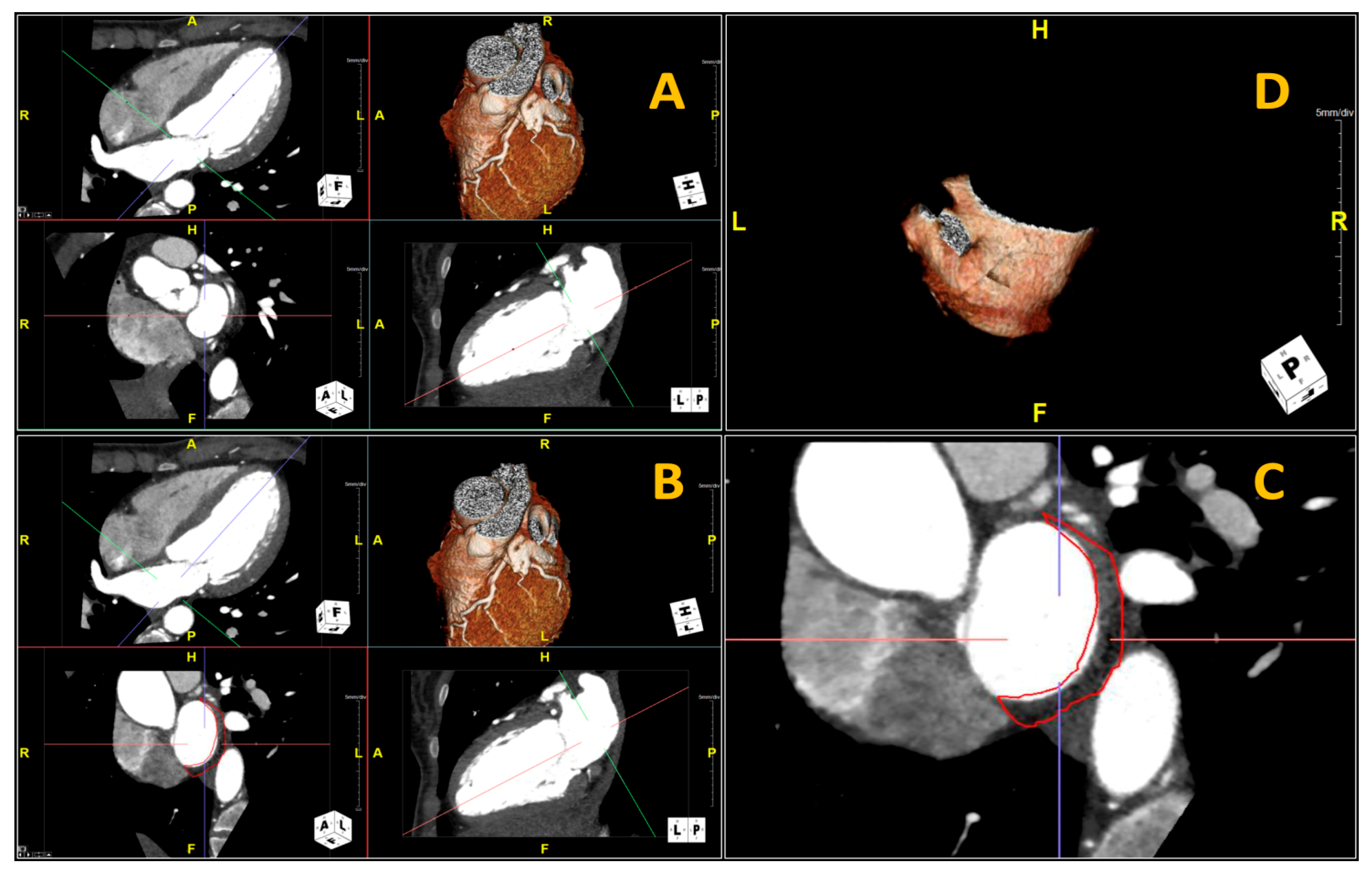
2.3. Fat-LA Volume and Attenuation
2.4. Statistical Analysis
3. Results
3.1. Patients
3.2. Posterior LA Adipose Tissue CTA Volume and Attenuation
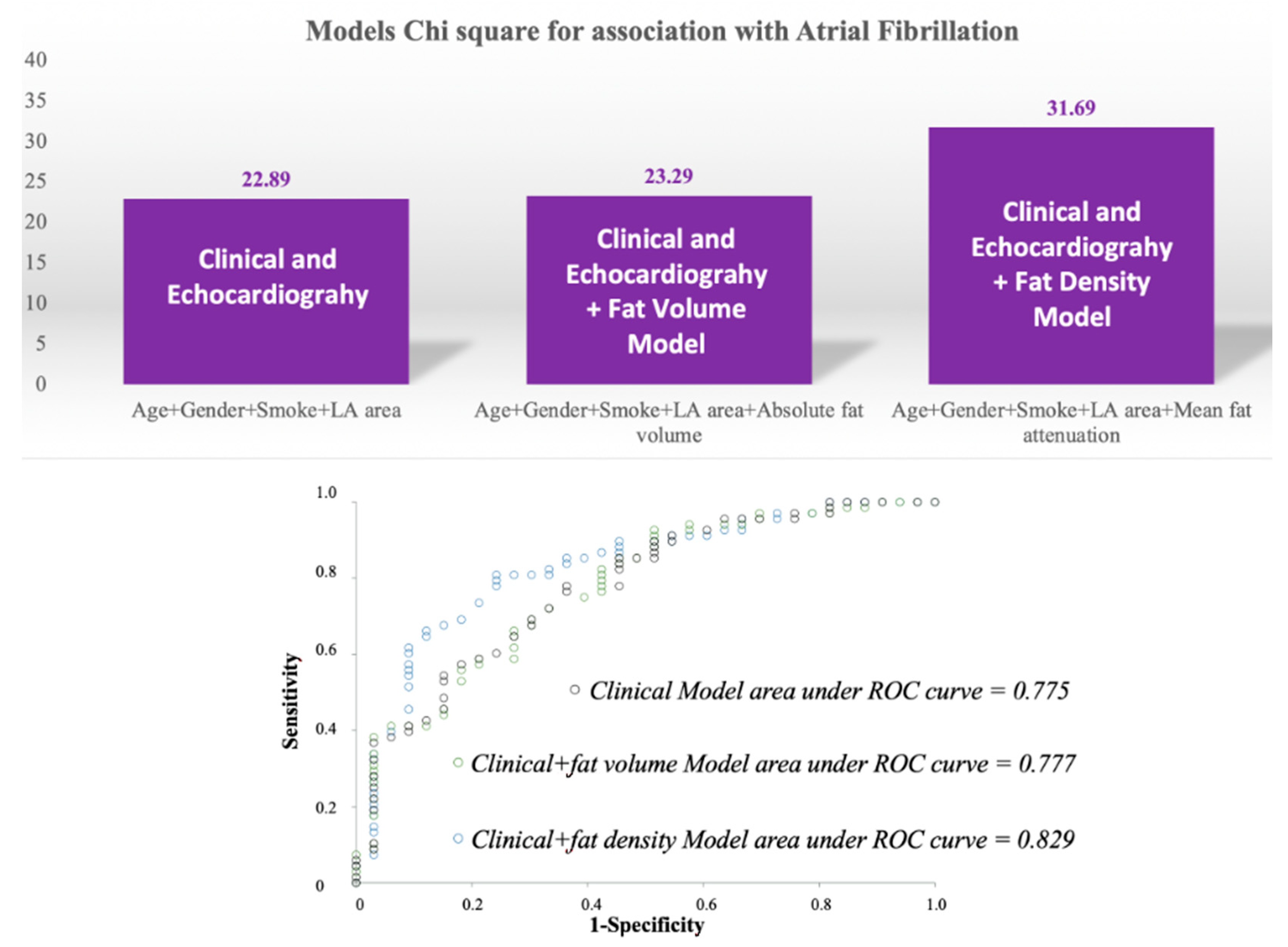
3.3. Clinical Variables and Fat-LA Volume/Density Logistic Regression Models
3.4. Comparison of Logistic Regression Models
4. Discussion
4.1. State of the Art
4.2. Fat Attenuation Versus Volume
5. Conclusions
Author Contributions
Funding
Institutional Review Board Statement
Informed Consent Statement
Data Availability Statement
Conflicts of Interest
References
- Al Chekakie, M.O.; Welles, C.C.; Metoyer, R.; Ibrahim, A.; Shapira, A.R.; Cytron, J.; Santucci, P.; Wilber, D.J.; Akar, J.G. Pericardial fat is independently associated with human atrial fibrillation. J. Am. Coll. Cardiol. 2010, 56, 784–788. [Google Scholar] [CrossRef] [Green Version]
- Thanassoulis, G.; Massaro, J.M.; O’Donnell, C.J.; Hoffmann, U.; Levy, D.; Ellinor, P.T.; Wang, T.J.; Schnabel, R.B.; Vasan, R.S.; Fox, C.S.; et al. Pericardial fat is associated with prevalent atrial fibrillation: The Framingham Heart Study. Circ. Arrhythm. Electrophysiol. 2010, 3, 345–350. [Google Scholar] [CrossRef] [PubMed] [Green Version]
- Yorgun, H.; Canpolat, U.; Aytemir, K.; Hazırolan, T.; Şahiner, L.; Kaya, E.B.; Kabakci, G.; Tokgözoğlu, L.; Özer, N.; Oto, A. Association of epicardial and peri-atrial adiposity with the presence and severity of non-valvular atrial fibrillation. Int. J. Cardiovasc. Imaging 2015, 31, 649–657. [Google Scholar] [CrossRef] [PubMed]
- Batal, O.; Schoenhagen, P.; Shao, M.; Ayyad, A.E.; Van Wagoner, D.R.; Halliburton, S.S.; Tchou, P.J.; Chung, M.K. Left atrial epicardial adiposity and atrial fibrillation. Circ. Arrhythm. Electrophysiol. 2010, 3, 230–236. [Google Scholar] [CrossRef] [Green Version]
- Tsao, H.M.; Hu, W.C.; Wu, M.H.; Tai, C.T.; Lin, Y.J.; Chang, S.L.; Lo, L.W.; Hu, Y.F.; Tuan, T.C.; Wu, T.J.; et al. Quantitative analysis of quantity and distribution of epicardial adipose tissue surrounding the left atrium in patients with atrial fibrillation and effect of recurrence after ablation. Am. J. Cardiol. 2011, 107, 1498–1503. [Google Scholar] [CrossRef] [PubMed]
- Van Rosendael, A.R.; Dimitriu-Leen, A.C.; van Rosendael, P.J.; Leung, M.; Smit, J.M.; Saraste, A.; Knuuti, J.; van der Geest, R.J.; van der Arend, B.W.; van Zwet, E.W.; et al. Association Between Posterior Left Atrial Adipose Tissue Mass and Atrial Fibrillation. Circ. Arrhythm. Electrophysiol. 2017, 10, e004614. [Google Scholar] [CrossRef] [PubMed]
- Nakamori, S.; Nezafat, M.; Ngo, L.H.; Manning, W.J.; Nezafat, R. Left Atrial Epicardial Fat Volume Is Associated with Atrial Fibrillation: A Prospective Cardiovascular Magnetic Resonance 3D Dixon Study. J. Am. Heart Assoc. 2018, 7, e008232. [Google Scholar] [CrossRef]
- Mahajan, R.; Lau, D.H.; Brooks, A.G.; Shipp, N.J.; Manavis, J.; Wood, J.P.; Finnie, J.W.; Samuel, C.S.; Royce, S.G.; Twomey, D.J.; et al. Electrophysiological, Electroanatomical, and Structural Remodeling of the Atria as Consequences of Sustained Obesity. J. Am. Coll. Cardiol. 2015, 66, 1–11. [Google Scholar] [CrossRef]
- Chung, M.K.; Martin, D.O.; Sprecher, D.; Wazni, O.; Kanderian, A.; Carnes, C.A.; Bauer, J.A.; Tchou, P.J.; Niebauer, M.J.; Natale, A.; et al. C-reactive protein elevation in patients with atrial arrhythmias: Inflammatory mechanisms and persistence of atrial fibrillation. Circulation 2001, 104, 2886–2891. [Google Scholar] [CrossRef] [Green Version]
- Guo, Y.; Lip, G.Y.; Apostolakis, S. Inflammation in atrial fibrillation. J. Am. Coll. Cardiol. 2012, 60, 2263–2270. [Google Scholar] [CrossRef] [Green Version]
- Marott, S.C.; Nordestgaard, B.G.; Zacho, J.; Friberg, J.; Jensen, G.B.; Tybjaerg-Hansen, A.; Benn, M. Does elevated C-reactive protein increase atrial fibrillation risk? A Mendelian randomization of 47,000 individuals from the general population. J. Am. Coll. Cardiol. 2010, 56, 789–795. [Google Scholar] [CrossRef] [Green Version]
- Conen, D.; Ridker, P.M.; Everett, B.M.; Tedrow, U.B.; Rose, L.; Cook, N.R.; Buring, J.E.; Albert, C.M. A multimarker approach to assess the influence of inflammation on the incidence of atrial fibrillation in women. Eur. Heart J. 2010, 31, 1730–1736. [Google Scholar] [CrossRef]
- Ishii, Y.; Abe, I.; Kira, S.; Harada, T.; Takano, M.; Oniki, T.; Kondo, H.; Teshima, Y.; Yufu, K.; Shuto, T.; et al. Detection of fibrotic remodeling of epicardial adipose tissue in patients with atrial fibrillation: Imaging approach based on histological observation. Heart Rhythm O2 2021, 2, 311–323. [Google Scholar] [CrossRef]
- Beyer, C.; Tokarska, L.; Stühlinger, M.; Feuchtner, G.; Hintringer, F.; Honold, S.; Fiedler, L.; Schönbauer, M.S.; Schönbauer, R.; Plank, F. Structural Cardiac Remodeling in Atrial Fibrillation. JACC Cardiovasc. Imaging 2021. [CrossRef]
- Antonopoulos, A.S.; Sanna, F.; Sabharwal, N.; Thomas, S.; Oikonomou, E.K.; Herdman, L.; Margaritis, M.; Shirodaria, C.; Kampoli, A.M.; Akoumianakis, I.; et al. Detecting human coronary inflammation by imaging perivascular fat. Sci. Transl. Med. 2017, 9, eaal2658. [Google Scholar] [CrossRef] [Green Version]
- Kwiecinski, J.; Dey, D.; Cadet, S.; Lee, S.E.; Otaki, Y.; Huynh, P.T.; Doris, M.K.; Eisenberg, E.; Yun, M.; Jansen, M.A.; et al. Peri-Coronary Adipose Tissue Density Is Associated With 18F-Sodium Fluoride Coronary Uptake in Stable Patients With High-Risk Plaques. JACC Cardiovasc. Imaging 2019, 12, 2000–2010. [Google Scholar] [CrossRef]
- Tuttolomondo, D.; Martini, C.; Nicolini, F.; Formica, F.; Pini, A.; Secchi, F.; Volpi, R.; De Filippo, M.; Gaibazzi, N. Perivascular Adipose Tissue Attenuation on Computed Tomography beyond the Coronary Arteries. A Systematic Review. Diagnostics 2021, 11, 1495. [Google Scholar] [CrossRef]
- Pasqualetto, M.C.; Tuttolomondo, D.; Cutruzzolà, A.; Niccoli, G.; Dey, D.; Greco, A.; Martini, C.; Irace, C.; Rigo, F.; Gaibazzi, N. Human coronary inflammation by computed tomography: Relationship with coronary microvascular dysfunction. Int. J. Cardiol. 2021, 336, 8–13. [Google Scholar] [CrossRef]
- Gaibazzi, N.; Sartorio, D.; Tuttolomondo, D.; Napolitano, F.; Siniscalchi, C.; Borrello, B.; Palumbo, A.A.; Nicolini, F. Attenuation of peri-vascular fat at computed tomography to measure inflammation in ascending aorta aneurysms. Eur. J. Prev. Cardiol. 2020. [Google Scholar] [CrossRef]
- Gaibazzi, N.; Tuttolomondo, D.; Nicolini, F.; Tafuni, A.; Sartorio, D.; Martini, C.; Maestri, F.; Gallingani, A.; De Filippo, M.; Corradi, D. The Histopathological Correlate of Peri-Vascular Adipose Tissue Attenuation on Computed Tomography in Surgical Ascending Aorta Aneurysms: Is This a Measure of Tissue Inflammation? Diagnostics 2021, 11, 1799. [Google Scholar] [CrossRef]
- Hindricks, G.; Potpara, T.; Dagres, N.; Arbelo, E.; Bax, J.J.; Blomström-Lundqvist, C.; Boriani, G.; Castella, M.; Dan, G.A.; Dilaveris, P.E.; et al. 2020 ESC Guidelines for the diagnosis and management of atrial fibrillation developed in collaboration with the European Association for Cardio-Thoracic Surgery (EACTS): The Task Force for the diagnosis and management of atrial fibrillation of the European Society of Cardiology (ESC) Developed with the special contribution of the European Heart Rhythm Association (EHRA) of the ESC. Eur. Heart J. 2021, 42, 373–498. [Google Scholar] [CrossRef]
- Gaibazzi, N.; Martini, C.; Botti, A.; Pinazzi, A.; Bottazzi, B.; Palumbo, A.A. Coronary Inflammation by Computed Tomography Pericoronary Fat Attenuation in MINOCA and Tako-Tsubo Syndrome. J. Am. Heart Assoc. 2019, 8, e013235. [Google Scholar] [CrossRef]
- Oikonomou, E.K.; Marwan, M.; Desai, M.Y.; Mancio, J.; Alashi, A.; Hutt Centeno, E.; Thomas, S.; Herdman, L.; Kotanidis, C.P.; Thomas, K.E.; et al. Non-invasive detection of coronary inflammation using computed tomography and prediction of residual cardiovascular risk (the CRISP CT study): A post-hoc analysis of prospective outcome data. Lancet 2018, 392, 929–939. [Google Scholar] [CrossRef] [Green Version]
- Lennerz, C.; Barman, M.; Tantawy, M.; Sopher, M.; Whittaker, P. Colchicine for primary prevention of atrial fibrillation after open-heart surgery: Systematic review and meta-analysis. Int. J. Cardiol. 2017, 249, 127–137. [Google Scholar] [CrossRef]
- Watanabe, E.; Miyagawa, M.; Uetani, T.; Kinoshita, M.; Kitazawa, R.; Kurata, M.; Ishimura, H.; Matsuda, T.; Tanabe, Y.; Kido, T.; et al. Positron emission tomography/computed tomography detection of increased 18F-fluorodeoxyglucose uptake in the cardiac atria of patients with atrial fibrillation. Int. J. Cardiol. 2019, 283, 171–177. [Google Scholar] [CrossRef] [Green Version]
- Mazurek, T.; Kiliszek, M.; Kobylecka, M.; Skubisz-Głuchowska, J.; Kochman, J.; Filipiak, K.; Królicki, L.; Opolski, G. Relation of proinflammatory activity of epicardial adipose tissue to the occurrence of atrial fibrillation. Am. J. Cardiol. 2014, 113, 1505–1508. [Google Scholar] [CrossRef]
- Lange, P.S.; Avramovic, N.; Frommeyer, G.; Wasmer, K.; Pott, C.; Eckardt, L.; Wenning, C. Routine 18F-FDG PET/CT does not detect inflammation in the left atrium in patients with atrial fibrillation. Int. J. Cardiovasc. Imaging 2017, 33, 1271–1276. [Google Scholar] [CrossRef] [Green Version]
- Al-Mashhadi, R.H.; Tolbod, L.P.; Bloch, L.Ø.; Bjørklund, M.M.; Nasr, Z.P.; Al-Mashhadi, Z.; Winterdahl, M.; Frøkiær, J.; Falk, E.; Bentzon, J.F. 18Fluorodeoxyglucose Accumulation in Arterial Tissues Determined by PET Signal Analysis. J. Am. Coll. Cardiol. 2019, 74, 1220–1232. [Google Scholar] [CrossRef] [PubMed]
- Kocyigit, D.; Gurses, K.M.; Yalcin, M.U.; Turk, G.; Evranos, B.; Yorgun, H.; Sahiner, M.L.; Kaya, E.B.; Hazirolan, T.; Tokgozoglu, L.; et al. Periatrial epicardial adipose tissue thickness is an independent predictor of atrial fibrillation recurrence after cryoballoon-based pulmonary vein isolation. J. Cardiovasc. Comput. Tomogr. 2015, 9, 295–302. [Google Scholar] [CrossRef] [PubMed]
- Ciuffo, L.; Nguyen, H.; Marques, M.D.; Aronis, K.N.; Sivasambu, B.; de Vasconcelos, H.D.; Tao, S.; Spragg, D.D.; Marine, J.E.; Berger, R.D.; et al. Periatrial Fat Quality Predicts Atrial Fibrillation Ablation Outcome. Circ. Cardiovasc. Imaging 2019, 12, e008764. [Google Scholar] [CrossRef] [PubMed]
- Sevinc, D.; Pasaoglu, L.; Coskun, R.; Atci, N.; Alimli, A.; Ucar, O. Relationships between left atrial pericardial fat and permanent atrial fibrillation: Results of a case-control study. Diagn. Interv. Imaging 2016, 97, 307–313. [Google Scholar] [CrossRef]
- Otsuka, N.; Okumura, Y.; Arai, M.; Kurokawa, S.; Nagashima, K.; Watanabe, R.; Wakamatsu, Y.; Yagyu, S.; Ohkubo, K.; Nakai, T.; et al. Effect of obesity and epicardial fat/fatty infiltration on electrical and structural remodeling associated with atrial fibrillation in a novel canine model of obesity and atrial fibrillation: A comparative study. J. Cardiovasc. Electrophysiol. 2021, 32, 889–899. [Google Scholar] [CrossRef]
- Gunturk, E.E.; Topuz, M.; Serhatlioğlu, F.; Akkaya, H. Echocardiographically Measured Epicardial Fat Predicts New-onset Atrial Fibrillation after Cardiac Surgery. Braz. J. Cardiovasc. Surg. 2020, 35, 339–345. [Google Scholar] [CrossRef]

| Total (n = 160) | Atrial Fibrillation (n = 80) | Sinus Rhythm (n = 80) | p-Value | |
|---|---|---|---|---|
| Age, years, mean, SD | 58.5 ± 12.5 | 61.4 ± 10.9 | 55.5 ± 12.9 | 0.004 |
| Female gender, n (%) | 67 (41.9) | 24 (30) | 43 (54) | 0.002 |
| Cardiovascular risk factors | ||||
| BMI, median, [lower-upper quartile] | 26.1 (24.1–29.2) | 27 (25–30) | 25.9 (23.7–28.6) | 0.189 |
| DM, n (%) | 9 (5.6) | 3 (3.8) | 6 (7.5) | 0.303 |
| HT, n (%) | 88 (55) | 49 (61.3) | 39 (48.8) | 0.112 |
| Dyslipidemia, n (%) | 62 (38.8) | 29 (36.3) | 33 (41.3) | 0.516 |
| Smoker, n (%) | 53 (33.1) | 35 (43.8) | 23 (28.7) | 0.070 |
| Medications | ||||
| Beta-blocker, n (%) | 78 (48.8) | 44 (55) | 34 (42.5) | 0.114 |
| ACE-I/ARBs, n (%) | 72 (45) | 41 (51.3) | 31 (38.8) | 0.112 |
| Statin, n (%) | 41 (25.6) | 18 (22.5) | 23 (28.8) | 0.365 |
| Ca-antagonist, n (%) | 18 (11.3) | 11 (13.8) | 7 (8.8) | 0.317 |
| Echocardiography | ||||
| LVEF, %, median, [lower-upper quartile] | 60 (55–64) | 60 (55–64) | 60 (55–63) | 0.723 |
| LA area, cm2, median, [lower-upper quartile] | 20 (17.2–24) | 22.4 (18.3–26) | 18 (15–19.5) | <0.001 |
| LAVi, mL/m2, median, [lower-upper quartile] | 34.1 (27–39) | 37 (33–43) | 33 (23.5–37.5) | 0.030 |
| Adipose tissue located posterior to the left atrium data on cardiac computed tomography angiography | ||||
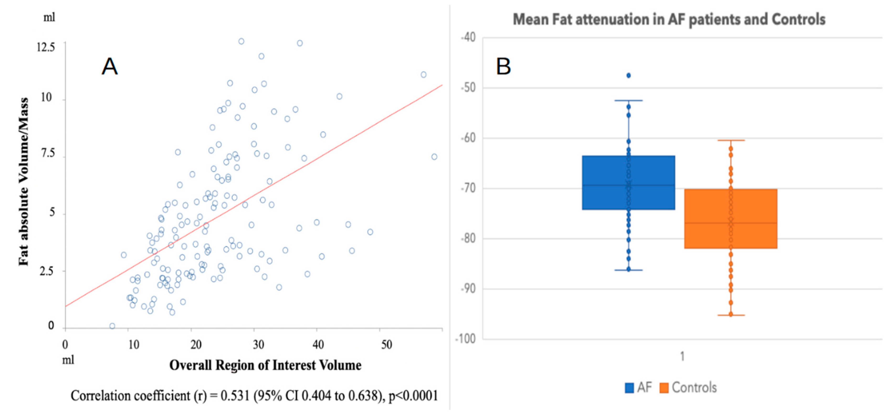
| Fat-LA volume, mL, median, [lower-upper quartile] | 4.2 (2.6–6.6) | 4.7 (3.2–7.5) | 3.6 (2.2–5.5) | 0.007 |
| ROI volume, mL, median, [lower-upper quartile] | 22.7 (16.7–28.1) | 26.4 (22.4–32.5) | 18 (14.1–23.6) | <0.001 |
| Fat-LA mass relative to ROI, %, median, [lower-upper quartile] | 19.8 (12.3–27.1) | 19.1 (11.8–26) | 21.3 (12.7–28.2) | 0.317 |
| Fat-LA attenuation, HU, SD | −73 ± 9.23 | −76.82 ± 8.54 | −69.15 ± 8.28 | <0.001 |
| Single Model | p-Value | Clinical and Echocardiography Model | p-Value | Clinical and Fat-LA Volume Model | p-Value | Clinical and Fat-LA Density Model | p-Value | |
|---|---|---|---|---|---|---|---|---|
| Univariable Odds Ratio (95% CI) | Multivariable Odds Ratio (95% CI) | Multivariable Odds Ratio (95% CI) | Multivariable Odds Ratio (95% CI) | |||||
| Age | 1.04 (1.01–1.07) | 0.005 | 1.02 (0.98–1.06) | 0.310 | 1.02 (0.98–1.06) | 0.381 | 1.03 (0.99–1.07) | 0.193 |
| Female gender | 0.35 (0.18–0.68) | 0.002 | 0.25 (0.18–1.40) | 0.189 | 0.54 (0.19–1.56) | 0.257 | 0.48 (0.16–1.41) | 0.184 |
| Cardiovascular risk factors | ||||||||
| BMI | 1.02 (0.98–1.09) | 0.633 | – | |||||
| DM | 0.59 (0.14–2.57) | 0.483 | – | – | – | – | – | – |
| HT | 1.70 (0.88–3.27) | 0.114 | – | – | – | – | – | – |
| Dyslipidemia | 0.73 (0.37–1.41) | 0.345 | – | – | – | – | – | – |
| Smoker | 2.54 (1.36–4.74) | 0.004 | 1.24 (0.52–2.94) | 0.624 | 1.25 (0.52–2.97) | 0.614 | 1.02 (0.41–2.51) | 0.972 |
| CHA2DS2-VASc score | 0.91 (0.70–1.18) | 0.458 | – | – | ||||
| Medications | ||||||||
| ACE-I/ARB | 1.65 (0.83–3.31) | 0.156 | – | – | – | – | – | – |
| Beta-blocker | 1.70 (0.85–3.38) | 0.133 | – | – | – | – | – | – |
| Statin | 0.65 (0.30–1.43) | 0.289 | – | – | – | – | – | – |
| Echocardiography | ||||||||
| LVEF | 1.02 (0.97–1.07) | 0.471 | – | – | – | – | – | – |
| LA area | 1.24 (1.11–1.38) | <0.001 | 1.18 (1.06–1.33) | 0.004 | 1.19 (1.06–1.34) | 0.004 | 1.17 (1.03–1.32) | 0.015 |
| Adipose tissue located posterior to the left atrium on cardiac computed tomography angiography | ||||||||
| Fat-LA volume | 1.18 (1.04–1.34) | 0.008 | 1.06 (0.88–1.27) | 0.541 | – | – | ||
| Fat-LA mean attenuation | 1.12 (1.07–1.17) | <0.001 | – | – | 1.09 (1.03–1.11) | 0.006 | ||
Publisher’s Note: MDPI stays neutral with regard to jurisdictional claims in published maps and institutional affiliations. |
© 2021 by the authors. Licensee MDPI, Basel, Switzerland. This article is an open access article distributed under the terms and conditions of the Creative Commons Attribution (CC BY) license (https://creativecommons.org/licenses/by/4.0/).
Share and Cite
Gaibazzi, N.; Martini, C.; Benatti, G.; Palumbo, A.A.; Cacciola, G.; Tuttolomondo, D. Atrial Fibrillation and Peri-Atrial Inflammation Measured through Adipose Tissue Attenuation on Cardiac Computed Tomography. Diagnostics 2021, 11, 2087. https://doi.org/10.3390/diagnostics11112087
Gaibazzi N, Martini C, Benatti G, Palumbo AA, Cacciola G, Tuttolomondo D. Atrial Fibrillation and Peri-Atrial Inflammation Measured through Adipose Tissue Attenuation on Cardiac Computed Tomography. Diagnostics. 2021; 11(11):2087. https://doi.org/10.3390/diagnostics11112087
Chicago/Turabian StyleGaibazzi, Nicola, Chiara Martini, Giorgio Benatti, Alessandro Anselmo Palumbo, Giovanna Cacciola, and Domenico Tuttolomondo. 2021. "Atrial Fibrillation and Peri-Atrial Inflammation Measured through Adipose Tissue Attenuation on Cardiac Computed Tomography" Diagnostics 11, no. 11: 2087. https://doi.org/10.3390/diagnostics11112087
APA StyleGaibazzi, N., Martini, C., Benatti, G., Palumbo, A. A., Cacciola, G., & Tuttolomondo, D. (2021). Atrial Fibrillation and Peri-Atrial Inflammation Measured through Adipose Tissue Attenuation on Cardiac Computed Tomography. Diagnostics, 11(11), 2087. https://doi.org/10.3390/diagnostics11112087