Article

Prevalence and Molecular Characterization of Parasitic Lice in Tibetan Yaks, Pigs and Sheep

1
College of Animal Science, Xizang Agriculture and Animal Husbandry University, Nyingchi 860000, China
2
Tibet Academy of Agriculture and Animal Husbandry Sciences, Lhasa 850009, China
3
Nierong County Agriculture and Animal Husbandry Science and Technology Service Station, Naqu 853500, China
4
College of Veterinary Medicine, Huazhong Agricultural University, Wuhan 430079, China
*
Authors to whom correspondence should be addressed.
Life 2025, 15(3), 444; https://doi.org/10.3390/life15030444
Submission received: 2 December 2024 / Revised: 24 February 2025 / Accepted: 5 March 2025 / Published: 12 March 2025

Abstract

:
The infestation of ectoparasites poses a severe problem in animal breeding, severely affecting animal health and causing substantial economic losses. However, limited information is available regarding lice infestations in plateau livestock. To address this, we collected and examined lice samples from Tibetan yaks, pigs and sheep, amplifying the rrnS and cox1 genes for evolutionary analysis. The results revealed that the prevalence of lice was 51.3% (95% CI: 44.0–58.6%) in yaks, Tibetan sheep and Tibetan pigs, with regional prevalence ranging from 7.7% to 67.5%. Morphometric analysis showed that female lice were bigger than male lice. In Tibetan pigs, females exhibited a prominent longer body length (p < 0.05), belly length (p < 0.01) and body length to body width ratio (p < 0.05). In yaks, females had longer body length (p < 0.01) and body width (p < 0.05). For Tibetan sheep, female lice had larger body length (p < 0.001), body width (p < 0.001), head length (p < 0.05) and belly length (p < 0.05). Additionally, molecular identification revealed that lice infesting wild and domestic yaks were Linognathus vituli with 96.59–98.78% (wild yaks) and 96.33–98.71% (yaks) similarity to the OL677823.1 isolate. Lice infesting Tibetan sheep were identified as Linognathus africanus, showing 99.02–99.76% similarity to the OP948898.1 isolate. Lice from Tibetan pigs were identified as Haematopinus apri, with 96.38–98.31% similarity to the ON000922.1 isolate. Moreover, cox1 gene analysis of lice from Tibetan pigs showed 96.97–97.72% similarity to the KC814616.1 isolate. It is concluded that these findings could provide valuable insights into the prevention and control of lice-related diseases in plateau animals, enhancing animal health and mitigating economic losses.

1. Introduction

The yak (Bos grunniens) is a long-haired, large ruminant breed [1], which is mainly distributed on the cold plateau regions of the Himalayas in South Asia with an average altitude of more than 3 km [2]. Globally, more than 16 million yaks inhabit the Qinghai–Tibet plateau in China [3]. In addition, yaks are crucial for both food and economic purposes, providing nutritious meat, high-protein milk, medicinally valuable fur and bones and dung used as fuel by local communities [4].
The Tibetan sheep (Ovis aries) is a representative indigenous herbivore inhabiting plateau regions at 3000 m above sea level (MASL) with over 23 million in China [5]. Analogous to yaks, Tibetan sheep have developed good physiological and genetic adaptations to overcome the plateau’s unfavorable environmental conditions, including extreme cold and hypoxia, through prolonged acclimatization. The acclimatization is achieved by several physiological adaptations, including elevated hemoglobin content and elevated mitochondrial content, and elevated oxygen-use efficiency in low oxygen states [6]. Moreover, Tibetan sheep provide a wide range of products, including high-quality meat, wool, fur and milk. These substantial contributions have played a vital role in supporting human civilization in the plateau regions, particularly among Tibetan communities [7]. The major types of sheep breeds found in the Tibetan Plateau region of China include Tibetan Sheep, Pianma Sheep, Ganjia Tibetan Sheep and Oula Sheep. In the present study, sampling was performed randomly.
The small Tibetan pig (Sus scrofa domesticus) is a unique local Tibet breed found in the high-altitude regions (3000 MASL) of the southeast Qinghai–Xizang plateau regions [3,8]. Through long-term domestication in harsh conditions characterized by hypoxia, extreme cold, high ultraviolet radiations and limited food resources, this economically significant, free-ranging species has developed exceptional resilience to disease and environmental stress [2]. These pigs produce not only lean meat with high yield but also high-quality, nutrient-rich products abundant in protein and essential amino acids [2]. However, the occurrence of infectious diseases in these animals poses a significant risk, leading to reduced productivity and substantial economic losses to animal husbandry [8,9,10].
Compulsory blood-sucking ectoparasites are widely recognized as a serious health threat to livestock, causing anemia and great economic losses by impairing rumination, reducing weight gain and lowering the quality and quantity of meat and milk production [11,12]. In Brazil, the estimated annual losses in the cattle industry due to ectoparasites was USD 6.86 billion [13], while Australia’s sheep industry suffered annual losses of USD 81 million [14]. Beyond their direct effects, many ectoparasites serve as vectors for blood-borne pathogens, promoting the spread of diseases among animals [15]. Among them, Linognathus vituli, a common ectoparasite of cattle, has a global prevalence ranging from 5% to 96% [16]. In addition to causing direct harm to ruminants and bringing economic losses to the cattle industry [17], this parasite is a crucial adjective vector of louse-borne pathogens such as Anaplasma marginale, Theileria orientalis, Rickettsia spp. and Coxiella burnetti [11,17,18]. Similarly, L. africanus is a major external parasite of sheep and goats [19], leading to significant economic losses for farmers and the tanning industry [20]. Alarmingly, zoonotic pathogens like Rickettsia sp. were detected in L. africanus, highlighting its public health risk [21]. Furthermore, the blood-sucking ectoparasite Haematopinus apri, commonly found in wild pigs, is a critical vector for pathogens such as the African swine fever virus, classical swine fever virus, Anaplasma spp. and the swinepox virus, posing a severe threat to livestock health and productivity [22].
Although lice are recognized as seriously harmful vermin, limited knowledge exists about L. vituli, L. africanus and H. apri infections in plateau animals (Family: Pedicinidae, Genus, Linognathus and Haematopinus). Therefore, the current study was conducted to investigate the prevalence and molecular characterization of these parasitic lice in yaks, Tibetan pigs and Tibetan sheep for the first time, assessing their impact on high-altitude yaks, pigs and sheep.

2. Materials and Methods

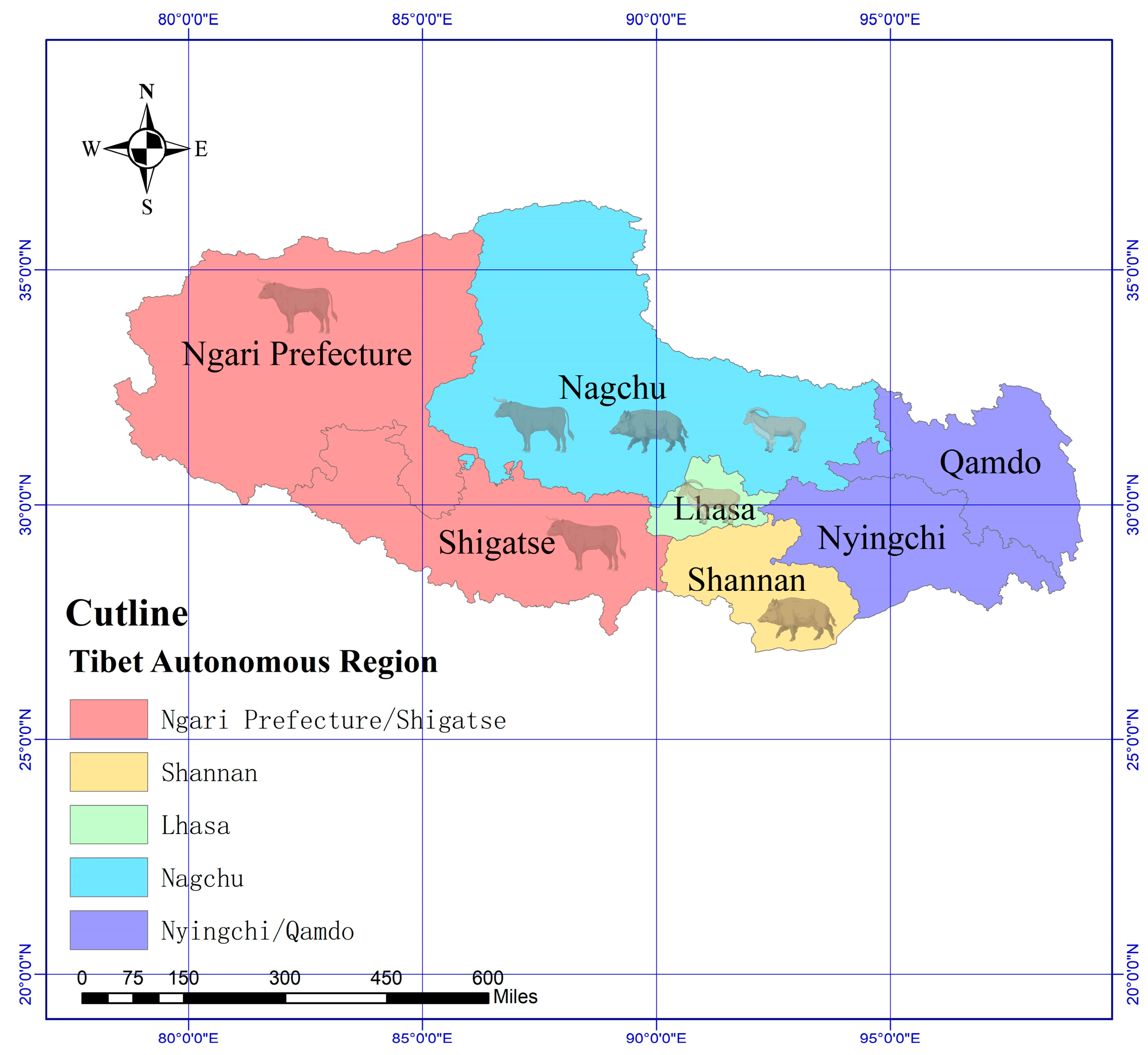
2.1. Sample Collection

Lice samples were collected from domestic yaks (n = 77), a wild yak (n = 1), Tibetan pigs (n = 20) and Tibetan sheep (n = 91) on the Tibetan plateau during 2022–2023. Samples were obtained by scraping the back skin of animals with sterile cotton swabs, while larger lice were directly picked using tweezers. All collected samples were fixed in 75% ethanol and stored at −20 °C for subsequent analysis [8]. The primers sequence is provided in Table 1 and the geographical locations of sampling are shown in Figure 1.

2.2. Lice Examination and Morphological Identification

All swab samples were examined using ZEN (blue edition) microscopy software (Version: 2.5) (ZEISS, Germany), and morphological identifications were performed following the methods described in previous studies [23,24]. A comprehensive measurement using morphometric analysis to obtain principal anatomical parameters of the specimens was undertaken including body length (BL/mm), body width (BW/mm), head length (HL/mm), belly length (BeL/mm), chest length (CL/mm) and breadth of breastplate (BBP/mm), and these specimens were carefully measured under the microscope to obtain accurate and reliable data. Additionally, the body length to body width ratio (LW) was calculated to assist in species characterization.

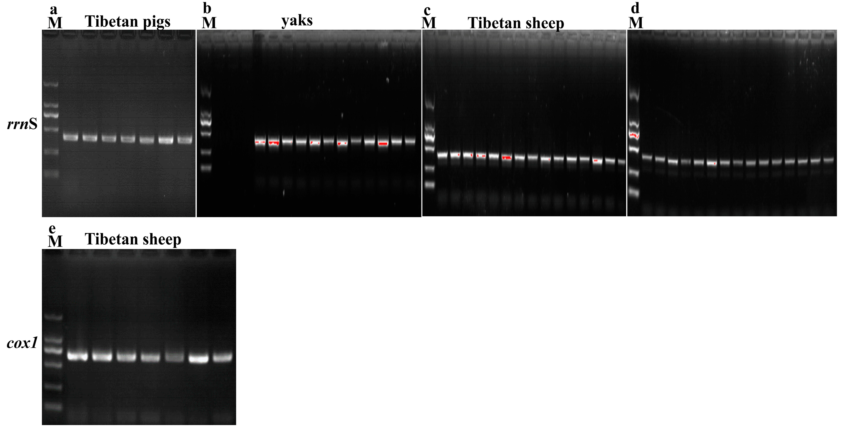
2.3. DNA Extraction and rrnS and cox1 Genes Amplification

Genomic DNA of yaks, Tibetan sheep and Tibetan pig lice were purified using the PureLink™ Genomic DNA Purification Kit (Invitrogen, Carlsbad, CA, USA) following the manufacturers’ strict protocol. The purified DNA thus acquired was then used to serve as templates for amplifying two candidate genes: mitochondrial ribosomal RNA small subunit (rrnS) and cytochrome c oxidase subunit 1 (cox1). The primer information is shown in Table 1. For PCR amplification, 50 μL reactions were prepared, including 25 μL 2 × Hieff® PCR Master Mix (Yeasen Biotechnology Co., Ltd., Shanghai, China), 2 μL DNA, 1 μL forward primer (10 μM), 1 μL reverse primer (10 μM) and 21 μL nuclease-free water. The PCR conditions were as follows: initial denaturation at 95 °C for 2 min, followed by 35 cycles of 95 °C for 30 s, annealing at 64 °C/cox1 (62 °C/rrnS) for 45 s, 72 °C for 30 s and a final extension at 72 °C for 10 min. PCR products were analyzed using 1.5% agarose gel electrophoresis to verify amplification.

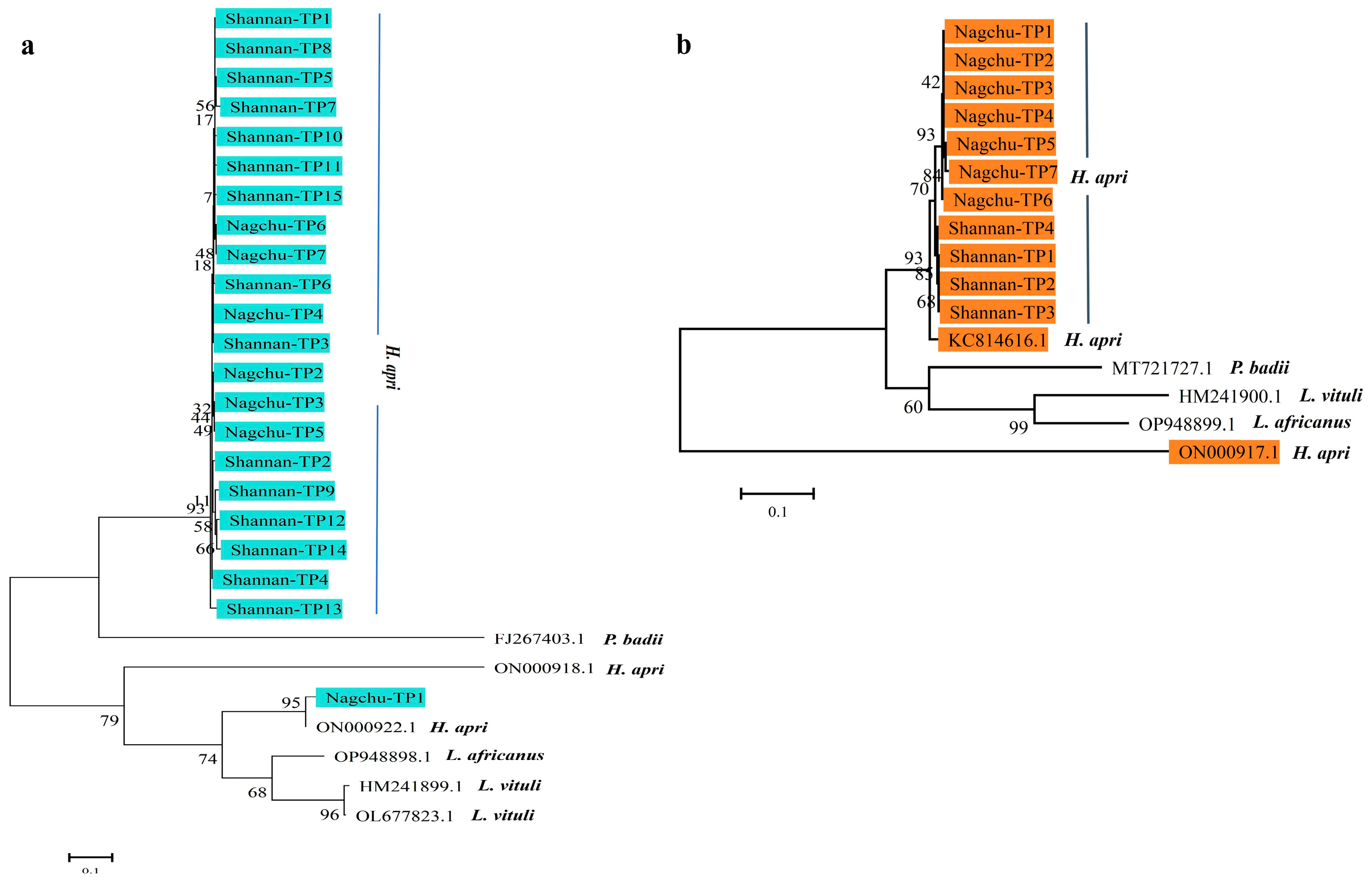
2.4. Sequencing and Phylogenetic Analyses

All the PCR positive bands were purified using the MolPure® Gel Extraction Kit (Molpure, Laguna Hills, CA, USA) and subsequently sent for commercial sequencing at Sangon Biotech (Shanghai, China). The sequences obtained then aligned with reference genes, including cox1 and rrnS, using NCBI BLASTn and MEGA (Version: 11.0). The reference genes for rrnS included sequences from Linognathus vituli (HM241899.1, L677823.1), Linognathus africanus (OP948898.1, HM171397.1), Linognathus spicatus (HM171399.1), Haematopinus apri (ON000922.1, ON000918.1) and Pedicinus badii (FJ267403.1). For cox1, the reference sequences were from Haematopinus apri (KC814616.1, ON000917.1), Linognathus vituli (HM241900.1), Linognathus africanus (OP948899.1) and Pedicinus badii (MT721727.1). Phylogenetic analysis of the cox1 and rrnS genes was carried out via MEGA (11.0) to assess the phylogenetic relationships between lice derived from plateau animals and other available lice isolates [25]. A phylogenetic tree was constructed using the maximum likelihood method, and the firmness of branches was evaluated by bootstrapping with 1000 times recalculation [26].

2.5. Statistical Analysis

The infestation rates of lice in different animals and regions, as well as body indexes of different lice species, were statistically analyzed using the chi-square test in SPSS (18.0) and the multiple unpaired t-test in GraphPad Prism (9.0). Confidence intervals were calculated using the Clopper–Pearson method. A significant difference was considered when p < 0.05.

3. Results

3.1. The Prevalence of Lice in Yaks, Tibetan Sheep and Tibetan Pigs

A total of 97 animals were examined, with an overall lice prevalence of 51.3% (95% CI: 44.0–58.6%). The prevalence varied across regions, ranging from 7.7% to 67.5%, showing a significant difference among different areas (p < 0.01). The prevalence of lice also differed significantly among animal species, ranging from 18.2% to 100.0% across different breeds (p < 0.01) (Table 2). Moreover, Figure 2a–c and Figure 3a–c show the morphological presentation of lice in yaks, Tibetan sheep, and pigs. Furthermore, Figure 4 shows the comparative analysis of the body index of lice from Tibetan pigs, yaks, and Tibetan sheep (Figure 4a–c), and also compares the female and male lice from different animals (Tibetan pigs, Yaks, and Tibetan sheep), (Figure 4d,e).

3.2. Molecular Characterization of Lice in Yaks, Tibetan Sheep and Tibetan Pigs

The mitochondrial rrnS gene of lice from Tibetan pigs, yaks and Tibetan sheep, as well as the cox1 gene of lice from Tibetan pigs, were successfully amplified (Figure 5). The PCR products corresponding to the cox1 gene were subsequently sequenced and submitted to the NCBI database with accession numbers of PQ615368-PQ615374 (Tibetan pigs from Nagchu) and PQ615375-PQ615378 (Tibetan pigs from Shannan). The rrnS sequences were assigned the following accession numbers: PQ622861-PQ622867 (Tibetan pigs from Nagchu), PQ622868-PQ622882 (Tibetan pigs from Shannan), PQ622839-PQ622845 (Tibetan sheep from Lhasa), PQ622846-PQ622860 (Tibetan sheep from Nagchu), PQ622800-PQ622814 (wild yaks from Ngari), PQ622815-PQ622829 (yaks from Nagchu) and PQ622830-PQ622838 (yaks from Ngari).
BLAST (version 2.15.0) analysis of these sequences with the NCBI database (https://blast.ncbi.nlm.nih.gov/Blast.cgi?PROGRAM=blastn&PAGE_TYPE=BlastSearch&LINK_LOC=blasthome. Accessed on 5 November 2023) revealed that lice from wild yaks and domestic yaks were identified as L. vituli with 96.59–98.78% (wild yaks) and 96.33–98.71% (domestic yaks) similarity to OL677823.1 isolated from cattle in Changsha, China, via the rrnS gene. The lice infected in Tibetan sheep were identified as L. africanus with 99.02–99.76% similarity to OP948898.1 isolated from Capra hircus in Pakistan via the rrnS gene. Lice infected in Tibetan pigs were identified as H. apri with 96.38–98.31% similarity to ON000922.1 isolated from wild boar in China via the rrnS gene. The cox1 gene from lice in Tibetan pigs was 96.97–97.72% similar to KC814616.1 isolated from Capra hircus in Pakistan.
Phylogenetic analysis of the rrnS gene showed that the L. vituli isolate from wild yaks (Ngari-WY15) and three isolates from yaks (Shigatsw-Y1, Shigatsw-Y2, Shigatsw-Y4) clustered with HM241899.1, while other isolates formed an independent branch (Figure 6a). Similarly, L. africanus isolates (Lhasa-TS1, Nagchu-TS5, Lhasa-TS6) from Tibetan sheep were located in the same cluster with OP948898.1, while other isolates formed a distinct cluster (Figure 6b). The H. apri isolate from Tibetan pigs (Nagchu-TP1) clustered with ON000922.1, while other isolates were placed in a separate cluster (Figure 7a). Additionally, all H. apri isolates from Tibetan pigs clustered with KC814616.1 based on the cox1 gene (Figure 7b).

4. Discussion

Food animals have a close relationship with the sustainable development of the economy and human civilization, and safeguarding animal health is meaningful and vitally important [27]. Despite their considerable impact as harmful ectoparasites, lice remained somewhat under-studied, particularly for their counterparts on agricultural livestock [28]. To the best of our knowledge, this current study is the initial probing of lice of veterinary importance on plateau animals regarding their molecular profiles in China.
The overall prevalence of lice in Tibetan livestock was determined to be 51.3%, comparable to the values reported in Irish cattle [29]. Caution must be exercised in directly comparing studies due to inherent variations in host species, breeds, age structure, management systems and geographic locations such as goats (97.4%) and sheep (100%) in Indonesia. For example, the prevalence of L. vituli in yaks, at 18.2%, was comparable to 19.3% in Ethiopian cattle [30,31]. However, differences in host physiology and environmental conditions may affect infestation patterns. Similarly, the prevalence of L. africanus in Tibetan sheep, at 93.5%, was considerably higher than that in goats of Rampur, at 11.2% [32], and in sheep (11.5%) and goats (27.9%) in Ethiopia [20]. The observed disparity may be due to variations in climatic conditions, ectoparasite control measures and differences in host susceptibility.
Comprehensive research has been conducted on H. suis in pigs [27,33]; however, information about H. apri in animals is limited. The prevalence of H. apri in Tibetan pigs was 50%, which was near H. suis in pigs (63.6%) in Rwanda [27], but higher than H. suis prevalence in pigs (15.3%) in Argentina [33]. The prevalence of lice varied significantly in different regions and breeds (Table 2) [31,34]. Our findings, along with previous studies, suggest that the occurrence of infestations in animals is determined by a range of factors, including both environmental and host-related factors. Specifically, free-grazing and direct contact with neighboring animals contribute most toward raising infestation intensity, through increased opportunity for parasite transmission. In addition, infestation trends follow a seasonal distribution, with increased infestations in winter in sheep and in goats in warmer environments. Herd size, geographical location and breed susceptibility have been determined to contribute toward infestations, with larger herds and native breed types having a relation with stocking density and genetic susceptibility toward infestations [1,2,3].
Previous studies have reported species such as H. eurysternus, H. quadripertusus, H. tuberculatus, L. vituli and Solenopotes capillatus in cattle [12,35]. However, all 39 isolates from wild and domestic yaks in our study were identified as L. vituli, which could be attributed to the fact that L. vituli is one of the most commonly reported lice species in cattle [36]. The lice from yaks exhibited high similarity (96.33–98.71%) to L. vituli (OL677823.1) isolated from adult cattle in Changsha, China, via blast analysis. This current study implies a potential genetic affinity between L. vituli from plateau yaks and the cattle isolate. However, in the phylogenetic tree based on the rrnS gene (Figure 6a), L. vituli isolates clustered into two distinct groups, possibly due to a few base differences among the isolates and the relatively short length of the rrnS gene.
Unlike previous studies, which primarily focused on H. suis in pigs [33,37], all 22 isolates from infected Tibetan pigs in our study were identified as H. apri with high similarity (96.38–98.31%) to the H. apri isolate (ON000922.1) from wild boar via the rrnS gene. These findings were further confirmed by analysis of the cox1 gene (Figure 7). H. apri is a louse species typically found in wild boars, and it shares both morphological and phylogenetic similarities with H. suis [38]. The occurrence of such a species of lice, endemic in feral pigs and wild boars naturally, can best be explained in terms of several ecologic and epidemiologic factors. One such reason is sympatric coexistence and reduced contact between feral and domestic pigs in a shared habitat with spillovers of parasites. Intermediate hosts such as birds and rodents can transmit infection through mechanically transporting lice and eggs between feral and domestic animals. In sheep and goats, common lice species include Bovicola caprae, B. limbata, B. crassipest, B. ovis, L. africanus and L. stenopsis [14,19]. In particular, B. ovis is frequently reported in sheep [39]. In contrast, all 22 isolates from Tibetan sheep in our study were identified as L. africanus, with high similarity (99.02–99.76%) to isolate (OP948898.1) from Capra hircus in Pakistan via the rrnS gene. L. africanus is a goat louse [40], which was reported in Capra hircus [28] and goats [19], and seldom detected in sheep. Our findings suggest that L. africanus may also infest sheep. The close cohabitation of Tibetan sheep and goats in shared grazing pastures, which could facilitate cross-species transmission, could be one of the possible reasons of the unusual presence of L. africanus. It is known that these plateau animals are social livestock that coexist with many wild animal species [1]. Lice are an important vector-borne reservoir for micro-pathogens [41]; hence, those infested animals may transmit lice and infectious microbes to other animals, particularly during warmer seasons. Zoonotic pathogens such as Rickettsia spp. and Bartonella spp. transmitted by lice may also pose a health threat to herders in plateau regions [28].
The small sample size of the wild yaks (n = 1) and Tibetan pigs (n = 20) puts limitations on the study’s validity. Sampling from the wild yaks was extremely limited due to their evasive behavior, harsh high-altitude habitat and their conservation status. Nonetheless, molecular characterization provides a useful baseline for future epidemiological investigations. An increase in the intensity of surveillance activities with larger sample sizes and the utilization of non-invasive genetic sampling techniques will be necessary to enhance our understanding of the dynamics of infestation by lice among populations of wild yaks.

5. Conclusions

In conclusion, we reported a high prevalence of lice infestation in yaks (18.2%), Tibetan sheep (93.5%) and Tibetan pigs (50.0%) on the Tibetan plateau. Through molecular analysis, lice were identified as L. vituli, L. africanus and H. apri, respectively, with a high genetic similarity to reference isolates. Notably, we confirmed for the first time that L. africanus, previously considered a goat louse, can also infest Tibetan sheep in the plateau environments. These findings may make a valuable contribution to the disease prevention and control of lice-related disease on plateau animals.

Author Contributions

Conceptualization, Q.W. and W.T.; methodology, W.L., X.Z., D.W. and B.S.; software, W.L.; validation, Q.W. and W.T.; formal analysis, W.L.; investigation, W.L., X.Z., D.W. and B.S.; resources, W.T.; data curation, W.L.; writing—original draft preparation, W.L., Q.W. and W.T.; writing—review and editing, Q.W., S.N. and W.T.; visualization, Q.W. and W.T.; supervision, Q.W. and W.T.; project administration, Q.W. and W.T.; funding acquisition, W.T. All authors have read and agreed to the published version of the manuscript.

Funding

This research was funded by Base and Talent Program of Science and Technology Plan in Tibet Autonomous Region (XZ202401JD0012).

Institutional Review Board Statement

Lice samples were collected accordance with guidelines and with the approval of the Ethics Committee of Xizang Agriculture and Animal Husbandry University and the Tibet Academy of Agriculture and Animal Husbandry Sciences (Permit number: 12540000MB0P024101, Approved on: 8 June 2022).

Informed Consent Statement

Not applicable.

Data Availability Statement

The rrnS and cox1 sequences of lice were deposited in GenBank with accession number: PQ622800-PQ622838, PQ622839-PQ622860 and PQ622861-622882.

Conflicts of Interest

The authors declare no conflicts of interest.

References

  1. Li, K.; Gao, J.; Shahzad, M.; Han, Z.; Nabi, F.; Liu, M.; Zhang, D.; Li, J. Seroprevalence of Toxoplasma gondii infection in yaks (Bos grunniens) on the Qinghai-Tibetan Plateau of China. Vet. Parasitol. 2014, 205, 354–356. [Google Scholar] [CrossRef] [PubMed]
  2. Chen, X.; Saeed, N.M.; Ding, J.; Dong, H.; Kulyar, M.F.; Bhutta, Z.A.; Mehmood, K.; Ali, M.M.; Irshad, I.; Zeng, J.; et al. Molecular Epidemiological Investigation of Cryptosporidium sp., Giardia duodenalis, Enterocytozoon bieneusi and Blastocystis sp. Infection in Free-ranged Yaks and Tibetan Pigs on the Plateau. Pak. Vet. J. 2022, 42, 533–539. [Google Scholar] [CrossRef] [PubMed]
  3. Li, K.; Zhang, L.; Zhang, H.; Lei, Z.; Luo, H.; Mehmood, K.; Shahzad, M.; Lan, Y.; Wang, M.; Li, J. Epidemiological investigation and risk factors of Echinococcus granulosus in yaks (Bos grunniens), Tibetan pigs and Tibetans on Qinghai Tibetan plateau. Acta Trop. 2017, 173, 147–152. [Google Scholar] [CrossRef]
  4. Peng, S.; Xu, C.; Saleem, M.U.; Babar, W.; Idrees, A.; Li, K. Epidemiological Investigation of Cryptosporidium Infection in Yaks in Chamdo, China. Pak. Vet. J. 2024, 2, 535–538. [Google Scholar] [CrossRef]
  5. Li, L.L.; Ma, S.K.; Peng, W.; Fang, Y.G.; Duo, H.R.; Fu, H.Y.; Jia, G.X. Genetic diversity and population structure of Tibetan sheep breeds determined by whole genome resequencing. Trop. Anim. Health Prod. 2021, 53, 1–9. [Google Scholar] [CrossRef]
  6. Cui, X.; Wang, Z.; Guo, P.; Li, F.; Chang, S.; Yan, T.; Zheng, H.; Hou, F. Shift of Feeding Strategies from Grazing to Different Forage Feeds Reshapes the Rumen Microbiota to Improve the Ability of Tibetan Sheep (Ovis aries) to Adapt to the Cold Season. Microbiol. Spectr. 2023, 11, 02816-22. [Google Scholar] [CrossRef]
  7. Shi, H.; Li, T.; Su, M.; Wang, H.; Li, Q.; Lang, X.; Ma, Y. Whole genome sequencing revealed genetic diversity, population structure, and selective signature of Panou Tibetan sheep. BMC Genom. 2023, 24, 50. [Google Scholar] [CrossRef]
  8. Li, K.; Luo, H.; Zhang, H.; Lan, Y.; Han, Z.; Shahzad, M.; Wang, X.; Qiu, G.; Huang, S.; Jiang, W.; et al. First report of Metastrongylus pudendotectus by the genetic characterization of mitochondria genome of cox1 in pigs from Tibet, China. Vet. Parasitol. 2016, 223, 91–95. [Google Scholar] [CrossRef]
  9. Wang, S.; Cao, Z.; Wu, Q.; Idrees, A.; Almutairi, M.H.; Dong, H. A Comparative Analysis and Verification of Differentially Expressed miRNAs could Provide New Insights for the Treatment of Endometritis in Yaks. Pak. Vet. J. 2023, 3, 486–492. [Google Scholar] [CrossRef]
  10. Ren, Y.; Zhaxi, Y.; Liu, M.; Idrees, A.; Li, K. Revealing the Fungi Microbiome Difference of Suffolk Cross with Tibetan Sheep on Plateau. Pak. Vet. J. 2023, 4, 748–756. [Google Scholar] [CrossRef]
  11. DeRosa, A.A.; Pullins, A.; Tena, J.K.; Holzmer, S.; Packianathan, R. Effectiveness of a fixed-dose combination injectable (0.2 mg/kg doramectin + 6.0 mg/kg levamisole hydrochloride) against Rhipicephalus microplus and sucking lice infesting cattle. Vet. Parasitol. 2023, 323, 110009. [Google Scholar] [CrossRef] [PubMed]
  12. Promrangsee, C.; Khositharattanakool, P.; Somwang, P.; Sunantaraporn, S.; Phumee, A.; Preativatanyou, K.; Tawatsin, A.; Brownell, N.; Siriyasatien, P. The Prevalence of Bartonella Bacteria in Cattle Lice Collected from Three Provinces of Thailand. Insects 2019, 10, 152. [Google Scholar] [CrossRef]
  13. Grisi, L.; Leite, R.C.; Martins, J.R.D.S.; Barros, A.T.M.D.; Andreotti, R.; Cançado, P.H.D.; León, A.A.P.D.; Pereira, J.B.; Villela, H.S. Reassessment of the potential economic impact of cattle parasites in Brazil. Rev. Bras. Parasitol. Vet. 2014, 23, 150–156. [Google Scholar] [CrossRef]
  14. Colvin, A.F.; Reeve, I.; Kahn, L.P.; Thompson, L.J.; Horton, B.J.; Walkden-Brown, S.W. Prevalence of sheep lice and trends in control practices across Australia—Australian sheep parasite control surveys from 2003 to 2019. Vet. Parasitol. Reg. Stud. Rep. 2022, 27, 100662. [Google Scholar] [CrossRef]
  15. Muhammad, A.; Bashir, R.; Mahmood, M.; Afzal, M.S.; Simsek, S.; Awan, U.A.; Khan, M.R.; Ahmed, H.; Cao, J. Epidemiology of Ectoparasites (Ticks, Lice, and Mites) in the Livestock of Pakistan: A Review. Front. Vet. Sci. 2021, 8, 780738. [Google Scholar] [CrossRef]
  16. Colwell, D. Life history parameters of the cattle long-nosed sucking louse, Linognathus vituli. Med. Vet. Entomol. 2014, 28, 432–437. [Google Scholar] [CrossRef] [PubMed]
  17. Hammer, J.F.; Jenkins, C.; Bogema, D.; Emery, D. Mechanical transfer of Theileria orientalis: Possible roles of biting arthropods, colostrum and husbandry practices in disease transmission. Parasites Vectors 2016, 9, 1–9. [Google Scholar] [CrossRef]
  18. Hornok, S.; Hofmann-Lehmann, R.; Fernández De Mera, I.G.; Meli, M.L.; Elek, V.; Hajtós, I.; Répási, A.; Gönczi, E.; Tánczos, B.; Farkas, R.; et al. Survey on blood-sucking lice (Phthiraptera: Anoplura) of ruminants and pigs with molecular detection of Anaplasma and Rickettsia spp. Vet. Parasitol. 2010, 174, 355–358. [Google Scholar] [CrossRef] [PubMed]
  19. Stefanov Nizamov, N. Identification of ectoparasitic insects among domestic goats in Bulgaria. Vet. World 2023, 16, 728–734. [Google Scholar] [CrossRef]
  20. Mulugeta, Y.; Yacob, H.T.; Ashenafi, H. Ectoparasites of small ruminants in three selected agro-ecological sites of Tigray Region, Ethiopia. Trop. Anim. Health Prod. 2010, 42, 1219–1224. [Google Scholar] [CrossRef]
  21. Ehlers, J.; Krüger, A.; Rakotondranary, S.J.; Ratovonamana, R.Y.; Poppert, S.; Ganzhorn, J.U.; Tappe, D. Molecular detection of Rickettsia spp., Borrelia spp., Bartonella spp. and Yersinia pestis in ectoparasites of endemic and domestic animals in southwest Madagascar. Acta Trop. 2020, 205, 105339. [Google Scholar] [CrossRef] [PubMed]
  22. Nie, Y.; Fu, Y.-T.; Wang, W.; Li, R.; Tang, W.-Q.; Liu, G.-H. Comparative analyses of the fragmented mitochondrial genomes of wild pig louse Haematopinus apri from China and Japan. Int. J. Parasitol. Parasites Wildl. 2022, 18, 25–29. [Google Scholar] [CrossRef]
  23. Perveen, N.; Muzaffar, S.B.; Al-Sabi, M.N.S.; Hamdan, L.; Aldarwich, A.; Iliashevich, D.; Mohteshamuddin, K.; Sparagano, O.A.; Willingham, A.L. The first record of ostrich feather louse (Struthiolipeurus struthionis) collected from farmed ostriches (Struthio camelus) in the United Arab Emirates. Vet. World 2024, 17, 125–130. [Google Scholar] [CrossRef]
  24. Brewer, P.J.; Sweet, A.D. Prevalence and diversity of parasitic bird lice (Insecta: Psocodea) in northeast Arkansas. Int. J. Parasitol. Parasites Wildl. 2023, 22, 205–215. [Google Scholar] [CrossRef] [PubMed]
  25. Yin, F.; Mu, D.; Tian, Z.; Li, D.; Ma, X.; Wang, J.; Guan, G.; Yin, H.; Li, F. Molecular Detection of Babesia gibsoni in Cats in China. Animals 2022, 12, 3066. [Google Scholar] [CrossRef] [PubMed]
  26. Tamura, K.; Stecher, G.; Kumar, S. MEGA11: Molecular Evolutionary Genetics Analysis Version 11. Mol. Biol. Evol. 2021, 38, 3022–3027. [Google Scholar] [CrossRef]
  27. Niyonsenga, E.; Twizerimana, J.; Mwabonimana, M.F. Haematopinus suis Infestation in Pig Farms in Busogo Sector, Rwanda. J. Trop. Med. 2023, 2023, 1–6. [Google Scholar] [CrossRef]
  28. Ballados-González, G.G.; Martínez-Hernández, J.M.; Martínez-Rodríguez, P.B.; Gamboa-Prieto, J.; González-Guzmán, S.; Paredes-Cervantes, V.; Grostieta, E.; Becker, I.; Aguilar-Domínguez, M.; Vieira, R.F.C.; et al. Molecular detection of hemotropic Mycoplasma and Bartonella species in lice from sheep and goats of Mexico. Vet. Parasitol. Reg. Stud. Rep. 2023, 44, 100921. [Google Scholar] [CrossRef]
  29. Mckiernan, F.; O’Connor, J.; Minchin, W.; O’Riordan, E.; Dillon, A.; Harrington, M.; Zintl, A. A pilot study on the prevalence of lice in Irish beef cattle and the first Irish report of deltamethrin tolerance in Bovicola bovis. Ir. Vet. J. 2021, 74, 20. [Google Scholar] [CrossRef]
  30. Insyari’ati, T.; Hamid, P.H.; Rahayu, E.T.; Sugar, D.L.; Rahma, N.N.; Kusumarini, S.; Kurnianto, H.; Wardhana, A.H. Ectoparasites Infestation to Small Ruminants and Practical Attitudes among Farmers toward Acaricides Treatment in Central Region of Java, Indonesia. Vet. Sci. 2024, 11, 162. [Google Scholar] [CrossRef]
  31. Kumsa, B.; Socolovschi, C.; Parola, P.; Rolain, J.; Raoult, D. Molecular Detection of AcinetobacterSpecies in Lice and Keds of Domestic Animals in Oromia Regional State, Ethiopia. PLoS ONE 2012, 12, e52377. [Google Scholar]
  32. Rashmi, A.; Saxena, A.K. Population levels of phthirapteran ectoparasites on the goats in Rampur (U.P.). J. Parasit. Dis. 2017, 41, 778–781. [Google Scholar] [CrossRef]
  33. Acosta, D.B.; Ruiz, M.; Sanchez, J.P. First molecular detection of Mycoplasma suis in the pig louse Haematopinus suis (Phthiraptera: Anoplura) from Argentina. Acta Trop. 2019, 194, 165–168. [Google Scholar] [CrossRef] [PubMed]
  34. Ajith, Y.; Dimri, U.; Gopalakrishnan, A.; Devi, G. A study on prevalence and factors associated with ectoparasitism in goats of two agro-climatic regions in India. J. Parasit. Dis. 2017, 41, 739–746. [Google Scholar] [CrossRef] [PubMed]
  35. Lakew, B.T.; Kheravii, S.K.; Wu, S.; Eastwood, S.; Andrew, N.R.; Nicholas, A.H.; Walkden-Brown, S.W. Detection and distribution of haematophagous flies and lice on cattle farms and potential role in the transmission of Theileria orientalis. Vet. Parasitol. 2021, 298, 109516. [Google Scholar] [CrossRef] [PubMed]
  36. Colwell, D.D. Persistent activity of moxidectin pour-on and injectable against sucking and biting louse infestations of cattle. Vet. Parasitol. 2002, 104, 319–326. [Google Scholar] [CrossRef]
  37. Braae, U.C.; Ngowi, H.A.; Johansen, M.V. Smallholder pig production: Prevalence and risk factors of ectoparasites. Vet. Parasitol. 2013, 196, 241–244. [Google Scholar] [CrossRef]
  38. Nishide, Y.; Oguchi, K.; Murakami, M.; Moriyama, M.; Koga, R.; Fukatsu, T. Endosymbiotic bacteria of the boar louse Haematopinus apri (Insecta: Phthiraptera: Anoplura). Front. Microbiol. 2022, 13, 962252. [Google Scholar] [CrossRef]
  39. Syamsul, V.S.; Okene, I.A.A.; Yahya, S.N.C.; Hamdan, R.H.; Lee, S.H.; Tan, L.P. Prevalence of Ectoparasitism on Small Ruminants in Kelantan, Malaysia. Trop. Life Sci. Res. 2020, 31, 45–56. [Google Scholar] [CrossRef]
  40. Fu, Y.-T.; Shao, R.; Suleman; Wang, W.; Wang, H.-M.; Liu, G.-H. The fragmented mitochondrial genomes of two Linognathus lice reveal active minichromosomal recombination and recombination hotspots. Iscience 2023, 26, 107351. [Google Scholar] [CrossRef]
  41. Ehounoud, C.B.; Fenollar, F.; Dahmani, M.; N’Guessan, J.D.; Raoult, D.; Mediannikov, O. Bacterial arthropod-borne diseases in West Africa. Acta Trop. 2017, 171, 124–137. [Google Scholar] [CrossRef]
Figure 1. Mapping the origins—geographical locations of sample collection.
Life 15 00444 g001
Figure 2. Morphological presentation of lice in Tibetan pigs: (a) female, (b) male, (c) ovum, (d) nymph.
Life 15 00444 g002
Figure 3. Morphological presentation of lice in yaks (a) and Tibetan sheep (b,c).
Life 15 00444 g003
Figure 4. Comparative analysis of body index of lice from different animals: (a) lice in Tibetan pigs, (b) lice in yaks, (c) lice in Tibetan sheep, (d) comparing female lice from different animals (Tibetan pigs, Yaks, and Tibetan sheep), (e) comparing male lice from different animals (Tibetan pigs, Yaks, and Tibetan sheep). * p < 0.05, ** p < 0.01, *** p < 0.001, **** p < 0.0001.
Life 15 00444 g004
Figure 5. PCR amplification results of the rrnS and cox1 genes of lice from yaks, Tibetan sheep and Tibetan pigs on 1.5% agarose gel: (a) rrnS gene of lice from Tibetan pigs, (b) rrnS gene of lice from yaks, (c) rrnS gene of lice from Tibetan sheep, (d) rrnS gene of lice from wild yak, (e) cox1 gene of lice from Tibetan sheep. Marker: 2000, 1000, 750, 500, 250, 100.
Life 15 00444 g005
Figure 6. Phylogenetic analysis of the current lice mt genomes of lice, focusing on the rrnS sequences using the maximum likelihood method. The resultant phylogenetic trees are presented with bootstrap values: (a) phylogenetic tree of lice from yaks based on the rrnS gene, (b) phylogenetic tree of lice from Tibetan sheep based on the rrnS gene.
Life 15 00444 g006
Figure 7. Phylogenetic analysis of the current lice mt genomes of the rrnS and cox1 sequences with references through the maximum likelihood method. The numbers at clades present bootstrap values: (a) phylogenetic tree of lice from Tibetan pigs based on the rrnS gene, (b) phylogenetic tree of lice from Tibetan pigs based on the cox1 gene.
Life 15 00444 g007
Table 1. Primer information of lice used in the current study.
Target GeneName of PrimerSequence (5′ to 3′)Size of Target Band (bp)
cox1HACX1FGCATATAATTATGGAGGAGAGAGGAA 657
HACX1R ATGACCAAAAAACCAGAATAGATGCTGG
rrnSHA12SFGGTCCATGAAAACTAATAACATATGGCGGT452
HA12RCATTGTATATAGTAGGGTATCTAATCCTAG
Table 2. The prevalence of lice in yak, Tibetan sheep and Tibetan pigs.
VariableCategoryNo. ExaminedNo. Positive% (95% CI)
Region aLhasa2627.7% (0.9–25.1%)
Ngari20315.0% (3.2–37.9%)
Shigatse9444.4% (13.7–78.8%)
Shannan11545.5% (16.7–76.6%)
Nagchu1238367.5% (58.4–75.6%)
Breed bDomestic yak771418.2% (10.3–28.6%)
Tibetan pig201050.0% (27.2–72.8%)
Tibetan sheep917293.5% (85.5–97.9%)
Wild yak11100.0% (2.5–100.0%)
Total 1899751.3% (44.0–58.6%)
a Significant difference was detected of the prevalence of lice in animals in different regions (p < 0.001, χ2 = 43.548). b Significant difference was detected of the prevalence of lice in animals in different regions (p < 0.001, χ2 = 62.962).
Disclaimer/Publisher’s Note: The statements, opinions and data contained in all publications are solely those of the individual author(s) and contributor(s) and not of MDPI and/or the editor(s). MDPI and/or the editor(s) disclaim responsibility for any injury to people or property resulting from any ideas, methods, instructions or products referred to in the content.

