Article

The Impact of Weight Loss during Chemoradiotherapy for Unresectable Esophageal Cancer: Real-World Results

1
Department of Radiation Oncology & Proton and Radiation Therapy Center, Kaohsiung Chang Gung Memorial Hospital and Chang Gung University College of Medicine, Kaohsiung 83301, Taiwan
2
Department of Computer Science, National Yang Ming Chiao Tung University, 1001 University Road, Hsinchu 30010, Taiwan
3
Department of Hematology-Oncology, Kaohsiung Chang Gung Memorial Hospital and Chang Gung University College of Medicine, Kaohsiung 83301, Taiwan
4
Department of Thoracic & Cardiovascular Surgery, Kaohsiung Chang Gung Memorial Hospital and Chang Gung University College of Medicine, Kaohsiung 83301, Taiwan
5
Division of Hepato-Gastroenterology, Department of Internal Medicine, Kaohsiung Chang Gung Memorial Hospital and Chang Gung University College of Medicine, Kaohsiung 83301, Taiwan
6
School of Traditional Chinese Medicine, Chang Gung University, Taoyuan 33302, Taiwan
*
Author to whom correspondence should be addressed.
Life 2022, 12(5), 706; https://doi.org/10.3390/life12050706
Submission received: 15 March 2022 / Revised: 3 May 2022 / Accepted: 5 May 2022 / Published: 8 May 2022

Abstract

:
Weight loss is a common phenomenon presented in unresectable esophageal cancer (EC) patients during their definitive chemoradiotherapy (dCRT) treatment course. This study explored the prognostic value of weight changes during dCRT in unresectable EC patients. From 2009 to 2017, 69 cT4b thoracic EC patients undergoing complete curative dCRT without baseline malnutrition were included. Clinical factors were analyzed via the Cox proportional hazards model and survival was analyzed by the Kaplan–Meier method. During dCRT, the median weight loss percentage was 5.51% (IQR = 2.77–8.85%), and the lowest body weight was reached at 35 days (IQR = 23–43 days). Median OS of these patients was 13.5 months. Both univariate and multivariate analysis demonstrated that weight loss ≤ 4% during dCRT was significantly associated with superior OS with a hazard ratio of 2.61 (95% CI: 1.40–4.85, p = 0.002). The median OS for patients with weight loss ≤ 4% and >4% during dCRT was 59.6 months and 9.7 months, respectively (p = 0.001). Our study demonstrated that weight loss ≤ 4% during dCRT course is a favorable prognostic factor for cT4b EC patients. This index could serve as a nutrition support reference for unresectable EC patients receiving dCRT in the future.

1. Introduction

Nutritional status is critical for esophageal cancer (EC) patients. Approximately 60–80% of EC patients are malnourished at diagnosis due to anorexia, dysphagia, reduced food intake, neuroendocrine changes, and elevated systemic inflammation induced by the tumor [1,2,3]. Baseline malnutrition in EC patients may lead to increased treatment-related toxicity [4], treatment interruption [5], more postoperative morbidity, and even worse overall survival [6,7,8,9].
In addition to baseline malnutrition, body weight loss (BWL) during cancer treatment is another important assessment tool for EC patients [10]. Although various tools have been proposed for nutritional status assessment [10,11,12], body weight loss has advantages as it is a simple, objective, and common method, which can serve as a surrogate for nutritional status during treatment.
Definitive chemoradiotherapy (dCRT) is a standard treatment modality for locally advanced and unresectable EC [13]. During the dCRT course, patients’ body weight may further deteriorate due to treatment-related toxicities such as esophagitis and anorexia [14,15,16]. Several studies have reported that BWL during chemoradiotherapy is correlated with poor survival in lung [17], head and neck [18], and rectal cancer patients [19]. However, the relationship between BWL during dCRT and treatment outcomes remains unclear for locally advanced EC patients. An optimal goal of nutrition support for these patients during their dCRT course has yet to be established. Therefore, we conducted this study to investigate the impact of BWL during dCRT on survival for cT4b EC patients without baseline malnutrition and to explore the optimal goal of body weight maintenance.

2. Materials and Methods

2.1. Patients

From 1 January 2009 to 31 December 2017, a total of 1120 consecutive patients with histologically proven thoracic EC were diagnosed and treated at our institution. Among this group, patients with thoracic cT4b EC who underwent curative dCRT were first selected for this retrospective review. Then, patients with a history of malignant disease, synchronous malignancy, or distant metastasis at an initial staging workup were excluded. Since this study was designed to evaluate nutritional changes during dCRT, patients with pre-treatment body mass index < 18.5 kg/m2 [10], body weight nadir in the first week of dCRT, and incomplete radiotherapy treatment course were also excluded for the following analyses, to avoid possible confounding on outcomes [20,21]. Our Institutional Review Board has approved this retrospective study.

2.2. Pretreatment Workup

A series of examinations including esophagogastroduodenoscopy (EGD) with biopsy, endoscopic ultrasound (EUS), and chest-computed tomography (CT) scans were performed for all patients at the time of initial diagnosis workup. Bronchoscopy was applied if clinical tracheobronchial tree invasion was suspected. For most patients diagnosed after late 2011, 18F-fluorodeoxyglucose (FDG) positron emission tomography/CT (PET/CT) was included in the initial staging workup. The diagnostic criteria for cT4b EC included tumor invading the great vessels, tracheobronchial tree, or vertebral bodies. Great vessel invasion was defined as the angle of fat plane obliteration by the tumor exceeding 90 degrees on contrasted chest CT, and EUS showing direct tumor invasion into the vessels. Tracheobronchial tree invasion was diagnosed when an irregular/deformed trachea or main bronchus caused by the tumor was identified on contrasted chest CT or direct invasion of the tracheobronchial tree was demonstrated by bronchoscopy. A multidisciplinary esophageal cancer team consisting of medical oncologists, surgical oncologists, and radiation oncologists was formed to discuss patient staging, treatment strategies, and post-treatment evaluation. The 7th edition of the American Joint Committee on Cancer (AJCC) Staging System [22] was used to determine the clinical stage.

2.3. Body Weight Measurement and Nutritional Counseling

The baseline body weight and height were measured before the first treatment using digital scales. During the dCRT course, body weight was recorded every week. Maximum weight change was calculated by baseline body weight minus lowest body weight during dCRT, divided by baseline body weight. Two sessions of nutritional counseling were scheduled for all patients; one was arranged after diagnosis and the other was arranged in the late period of dCRT by a licensed nutritionist.

2.4. Treatment

Our treatment and follow-up protocols for esophageal cancer have been reported [23], and are briefly summarized here. All patients underwent concurrent chemoradiotherapy with cisplatin (75 mg/m2; 4 h infusion; on day 1) and 5-fluorouracil (1000 mg/m2; continuous infusion on days 1–4) every 4 weeks. For patients with impaired renal function, defined as creatinine clearance rate <60 mL/min, Carboplatin was used instead.
For radiotherapy, the prescribed dose was 50–50.4 Gy in 25–28 daily fractions, 5 days per week. Gross tumor volumes (GTVs) were delineated by the gross tumor and lymph nodes based on chest CT scan and PET-CT studies. The clinical target volume (CTV) included the GTV with comprehensive regional lymph drain coverage; then, a three-dimensional expansion with 0.5–1.0 cm was used to generate planning target volume (PTV). Additional 10–16 Gy in 5–8 daily fractions were prescribed for patients with gross supraclavicular fossa lymph nodes metastasis. Most treatment plans used intensity-modulated radiation therapy (IMRT) with a 6- or 10 MV photon beam.

2.5. Follow-Up Schedule

Weekly outpatient clinic visit was arranged for all patients during dCRT course to monitor body weight change, treatment toxicities, and their general condition. The first follow-up after dCRT completion was arranged 6 weeks later and every 3–4 months for the first two years, then every 6 months for the next 3 years, then once each year. Follow-up workup included toxicity evaluations, physical examinations, chest CT images, EGD with biopsies, and/or PET/CT studies. Treatment toxicity was evaluated by the Common Toxicity Criteria for Adverse Events (CTCAE) ver. 4.0 [24]. Response Evaluation Criteria for Solid Tumors (RECIST) ver. 1.1 [25] was used to evaluate the clinical tumor response based on the first two follow-up chest CT scans within 4 months after completion of dCRT treatment. Clinical complete response (CR) was defined as no detectable residual tumor/ulceration with negative biopsy upon EGD examination, no regional lymph nodes with a short axis ≥ 10 mm in diameter, and no distant metastasis on chest CT studies. Metabolic complete response was defined as a physiologic level of FDG uptake on the primary tumor and regional lymph nodes.

2.6. Statistical Analysis

Overall survival (OS) was estimated by the Kaplan–Meier method with the log-rank test for significance. Univariable and multivariable analyses were performed using the Cox proportional hazards model. The optimal cut point for body weight change [26,27], data processing, and computation were performed by R, version 4.0.2 (The R Foundation for Statistical Computing, Vienna, Austria) and SPSS, version 22.0, software (SPSS, Chicago, IL, USA). A two-sided p-value lower than 0.05 was considered statistically significant.

3. Results

3.1. Patient Characteristics

A total of 69 patients were finally included in this study, the median age was 58.5 years, 98.6% patients were male, and 98.6% were diagnosed with squamous cell carcinoma. Over 90% of patients had a history of smoking and alcohol drinking. All patients had regional lymph node metastasis and two-thirds of them (45/69) had cN2-3 disease. The complete patient characteristics are listed in Table 1.

3.2. BMI, Body Weight Change, Optimal Cut Point for Weight Change, and Feeding Condition

The mean pretreatment BMI of the patients was 22.8 ± 3.4 kg/m2 (range: 18.5–32.3). Sixty-two patients (89.9%) experienced weight loss throughout dCRT. The median percentage of weight loss during dCRT was 5.5% (interquartile range (IQR) = 2.77–8.85%) and the lowest weight was reached at median 35 days (IQR = 23–43 days) after dCRT. The optimal cut point for weight change was 3.61%. For simplicity, we rounded off this number at 4%. Twenty-nine patients (42.0%) maintained oral intake throughout the treatment course, 34 patients (49.3%) had nasogastric (NG) tube intubation, and 6 patients (8.7%) had jejunostomy tube placement before dCRT started.
Table 1. Demographics (N = 69).
CharacteristicValue
Age
 Mean58.5 ± 10.0 (38.8–78.7)
 ≤6550 (72.5%)
 >6519 (27.5%)
Gender
 Male68 (98.6%)
 Female1 (1.4%)
BMI
 Mean22.8 ± 3.4 (18.5–32.3)
Comorbidities
 Hypertension17 (24.6%)
 Diabetes mellitus5 (7.2%)
 COPD4 (5.8%)
 Liver disease
  Cirrhosis6 (8.7%)
  Chronic HBV5 (7.2%)
 Chronic HCV2 (2.9%)
 Chronic renal disease1 (1.4%)
Personal history
 Smoking63 (91.3%)
 Betel nut use42 (60.9%)
 Alcohol use63 (91.3%)
Feeding route
 Oral29 (42.0%)
 Nasogastric tube34 (49.3%)
 Jejunostomy tube6 (8.7%)
Histology type
 SCC68 (98.6%)
 Adenosquamous1 (1.4%)
 Adenocarcinoma0 (0.0%)
Clinical lymph node category
 124 (34.8%)
 230 (43.5%)
 315 (21.7%)
Tumor location
 Upper thoracic esophagus21 (30.4%)
 Middle thoracic esophagus31 (44.9%)
 Lower thoracic esophagus17 (24.7%)
Tumor invasion site
 Great vessels28 (40.6%)
 Airway23 (33.3%)
 Both18 (26.1%)
Tumor length
 ≤6 cm33 (47.8%)
 >6 cm36 (52.2%)
Values are number (%) or mean ± SD (range). Abbreviations: BMI = body mass index; dCRT = definitive chemoradiotherapy; HBV = Hepatitis B; HCV = Hepatitis C; COPD = chronic obstructive pulmonary disease SCC = squamous cell carcinoma.

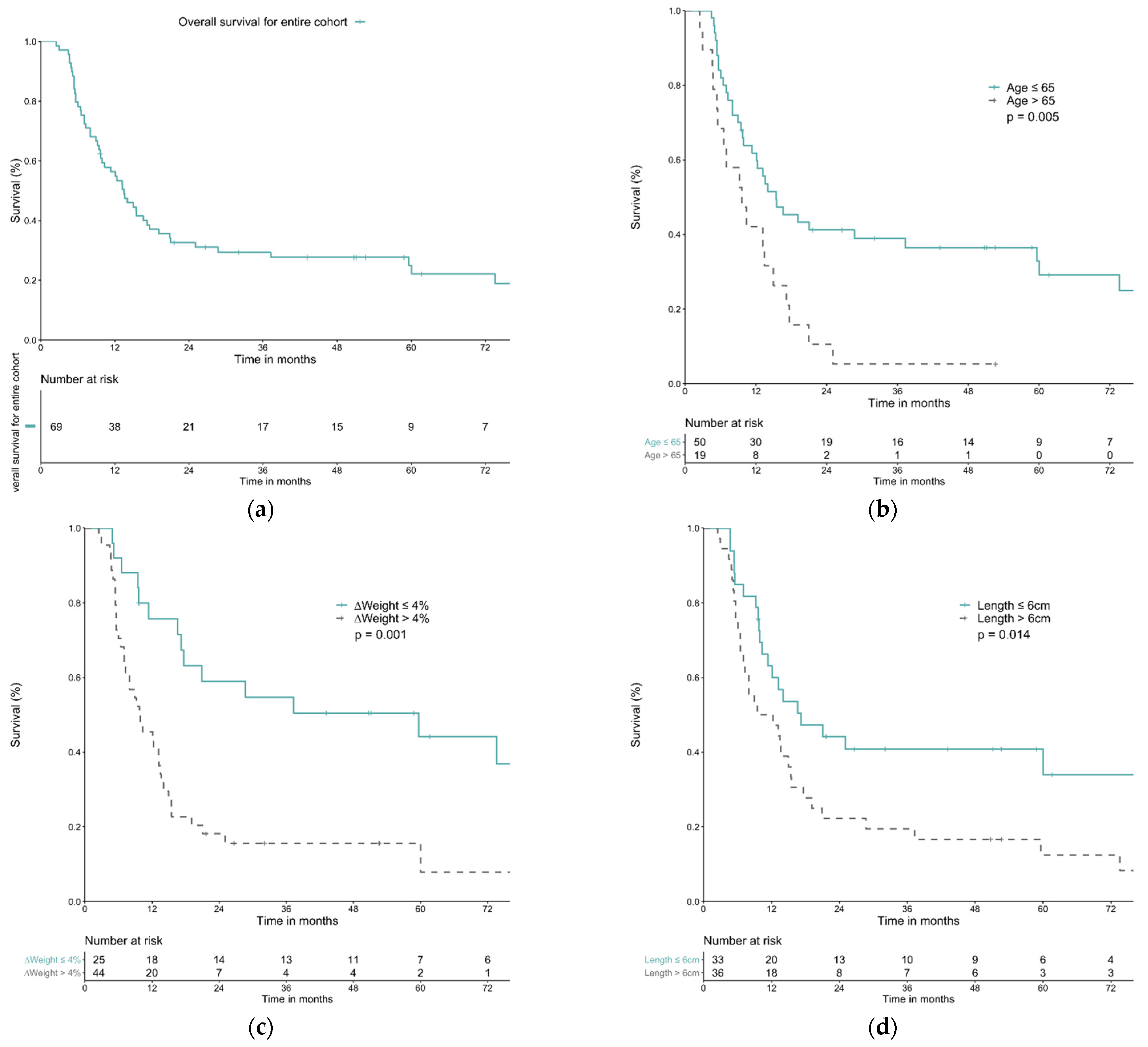
3.3. Treatment Response, Toxicities, and Survival Outcomes

Treatment responses were observed in 81.2% patients, and the CR rate was 30.4% (21 patients). Four (5.8%) patients developed grade 3 esophagitis, no patient had greater or equal to grade 3 radiation pneumonitis, 5 patients developed tracheo-esophageal fistula, and 1 patient had tracheo-aortic fistula (Table 2).
The median follow-up time was 61.7 months (range: from 2.5 months to 96.3 months). The median OS for the entire cohort was 13.5 months. The 1-year, 3-year, and 5-year OS rates for these patients were 56.4%, 29.5%, and 24.9%, respectively.
Forty-four patients (63.8%) experienced weight loss > 4% during dCRT course. The median OS was 59.6 months for patients with weight loss ≤ 4% and 9.6 months for patients with weight loss > 4%. The OS rate for weight loss ≤ 4% group at 1, 3, and 5 years was 75.8%, 54.7%, 44.2%, respectively, compared with 45.5%, 15.6%, 15.6% in weight loss > 4% group (p = 0.001). Only 5 out of 44 patients achieved CR in the weight loss > 4% group, while 16 out of 25 patients achieved CR in the weight loss ≤ 4% group (Fisher’s exact test, p = 0.000). In both univariable and multivariable analysis, age ≤ 65 years old (hazard ratio (HR) 1.91, 95% confidence interval (CI) 1.06–3.44, p = 0.032), weight change ≤ 4% (HR 2.61, 95% CI 1.40–4.85, p = 0.002), and tumor length ≤ 6 cm (HR 1.83, 95% CI 1.05–3.22, p = 0.035) were significant favorable prognostic factors (Figure 1 and Table 3).

4. Discussion

Our study demonstrated that maintaining a BWL less than 4% during dCRT course is critical for survival in cT4b EC patients. The 5-year OS rate was 44.2% for patients with BWL ≤ 4%, compared to 15.6% for those with BWL > 4%. Both univariable and multivariable analyses confirmed that BWL ≤ 4% was a significant favorable prognostic factor. Furthermore, BWL ≤ 4% during dCRT course was also significantly associated with clinical complete response status, a known positive prognostic factor for T4b EC patients [23]. Therefore, BWL during dCRT is a crucial yet adjustable factor in cT4b EC treatment.
Here, we must emphasize that BWL during dCRT, a dynamic change in nutritional status, differs from baseline nutrition status such as pretreatment BMI, a widely reported prognostic factor for EC patients [8,9,12,20,21,28]. Therefore, in this study, we excluded patients with pretreatment BMI < 18.5 kg/m2 in our cohort to avoid the potential confounding effect and aimed at an adjustable factor: changes in body weight during dCRT for EC patients. To our knowledge, this is the first report in the literature to demonstrate the association between body weight change during dCRT and survival for EC patients.
Since BWL was the only modifiable prognosticator for cT4b EC patients found in our institutional analysis, and previous studies have demonstrated that intensive nutritional management could improve nutritional status for EC patients during their dCRT course [29,30,31], a nutritional support program for EC patients has been embedded into our institution multi-modality treatment protocol for EC patients to prevent BWL since the late 2010s. This nutritional support program includes two primary strategies. The first strategy is to establish a pre-treatment enteral feeding route for reliable nutritional intake, preferably feeding jejunostomy, and a nasogastric tube. In a recent prospective multi-center randomized clinical trial, enteral nutrition was proven to alleviate BWL, reduce serum albumin/hemoglobin decline, and decrease grade 3/4 leukopenia rate during dCRT for unresectable EC patients. A nutrition formula with high-proteins and polyunsaturated fat was suggested, while enteral immunonutrition might further maintain nutritional status for EC patients during dCRT course [32,33]. The second strategy was to arrange at least two planned sessions of nutritional consultation by certified clinical nutritionists during the treatment course. The first session was arranged soon after diagnosis for an evaluation of patients’ baseline nutritional status and to design a personalized nutritional support plan based on individual nutritional status, feeding condition, and physical activities. The second session was arranged around the 4th week during dCRT to adjust patients’ personalized nutritional support plans. Early and periodic nutrition assessments/interventions had been shown to improve survival outcomes and significantly reduce weight loss, de-crease unplanned hospital admissions, and increase RT completion rates for EC patients who underwent dCRT [12,31].
Although BWL was a critical prognostic factor in our study, the direct mechanism that causes worse survival for patients who have BWL during treatment remains unclear. Several hypotheses had been proposed to explain this association. First, impaired immunity may play a major role. The immune system is critical to tumor development [34] and deficiency of nutrients such as amino acids, vitamins, minerals, and trace elements in patients who suffer from excessive BWL might have a detrimental impact on immune function [35,36,37,38]. Another possible reason is skeletal muscle loss. Loss of skeletal muscle is commonly observed in EC patients after chemoradiotherapy and has been reported as a poor prognostic factor [39,40,41,42]. Moreover, BWL might cause anatomical changes, increase setup error, and alter the dose distribution of radiotherapy [43,44]. All these problems could increase treatment toxicities and reduce accuracy. Finally, the dysregulated inflammatory response in cachectic patients may induce radiation resistance [45,46]. Elevated circulating cytokines such as TNF-α and IL-6 may activate downstream effectors NF-κB and STAT3 and lead to increased radioresistance by producing growth factors and angiogenic factors [47,48,49].
This present study has several limitations that should be addressed. First, as it was a retrospective study, selection bias and missing data exist. Several nutritional assessment biochemical markers/tools, such as serum albumin, fat-free body mass, or patient-generated subjective global assessment, could not be completely collected and were not included in this study. Second, a strict guideline for feeding route, nasogastric tube placement, or feeding jejunostomy did not exist during the study period. The choice of feeding route largely depended on patients’ preference and physicians’ choice. Third, patients with baseline malnutrition were not included in this study; the optimal body-weight monitoring index for this group of patients remains unclear. Lastly, whether our nutritional support protocol may improve clinical outcomes remains unclear due to the short follow-up time and limited case numbers. A larger cohort and long-term follow-up are warranted to confirm the potential benefits of our protocol.

5. Conclusions

Maintaining a cT4b EC patients’ BWL of less than 4% of their initial body weight during dCRT is a significant prognostic factor for OS and could serve as an individualized goal for nutritional support. Future prospective studies are warranted to validate our findings.

Author Contributions

Conceptualization, T.-T.H. and Y.-M.W.; data curation, T.-T.H., S.-H.L. and Y.-M.W.; formal analysis, T.-T.H. and Y.-M.W.; investigation, T.-T.H., S.-Y.C. and Y.-H.L.; methodology, T.-T.H. and Y.-M.W.; project administration, Y.-M.W.; resources, S.-Y.C., Y.-H.L., S.-H.L., Y.-H.C., H.-I.L., C.-M.L., F.-M.F., Y.-C.C., Y.-P.C. and Y.-M.W.; validation, S.-Y.C., Y.-H.L. and Y.-M.W.; visualization, T.-T.H.; writing—original draft, T.-T.H. and Y.-M.W.; writing—review and editing, T.-T.H., S.-Y.C., Y.-H.L., S.-H.L., Y.-H.C., H.-I.L., C.-M.L., F.-M.F., Y.-C.C., Y.-P.C. and Y.-M.W. All authors have read and agreed to the published version of the manuscript.

Funding

This research was partially funded by a grant from the Chang-Gung Medical Research Project (CMRPG8J0561).

Institutional Review Board Statement

This study was conducted according to the guidelines of the Declaration of Helsinki and approved by the Institutional Review Board of Chang Gung Memorial Hospital (201900218B0D001).

Informed Consent Statement

Patient consent was waived by the IRB, as this study was a retrospective study and all cases were treated by clinical routines.

Data Availability Statement

The data presented in this study are available on request from the corresponding author. The data are not publicly available due to the nature of this research; participants of this study did not agree for their data to be shared publicly.

Acknowledgments

We appreciated the help from the Biostatistics Center, Kaohsiung Chang Gung Memorial Hospital with the statistics.

Conflicts of Interest

The authors declare no conflict of interest.

References

  1. Anandavadivelan, P.; Lagergren, P. Cachexia in patients with oesophageal cancer. Nat. Rev. Clin. Oncol. 2015, 13, 185–198. [Google Scholar] [CrossRef] [PubMed]
  2. Hébuterne, X.; Lemarié, E.; Michallet, M.; De Montreuil, C.B.; Schneider, S.; Goldwasser, F. Prevalence of Malnutrition and Current Use of Nutrition Support in Patients with Cancer. J. Parenter. Enter. Nutr. 2014, 38, 196–204. [Google Scholar] [CrossRef] [PubMed]
  3. Riccardi, D.; Allen, K. Nutritional Management of Patients with Esophageal and Esophagogastric Junction Cancer. Cancer Control 1999, 6, 64–72. [Google Scholar] [CrossRef] [PubMed] [Green Version]
  4. Miyata, H.; Yano, M.; Yasuda, T.; Hamano, R.; Yamasaki, M.; Hou, E.; Motoori, M.; Shiraishi, O.; Tanaka, K.; Mori, M.; et al. Randomized study of clinical effect of enteral nutrition support during neoadjuvant chemotherapy on chemotherapy-related toxicity in patients with esophageal cancer. Clin. Nutr. 2011, 31, 330–336. [Google Scholar] [CrossRef]
  5. Bansal, A.; Kapoor, R.; Kumar, S.; Miriyala, R.T. Factors influencing compliance to radical treatment of middle thoracic esophageal cancer: An audit from a regional cancer centre. Indian J. Palliat. Care 2016, 22, 288–294. [Google Scholar] [CrossRef]
  6. Yu, X.-L.; Yang, J.; Chen, T.; Liu, Y.-M.; Xue, W.-P.; Wang, M.-H.; Bai, S.-M. Excessive Pretreatment Weight Loss Is a Risk Factor for the Survival Outcome of Esophageal Carcinoma Patients Undergoing Radical Surgery and Postoperative Adjuvant Chemotherapy. Can. J. Gastroenterol. Hepatol. 2018, 2018, 6075207. [Google Scholar] [CrossRef] [Green Version]
  7. Arends, J.; Bachmann, P.; Baracos, V.; Barthelemy, N.; Bertz, H.; Bozzetti, F.; Fearon, K.; Hütterer, E.; Isenring, E.; Kaasa, S.; et al. ESPEN guidelines on nutrition in cancer patients. Clin. Nutr. 2017, 36, 11–48. [Google Scholar] [CrossRef] [Green Version]
  8. Ji, W.; Zheng, W.; Li, B.; Cao, C.; Mao, W. Influence of body mass index on the long-term outcomes of patients with esophageal squamous cell carcinoma who underwent esophagectomy as a primary treatment. Medicine 2016, 95, e4204. [Google Scholar] [CrossRef]
  9. Zhang, S.S.; Yang, H.; Luo, K.J.; Huang, Q.Y.; Chen, J.Y.; Yang, F.; Cai, X.L.; Xie, X.; Liu, Q.W.; E Bella, A.; et al. The impact of body mass index on complication and survival in resected oesophageal cancer: A clinical-based cohort and meta-analysis. Br. J. Cancer 2013, 109, 2894–2903. [Google Scholar] [CrossRef] [Green Version]
  10. Cederholm, T.; Bosaeus, I.; Barazzoni, R.; Bauer, J.; Van Gossum, A.; Klek, S.; Muscaritoli, M.; Nyulasi, I.; Ockenga, J.; Schneider, S.; et al. Diagnostic criteria for malnutrition—An ESPEN Consensus Statement. Clin. Nutr. 2015, 34, 335–340. [Google Scholar] [CrossRef]
  11. Cederholm, T.; Barazzoni, R.; Austin, P.; Ballmer, P.; Biolo, G.; Bischoff, S.C.; Compher, C.; Correia, I.; Higashiguchi, T.; Holst, M.; et al. ESPEN guidelines on definitions and terminology of clinical nutrition. Clin. Nutr. 2017, 36, 49–64. [Google Scholar] [CrossRef] [PubMed]
  12. Cox, S.; Powell, C.; Carter, B.; Hurt, C.; Mukherjee, S.; Crosby, T.D.L. Role of nutritional status and intervention in oesophageal cancer treated with definitive chemoradiotherapy: Outcomes from SCOPE1. Br. J. Cancer 2016, 115, 172–177. [Google Scholar] [CrossRef] [PubMed] [Green Version]
  13. Netwrok, N.C.C. Esophageal and Esophagogastric Junction Cancers. 2022; Version 2. 2022. Available online: https://www.nccn.org/professionals/physician_gls/pdf/esophageal.pdf (accessed on 18 February 2022).
  14. Pan, P.; Tao, G.; Sun, X. Subjective global assessment and prealbumin levels of esophageal cancer patients undergoing concurrent chemoradiotherapy. Nutr. Hosp. 2015, 31, 2167–2173. [Google Scholar] [CrossRef] [PubMed]
  15. Deans, D.A.C.; Tan, B.H.; Wigmore, S.J.; A Ross, J.; De Beaux, A.C.; Paterson-Brown, S.; Fearon, K.C.H. The influence of systemic inflammation, dietary intake and stage of disease on rate of weight loss in patients with gastro-oesophageal cancer. Br. J. Cancer 2009, 100, 63–69. [Google Scholar] [CrossRef]
  16. Bower, M.R.; Martin, R.C. Nutritional management during neoadjuvant therapy for esophageal cancer. J. Surg. Oncol. 2009, 100, 82–87. [Google Scholar] [CrossRef]
  17. Sanders, K.J.; Hendriks, L.E.; Troost, E.G.; Bootsma, G.P.; Houben, R.M.; Schols, A.M.; Dingemans, A.-M.C. Early Weight Loss during Chemoradiotherapy Has a Detrimental Impact on Outcome in NSCLC. J. Thorac. Oncol. 2016, 11, 873–879. [Google Scholar] [CrossRef] [Green Version]
  18. Capuano, G.; Grosso, A.; Gentile, P.C.; Battista, M.; Bianciardi, F.; Di Palma, A.; Pavese, I.; Satta, F.; Tosti, M.; Rn, A.P.; et al. Influence of weight loss on outcomes in patients with head and neck cancer undergoing concomitant chemoradiotherapy. Head Neck 2007, 30, 503–508. [Google Scholar] [CrossRef]
  19. Lin, J.; Peng, J.; Qdaisat, A.; Li, L.; Chen, G.; Lu, Z.; Wu, X.; Gao, Y.; Zeng, Z.; Ding, P.; et al. Severe weight loss during preoperative chemoradiotherapy compromises survival outcome for patients with locally advanced rectal cancer. J. Cancer Res. Clin. Oncol. 2016, 142, 2551–2560. [Google Scholar] [CrossRef] [Green Version]
  20. Clavier, J.-B.; Antoni, D.; Atlani, D.; Ben Abdelghani, M.; Schumacher, C.; Dufour, P.; Kurtz, J.-E.; Noel, G. Baseline nutritional status is prognostic factor after definitive radiochemotherapy for esophageal cancer. Dis. Esophagus 2012, 27, 560–567. [Google Scholar] [CrossRef]
  21. Di Fiore, F.; Lecleire, S.; Pop, D.; Rigal, O.; Hamidou, H.; Paillot, B.; Ducrotté, P.; Lerebours, E.; Michel, P. Baseline Nutritional Status Is Predictive of Response to Treatment and Survival in Patients Treated by Definitive Chemoradiotherapy for a Locally Advanced Esophageal Cancer. Am. J. Gastroenterol. 2007, 102, 2557–2563. [Google Scholar] [CrossRef]
  22. Edge, S.B.; Byrd, D.R.; Compton, C.C.; Fritz, A.G.; Trotti, A.L.; Greene, F.L. AJCC Cancer Staging Manual, 7th ed.; Springer: New York, NY, USA, 2010. [Google Scholar]
  23. Huang, T.-T.; Li, S.-H.; Chen, Y.-H.; Lu, H.-I.; Lo, C.-M.; Fang, F.-M.; Chou, S.-Y.; Chiu, Y.-C.; Chou, Y.-P.; Wang, Y.-M. Definitive chemoradiotherapy for clinical T4b esophageal cancer—Treatment outcomes, failure patterns, and prognostic factors. Radiother. Oncol. 2021, 157, 56–62. [Google Scholar] [CrossRef] [PubMed]
  24. Common Terminology Criteria for Adverse Events (CTCAE) Version 4.0. 2009. Available online: https://www.eortc.be/services/doc/ctc/CTCAE_4.03_2010-06-14_QuickReference_5x7.pdf (accessed on 7 December 2021).
  25. Eisenhauer, E.A.; Therasse, P.; Bogaerts, J.; Schwartz, L.H.; Sargent, D.; Ford, R.; Dancey, J.; Arbuck, S.; Gwyther, S.; Mooney, M.; et al. New response evaluation criteria in solid tumours: Revised RECIST guideline (version 1.1). Eur. J. Cancer 2009, 45, 228–247. [Google Scholar] [CrossRef] [PubMed]
  26. Ogłuszka, M.; Orzechowska, M.; Jędroszka, D.; Witas, P.; Bednarek, A.K. Evaluate Cutpoints: Adaptable continuous data distribution system for determining survival in Kaplan-Meier estimator. Comput. Methods Programs Biomed. 2019, 177, 133–139. [Google Scholar] [CrossRef] [PubMed]
  27. Meyers, J.P.; Mandrekar, J. Cutpoint Determination Methods in Survival Analysis using SAS®: Updated %FINDCUT macro. In Proceedings of the SAS Global Forum 2015, Dallas, TX, USA, 26–29 April 2015. [Google Scholar]
  28. Smith, M.; Zhou, M.; Whitlock, G.; Yang, G.; Offer, A.; Hui, G.; Peto, R.; Huang, Z.; Chen, Z. Esophageal cancer and body mass index: Results from a prospective study of 220,000 men in China and a meta-analysis of published studies. Int. J. Cancer 2007, 122, 1604–1610. [Google Scholar] [CrossRef]
  29. Qiu, Y.; You, J.; Wang, K.; Cao, Y.; Hu, Y.; Zhang, H.; Fu, R.; Sun, Y.; Chen, H.; Yuan, L.; et al. Effect of whole-course nutrition management on patients with esophageal cancer undergoing concurrent chemoradiotherapy: A randomized control trial. Nutrition 2019, 69, 110558. [Google Scholar] [CrossRef]
  30. Chen, M.-J.; Wu, I.-C.; Chen, Y.-J.; Wang, T.-E.; Chang, Y.-F.; Yang, C.-L.; Huang, W.-C.; Chang, W.-K.; Sheu, B.-S.; Wu, M.-S.; et al. Nutrition therapy in esophageal cancer—Consensus statement of the Gastroenterological Society of Taiwan. Dis. Esophagus 2018, 31, doy016. [Google Scholar] [CrossRef]
  31. Odelli, C.; Burgess, D.; Bateman, L.; Hughes, A.; Ackland, S.; Gillies, J.; Collins, C.E. Nutrition Support Improves Patient Outcomes, Treatment Tolerance and Admission Characteristics in Oesophageal Cancer. Clin. Oncol. 2005, 17, 639–645. [Google Scholar] [CrossRef]
  32. Lyu, J.; Li, T.; Xie, C.; Li, J.; Xing, L.; Zhang, X.; Shen, L.; Zhao, K.; Zhao, R.; Yang, D.; et al. Enteral nutrition in esophageal cancer patients treated with radiotherapy: A Chinese expert consensus 2018. Futur. Oncol. 2019, 15, 517–531. [Google Scholar] [CrossRef]
  33. Vasson, M.-P.; Talvas, J.; Perche, O.; Dillies, A.-F.; Bachmann, P.; Pezet, D.; Achim, A.-C.; Pommier, P.; Racadot, S.; Weber, A.; et al. Immunonutrition improves functional capacities in head and neck and esophageal cancer patients undergoing radiochemotherapy: A randomized clinical trial. Clin. Nutr. 2013, 33, 204–210. [Google Scholar] [CrossRef]
  34. Gonzalez, H.; Hagerling, C.; Werb, Z. Roles of the immune system in cancer: From tumor initiation to metastatic progression. Genes Dev. 2018, 32, 1267–1284. [Google Scholar] [CrossRef] [Green Version]
  35. Sikalidis, A.K. Amino Acids and Immune Response: A Role for Cysteine, Glutamine, Phenylalanine, Tryptophan and Arginine in T-cell Function and Cancer? Pathol. Oncol. Res. 2015, 21, 9–17. [Google Scholar] [CrossRef] [PubMed]
  36. Mora, J.R.; Iwata, M.; von Andrian, U.H. Vitamin effects on the immune system: Vitamins A and D take centre stage. Nat. Rev. Immunol. 2008, 8, 685–698. [Google Scholar] [CrossRef] [PubMed] [Green Version]
  37. Wintergerst, E.S.; Maggini, S.; Hornig, D.H. Contribution of Selected Vitamins and Trace Elements to Immune Function. Ann. Nutr. Metab. 2007, 51, 301–323. [Google Scholar] [CrossRef] [PubMed] [Green Version]
  38. Li, P.; Yin, Y.-L.; Li, D.; Kim, S.W.; Wu, G. Amino acids and immune function. Br. J. Nutr. 2007, 98, 237–252. [Google Scholar] [CrossRef] [Green Version]
  39. Yoon, H.G.; Oh, D.; Ahn, Y.C.; Noh, J.M.; Pyo, H.; Cho, W.K.; Song, Y.M.; Park, M.; Hwang, N.Y.; Sun, J.-M.; et al. Prognostic Impact of Sarcopenia and Skeletal Muscle Loss During Neoadjuvant Chemoradiotherapy in Esophageal Cancer. Cancers 2020, 12, 925. [Google Scholar] [CrossRef] [Green Version]
  40. Impact of Sarcopenia in Patients with Unresectable Locally Advanced Esophageal Cancer Receiving Chemoradiotherapy. In Vivo 2018, 32, 603–610. [CrossRef]
  41. Järvinen, T.; Ilonen, I.; Kauppi, J.; Salo, J.; Räsänen, J. Loss of skeletal muscle mass during neoadjuvant treatments correlates with worse prognosis in esophageal cancer: A retrospective cohort study. World J. Surg. Oncol. 2018, 16, 27. [Google Scholar] [CrossRef]
  42. Reisinger, K.W.; Bosmans, J.W.A.M.; Uittenbogaart, M.; Gs, A.A.; Poeze, M.; Sosef, M.N.; Derikx, J.P.M. Loss of Skeletal Muscle Mass During Neoadjuvant Chemoradiotherapy Predicts Postoperative Mortality in Esophageal Cancer Surgery. Ann. Surg. Oncol. 2015, 22, 4445–4452. [Google Scholar] [CrossRef] [Green Version]
  43. Radaideh, K.M. Dosimetric impact of weight loss and anatomical changes at organs at risk during intensity-modulated radiotherapy for head-and-neck cancer. J. Radiat. Res. Appl. Sci. 2020, 13, 301–308. [Google Scholar] [CrossRef] [Green Version]
  44. Hou, W.-H.; Wang, C.-W.; Tsai, C.-L.; Hsu, F.-M.; Cheng, J.C.-H. The ratio of weight loss to planning target volume significantly impacts setup errors in nasopharyngeal cancer patients undergoing helical tomotherapy with daily megavoltage computed tomography. Radiol. Oncol. 2016, 50, 427–432. [Google Scholar] [CrossRef] [Green Version]
  45. Sakanaka, K.; Fujii, K.; Ishida, Y.; Miyamoto, S.; Horimatsu, T.; Muto, M.; Mizowaki, T. Nutritional and clinical outcomes of chemoradiotherapy for clinical T1N0M0 esophageal carcinoma. Cancer Manag. Res. 2019, 11, 3623–3630. [Google Scholar] [CrossRef] [PubMed] [Green Version]
  46. Laine, A.; Iyengar, P.; Pandita, T.K. The Role of Inflammatory Pathways in Cancer-Associated Cachexia and Radiation Resistance. Mol. Cancer Res. 2013, 11, 967–972. [Google Scholar] [CrossRef] [Green Version]
  47. Hinz, M.; Krappmann, D.; Eichten, A.; Heder, A.; Scheidereit, C.; Strauss, M. NF-κB Function in Growth Control: Regulation of Cyclin D1 Expression and G0/G1-to-S-Phase Transition. Mol. Cell. Biol. 1999, 19, 2690–2698. [Google Scholar] [CrossRef] [PubMed] [Green Version]
  48. Harant, H.; de Martin, R.; Andrew, P.J.; Foglar, E.; Dittrich, C.; Lindley, I.J.D. Synergistic Activation of Interleukin-8 Gene Transcription by All-trans-retinoic Acid and Tumor Necrosis Factor-α Involves the Transcription Factor NF-κB. J. Biol. Chem. 1996, 271, 26954–26961. [Google Scholar] [CrossRef] [PubMed] [Green Version]
  49. Betts, J.C.; Cheshire, J.K.; Akira, T.; Kishimoto, T.; Woo, P. The role of NF-kappa B and NF-IL6 transactivating factors in the synergistic activation of human serum amyloid A gene expression by interleukin-1 and interleukin-6. J. Biol. Chem. 1993, 268, 25624–25631. [Google Scholar] [CrossRef]
Figure 1. Overall survival curves of patients with T4b esophageal cancer without baseline body mass index < 18.5 kg/m2 and OS curves according to different parameters. (a) Overall survival curve of the entire cohort. (b) Overall survival curve according to age ≤ 65 or >65. (c) Overall survival curve according to weight loss during treatment ≤ 4% or >4%. (d) Overall survival curve according to tumor length ≤ 6 cm or >6 cm.
Life 12 00706 g001
Table 2. Summary of dCRT treatment.
VariableNo. (%)
Weight change during
 ≤4%25 (36.2%)
 >4%44 (63.8%)
RT modality
 3D-CRT1 (1.4%)
 2D + IMRT12 (17.4%)
 IMRT56 (81.2%)
Post-dCRT complication
 ≥Grade 3 esophagitis4 (5.8%)
 ≥Grade 3 radiation pneumonitis0 (0%)
 Tracheo-esophageal fistula5 (7.2%)
 Tracheo-aortic fistula1 (1.4%)
Treatment response
 Complete response21 (30.4%)
 Partial response35 (50.8%)
 Stable/progression disease9 (13.0%)
 Non-accessible4 (5.8%)
Abbreviations: dCRT = definitive chemoradiotherapy; RT = radiation therapy; 3D-CRT = 3-dimensional conformal RT; IMRT = intensity-modulated RT.
Table 3. Overall/median survival and univariable/multivariable analysis of clinical parameters predicting overall survival.
VariableNOS (%)MS
(Months)
p ValueUnivariable AnalysisMultivariable Analysis
1-Year3-Year5-YearHR95% CIp ValueHR95% CIp Value
All6956.4%29.5%24.9%13.5
Age
≤655061.8%38.9%32.8%15.40.005 *2.281.26–4.100.006 *1.911.06–3.440.032 *
>651942.1%5.3%5.3%9.6
Weight change
≤4%2575.8%54.7%44.2%59.60.001 *2.831.53–5.260.001 *2.611.40–4.850.002 *
>4%4445.5%15.6%15.6%9.7
Clinical N stage
cN12470.6%52.9%45.4%59.60.010 *2.221.19–4.130.012 *N.S.
cN2-34548.9%16.9%14.1%11.4
Tumor location
U/3 EC2157.1%23.8%23.8%13.20.377N.S.N.S.
M/3 EC3158.1%41.4%31.0%16.6
L/3 EC1751.8%12.9%12.9%12.1
Tumor invasion
Great vessels2860.3%22.6%18.1%13.50.457N.S.N.S.
Airway2365.2%38.6%30.9%17.6
Both1838.9%27.8%27.8%8.0
Tumor length
≤6 cm3363.1%40.8%40.8%17.20.014 *1.981.13–3.460.016 *1.831.05–3.220.035 *
>6 cm3650.0%19.4%12.5%9.5
Abbreviations: N = number; OS = overall survival rate; MS = median survival; HR = hazard ratio; CI = confidence interval; BMI = body mass index; U/3 = upper third; M/3 = middle third; L/3 = lower third; EC = esophageal cancer; * = statistically significant; N.S = not statistically significant.
Publisher’s Note: MDPI stays neutral with regard to jurisdictional claims in published maps and institutional affiliations.

