Article

The Impact of Intermittent Hypoxemia on Left Atrial Remodeling in Patients with Obstructive Sleep Apnea Syndrome

1
Department of Internal Medicine, Division of Cardiology, Kaohsiung Chang Gung Memorial Hospital, Kaohsiung 833, Taiwan
2
School of Medicine, College of Medicine, Chang Gung University, Taoyuan 333, Taiwan
3
Graduate Institute of Clinical Medical Sciences, College of Medicine, Chang Gung University, Taoyuan 333, Taiwan
4
Department of Internal Medicine, Division of Pulmonary & Critical Care Medicine, Kaohsiung Chang Gung Memorial Hospital, Kaohsiung 833, Taiwan
5
Emergency Department, Kaohsiung Chang Gung Memorial Hospital, Kaohsiung 833, Taiwan
6
Department of Neurology, Kaohsiung Chang Gung Memorial Hospital, Kaohsiung 833, Taiwan
*
Author to whom correspondence should be addressed.
Life 2022, 12(2), 148; https://doi.org/10.3390/life12020148
Submission received: 8 December 2021 / Revised: 13 January 2022 / Accepted: 19 January 2022 / Published: 20 January 2022
(This article belongs to the Section Medical Research)

Abstract

:
Obstructive sleep apnea syndrome (OSAS) is a significant risk factor for left atrial (LA) remodeling. Intermittent hypoxemia occurs during the sleep cycle in patients with OSAS and plays a crucial role in cardiovascular pathologies such as stroke, arrhythmia, and coronary artery disease. However, there is very little information about the role of intermittent hypoxemia in LA remodeling in patients with OSAS. In total, 154 patients with sleep-related breathing disorders (SRBD) were prospectively recruited for this study. All enrolled SRBD patients underwent polysomnography and echocardiography. Significant OSAS was defined as an oxygen desaturation index (ODI) of ≥10 per hour. Intermittent hypoxia/reoxygenation (IHR) stimulation was used to test the effect of hypoxia on the viability, reactive oxygen species, apoptosis, and inflammation-associated cytokine expression in the HL-1 cell line. To investigate the effect of patients’ exosomes on HIF-1 and inflammation-associated cytokine expression, as well as the relationship between ODI and their expression, exosomes were purified from the plasma of 95 patients with SRBD and incubated in HL-1 cells. The LA size was larger in patients with significant OSAS than in those without. There was a significant association between ODI, lowest SpO2, mean SpO2, and LA size (all p < 0.05) but not between the apnea–hypopnea index and LA size. IHR condition caused increased LDH activity, reactive oxygen species (ROS) levels, and apoptosis in HL-1 cells and decreased cellular viability (all p < 0.05). The expression of HIF-1α, TNF-α, IL-6, and TGF-β increased in the IHR condition compared with the control (all p < 0.05). The expression of HIF-1α, IL-1β, and IL-6 increased in the HL-1 cells incubated with exosomes from those patients with significant OSAS than those without (all p < 0.05). There was a significantly positive correlation between ODI and the expression of HIF-1α, TNF-α, IL-1β, IL-6, and TGF-β; a significantly negative correlation between mean SpO2 and IL-6 and TGF-β; and a significantly negative correlation between the lowest SpO2 and HIF-1α (all p < 0.05). In conclusion, intermittent hypoxemia was strongly associated with LA remodeling, which might be through increased ROS levels, LDH activity, apoptosis, and the expression of HIF-1α and inflammation-associated cytokines.

1. Introduction

Left atrial (LA) remodeling is linked to cardiovascular disease, ischemic stroke, and mortality [1,2,3]. The most prevalent and easily observable clinical characteristic of structural remodeling is LA enlargement, which is triggered by LA pressure and volume overload [4]. Inflammation cytokines, inflammation-related structural alterations, and fibrosis have all been linked to LA remodeling [5]. Several inflammatory cytokines, including interleukin (IL)-1β, IL-6, tumor necrosis factor (TNF)-α, and transforming growth factor (TGF)-β, were discovered to be linked to atrial fibrosis and could be utilized to predict clinical outcomes [6,7,8,9,10].
The most common type of sleep-related breathing disorder (SRBD) is obstructive sleep apnea syndrome (OSAS). OSAS has been linked to hypertension, as well as an increased risk of arrhythmia, heart failure, coronary artery disease, stroke, and even mortality [4,11,12,13,14]. Cross-sectional studies in patients with coronary artery disease, heart failure, end-stage renal disease, and the general elderly population have found an association between LA size and OSAS [15,16,17,18]. OSAS is characterized by recurrent episodes of pharyngeal obstruction, either partial or complete, and produces negative intrathoracic pressure, which may lead to changes in the volume and stretching of the atria [4,19]. Additionally, OSAS results in intermittent hypoxemia and hypercapnia [4]. Intermittent hypoxia and post-apnea reoxygenation increase the myocardium’s oxidative stress, resulting in myocardial inflammation and remodeling [4,20]. Nocturnal hypoxemia episodes are one of the main reasons for developing complications associated with OSAS and are associated with an increase in mortality in patients with stable heart failure [21,22,23,24]. Additionally, the intermittent hypoxia and inflammation that occur in patients with OSAS activate hypoxia-inducible factors (HIF)-1, which may be associated with LA remodeling [25]. However, the detailed mechanism has not been thoroughly established until today. Because the apnea–hypopnea index (AHI) does not account for apnea depth and time, the oxygen desaturation index (ODI) and other desaturation severity measures may be better predictive of morbidity and death in individuals with OSAS than the AHI [22]. Given the importance of hypoxemic events in patients with OSAS, we set out to investigate the relationship between the ODI, the AHI, and other desaturation severity indices and LA remodeling in OSAS patients, as well as the possible mechanisms underlying hypoxemia and reoxygenation and their impact on LA remodeling.
Exosomes are released by a variety of cell types and are found in nearly all bodily fluids [26,27,28]. Increased interest in them has been sparked by the discovery of their role in intercellular genetic communication. HL-1 cells are contractile and exhibit phenotypic characteristics similar to those of adult atrial cardiomyocytes [29]. Our previous study demonstrated that exosomes isolated from the plasma of OSAS patients with AF influenced GJA1 gene expression in HL-1 cells [30]. The purpose of this study was to determine the effect of exosomes from patients with and without significant OSAS on LA remodeling by analyzing the expression of genes in HL-1 cells treated with exosomes. In OSAS patients, we hypothesized that intermittent hypoxia/reoxygenation (IHR) stimulation would increase reactive oxygen species (ROS) production and inflammatory cytokine release, resulting in LA apoptosis and remodeling. The purpose of this study was to determine the effect of IHR on the expression of HIF-1α and inflammation-associated cytokines in HL-1 cells and LA remodeling in OSAS patients, as well as the effect of exosomes from patients with and without significant OSAS on the expression of HIF-1α and inflammation-associated cytokines in order to better understand their relationship to LA remodeling.

2. Materials and Methods

2.1. Patient Enrollment and Sample Management

From May 2019 through to October 2021, patients with SRBD were enrolled in this study. Patients who had an acute infection, autoimmune disease, or malignancy or who were under the age of 30 were excluded from the study. Following enrollment in the study, participants underwent polysomnography (PSG) and peripheral blood (PB) sampling. Plasma from the PB was used to quantify and purify exosomes. The patients’ clinical features were analyzed, including age, sex, comorbidities, PSG, and LA size as assessed by echocardiography. Written informed consent was obtained from the participants before starting the study. This study was approved by the Institutional Review Board of Chang Gung Memorial Hospital (IRB number: 201801943B0, 7 January 2019) and conformed to the guidelines set forth by the Declaration of Helsinki.

2.2. Overnight PSG Study and Definition of SRBD Metrics

An overnight PSG study was performed on all individuals who were enrolled in this trial. The PSG procedures were described in detail in our previous research [30,31]. To summarize, the overnight PSG study was conducted in the sleep center of our hospital utilizing a commercial suite that has been standardized (Sandman Elite, Mallinckrodt Inc., St. Louis, MO, USA). Experienced technicians used defined criteria to record, analyze, and identify sleep parameters [32]. The absence of nasal airflow for at least 10 s was defined as apnea. Hypopnea was defined as a drop in nasal airflow of 30% or more for at least 10 s, accompanied by a decline in arterial SpO2 of more than 4% of the baseline level or arousal. Significant OSAS was defined as an ODI of ≥10 per hour associated with daytime drowsiness and sleep apneas. The ODI was calculated as the average number of desaturation occurrences each sleeping hour. Desaturation episodes were defined as a drop in O2 saturation of 4% or more in comparison with the pre-event baseline value lasting for at least 10 s.

2.3. Definition of LA Size by Echocardiography Measurement

In accordance with our previous echocardiography protocol, we used a Sonos 7500 (Live 3D Echo; Philips Medical Systems, Amsterdam, The Netherlands) to measure the size of the LA. This was carried out with conventional M-mode echocardiography that was performed perpendicular to the LA posterior wall’s long axis from the inner edge to inner edge, at the level of the aortic sinuses [33,34].

2.4. HL-1 Cell Culture, IHR Stimulation, and Measurement of Cell Viability, LDH Cytotoxicity, Intracellular ROS, and Cell Apoptosis

2.4.1. HL-1 Cell Culture

Pooled primary cells of the HL-1 Cardiac Muscle Cell Line were purchased from SIGMA (SIGMA-ALDRICH Corp., St. Louis, MO, USA) and cultured in a mixture of Claycomb Basal Medium as previously described [35].

2.4.2. In Vitro IHR Stimulation

HL-1 were seeded in 6-well plates (4 × 10⁵ cells/well) and then exposed to either normoxia or intermittent hypoxia/reoxygenation (IHR) stimulation. The custom-designed incubation chambers were connected to an external computer-controlled O2-CO2-N2 system. The detailed settings were described previously [36].

2.4.3. Measurement of Cell Viability by a WST-1 Assay

For the last 2 h, WST-1 reagent was added as directed in the instruction manual (Roche, Mannheim, Germany). The viable cell mass was determined using an optical density measurement at 450 nm, with 600 nm serving as a reference wavelength.

2.4.4. Measurement of Cytotoxicity by LDH Assay

The Pierce™ LDH Cytotoxicity Assay Kit was used to determine the cytotoxicity of the cells by measuring the amount of lactate dehydrogenase (LDH) released from the cytoplasm (Thermo Scientific, Waltham, MA, USA). The absorbance at 490 and 680 nm was determined using an ELISA reader.

2.4.5. Measurement of Intracellular ROS

We used the ROS-sensitive probe 2′,7′-dichlorodihydrofluorescein diacetate to determine the intracellular ROS levels (H2DCFDA, SIGMA-ALDRICH Corp., St. Louis, MO USA). Following intermittent hypoxia/reoxygenation (IHR) stimulation, cells were incubated in the dark for 30 min with 5 M H2DCFDA and immediately analyzed using a flow cytometer set to 488 and 535 nm excitation and emission wavelengths, respectively (Beckman Cytomics™ FC500, Beckman Coulter, Brea, CA, USA).

2.4.6. Flow Cytometry Analysis for Cell Apoptosis Measurement

The apoptosis rates of HL-1 cells were determined using flow cytometry following IHR stimulation using the CF™ 488A Annexin V/Propidium iodide (PI) apoptosis detection kit (Biotium, Fremont, CA, USA). The percentage of Annexin V and propidium iodide double-positive cells (late apoptosis) was determined in the FL1 channel using the Beckman Cytomics™ FC500 at excitation and emission wavelengths of 488 and 530 nm, respectively.

2.5. Exosome Isolation, Purification, Quantification, and Incubation in HL-1 Cells

The isolation, purification, and quantification of exosomes were carried out as previously described [37,38,39]. PB samples were collected from 95 patients with SRBD. The HL-1 cells were then treated for 24 h at 37 °C with 10 ug/mL exosomes obtained from patients with and without significant OSAS.

2.6. Real-Time Quantitative Reverse Transcriptase–Polymerase Chain Reaction Analysis (qRT-PCR) of mRNA Expression in HL-1 Cells

As previously described, HL-1 cells were isolated, RNA was extracted, cDNA was synthesized, and qRT-PCR was performed [40,41]. Supplementary Table S1 contains the sequences of the PCR primers. To obtain the relative threshold cycle (ΔCt), the expression levels of HIF-1α and inflammation-associated cytokines were normalized to the internal control GAPDH. Each reaction was carried out in duplicate.

2.7. Statistical Analysis

For all patients and patient subgroups, descriptive summaries are provided. Quantitative data are expressed as percentages, means ± standard deviation, or medians (interquartile range). The chi-square or Fisher’s exact test was used to compare categorical variables between patients with and without significant OSAS. Student’s t-test or the Mann–Whitney U-test were used to compare differences in continuous data. The 2ΔΔCt calculation was used to determine the fold changes in mRNA gene expression in HL-1 cells. Pearson’s correlation was used to examine the relationship between LA size in SRBD patients, mRNA gene expression in HL-1 cells, and PSG metrics in SRBD patients. We conducted all statistical analyses using SPSS version 17.0 software (SPSS, Chicago, IL, USA).

3. Results

3.1. Baseline Characteristics of Patients with and without Significant OSAS

Between May 2019 and October 2021, 154 patients with SRBD were prospectively enrolled. One hundred and nine (70.8%) had significant OSAS (ODI ≥ 10 per hour), and 45 (29.2%) did not. The differences between patients with and without significant OSAS are summarized in Table 1. Briefly, the patients with significant OSAS had a higher body mass index, AHI, and ODI; a lower lowest oxygen saturation (SpO2) and mean SpO2; and a larger LA size (all p < 0.05). Patients with and without significant OSAS had the same baseline characteristics, including age, sex, smoking habit, waistline, diabetes mellitus, hypertension, heart failure, stroke, coronary artery disease, thyroid disorder, and Epworth sleepiness scale.

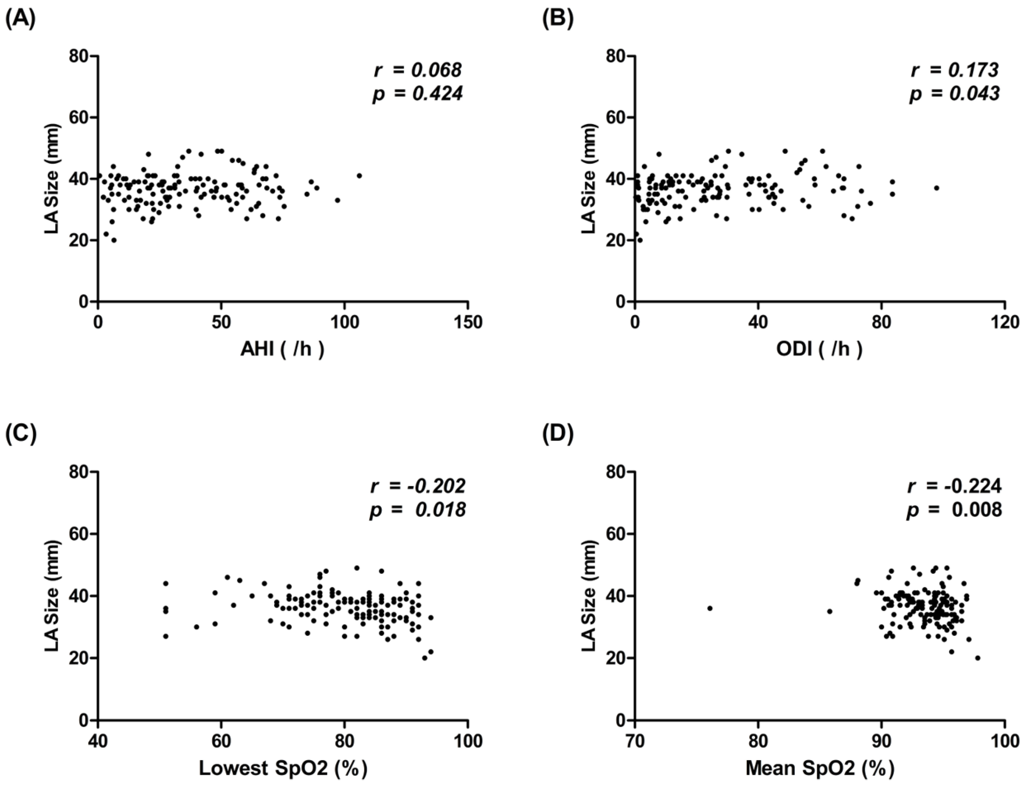
3.2. Correlation between Polysomnography (PSG) Metrics and LA Size in Patients with SRBD

To evaluate the importance of hypoxemic events associated with LA remodeling in patients with SRBD, we analyzed the association between different PSG metrics and LA size. We found there was a positive correlation between LA size and ODI (r = 0.173, p = 0.043) and a negative correlation between LA size and the lowest SpO2 (r = −0.202, p = 0.018) and mean SpO2 (r = −0.224, p = 0.008) (Figure 1). There was no significant correlation between LA size and AHI.

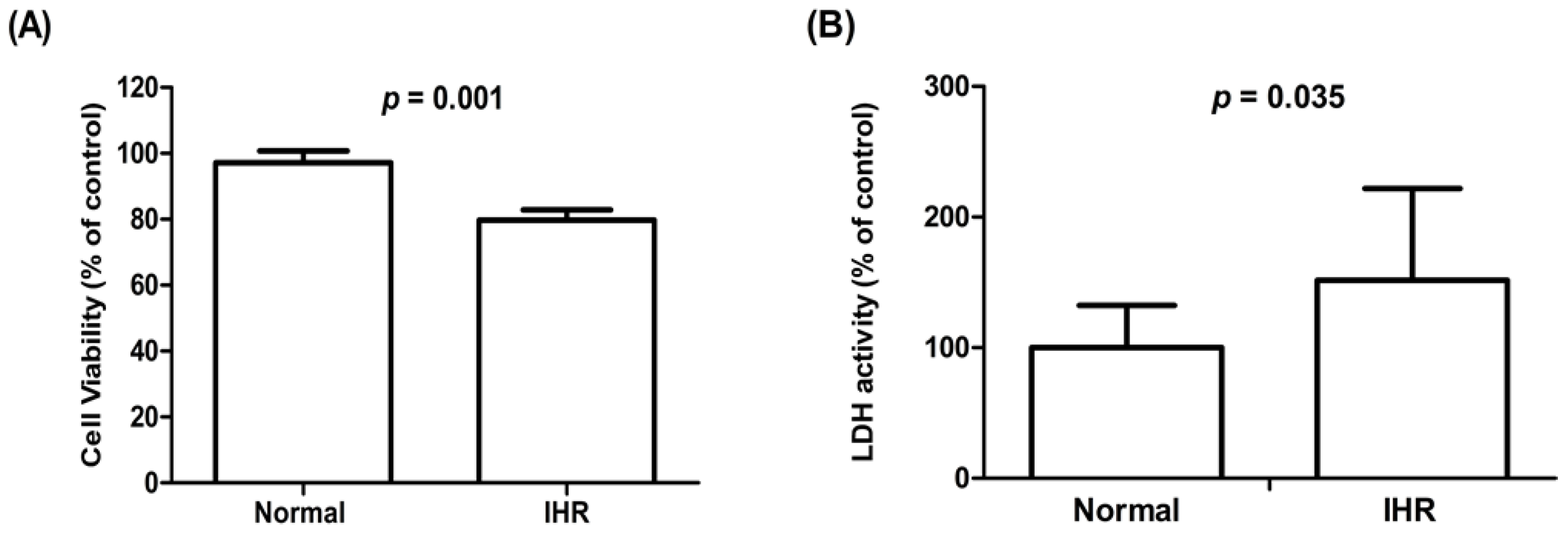
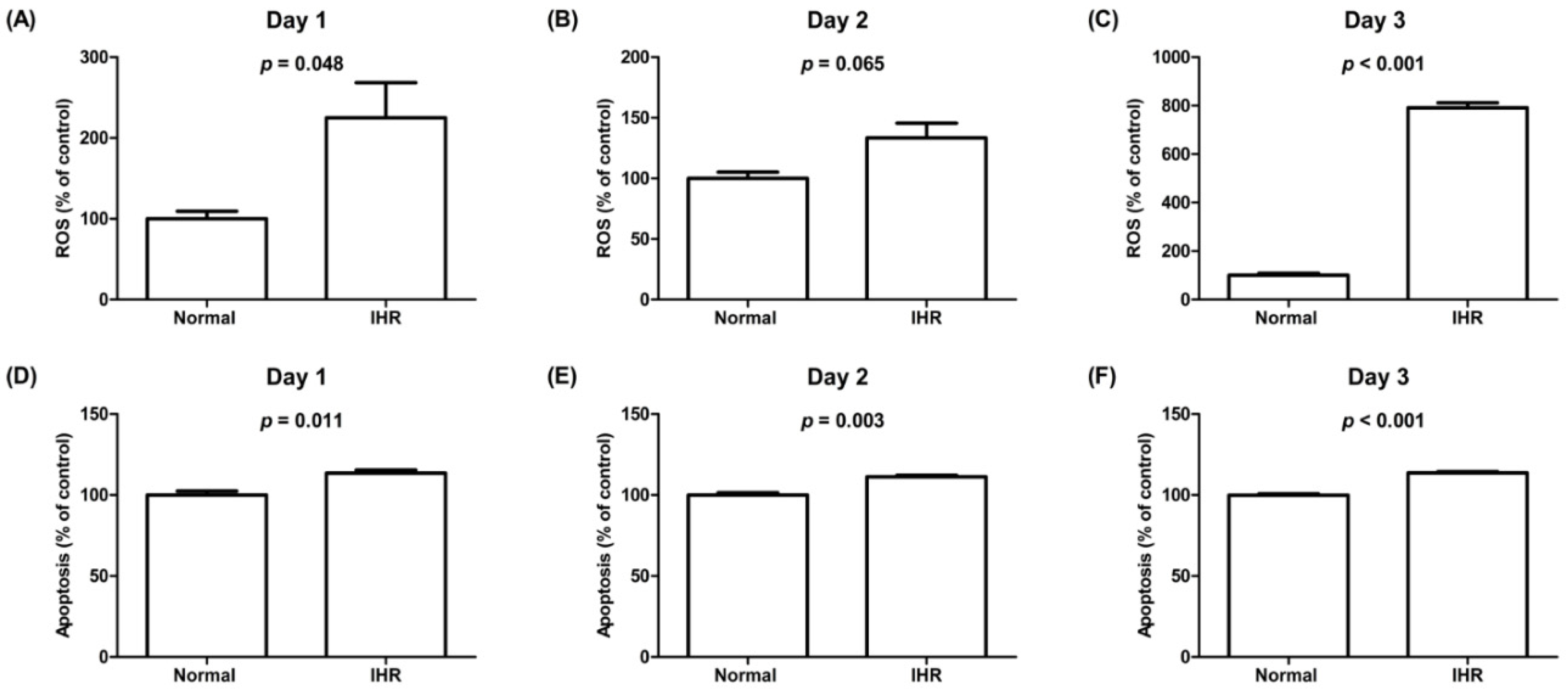
3.3. Cell Viability, Lactate Dehydrogenase (LDH) Activity, ROS Levels, Apoptosis, and the Expression of HIF-1α and Inflammatory Cytokines of HL-1 Cells Exposed to IHR Stimulation or Normoxia

To evaluate the impact of IHR on the morphologic and functional changes, we exposed HL-1 cells to IHR stimulation or normoxia, which showed decreased cell viability and increased LDH activity on Day 3, increased ROS levels on Day 1 and Day 3, and apoptosis from Day 1 to Day 3 in HL-1 cells exposed to IHR compared with normoxia (Figure 2 and Figure 3). The expression levels of HIF-1α, TNF-α, IL-6, and TGF-β were significantly higher in HL-1 cells exposed to IHR stimulation rather than normoxia (all p < 0.05) (Figure 4).

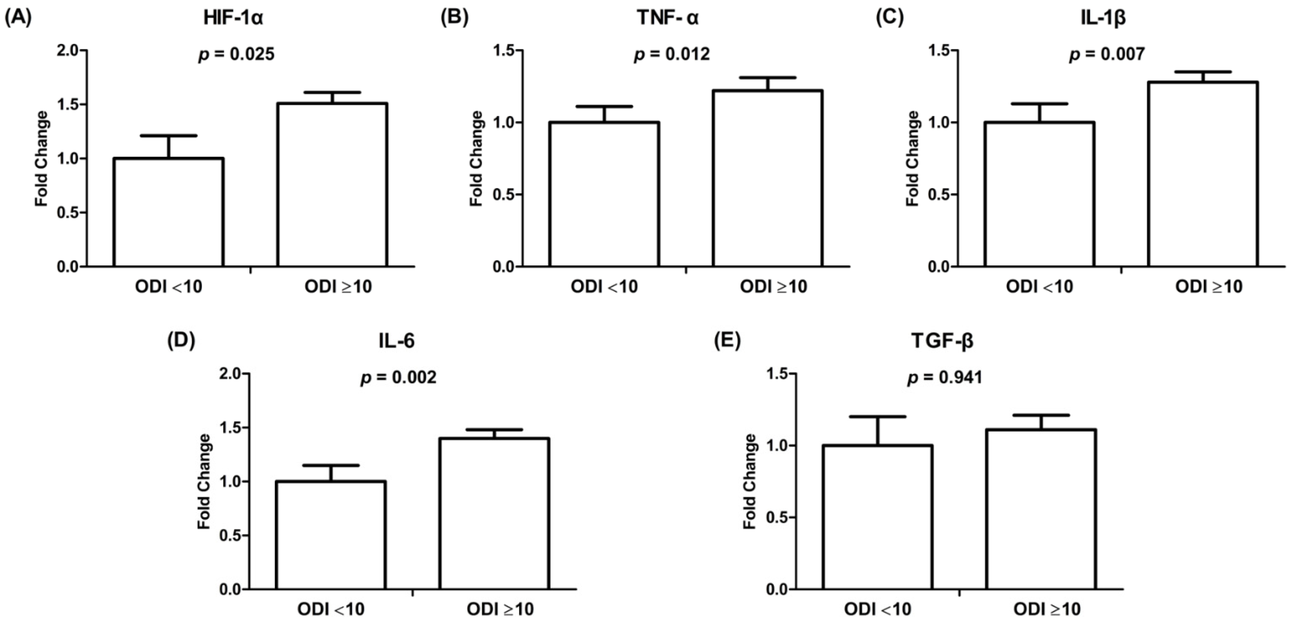
3.4. mRNA Gene Expression of HIF-1α and Inflammatory Cytokines in HL-1 Cells Treated with Exosomes Derived from Patients with and without Significant OSAS

To evaluate the effect and mechanism of intermittent hypoxemic events on atrial cardiomyocytes, HL-1 cells were treated with exosomes derived from 95 patients with and without significant OSAS. Patients with significant OSAS had higher mRNA expression levels of HIF-1α, TNF-α, IL-1β, and IL-6 (both p < 0.05) than patients without significant OSAS (Figure 5). There was no difference in the mRNA expression of TGF-β between patients with and without significant OSAS.

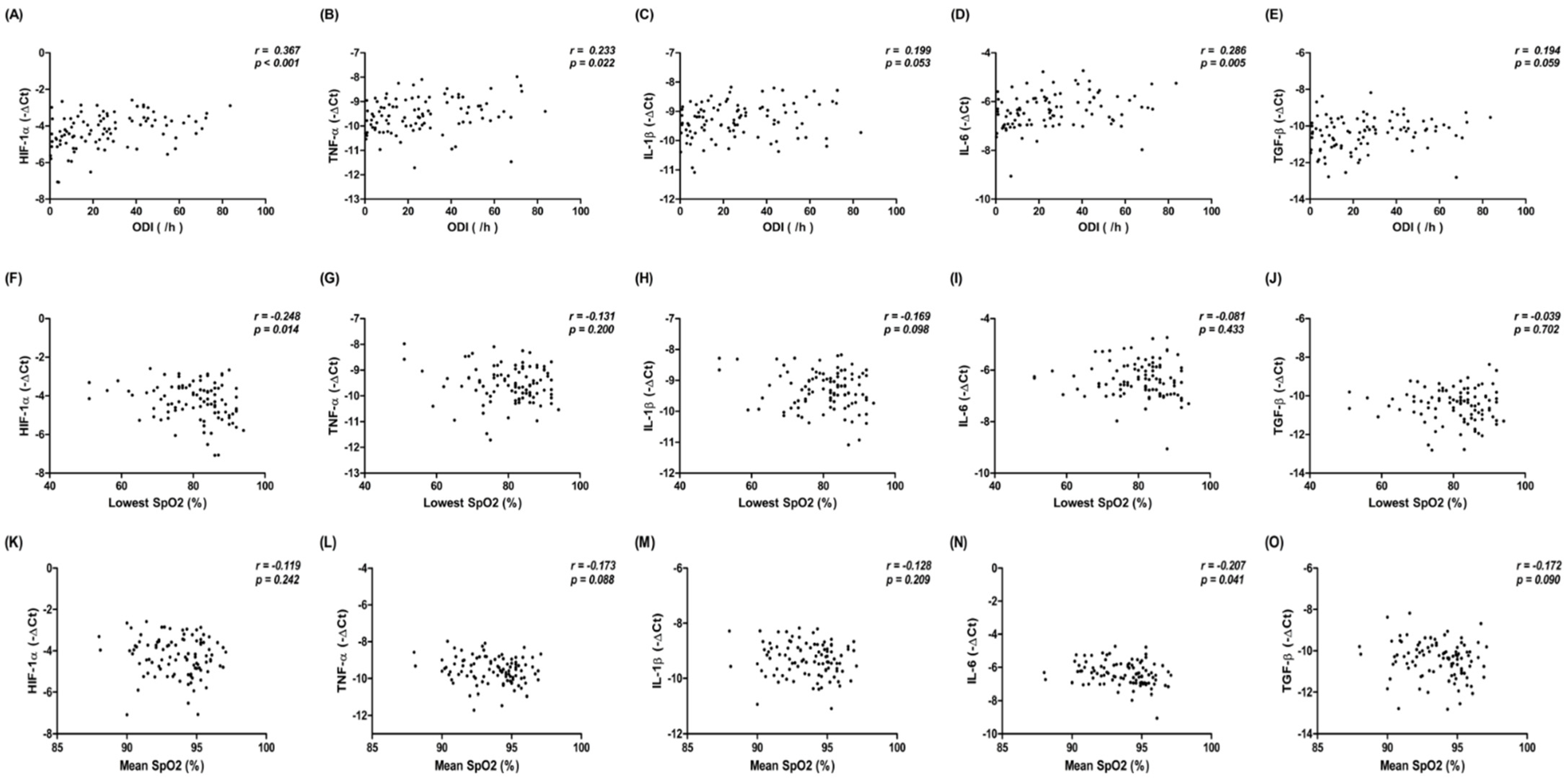
3.5. Correlation between mRNA Gene Expression of HIF-1α and Inflammatory Cytokines in HL-1 Cells Treated with Exosomes from Patients with SRDB, and PSG Metrics in Patients with SRDB

To investigate the possible impact of the severity of intermittent hypoxemia in 95 patients with SRDB on the expression of HIF-1a and inflammatory cytokines in HL-1 cells, we analyzed the correlation between PSG metrics in patients with SRDB and the expression of HIF-1a and inflammatory cytokines in HL-1 cells. We found there were significantly positive correlations between ODI and the expression of HIF-1α (r = 0.367, p < 0.001), TNF-α (r = 0.233, p = 0.022), and IL-6 (r = 0.286, p = 0.005); a significantly negative correlation between the lowest SpO2 and HIF-1a (r = −0.248, p = 0.014); and a significantly negative correlation between mean SpO2 and IL-6 (r = −0.207, p = 0.041). There was no significant correlation between ODI and the expression of IL-1β and TGF-β; between the lowest SpO2 and the expression of TNF-α, IL-1β, IL-6, and TGF-β; and between mean SpO2 and the expression of HIF-1α, TNF-α, IL-1β, and TGF-β (all p > 0.05) (Figure 6).

4. Discussion

There were several significant findings in this study. First, patients with significant OSAS had a larger LA size, and larger LA size was significantly associated with higher ODI, and lower lowest SpO2 and mean SpO2, but was not associated with AHI. Second, there were increases in the amount of LDH activity, ROS levels, apoptosis, and the expression of HIF-1α, TNF-α, IL-6, and TGF-β, and decreased cellular viability in HL-1 cells exposed to IHR stimulation compared with normoxia. Third, the expression of HIF-1α, TNF-α, IL-1β, and IL-6 was significantly increased in HL-1 cells treated with exosomes from patients with significant OSAS compared with those without significant OSAS. There was a significantly positive correlation between higher ODI and the increased expression of HIF-1α, TNF-α, and IL-6; a significantly negative correlation between a lower lowest SpO2 and increased expression of HIF-1α; and a significantly negative correlation between lower mean SpO2 and increased expression of IL-6.
A previous study showed that LA size was associated with the severity of OSAS and that AHI ≥ 15 was a significant predictor of LA enlargement [18]. In this study, ODI, mean SpO2, and lowest SpO2 but not AHI were significantly correlated with LA size. Respiratory arrest during sleep causes CO2 retention and hypoxemia, and desaturation episodes are one of the leading causes of the development of complications associated with OSAS [21,22,23]. Previous studies showed that ODI or desaturation severity was a better predictor than AHI for evaluating the clinical symptoms and risk of cerebrovascular and cardiovascular morbidities and mortality rates in patients with OSAS [22,42,43,44] The most critical limitations regarding AHI are that the morphological characteristics of abnormal respiratory events (i.e., duration and depth) are not taken into consideration. The physiological stress levels of different patients with similar AHI levels can be very different [22]. While the duration and depth of the apnea episodes increase, the frequency of AHI may decrease. Therefore, ODI and desaturation severity (i.e., lowest SpO2 and mean SpO2), which could be determined using a nocturnal pulse oximeter, maybe an earlier and better way to evaluate the pathophysiologic changes in patients with OSAS. Our study discovered that LA size was significantly associated with ODI but not with AHI in patients with SRBD, indicating that intermittent hypoxia plays a critical role in LA remodeling in these patients. Our findings suggest that in the study of LA remodeling, ODI may be a more useful and significant parameter for identifying high-risk patients with SRBD.
HIF-1 is activated under hypoxic conditions in various heart diseases, such as ischemic heart disease or heart failure, according to several studies [45,46,47]. In patients with OSAS, serum HIF-1α was found to be upregulated in recent studies [25,48,49]. HIF-1α expression was also linked to atrial fibrosis in rats induced with isoproterenol in a previous study [10]. Intermittent hypoxia can cause non-lethal ROS levels, which activate the HIF-1α promoter and drive HIF-1α transcription and function [50,51]. HIF is the master regulator of the hypoxia response and coordinates a transcriptional program that ensures optimal cellular adaptation to oxygen deprivation [52,53]. This study showed that IHR caused apoptosis and cellular damage in HL-1 cells with increased ROS levels, and the mRNA expression of HIF-1α. In addition, cell viability, LDH activity, and HIF-1α increased in HL-1 cells treated with exosomes from patients with significant OSAS compared with patients without OSAS. Moreover, the expression of HIF-1α was significantly and positively correlated with higher ODI and negatively correlated with the lowest SpO2 of patients with SRBD. Our study and previous studies revealed that intermittent hypoxemia-induced activation of HIF-1α signaling might play an essential role in the LA remodeling in patients with OSAS. However, it is unknown whether HIF-1α activation plays a critical role in myocardial protection against chronic hypoxemia to provide tissue tolerance against hypoxic injury. Further studies may investigate if targeting HIF-1α therapy could prevent LA remodeling in OSAS patients.
IHR activates pro-inflammatory cytokines including TNF-α and IL-1β, which have been demonstrated to promote HIF-1α accumulation and transcriptional activity [54]. In addition, through a signal transduction and activator of transcription (STAT3)-dependent mechanism, IL-6 and TGF increase the mRNA expression of HIF-1α [55,56]. TGF-β also enhances HIF-1α protein stability by decreasing PHD2 expression via Smads [57]. Activation of HIF-1α in mast cells, on the other hand, promoted the expression of IL-6 and TNF-α [58], but reduced HIF-1α expression, causing IL-1β expression to be hampered [59]. To summarize, hypoxia-induced HIF signaling and inflammation are mediated by the immune system’s complicated activities [53]. This study confirmed previous studies showing that IHR increases inflammatory cytokine expression. In individuals with SRBD, the production of inflammatory cytokines was linked to the severity of hypoxemia. More research is needed to determine the influence and interaction between HIF-1 and inflammation on LA remodeling in OSAS patients.
This study had some limitations. First, the sample size was insufficient to assess HIF-1’s predictive usefulness in LA remodeling. To corroborate the finding, larger cohort studies concentrating on verifying the role of HIF-1α expression in predicting LA remodeling should be conducted. Second, we did not look into the impact of therapy or other risk reduction strategies on HIF-1α, the expression of other inflammatory cytokines, and LA remodeling in OSAS patients. More research is needed to determine the impact of OSAS treatment on these biomarkers and clinical outcomes. Third, the left atrial volume index may be a more reliable method for determining the size of the left atrium than the LA dimension measured with conventional M-mode echocardiography. Regrettably, we were unable to provide data on LAVI in this study because it is not routinely measured in our daily practice. However, previous research and our previous study both indicated that there was a strong correlation between the LA’s dimension and left atrial volume index (r = 0.860, p < 0.001 in our previous study) [60,61]. Finally, we defined an ODI of >10 as moderate to severe OSAS according to a previous publication which demonstrated a sensitivity of 93% [62]. Although the results were statistically significant, underpowering may have occurred in the group with ODI < 10 due to the smaller number of cases (n = 45).

5. Conclusions

In conclusion, our study showed that intermittent hypoxemia plays an important role in LA remodeling in patients with SRBD. The ODI and other hypoxemia indices were significantly associated with LA remodeling. IHR stimulation and exosomes from patients with significant OSAS caused increased apoptosis, cellular damage, and ROS levels and decreased cellular viability in HL-1 cells. IHR stimulation and exosomes from significant OSAS patients also activated HIF-1α signaling and inflammatory cytokines, which were also significantly associated with ODI and other hypoxemia indices in patients with SRBD.

Supplementary Materials

The following are available online at https://www.mdpi.com/article/10.3390/life12020148/s1. Table S1: Quantitative real-time polymerase chain reaction primers.

Author Contributions

Y.-L.C. and M.-C.L. led the design of the study and revised the draft of the manuscript. Y.-L.C., Y.-C.C., W.-H.L., P.-T.L., Y.-N.F. and P.-Y.H. collected the research data and prepared the draft of the manuscript. Y.-T.C., H.-T.W., S.H. and K.-T.H. performed the clinical work and organized the collected data. Y.-L.C., H.-T.W. and M.-C.S. performed the statistical analysis. M.-C.L. supervised and validated the clinical work and results. All authors have read and agreed to the published version of the manuscript.

Funding

This study was supported by grants CMRPG8J0221, CMRPG8J0222, and CMRPG8J0223 from Kaohsiung Chang Gung Memorial Hospital, Taiwan.

Institutional Review Board Statement

This study was approved by the Institutional Review Board of Chang Gung Memorial Hospital (IRB number: 201801943B0, 7 January 2019) and conformed to the guidelines set forth by the Declaration of Helsinki.

Informed Consent Statement

Written informed consent was obtained from the participants before starting the study.

Data Availability Statement

The data underlying this article will be shared on reasonable request to the corresponding author.

Conflicts of Interest

The authors declare no conflict of interest.

References

  1. Tsang, T.S.; Barnes, M.E.; Gersh, B.J.; Takemoto, Y.; Rosales, A.G.; Bailey, K.R.; Seward, J.B. Prediction of risk for first age-related cardiovascular events in an elderly population: The incremental value of echocardiography. J. Am. Coll. Cardiol. 2003, 42, 1199–1205. [Google Scholar] [CrossRef] [Green Version]
  2. Pritchett, A.M.; Mahoney, D.W.; Jacobsen, S.J.; Rodeheffer, R.J.; Karon, B.L.; Redfield, M.M. Diastolic dysfunction and left atrial volume: A population-based study. J. Am. Coll. Cardiol. 2005, 45, 87–92. [Google Scholar] [CrossRef] [Green Version]
  3. Hoit, B.D. Left Atrial Remodeling: More Than Just Left Atrial Enlargement. Circ. Cardiovasc. Imaging 2017, 10, e006036. [Google Scholar] [CrossRef] [PubMed] [Green Version]
  4. Somers, V.K.; White, D.P.; Amin, R.; Abraham, W.T.; Costa, F.; Culebras, A.; Daniels, S.; Floras, J.S.; Hunt, C.E.; Olson, L.J.; et al. Sleep apnea and cardiovascular disease: An American Heart Association/american College Of Cardiology Foundation Scientific Statement from the American Heart Association Council for High Blood Pressure Research Professional Education Committee, Council on Clinical Cardiology, Stroke Council, and Council On Cardiovascular Nursing. In collaboration with the National Heart, Lung, and Blood Institute National Center on Sleep Disorders Research (National Institutes of Health). Circulation 2008, 118, 1080–1111. [Google Scholar] [CrossRef]
  5. Molina, C.E.; Abu-Taha, I.H.; Wang, Q.; Rosello-Diez, E.; Kamler, M.; Nattel, S.; Ravens, U.; Wehrens, X.H.T.; Hove-Madsen, L.; Heijman, J.; et al. Profibrotic, Electrical, and Calcium-Handling Remodeling of the Atria in Heart Failure Patients With and Without Atrial Fibrillation. Front. Physiol. 2018, 9, 1383. [Google Scholar] [CrossRef] [Green Version]
  6. Wu, N.; Xu, B.; Xiang, Y.; Wu, L.; Zhang, Y.; Ma, X.; Tong, S.; Shu, M.; Song, Z.; Li, Y.; et al. Association of inflammatory factors with occurrence and recurrence of atrial fibrillation: A meta-analysis. Int. J. Cardiol. 2013, 169, 62–72. [Google Scholar] [CrossRef] [PubMed]
  7. Alegret, J.M.; Aragones, G.; Elosua, R.; Beltran-Debon, R.; Hernandez-Aguilera, A.; Romero-Menor, C.; Camps, J.; Joven, J. The relevance of the association between inflammation and atrial fibrillation. Eur. J. Clin. Investig. 2013, 43, 324–331. [Google Scholar] [CrossRef] [PubMed]
  8. Liuba, I.; Ahlmroth, H.; Jonasson, L.; Englund, A.; Jonsson, A.; Safstrom, K.; Walfridsson, H. Source of inflammatory markers in patients with atrial fibrillation. Europace 2008, 10, 848–853. [Google Scholar] [CrossRef]
  9. Guo, Y.; Apostalakis, S.; Blann, A.D.; Lip, G.Y. Plasma CX3CL1 levels and long term outcomes of patients with atrial fibrillation: The West Birmingham Atrial Fibrillation Project. Cerebrovasc. Dis. 2014, 38, 204–211. [Google Scholar] [CrossRef] [PubMed]
  10. Su, F.; Zhang, W.; Chen, Y.; Ma, L.; Zhang, H.; Wang, F. Significance of hypoxia-inducible factor-1alpha expression with atrial fibrosis in rats induced with isoproterenol. Exp. Ther. Med. 2014, 8, 1677–1682. [Google Scholar] [CrossRef] [PubMed] [Green Version]
  11. Pepperell, J.C.; Ramdassingh-Dow, S.; Crosthwaite, N.; Mullins, R.; Jenkinson, C.; Stradling, J.R.; Davies, R.J. Ambulatory blood pressure after therapeutic and subtherapeutic nasal continuous positive airway pressure for obstructive sleep apnoea: A randomised parallel trial. Lancet 2002, 359, 204–210. [Google Scholar] [CrossRef]
  12. Gami, A.S.; Pressman, G.; Caples, S.M.; Kanagala, R.; Gard, J.J.; Davison, D.E.; Malouf, J.F.; Ammash, N.M.; Friedman, P.A.; Somers, V.K. Association of atrial fibrillation and obstructive sleep apnea. Circulation 2004, 110, 364–367. [Google Scholar] [CrossRef] [PubMed]
  13. Gami, A.S.; Hodge, D.O.; Herges, R.M.; Olson, E.J.; Nykodym, J.; Kara, T.; Somers, V.K. Obstructive sleep apnea, obesity, and the risk of incident atrial fibrillation. J. Am. Coll. Cardiol. 2007, 49, 565–571. [Google Scholar] [CrossRef] [Green Version]
  14. Yaggi, H.K.; Concato, J.; Kernan, W.N.; Lichtman, J.H.; Brass, L.M.; Mohsenin, V. Obstructive sleep apnea as a risk factor for stroke and death. N. Engl. J. Med. 2005, 353, 2034–2041. [Google Scholar] [CrossRef] [PubMed] [Green Version]
  15. Glantz, H.; Thunstrom, E.; Johansson, M.C.; Wallentin Guron, C.; Uzel, H.; Ejdeback, J.; Nasic, S.; Peker, Y. Obstructive sleep apnea is independently associated with worse diastolic function in coronary artery disease. Sleep Med. 2015, 16, 160–167. [Google Scholar] [CrossRef] [PubMed] [Green Version]
  16. Arzt, M.; Woehrle, H.; Oldenburg, O.; Graml, A.; Suling, A.; Erdmann, E.; Teschler, H.; Wegscheider, K.; Schla, H.F.I. Prevalence and Predictors of Sleep-Disordered Breathing in Patients With Stable Chronic Heart Failure: The SchlaHF Registry. JACC Heart Fail. 2016, 4, 116–125. [Google Scholar] [CrossRef]
  17. Lyons, O.D.; Chan, C.T.; Elias, R.M.; Bradley, T.D. Relationship of left atrial size to obstructive sleep apnea severity in end-stage renal disease. Sleep Med. 2014, 15, 1314–1318. [Google Scholar] [CrossRef]
  18. Holtstrand Hjalm, H.; Fu, M.; Hansson, P.O.; Zhong, Y.; Caidahl, K.; Mandalenakis, Z.; Morales, D.; Ergatoudes, C.; Rosengren, A.; Grote, L.; et al. Association between left atrial enlargement and obstructive sleep apnea in a general population of 71-year-old men. J. Sleep Res. 2018, 27, 252–258. [Google Scholar] [CrossRef]
  19. Peppard, P.E.; Young, T.; Barnet, J.H.; Palta, M.; Hagen, E.W.; Hla, K.M. Increased prevalence of sleep-disordered breathing in adults. Am. J. Epidemiol. 2013, 177, 1006–1014. [Google Scholar] [CrossRef] [Green Version]
  20. Burstein, B.; Nattel, S. Atrial fibrosis: Mechanisms and clinical relevance in atrial fibrillation. J. Am. Coll. Cardiol. 2008, 51, 802–809. [Google Scholar] [CrossRef] [Green Version]
  21. Konecny, T.; Kara, T.; Somers, V.K. Obstructive sleep apnea and hypertension: An update. Hypertension 2014, 63, 203–209. [Google Scholar] [CrossRef] [PubMed] [Green Version]
  22. Kulkas, A.; Tiihonen, P.; Julkunen, P.; Mervaala, E.; Toyras, J. Novel parameters indicate significant differences in severity of obstructive sleep apnea with patients having similar apnea-hypopnea index. Med. Biol. Eng. Comput. 2013, 51, 697–708. [Google Scholar] [CrossRef]
  23. Mediano, O.; Barcelo, A.; de la Pena, M.; Gozal, D.; Agusti, A.; Barbe, F. Daytime sleepiness and polysomnographic variables in sleep apnoea patients. Eur. Respir. J. 2007, 30, 110–113. [Google Scholar] [CrossRef] [PubMed] [Green Version]
  24. Oldenburg, O.; Wellmann, B.; Buchholz, A.; Bitter, T.; Fox, H.; Thiem, U.; Horstkotte, D.; Wegscheider, K. Nocturnal hypoxaemia is associated with increased mortality in stable heart failure patients. Eur. Heart J. 2016, 37, 1695–1703. [Google Scholar] [CrossRef] [PubMed]
  25. Gabryelska, A.; Szmyd, B.; Szemraj, J.; Stawski, R.; Sochal, M.; Bialasiewicz, P. Patients with obstructive sleep apnea present with chronic upregulation of serum HIF-1alpha protein. J. Clin. Sleep Med. 2020, 16, 1761–1768. [Google Scholar] [CrossRef]
  26. Gross, J.C.; Chaudhary, V.; Bartscherer, K.; Boutros, M. Active Wnt proteins are secreted on exosomes. Nat. Cell Biol. 2012, 14, 1036–1045. [Google Scholar] [CrossRef]
  27. Mathivanan, S.; Ji, H.; Simpson, R.J. Exosomes: Extracellular organelles important in intercellular communication. J. Proteom. 2010, 73, 1907–1920. [Google Scholar] [CrossRef]
  28. Liao, J.; Liu, R.; Yin, L.; Pu, Y. Expression profiling of exosomal miRNAs derived from human esophageal cancer cells by Solexa high-throughput sequencing. Int. J. Mol. Sci. 2014, 15, 15530–15551. [Google Scholar] [CrossRef] [PubMed] [Green Version]
  29. Claycomb, W.C.; Lanson, N.A., Jr.; Stallworth, B.S.; Egeland, D.B.; Delcarpio, J.B.; Bahinski, A.; Izzo, N.J., Jr. HL-1 cells: A cardiac muscle cell line that contracts and retains phenotypic characteristics of the adult cardiomyocyte. Proc. Natl. Acad. Sci. USA 1998, 95, 2979–2984. [Google Scholar] [CrossRef] [PubMed] [Green Version]
  30. Chen, Y.L.; Chen, Y.C.; Chang, Y.T.; Wang, H.T.; Liu, W.H.; Chong, S.Z.; Lin, P.T.; Hsu, P.Y.; Su, M.C.; Lin, M.C. GJA1 Expression and Left Atrial Remodeling in the Incidence of Atrial Fibrillation in Patients with Obstructive Sleep Apnea Syndrome. Biomedicines 2021, 9, 1463. [Google Scholar] [CrossRef]
  31. Chen, Y.L.; Su, M.C.; Liu, W.H.; Wang, C.C.; Lin, M.C.; Chen, M.C. Influence and predicting variables of obstructive sleep apnea on cardiac function and remodeling in patients without congestive heart failure. J. Clin. Sleep Med. 2014, 10, 57–64. [Google Scholar] [CrossRef] [PubMed] [Green Version]
  32. Redline, S.; Budhiraja, R.; Kapur, V.; Marcus, C.L.; Mateika, J.H.; Mehra, R.; Parthasarthy, S.; Somers, V.K.; Strohl, K.P.; Sulit, L.G.; et al. The scoring of respiratory events in sleep: Reliability and validity. J. Clin. Sleep Med. 2007, 3, 169–200. [Google Scholar] [CrossRef] [PubMed]
  33. Chen, Y.L.; Wang, H.T.; Chen, H.C.; Chai, H.T.; Lee, Y.W.; Liu, W.H. Localization of right ventricular non-apical lead position: Comparison of three-dimensional echocardiography, computed tomography, and fluoroscopic imaging. J. Int. Med. Res. 2021, 49, 300060521996159. [Google Scholar] [CrossRef] [PubMed]
  34. Liu, W.H.; Guo, B.F.; Chen, Y.L.; Tsai, T.H.; Fu, M.; Chua, S.; Chen, M.C. Right ventricular outflow tract pacing causes intraventricular dyssynchrony in patients with sick sinus syndrome: A real-time three-dimensional echocardiographic study. J. Am. Soc. Echocardiogr. 2010, 23, 599–607. [Google Scholar] [CrossRef] [PubMed]
  35. Kuznetsov, A.V.; Javadov, S.; Sickinger, S.; Frotschnig, S.; Grimm, M. H9c2 and HL-1 cells demonstrate distinct features of energy metabolism, mitochondrial function and sensitivity to hypoxia-reoxygenation. Biochim. Biophys. Acta 2015, 1853, 276–284. [Google Scholar] [CrossRef] [PubMed] [Green Version]
  36. Chen, Y.C.; Hsu, P.Y.; Su, M.C.; Chin, C.H.; Liou, C.W.; Wang, T.Y.; Lin, Y.Y.; Lee, C.P.; Lin, M.C.; Hsiao, C.C. miR-21-5p Under-Expression in Patients with Obstructive Sleep Apnea Modulates Intermittent Hypoxia with Re-Oxygenation-Induced-Cell Apoptosis and Cytotoxicity by Targeting Pro-Inflammatory TNF-alpha-TLR4 Signaling. Int. J. Mol. Sci. 2020, 21, 999. [Google Scholar] [CrossRef] [Green Version]
  37. Tauro, B.J.; Greening, D.W.; Mathias, R.A.; Ji, H.; Mathivanan, S.; Scott, A.M.; Simpson, R.J. Comparison of ultracentrifugation, density gradient separation, and immunoaffinity capture methods for isolating human colon cancer cell line LIM1863-derived exosomes. Methods 2012, 56, 293–304. [Google Scholar] [CrossRef]
  38. Lydic, T.A.; Townsend, S.; Adda, C.G.; Collins, C.; Mathivanan, S.; Reid, G.E. Rapid and comprehensive ‘shotgun’ lipidome profiling of colorectal cancer cell derived exosomes. Methods 2015, 87, 83–95. [Google Scholar] [CrossRef] [Green Version]
  39. Huang, C.; Fisher, K.P.; Hammer, S.S.; Navitskaya, S.; Blanchard, G.J.; Busik, J.V. Plasma Exosomes Contribute to Microvascular Damage in Diabetic Retinopathy by Activating the Classical Complement Pathway. Diabetes 2018, 67, 1639–1649. [Google Scholar] [CrossRef] [Green Version]
  40. Hsu, P.Y.; Hsi, E.; Wang, T.M.; Lin, R.T.; Liao, Y.C.; Juo, S.H. MicroRNA let-7g possesses a therapeutic potential for peripheral artery disease. J. Cell Mol. Med. 2017, 21, 519–529. [Google Scholar] [CrossRef] [Green Version]
  41. Nguyen, S.V.; Claycomb, W.C. Hypoxia regulates the expression of the adrenomedullin and HIF-1 genes in cultured HL-1 cardiomyocytes. Biochem. Biophys. Res. Commun. 1999, 265, 382–386. [Google Scholar] [CrossRef] [Green Version]
  42. Kainulainen, S.; Toyras, J.; Oksenberg, A.; Korkalainen, H.; Sefa, S.; Kulkas, A.; Leppanen, T. Severity of Desaturations Reflects OSA-Related Daytime Sleepiness Better Than AHI. J. Clin. Sleep Med. 2019, 15, 1135–1142. [Google Scholar] [CrossRef]
  43. Jean-Louis, G.; Brown, C.D.; Zizi, F.; Ogedegbe, G.; Boutin-Foster, C.; Gorga, J.; McFarlane, S.I. Cardiovascular disease risk reduction with sleep apnea treatment. Expert Rev. Cardiovasc. Ther. 2010, 8, 995–1005. [Google Scholar] [CrossRef] [Green Version]
  44. Liu, J.F.; Tsai, C.M.; Su, M.C.; Lin, M.C.; Lin, H.C.; Lee, W.J.; Hsieh, K.S.; Niu, C.K.; Yu, H.R. Application of desaturation index in post-surgery follow-up in children with obstructive sleep apnea syndrome. Eur. Arch. Otorhino-Laryngol. 2017, 274, 375–382. [Google Scholar] [CrossRef] [PubMed]
  45. Tian, X.; Zhou, N.; Yuan, J.; Lu, L.; Zhang, Q.; Wei, M.; Zou, Y.; Yuan, L. Heat shock transcription factor 1 regulates exercise-induced myocardial angiogenesis after pressure overload via HIF-1alpha/VEGF pathway. J. Cell Mol. Med. 2020, 24, 2178–2188. [Google Scholar] [CrossRef]
  46. Morand, J.; Arnaud, C.; Pepin, J.L.; Godin-Ribuot, D. Chronic intermittent hypoxia promotes myocardial ischemia-related ventricular arrhythmias and sudden cardiac death. Sci. Rep. 2018, 8, 2997. [Google Scholar] [CrossRef] [PubMed] [Green Version]
  47. Chen, Y.F.; Pandey, S.; Day, C.H.; Chen, Y.F.; Jiang, A.Z.; Ho, T.J.; Chen, R.J.; Padma, V.V.; Kuo, W.W.; Huang, C.Y. Synergistic effect of HIF-1alpha and FoxO3a trigger cardiomyocyte apoptosis under hyperglycemic ischemia condition. J. Cell Physiol. 2018, 233, 3660–3671. [Google Scholar] [CrossRef]
  48. Lu, D.; Li, N.; Yao, X.; Zhou, L. Potential inflammatory markers in obstructive sleep apnea-hypopnea syndrome. Bosn. J. Basic Med. Sci. 2017, 17, 47–53. [Google Scholar] [CrossRef] [Green Version]
  49. Gabryelska, A.; Stawski, R.; Sochal, M.; Szmyd, B.; Bialasiewicz, P. Influence of one-night CPAP therapy on the changes of HIF-1alpha protein in OSA patients: A pilot study. J. Sleep Res. 2020, 29, e12995. [Google Scholar] [CrossRef]
  50. Bonello, S.; Zahringer, C.; BelAiba, R.S.; Djordjevic, T.; Hess, J.; Michiels, C.; Kietzmann, T.; Gorlach, A. Reactive oxygen species activate the HIF-1alpha promoter via a functional NFkappaB site. Arterioscler. Thromb. Vasc. Biol. 2007, 27, 755–761. [Google Scholar] [CrossRef] [PubMed] [Green Version]
  51. Chen, T.I.; Hsu, Y.C.; Lien, C.F.; Lin, J.H.; Chiu, H.W.; Yang, K.T. Non-lethal levels of oxidative stress in response to short-term intermittent hypoxia enhance ca(2)(+) handling in neonatal rat cardiomyocytes. Cell Physiol. Biochem. 2014, 33, 513–527. [Google Scholar] [CrossRef]
  52. Semenza, G.L. Oxygen sensing, homeostasis, and disease. N. Engl. J. Med. 2011, 365, 537–547. [Google Scholar] [CrossRef] [Green Version]
  53. Corrado, C.; Fontana, S. Hypoxia and HIF Signaling: One Axis with Divergent Effects. Int. J. Mol. Sci. 2020, 21, 5611. [Google Scholar] [CrossRef] [PubMed]
  54. Jung, Y.; Isaacs, J.S.; Lee, S.; Trepel, J.; Liu, Z.G.; Neckers, L. Hypoxia-inducible factor induction by tumour necrosis factor in normoxic cells requires receptor-interacting protein-dependent nuclear factor kappa B activation. Biochem. J. 2003, 370, 1011–1017. [Google Scholar] [CrossRef]
  55. Dang, E.V.; Barbi, J.; Yang, H.Y.; Jinasena, D.; Yu, H.; Zheng, Y.; Bordman, Z.; Fu, J.; Kim, Y.; Yen, H.R.; et al. Control of T(H)17/T(reg) balance by hypoxia-inducible factor 1. Cell 2011, 146, 772–784. [Google Scholar] [CrossRef] [Green Version]
  56. Ikejiri, A.; Nagai, S.; Goda, N.; Kurebayashi, Y.; Osada-Oka, M.; Takubo, K.; Suda, T.; Koyasu, S. Dynamic regulation of Th17 differentiation by oxygen concentrations. Int. Immunol. 2012, 24, 137–146. [Google Scholar] [CrossRef] [Green Version]
  57. McMahon, S.; Charbonneau, M.; Grandmont, S.; Richard, D.E.; Dubois, C.M. Transforming growth factor beta1 induces hypoxia-inducible factor-1 stabilization through selective inhibition of PHD2 expression. J. Biol. Chem. 2006, 281, 24171–24181. [Google Scholar] [CrossRef] [PubMed] [Green Version]
  58. Jeong, H.J.; Chung, H.S.; Lee, B.R.; Kim, S.J.; Yoo, S.J.; Hong, S.H.; Kim, H.M. Expression of proinflammatory cytokines via HIF-1alpha and NF-kappaB activation on desferrioxamine-stimulated HMC-1 cells. Biochem. Biophys. Res. Commun. 2003, 306, 805–811. [Google Scholar] [CrossRef]
  59. Fang, H.Y.; Hughes, R.; Murdoch, C.; Coffelt, S.B.; Biswas, S.K.; Harris, A.L.; Johnson, R.S.; Imityaz, H.Z.; Simon, M.C.; Fredlund, E.; et al. Hypoxia-inducible factors 1 and 2 are important transcriptional effectors in primary macrophages experiencing hypoxia. Blood 2009, 114, 844–859. [Google Scholar] [CrossRef] [PubMed] [Green Version]
  60. Bhopalwala, H.; Dewaswala, N.; Liu, S.; Scott, C.G.; Welper, J.M.; Akinnusotu, O.; Bos, J.M.; Ommen, S.R.; Ackerman, M.J.; Pellikka, P.A.; et al. Conversion of left atrial volume to diameter for automated estimation of sudden cardiac death risk in hypertrophic cardiomyopathy. Echocardiography 2021, 38, 183–188. [Google Scholar] [CrossRef] [PubMed]
  61. Chen, Y.L.; Chuang, J.H.; Wang, H.T.; Chen, H.C.; Liu, W.H.; Yang, M.Y. Altered Expression of Circadian Clock Genes in Patients with Atrial Fibrillation Is Associated with Atrial High-Rate Episodes and Left Atrial Remodeling. Diagnostics 2021, 11, 90. [Google Scholar] [CrossRef] [PubMed]
  62. Chung, F.; Liao, P.; Elsaid, H.; Islam, S.; Shapiro, C.M.; Sun, Y. Oxygen desaturation index from nocturnal oximetry: A sensitive and specific tool to detect sleep-disordered breathing in surgical patients. Anesth. Analg. 2012, 114, 993–1000. [Google Scholar] [CrossRef] [PubMed]
Figure 1. Correlation between LA size and the (A) AHI, (B) ODI, (C) lowest SpO2, and (D) mean SpO2 in patients with SRBD. The correlation was assessed by bivariate correlation analysis. The r and p-values indicate the correlation between LA size (expressed in units of mm) and AHI (expressed in units per hour), ODI (expressed in units per hour), lowest SpO2 (expressed in %), and mean SpO2 (expressed in). AHI, apnea–hypopnea index; LA, left atrial; ODI, oxygen desaturation index; SpO2, oxygen saturation; SRBD, sleep-related breathing disorder.
Life 12 00148 g001
Figure 2. Cell viability (A) and LDH activity (B) in HL-1 cells exposed to IHR stimulation or normoxia on Day 3. The y-axis represents cell viability (% of control) in (A) and LDH activity (% of control) in (B). IHR, intermittent hypoxia/reoxygenation; LDH, lactate dehydrogenase.
Life 12 00148 g002
Figure 3. ROS activity (AC) and apoptosis rates (DF) in HL-1 cells exposed to IHR stimulation or normoxia on Days 1–3. The y-axis represents ROS levels (% of control) in (AC) and apoptosis rates (% of control) in (DF). IHR, intermittent hypoxia/reoxygenation; ROS, reactive oxygen species.
Life 12 00148 g003
Figure 4. mRNA expression of HIF-1α (A) and inflammatory cytokines (BE) of HL-1 cells exposed to IHR or normoxia. The y-axis represents the fold change in the mRNA expression level in HL-1 cells exposed to IHR stimulation compared with normoxia. HIF, hypoxia-inducible factor; IHR, intermittent hypoxia/reoxygenation; IL, interleukin; TGF, transforming growth factor; TNF, tumor necrosis factor.
Life 12 00148 g004
Figure 5. mRNA gene expression of HIF-1α (A) and inflammatory cytokines (BE) in HL-1 cells treated with exosomes derived from patients with and without significant OSAS. The y-axis represents the fold change on mRNA expression levels in HL-1 cells incubated with exosomes derived from patients with significant OSAS (n = 74) compared with those without OSAS (n = 21). HIF, hypoxia-inducible factors; IL, interleukin; OSAS, obstructive sleep apnea syndrome; TGF, transforming growth factor; TNF, tumor necrosis factor.
Life 12 00148 g005
Figure 6. Correlation between the mRNA gene expression of HIF-1α and inflammatory cytokines in HL-1 cells treated with exosomes from 95 patients with SRDB, and PSG metrics, including ODI (AE), lowest SpO2 (FJ), and mean SpO2 (KO) in patients with SRDB. PSG, polysomnography; ODI, oxygen desaturation index; SpO2, oxygen desaturation; SRDB, sleep-related breathing disorder.
Life 12 00148 g006
Table 1. Baseline characteristics of obstructive sleep apnea syndrome patients with and without ODI ≥ 10.
VariablesODI ≥ 10 (per hour) (n = 109)ODI < 10 (per hour) (n = 45)p-Value
Age55.8 ± 11.053.3 ± 9.80.188
Sex (male)81 (74.3%)30 (66.7%)0.336
Smoking20 (18.3%)11 (24.4%)0.391
BMI26.5 ± 3.324.2 ± 3.1<0.001
Waistline85.6 ± 8.381.7 ± 8.20.063
DM13 (11.9%)3 (6.7%)0.331
HTN42 (38.5%)15 (33.3%)0.543
HF3 (2.8%)0 (0%)0.261
Stroke4 (3.7%)2 (4.4%)0.821
CAD5 (4.6%)2 (4.4%)0.969
AF20 (18.3%)6 (13.3)0.518
Paroxysmal AF9 (8.3%)4 (8.9%)
Persistent AF11 (10.1%)2 (4.4%)
Thyroid disorder2 (1.8%)0 (0%)0.360
Polysomnography
Epworth sleepiness scale9.3 (6.0–12.0)8.0 (5.0–12.0)0.254
AHI (per hour)45.4 (27.7–43.2)13.2 (5.7–20.2)<0.001
ODI (per hour)36.5 (19.7–48.4)4.8 (2.7–7.1)<0.001
Lowest SpO2 (%)76.7 (72.0–84.0)87.9 (86.0–91.0)<0.001
Mean SpO2 (%)92.8 (91.8–94.6)94.7 (93.6–96.0)<0.001
LA size by echocardiography37.0 ± 5.034.6 ± 5.60.021
Data are expressed as the mean ± standard deviation, median (interquartile range), or number (%). AF, atrial fibrillation; AHI, apnea–hypopnea index; BMI, body mass index; CAD, coronary artery disease; DM, diabetes mellitus; HF, heart failure; HTN, hypertension; LA, left atrial; ODI, oxygen desaturation index; SpO2, oxygen saturation.
Publisher’s Note: MDPI stays neutral with regard to jurisdictional claims in published maps and institutional affiliations.

