Review

The Conserved Family of the Pyridoxal Phosphate-Binding Protein (PLPBP) and Its Cyanobacterial Paradigm PipY

1
Dto. de Fisiología, Genética y Microbiología, Universidad de Alicante, 03690 San Vicente del Raspeig, Spain
2
Instituto de Biomedicina de Valencia, Consejo Superior de Investigaciones Científicas (IBV-CSIC), 46010 Valencia, Spain
3
Group CB06/07/0077 at the Instituto de Biomedicina de Valencia (IBV-CSIC) of CIBERER-ISCIII, Centro de Investigación Biomédica en Red de Enfermedades Raras, 28029 Madrid, Spain
*
Author to whom correspondence should be addressed.
Life 2022, 12(10), 1622; https://doi.org/10.3390/life12101622
Submission received: 20 September 2022 / Revised: 5 October 2022 / Accepted: 15 October 2022 / Published: 17 October 2022
(This article belongs to the Special Issue Molecular Genetics of Cyanobacteria)

Abstract

:
The PLPBP family of pyridoxal phosphate-binding proteins has a high degree of sequence conservation and is represented in all three domains of life. PLPBP members, of which a few representatives have been studied in different contexts, are single-domain proteins with no known enzymatic activity that exhibit the fold type III of PLP-holoenzymes, consisting in an α/β barrel (TIM-barrel), where the PLP cofactor is solvent-exposed. Despite the constant presence of cofactor PLP (a key catalytic element in PLP enzymes), PLPBP family members appear to have purely regulatory functions affecting the homeostasis of vitamin B6 vitamers and amino/keto acids. Perturbation of these metabolites and pleiotropic phenotypes have been reported in bacteria and zebrafish after PLPBP gene inactivation as well as in patients with vitamin B6-dependent epilepsy that results from loss-of-function mutations at the PLPBP. Here, we review information gathered from diverse studies and biological systems, emphasizing the structural and functional conservation of the PLPBP members and discussing the informative nature of model systems and experimental approaches. In this context, the relatively high level of structural and functional characterization of PipY from Synechococcus elongatus PCC 7942 provides a unique opportunity to investigate the PLPBP roles in the context of a signaling pathway conserved in cyanobacteria.

1. Introduction

Cyanobacteria, phototrophic organisms performing oxygenic photosynthesis, constitute an ecologically and biotechnologically important phylum, responsible for the evolution of the oxygenic atmosphere, being the main contributors to marine primary production [1]. Their photosynthetic lifestyle and ease of cultivation make them ideal production systems for several high-value compounds, including biofuels [2]. Despite important breakthroughs in the genetic analysis of cyanobacteria, there is still a remarkable proportion of genes of unknown function in this phylum, many of which are presumably relevant to the biology of cyanobacteria.
The cyanobacterium Synechococcus elongatus PCC7942 (hereafter S. elongatus), the only photosynthetic organism for which the contribution of each gene to fitness has been evaluated so far [3], is being used as a model system to address fundamental questions concerning the photosynthetic lifestyle. More recently, the S. elongatus genome has been used as the reference organism to create a database for the Cyanobacterial-Linked Genome [4], accessible through an interactive platform “https://dfgm.ua.es/es/cyanobacterial-genetics/dclg/index.htm (accesed on 1 August 2022)”.
In bacteria and plants, 2-oxoglutarate (2-OG), a key metabolic signal of the intracellular carbon-to-nitrogen balance, is sensed by the highly conserved and widely distributed signal transduction protein PII. PII regulates the activity of proteins involved in nitrogen metabolism by direct protein–protein interactions [5]. In S. elongatus PII interacts with a small (89 residues) protein called PipX (PII-interacting protein X), which was initially identified in yeast two-hybrid analyses [6,7].
PipX was also found in searches for proteins interacting with NtcA, the global transcriptional regulator involved in nitrogen assimilation in cyanobacteria [8]. PipX stabilizes the conformation of NtcA which is transcriptionally active and probably helps the local recruitment of RNA polymerase to NtcA-dependent promoters [9]. At low 2-OG concentrations corresponding to nitrogen-excess conditions, the sequestration of PipX by PII renders PipX unavailable for NtcA binding and activation, reducing the expression of NtcA-dependent gene targets [9,10,11,12,13]. Partner swapping by PipX is enabled by its N-terminal Tudor-like domain (TLD/KOW), which provides contacts for both NtcA and PII. Complex formation with PipX increases the affinity of PII for ADP [9], and, conversely, the interaction between PII and PipX is highly sensitive to fluctuations in the ATP/ADP ratio [14]. Thus, PipX partner swapping between PII and NtcA integrates signaling of the carbon-to-nitrogen ratio and the energy status by PII with the regulation of nitrogen-responsive genes controlled by NtcA [10,15,16].
Interestingly, a high PipX/PII ratio prevents growth [11,17] and, consistent with this, cyanobacterial genomes always contain at least as many copies of glnB as of pipX [18], suggesting that a relatively high ratio of PII over PipX is required to counteract unwanted interactions with low-affinity PipX partners.
In S. elongatus pipX is co-transcribed with the downstream gene pipY. This last gene belongs to the widely distributed and highly conserved pyridoxal phosphate (PLP)-binding protein (COG0325/PLPBP) family that is involved in vitamin B6 and amino acid homeostasis [19]. The PLPBP family (also termed ProsC/PROSC or COG0325 family) members are found in all kingdoms of life, exemplified by the proteins YBL036C (yeast), YggS (Gram-negative bacteria), YlmE (Gram-positive bacteria), PipY (cyanobacteria), PLPHP (humans) and HTH5 (rice). These are all single-domain proteins exhibiting the fold type III of PLP-holoenzymes [20,21,22,23,24] with no known enzymatic activity.
The association of pipY with pipX in an operon provides a unique opportunity to investigate the roles of PLPBP proteins in the context of a signaling pathway conserved in cyanobacteria, which is interconnected with the well-characterized nitrogen regulation network [10]. Importantly, PipY from S. elongatus is one of the best-characterized PLPBP members and can be regarded as a paradigm for PLPBP proteins [19,22,25].

2. Structural and Functional Features of PLPBPs

2.1. PLP Is Solvent-Exposed in PLPBP Structures

The vitamin B6 vitamer PLP is used as a cofactor for enzyme-catalyzed reactions which include transamination, decarboxylation, racemization, aldol cleavage, or replacement reactions among others [26]. Since amino acid metabolism and other essential processes require PLP-dependent enzymes [27,28], PLP availability is of paramount importance to supply cofactors to activate newly synthesized apo-B6 enzymes. PLP is also required as a cofactor of glycogen phosphorylase [29] and certain transcriptional factors and regulators [28]. However, its aldehyde group endows PLP with high chemical reactivity, sometimes causing the inactivation of proteins (see for example, [30]), and therefore additional mechanisms are required for keeping the levels of free PLP low in cells and tissues. In the first report of a member of this family, Eswaramoorthy et al. (2003) documented structural parallelisms between the yeast protein YBL036C and the N-terminal domain of alanine racemases, leading them to infer (and even to provide some experimental hints for it) that PLPBP had alanine racemase activity [21]. However, no amino acid racemase, decarboxylase, deaminase, or transaminase activities were found for E. coli or human proteins [31], and although crystal structures of alanine racemase with bound substrates (D-ala) or inhibitors (D-cycloserine) have been determined [32], extensive crystallization attempts with these molecules did not detect any binding to PipY [22]. Furthermore, in vivo work did not support alanine racemase activity for S. elongatus PipY [19]. Therefore, despite the key importance of the PLP cofactor for PLPBP function (see below), PLP appears to have no catalytic function in the PLPBP family.
Structures of six PLPBP members have been determined and deposited in the Protein DataBank (PDB, “https://www.rcsb.org/ (accesed on 1 September 2022)”) (Table 1). All of these structures correspond to single-domain chains folded according to the triose phosphate isomerase (TIM) barrel typically found in the fold type III of PLP-dependent enzymes. The only ones reported to date from a eukaryotic organism correspond to yeast protein YBL036C. The others are from a Gram-positive bacterium (Bifidobacterium adolescentis), and four Gram-negative bacteria including the cyanobacterium S. elongatus (Table 1). S. elongatus PipY structures with and without PLP offer high resolution and have been used to estimate the effects of clinical missense mutations found in the PLPBP human gene in patients with vitamin B6-dependent epilepsy [22,23]. Here, we use PipY as a reference for the additional discussion on structural and functional details concerning studied members of the protein family. Figure 1 shows the structure of PipY containing PLP (PDB file 5NM8).
The TIM-barrel fold, initially described for triosephosphate isomerase [33], is a highly widespread protein fold, generally reported as consisting of eight α helices that alternate with parallel β strands of a circularly closed β-sheet, in which the helices encircle the sheet (reviewed in [34]). The TIM-barrel of PLP proteins, first described for alanine racemase [32], characterizes the fold type III of PLP-dependent enzymes and presents an extra N-terminal α helix preceding the first of the eight βα repeats. However, while this modified TIM-barrel is part of a two-domain subunit forming homodimers in alanine racemase, ornithine decarboxylase, and the broad specificity amino acid racemase [20,26,35], PLPBP members are single-domain proteins that appear to be mainly monomers (but see discussion below) ([21,22]; and other PDBs in Table 1).
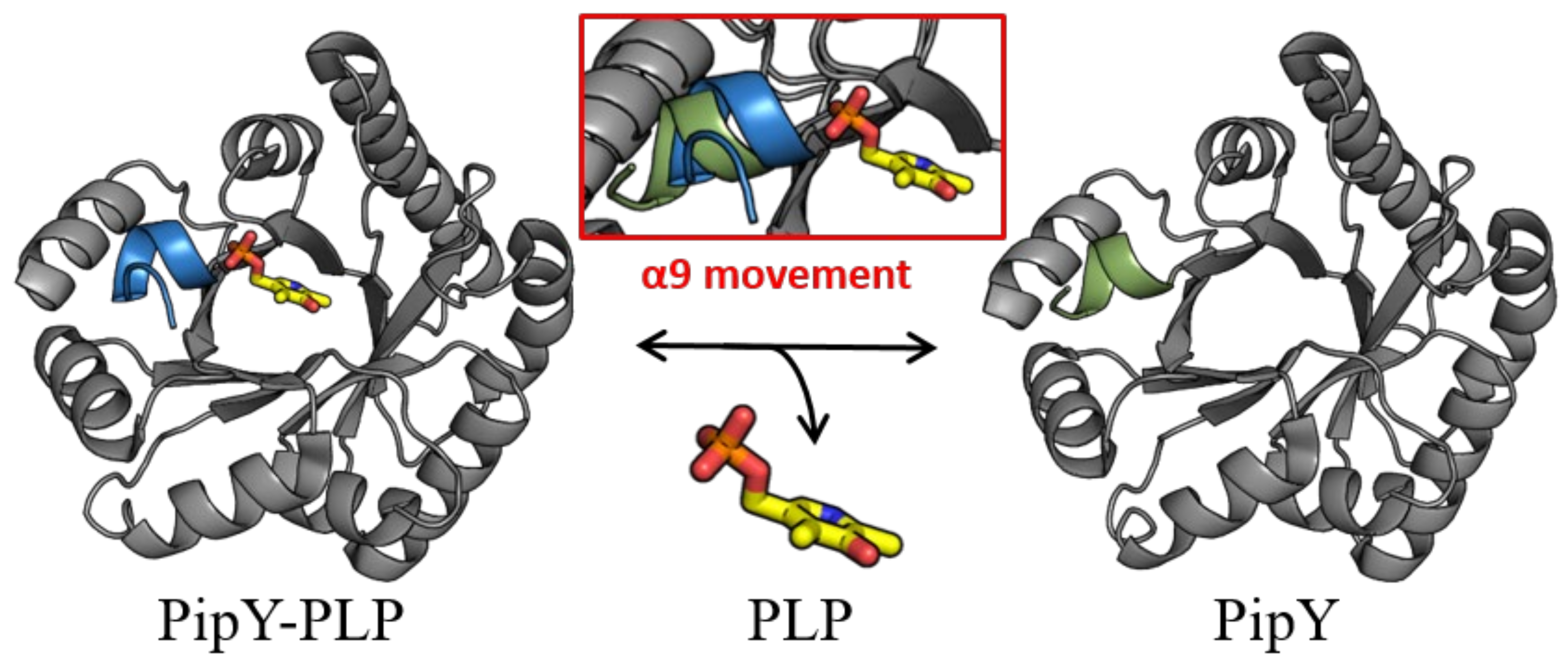
While PLP is found in PLPBP structures in the same location that it is found in the fold type III of PLP enzymes, in all PLPBP structures the PLP cofactor is solvent-exposed and highly accessible, thus being appropriately positioned for a role of PLPBP as a PLP delivering device in cells. In this context, the role of the C-terminal α helix (helix 9) of PipY in anchoring the phosphate of PLP, and the relatively large changes in helix 9 orientation depending on the presence or absence of PLP have led to the hypothesizing [22] that this helix may have a role in being a trigger for the binding and release of PLP (Figure 2).
The available data so far suggest that the proteins of this family might act as PLP carriers which supply the cofactor to PLP-dependent enzymes, shielding this cofactor from unwanted reactions with other molecules, although this has not been strictly proven and the mechanisms involved remain unclarified.

2.2. Dimerization of Just Some PLPBP Family Members?

While all available crystal structures are consistent with PLPBP family members being monomers, size-exclusion chromatography of human PLPHP, performed in two different studies [23,36] revealed a second peak corresponding to dimers. In [23], the minor peak was shown to depend on disulfide bridges, a result interpreted as PLPHP being mainly monomeric with the possibility of stable dimerization via the formation of a disulfide bridge between exposed cysteines. Consistent with this, human mutation Tyr69Cys increased dimer formation (Table 2). However, Fux and Sieber [36] challenged this view, reporting that their PLPHP preparation was predominantly dimeric even under reducing conditions [36]. They also suggested that discrepancies with the previous work may be due to differences in expression strains or purification strategies. It is worth noting that while the human protein contains five cysteine residues, the amino acid chains of S. elongatus PipY and E. coli YggS have just one or two cysteines, respectively, and thus they would be less prone to making disulfide bonds under oxidative conditions.
Although the relative importance of dimeric forms of PLPBP family members in cell systems remains to be determined, it is worth noting that dimer formation could significantly enhance the putative function of PLPBPs in shielding PLP transport or storage, which are the most likely functions attributed to PLPBPs so far. Importantly, the identification of dimeric forms of human PLPHP opens the possibility that at least two different forms of PLPBPs (monomeric and dimeric), perhaps with different regulatory properties, might be found in cells. Whether dimer formation is a property of just some PLPBP members or a universal feature of the family, and whether in vivo dimer formation requires cysteines in critical positions or can also be induced by other effectors or cell components are open questions that require further investigation.

3. PLPBP-Related Phenotypes

3.1. Null Mutations, Heterologous Complementation, and Animal Disease Models Support Universal Functions of PLPBP Family Members

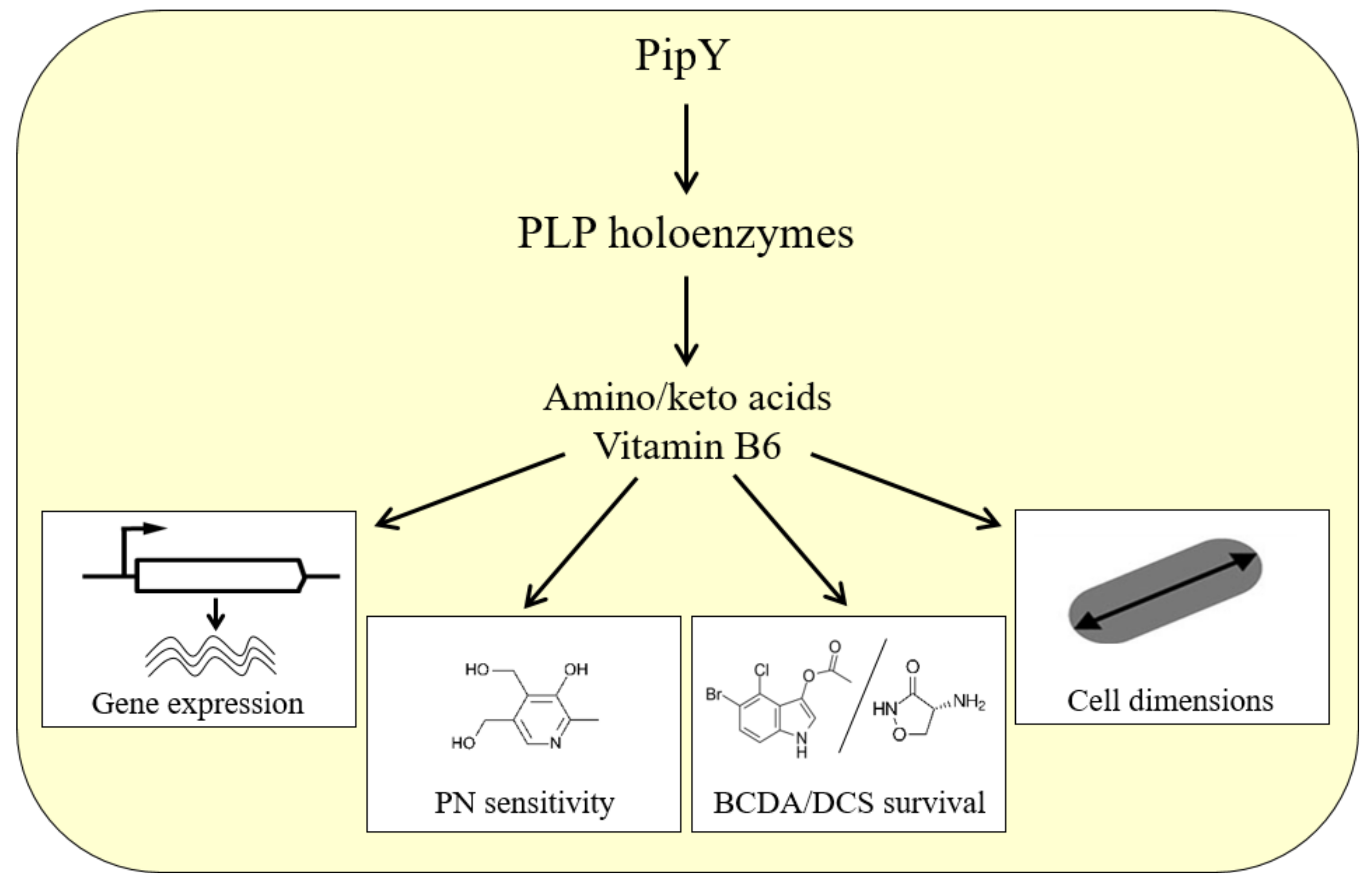
Consistent with a key role of PLPBPs in PLP homeostasis, inactivation of the corresponding genes results in alterations of the relative levels of B6 vitamers and amino/keto acids in all organisms investigated. The null mutants of E. coli, S. elongatus, Salmonella enterica, Acidovorax citrulli, S. cerevisiae, and Danio rerio (zebrafish) showed sensitivity to pyridoxine (PN), an overaccumulation of PNP and/or an imbalance of the amino/keto acid pools ([37] and references within it). As a consequence of these metabolic alterations, in which PLP-dependent enzymes are presumably involved, pleiotropic phenotypes have been associated with PLPBP deficiency, which may alter different cellular processes in different organisms. The phenotypic changes observed (at least) in cyanobacteria are summarized in the general model for PLPBP function illustrated in Figure 3.
As an example, S. elongatus pipY and A. citrulli yggS null mutants both show high sensitivity to the antibiotic β-chloro-D-alanine (BCDA), while S. elongatus pipY mutants are also sensitive to D-cycloserine (DCS) [19,38]. Importantly, PipY overexpression did not increase resistance to either of these antibiotics, as would be expected for a direct antibiotic target. Instead, PLPBPs indirectly confer antibiotic resistance by protecting the essential, high-affinity antibiotic targets specific for their cognate PLP-dependent enzymes. The specific targets in this example are glutamate racemases for BCDA [39] and alanine racemases for both DCS and BCDA [40]. The indirect role of antibiotic resistance also agrees with the reported failure to obtain crystals of PipY with D-ala or DCS [22].
Multiple lines of evidence reinforce the idea that diverse PLPBPs share common functions in cells. Distantly related PLPBP members were able to rescue strain-specific defects, such as Val overproduction and the PN sensitivity phenotypes of E. coli yggS mutants. These two E. coli yggS mutant phenotypes were both rescued by the heterologous expression of orthologs from bacilli, yeast, plants (Zea mays, Arabidopsis thaliana), or humans [31,41,42]. The introduction of human PLPHP into the corresponding null mutants of yeast partially rescued the growth phenotype on poor carbon sources [43]. Last but not least, PLPHP deficiency reproduced the human disease in zebrafish larvae; the knockout mutants showed multiple signs of seizure activity before dying, while pyridoxine treatment improved the epileptic phenotype and prolonged lifespan [43].

3.2. PLPBP Mutations Cause B6-Dependent Epilepsy in Humans

Relatively recently, recessive mutations affecting human PLPBP were reported as causal in patients with vitamin B6-dependent epilepsy [41]. Since then, many others have reported familial cases of vitamin B6-dependent epilepsy associated with missense, nonsense, and splice-site mutations at the PLPBP locus [43,44,45,46,47,48,49,50,51,52,53,54]. Some of the PLPHP mutant proteins have been studied in vitro and found to decrease protein levels or PLP cofactor-binding [22,23,41], and Table 2. Furthermore, the missense mutations found in patients target highly conserved residues (except for residues Pro87 and His275), as shown in Figure 1, and Table 2.
As already indicated, most mutations decreased the binding of the PLP cofactor, protein levels, and/or thermal stability. That is the case for mutations Pro40Leu, Tyr69Cys, Pro87Leu, Arg205Gln, Leu175Pro and Arg241Gln [22,23], Arg41Gln, Val45Asp and Glu67Lys [36] and Ile94Phe, Thr116Ile and Gly224Ala [43]. It is worth noting that although Pro87 is a variable residue, the restriction in chain stereochemistry imposed by the imino acid nature of proline explains the frequently drastic structural effects resulting from proline substitutions. Thus, it is not surprising that despite the lack of conservation, Pro87Leu also results in the loss-of-function of PLPHP. On the other hand, His275Asp targets the non-conserved C-terminal extension of the human protein and it is unlikely to directly affect PLP binding, raising questions on the function of this extension.
In summary, the reported mutations at the PLPBP locus causing vitamin B6-dependent epilepsy are all consistent with the loss-of-function of PLPHP. To our knowledge, no gain-of-function mutations or mutations consistent with increased expression of PLPHP have been reported, raising the question of whether such mutations would result in a pathological consequence or cause a different type of disease pathology.
Table 2. Missense mutations reported in human PLPHP associated with vitamin B6-dependent epilepsy, as well as two experimental mutations in orthologous bacterial proteins. Molecular mechanisms of damage.
Amino Acid Change in HuPLPHPMolecular Mechanism: Effect on PLPBP Protein
Amino acidConserv. Score 1ChangeClinical effectsReported inNumber of patients Observed effectInferred fromRef.
P407P40LSeizures[44]
[51]
1 (P40L/R241Q)
1 (P40L/splicing)
↓ thermostabilityIn vitro studies on rHuPLPHP[23]
R414R41QSeizures
Mild disease 2
[54]
[43]
2 (homozygous;R41Q/V45D)
3 (homozygous)
↓yield/misfolding?
↓thermostability
In vitro studies on rHuPLPHP[36]
R41WSeizures, death[50] 1 (homozygous)NTNTNT
V459V45DSeizures[54] 1 (R41Q/V45D)↓↓PLP content
↓ thermostability
In vitro studies on rHuPLPHP[36]
K479K47ANot reported in humans (prenatally lethal?)lack of PLPrEcyggSK36A[31]
E679E67KSeizures
Severe disease 2
[54]
[43]
1 (homozygous)
3 (homozygous)
MisfoldingIn vitro studies on rHuPLPHP[36]
Y697Y69CSeizures
Moderate disease 2
[44]1 (homozygous)Higher dimerization
↓PLP content
In vitro studies on rHuPLPHP[23]
P871P87LSeizures
Severe disease 2
[41]
[44]
[53]
1(P87L/R241Q)
1 (homozygous)
1 (P87L/splicing)
↓solubility/misfoldingIn vitro studies on rHuPLPHP[23]
I948I94FSeizures
Mild disease 2
[43]1 (homozygous)Proposed ↓ in PLP saturationStructural modeling of HuPLPHP[43]
M1136M113TSeizures[51]1 (M113T/C15X)NTNTNT
T1167T116ISeizures
Severe disease 2
[43]2 (1 homozygous;
1 homozygous (T116I/H275D))
Proposed ↓ in PLP saturationStructural modeling of HuPLPHP[43]
L1756L175PSeizures
Severe disease 2
[41]1 (homozygous)MisfoldingIn vitro studies on rHuPLPHP[23]
R2057R205QSeizures
Moderate disease 2
[44]
[54]
1 (R205Q/null)
1 (homozygous)
↓thermostabilityIn vitro studies on rHuPLPHP[23]
G2249G224ASeizures
Severe disease 2
[43]1 (G224A/splicing)Proposed ↓ in PLP saturationStructural modeling of HuPLPHP[43]
S2269S226ANot reported in humans (prenatally lethal?)↓PLP saturationrFnS201A[24]
R2419R241QSeizures[41]
[44]
[52]
1 (P87L/R241Q)
1 (P40L/R241Q)
1(R241Q/splicing)
↓solubility
↓thermostability
↓PLP binding
In vitro studies on rHuPLPHP
In vitro studies on rSePipYR210Q
[22,23]
I2427I242TSeizures[45]1 (homozygous)NTNTNT
H275NAH275DSeizures[43]1 (homozygous T116I/H275D)Variant of uncertain significance (VUS)
Structural modeling of HuPLPHP[43]
NA: not applicable. NT: not tested. HuPLPHP: Human PLPHP. rHuPLPHP: recombinant human PLPHP. rEcyggSK36A: recombinant E. coli yggS K36A mutant. rSePipYR210Q: recombinant S. elongatus PipY R210Q mutant. rFnS201A: recombinant F. nucleatum yggS S201A mutant. 1 Conservation score as given in Figure 1. 2 Severity score given in [43].

3.3. Phenotypes Associated with PLPBP Excess

There are, to the best of our knowledge, three reports dealing with the phenotypic effects of increasing the levels of proteins in this family. In two of them, bacterial cells were engineered for PLPBP overexpression to address specific questions in the context of neighbor genes (see Section 4.1). However, the third report constitutes a unique example of an adaptative spontaneous mutation in a cereal crop, dealing with a natural variation in wild rice (Oryza rufipogon) conferring high-temperature tolerance at the phenological heading stage [55]. A map-based cloning approach for heat tolerance specifically at the panicle development stage identified qHTH5 as a major quantitative trait locus (QTL) and the target gene (encoding a PLPBP ortholog) was named HTH5. The corresponding change was an up promoter mutation that increased transcript levels from HTH5. Although the exact mechanism of the heat tolerance phenotype was not ascertained, the authors suggested that it could be related to altered reactive oxygen species (ROS) homeostasis, thus expanding the putative functions of PLPBP.
It is worth noting that there are no studies so far dealing with the metabolic consequences of increased cellular levels of PLPBP family proteins, in contrast with the numerous reports describing metabolic changes corresponding to loss-of-function mutations in bacterial systems and human patients.

3.4. Synthetic Lethality between PLPBP Family Members and PLP-Holoenzymes Supports Some Functional Redundancy

Synthetic lethality between PLPBP and PLP-holoenzymes has been reported in E. coli for yggS with both glyA [42,56] and serA [57], in S. enterica for yggS and aspC [58] and S. elongatus for pipY and cysK [19]. Synthetic lethality probably reflects the common involvement of the corresponding protein pairs in amino acid and PLP homeostasis, but also supports the idea that any relatively abundant PLP-dependent protein (in addition to PLPBP family members) could fulfill a role as a PLP reservoir to prevent the possible consequences of an excess of unbound PLP. In line with this, PipY and CysK, the latter encoded by a non-essential gene with three paralogs in S. elongatus, are the two most abundant PLP-binding proteins in this cyanobacterium [19]. It is worth noting that PLPBPs appear to be relatively abundant proteins in many organisms, as is also the case with yeast or HeLa cells, where levels of PLPBPs are almost 10-fold higher compared to the median protein copy number [59].
Functional redundancy between PLPBP family members and PLP-holoenzymes, supported by synthetic lethality in distantly related bacterial groups, suggests that at least some of the multiple PLP-containing proteins expressed in living cells participate in PLP homeostasis. It also explains that, despite the universality of PLP-derived challenges in all types of cells, PLPBP members are neither essential nor ubiquitous in the cellular systems characterized so far [42]. Functional redundancy among PLP-holoenzymes appears to occur even in organisms with relatively small genomes, such as S. elongatus, where out of 41 PLP-binding protein sequences [60], 11 corresponded to non-essential and 6 to beneficial genes (their inactivation slows the growth of cultures) under standard photoautotrophic conditions [3].

4. Guilty by Association Strategies to Get Insights into PLPBP Family Functions

The lack of knowledge of the molecular mechanisms involved in the cellular functions of PLPBPs has probably contributed to the use of guilt by association strategies to establish connections between PLPBP members and specific biological processes. Gene synteny, and more recently protein interaction approaches have provided useful information in this context.

4.1. Genes of the PLPBP Family in Clusters and Operons from Bacteria

Bacterial genomes offer an opportunity to apply the principle of guilt by association to search for characterized proteins that may belong to metabolic or signaling pathways involving PLPBP members. In this context, the distribution of PLPBP genes and physical clustering with genes of known function in bacteria was first investigated and discussed in detail by [42]. In addition to the association with proC (Pyrroline-5-carboxylate reductase, EC 1.5.1.2) [61], which is responsible for the prosC denomination [62,63], PLPBP genes also clustered with cell division and cell wall biosynthesis (dcw, in Gram-positive bacteria and mycobacteria), PLP salvage, surface motility, secretion, amino acid metabolism, and translation genes [42].
In S. elongatus and a majority of the available cyanobacterial genomes, the genes sepF (involved in cell division and restricted to Gram-positive bacteria and cyanobacteria) and proC were found at short distances downstream of pipY, while the arrangements pipY-sepF and sepF-proC were also frequently found in the available cyanobacterial genomes. The precise arrangement of pipY-sepF-proC and the linkage to the phylum-restricted gene pipX are both cyanobacterial hallmarks [19].
Several reports explored a putative role for PLPBP family members in cell division and cell wall functions by describing cell size or cell wall-related phenotypes in null or overexpressing strains. Streptococcus pneumoniae ylmE cells were slightly larger than normal [64], while normal cell dimensions were reported for E. coli yggS [42] and S. elongatus pipY under standard culture conditions. On the other hand, overexpressing YlmE in B. subtilis blocked biofilm formation [65], while the overexpression of PipY increased S. elongatus cell length up to 28% [19]. Taken together, these findings support the notion that, at least in some bacterial groups, perturbations of the amino/keto acid pool may result in the accumulation of metabolic signals affecting cell wall metabolism or cell division. In addition, the possibility of physical interaction between a given PLPBP family protein and cell wall-related proteins cannot be excluded at present.
Very recently, it has been noted that the linkage with pilT, encoding an ATPase mediating pilus retraction and disassembly, is typical of phytopathogenic bacteria, where yggS and pilT are contiguous and divergently transcribed [38]. Importantly, yggS inactivation in Acidovorax citrulli significantly affected type III secretion, reduced swimming motility, and resulted in attenuated virulence. Thus, despite the lack of molecular details, the functional connection established between the yggS and pilT genes of phytopathogenic bacteria also supports the use of synteny approaches to gain additional insights into the role of PLPBP family members in particular biological processes or environmental contexts.

4.2. The Close Relationship between PipX and PipY in Cyanobacteria

Since most of the mRNA transcripts identified in cyanobacteria are monocistronic (approximately 62% in S. elongatus), co-transcription of pipX and pipY is a strong indication of functional association [66,67]. Furthermore, close to 80% of pipX genes are found adjacent to pipY genes, presumably as part of bicistronic pipXY operons. In addition, tight co-regulation and even translational coupling can be inferred by the relatively short or non-existent intergenic distances found between contiguous pipX and pipY coding sequences, strongly suggesting a functional interaction between PipX and PipY in most, if not all, cyanobacteria [19]. When not adjacent, as in Synechocystis strains, pipX, and pipY genes appear to be monocistronic or with no clear linkage to particular genes [25]. Another indication of the tight relationship between pipX and pipY genes in cyanobacteria is the finding that PipX increases the expression of either pipY or a reporter gene occupying the pipY locus in S. elongatus, suggesting the importance of the PipX-PipY balance in cyanobacteria [25].
While all this is strongly suggestive of physical interaction between PipX and PipY proteins in cyanobacteria, no evidence could be obtained for this interaction using yeast two-hybrid assays [19]. However, false negatives are not rare in this genetic system [6,68], and interactions between PipX and PipY proteins may depend on the factor(s) present in S. elongatus but absent from yeast nuclei, or present only under certain physiological or environmental conditions. As noted above, PipX is part of a complex partner-swapping network governed by molecule effectors signaling the ATP/ADP ratio (PII), 2-OG levels (PII, NtcA), or the GTP/GDP ratio (EngA) [10,16,69]. The levels of these metabolites and the relative abundance of the PipX interactants will influence the formation of complexes with different partners, raising the question of whether the putative PipX-PipY complexes would depend on an additional effector, and if the role played by PLP is as a cofactor of PipY, questions that deserve further investigation.

4.3. PLPHP Cellular Interaction Network

Co-immunoprecipitation with PLPHP after the crosslinking of proteins from a human embryonic kidney cell line (HEK293) revealed the presence of PLPHP in both the cytosol and mitochondria [36]. Contrary to the author’s expectations, an overrepresentation of PLP-dependent enzymes was not observed. Instead, a high number of proteins involved in cytoskeleton organization was detected, including components of the γ-tubulin ring complex necessary for microtubule nucleation at the centrosome, proteins involved in centriole and spindle formation, or several subunits of the f-actin capping complex which regulates the growth of actin filaments. The significance of these findings remains to be addressed.

5. Regulation of Gene Expression by PLPBP

To our knowledge, there are only two reports analyzing differentially expressed transcripts in PLPBP protein-deficient mutants. The first one investigated the genetic interactions between PipX and PipY (see below). A more recent study presented results obtained from the yggS mutant of A. citrulli in the context of virulence and other related phenotypic features [38].
Another study investigated the effect of PLPBP inactivation on the proteome of the HEK293 cell line, which resulted in the significant upregulation of proteins associated with cytoskeleton organization and cell division as well as downregulation of two PLP-dependent enzymes involved in H2S synthesis [36].
While these studies are not easily comparable, the relatively high number of differentially expressed transcripts in bacteria and the proteins in the human cell line study emphasize the importance of PLP homeostasis in the global regulation of gene expression in the different cellular systems analyzed.
Role of PipY in the Context of Cyanobacterial Nitrogen Regulation
The apparent recruitment of PipY, via PipX, into the 2-OG-dependent nitrogen interaction network of S. elongatus [10] provides a unique opportunity to investigate the functions of PipY, a paradigm PLPBP member, in the context of a relatively well-characterized signaling network in a cyanobacterial model system.
Transcriptomic analysis with S. elongatus pipX null and gain-of-function mutant derivatives showed that NtcA targets were only a minor fraction of the affected transcripts [70] and are consistent with PipX having a role as a repressor of many photosynthesis and translation-related genes. The model emerging from this analysis is that PipX participates in at least four types of regulatory complexes, including among them the already characterized transcriptional complex NtcA-PipX. Importantly, transcriptomic analysis with single and double pipX and pipY null mutants revealed a PipY-dependent induction of NtcA-activated transcripts as well as the implication of both PipX and PipY in the regulation of NtcA-independent transcripts [19], where they could have similar or opposite effects, suggesting a rather complex regulation. The only other transcriptional regulator known to interact with PipX is PlmA [71], which so far is the best candidate to mediate one of the PipX-regulated transcriptional responses independent of NtcA. However, signals regulating PlmA activity remain to be discovered.
PipY may alter nitrogen signaling in cyanobacteria by affecting the levels of amino/keto acids, including 2-OG, a possibility suggested by the positive role of PipY at NtcA-activated transcripts, which are dependent on 2-OG. On the other hand, the expression of the NtcA-independent transcripts that are co-regulated by PipX-PipY would require a transcriptional regulator that can also respond to amino/keto acid effectors, or perhaps to PLP or related compounds, a possibility worth exploring.

6. Concluding Remarks

The structural and functional information gathered from different systems and heterologous complementation analyses of null mutants emphasize the importance of the conserved role of the PLPBP family in vitamin B6 vitamers and amino/keto acid homeostasis, providing a rationale for the pleiotropic phenotypes and changes in gene expression patterns caused by PLPBP deficiency.
Despite growing interest in the field, recently fueled by the identification of familial mutations of the PLPBP causing vitamin B6-dependent epilepsy in humans, fundamental questions concerning the mechanism of action of PLPBPs remain to be answered and additional lines of investigation are required to advance knowledge. Bacterial systems can provide further insights into the function and conserved structural features of PLPBPs in cases where site-directed mutagenesis and genetic manipulation are rapid and tractable. The metabolic effects of PLPBP overexpression and relevant phenotypic effects of putative gain-of-function mutations should also be addressed. PLPBP members with tight functional connections with known genes also deserve attention. In this context, the tight association of pipY with pipX, encoding a key component of the cyanobacterial nitrogen interaction network, provides a unique opportunity to investigate the roles of the PLPBP family proteins in the context of a signaling pathway conserved in cyanobacteria that would provide valuable lessons on both universal and specific functions of this yet mysterious protein family.

Author Contributions

Conceptualization, A.C., L.T. and V.R.; writing—original draft preparation, A.C.; writing—review and editing L.T., A.L., V.R. and A.C.; funding acquisition, A.C. and V.R. All authors have read and agreed to the published version of the manuscript.

Funding

Work in our laboratories were supported by grants PID220-118816GB-I00 and BFU2015-66360-P from the Spanish Government (MICINN), grants GRE20-04-C, VIGROB20-126, UADIF20-34 and UADIF19-36 from the University of Alicante (to AC) and grant CIVP-20A6610 from the Fundación Ramón Areces (to VR).

Institutional Review Board Statement

Not applicable.

Informed Consent Statement

Not applicable.

Data Availability Statement

Not applicable.

Acknowledgments

We would like to thank R. Dixon, A. Forcada-Nadal, P. Salinas, and R. Cantos for their critical reading of the manuscript.

Conflicts of Interest

The authors declare no conflict of interest.

References

  1. Blank, C.E.; Sánchez-Baracaldo, P. Timing of Morphological and Ecological Innovations in the Cyanobacteria--a Key to Understanding the Rise in Atmospheric Oxygen. Geobiology 2010, 8, 1–23. [Google Scholar] [CrossRef] [PubMed]
  2. Khan, S.; Fu, P. Biotechnological Perspectives on Algae: A Viable Option for Next Generation Biofuels. Curr. Opin. Biotechnol. 2020, 62, 146–152. [Google Scholar] [CrossRef] [PubMed]
  3. Rubin, B.E.; Wetmore, K.M.; Price, M.N.; Diamond, S.; Shultzaberger, R.K.; Lowe, L.C.; Curtin, G.; Arkin, A.P.; Deutschbauer, A.; Golden, S.S. The Essential Gene Set of a Photosynthetic Organism. Proc. Natl. Acad. Sci. USA 2015, 112, E6634–E6643. [Google Scholar] [CrossRef] [PubMed] [Green Version]
  4. Labella, J.I.; Llop, A.; Contreras, A. The Default Cyanobacterial Linked Genome: An Interactive Platform Based on Cyanobacterial Linkage Networks to Assist Functional Genomics. FEBS Lett. 2020, 594, 1661–1674. [Google Scholar] [CrossRef]
  5. Forchhammer, K.; Selim, K.A. Carbon/Nitrogen Homeostasis Control in Cyanobacteria. FEMS Microbiol. Rev. 2020, 44, 33–53. [Google Scholar] [CrossRef]
  6. Burillo, S.; Luque, I.; Fuentes, I.; Contreras, A. Interactions between the Nitrogen Signal Transduction Protein PII and N -Acetyl Glutamate Kinase in Organisms That Perform Oxygenic Photosynthesis. J. Bacteriol. 2004, 186, 3346–3354. [Google Scholar] [CrossRef] [Green Version]
  7. Espinosa, J.; Forchhammer, K.; Burillo, S.; Contreras, A. Interaction Network in Cyanobacterial Nitrogen Regulation: PipX, a Protein That Interacts in a 2-Oxoglutarate Dependent Manner with PII and NtcA. Mol. Microbiol. 2006, 61, 457–469. [Google Scholar] [CrossRef]
  8. Herrero, A.; Muro-Pastor, A.M.; Flores, E. Nitrogen Control in Cyanobacteria. J. Bacteriol. 2001, 183, 411–425. [Google Scholar] [CrossRef] [Green Version]
  9. Llácer, J.L.; Espinosa, J.; Castells, M.A.; Contreras, A.; Forchhammer, K.; Rubio, V. Structural Basis for the Regulation of NtcA-Dependent Transcription by Proteins PipX and PII. Proc. Natl. Acad. Sci. USA 2010, 107, 15397–15402. [Google Scholar] [CrossRef] [Green Version]
  10. Forcada-Nadal, A.; Llácer, J.L.; Contreras, A.; Marco-Marín, C.; Rubio, V. The PII-NAGK-PipX-NtcA Regulatory Axis of Cyanobacteria: A Tale of Changing Partners, Allosteric Effectors and Non-Covalent Interactions. Front. Mol. Biosci. 2018, 5, 91. [Google Scholar] [CrossRef]
  11. Espinosa, J.; Castells, M.A.; Laichoubi, K.B.; Forchhammer, K.; Contreras, A. Effects of Spontaneous Mutations in PipX Functions and Regulatory Complexes on the Cyanobacterium Synechococcus elongatus Strain PCC 7942. Microbiology 2010, 156, 1517–1526. [Google Scholar] [CrossRef] [Green Version]
  12. Espinosa, J.; Forchhammer, K.; Contreras, A. Role of the Synechococcus PCC 7942 Nitrogen Regulator Protein PipX in NtcA-Controlled Processes. Microbiology 2007, 153, 711–718. [Google Scholar] [CrossRef] [Green Version]
  13. Laichoubi, K.B.; Espinosa, J.; Castells, M.A.; Contreras, A. Mutational Analysis of the Cyanobacterial Nitrogen Regulator PipX. PLoS ONE 2012, 7, e35845. [Google Scholar] [CrossRef] [Green Version]
  14. Zeth, K.; Fokinas, O.; Forchhammers, K. Structural Basis and Target-Specific Modulation of ADP Sensing by the Synechococcus elongatus PII Signaling Protein. J. Biol. Chem. 2014, 289, 8960–8972. [Google Scholar] [CrossRef] [Green Version]
  15. Espinosa, J.; Labella, J.I.; Cantos, R.; Contreras, A. Energy Drives the Dynamic Localization of Cyanobacterial Nitrogen Regulators during Diurnal Cycles. Env. Microbiol. 2018, 20, 1240–1252. [Google Scholar] [CrossRef] [Green Version]
  16. Labella, J.I.; Cantos, R.; Salinas, P.; Espinosa, J.; Contreras, A. Distinctive Features of PipX, a Unique Signaling Protein of Cyanobacteria. Life 2020, 10, 79. [Google Scholar] [CrossRef]
  17. Espinosa, J.; Castells, M.A.; Laichoubi, K.B.; Contreras, A. Mutations at PipX Suppress Lethality of PII -Deficient Mutants of Synechococcus elongatus PCC 7942. J. Bacteriol. 2009, 191, 4863–4869. [Google Scholar] [CrossRef] [Green Version]
  18. Laichoubi, K.B.; Beez, S.; Espinosa, J.; Forchhammer, K.; Contreras, A. The Nitrogen Interaction Network in Synechococcus WH5701, a Cyanobacterium with Two PipX and Two PII-like Proteins. Microbiology 2011, 157, 1220–1228. [Google Scholar] [CrossRef] [Green Version]
  19. Labella, J.I.; Cantos, R.; Espinosa, J.; Forcada-Nadal, A.; Rubio, V.; Contreras, A. PipY, a Member of the Conserved COG0325 Family of PLP-Binding Proteins, Expands the Cyanobacterial Nitrogen Regulatory Network. Front. Microbiol. 2017, 8, 1244. [Google Scholar] [CrossRef] [Green Version]
  20. Schneider, G.; Käck, H.; Lindqvist, Y. The Manifold of Vitamin B6 Dependent Enzymes. Structure 2000, 8, R1–R6. [Google Scholar] [CrossRef]
  21. Eswaramoorthy, S.; Gerchman, S.; Graziano, V.; Kycia, H.; Studier, F.W.; Swaminathan, S. Structure of a Yeast Hypothetical Protein Selected by a Structural Genomics Approach. Acta. Cryst. D Biol. Cryst. 2003, 59, 127–135. [Google Scholar] [CrossRef] [PubMed]
  22. Tremiño, L.; Forcada-Nadal, A.; Contreras, A.; Rubio, V. Studies on Cyanobacterial Protein PipY Shed Light on Structure, Potential Functions, and Vitamin B6-Dependent Epilepsy. FEBS Lett. 2017, 591, 3431–3442. [Google Scholar] [CrossRef] [PubMed] [Green Version]
  23. Tremiño, L.; Forcada-Nadal, A.; Rubio, V. Insight into Vitamin B6-Dependent Epilepsy Due to PLPBP (Previously PROSC) Missense Mutations. Hum. Mutat. 2018, 39, 1002–1013. [Google Scholar] [CrossRef] [PubMed]
  24. He, S.; Chen, Y.; Wang, L.; Bai, X.; Bu, T.; Zhang, J.; Lu, M.; Ha, N.-C.; Quan, C.; Nam, K.H.; et al. Structural and Functional Analysis of the Pyridoxal Phosphate Homeostasis Protein YggS from Fusobacterium nucleatum. Molecules 2022, 27, 4781. [Google Scholar] [CrossRef]
  25. Cantos, R.; Labella, J.I.; Espinosa, J.; Contreras, A. The Nitrogen Regulator PipX Acts in Cis to Prevent Operon Polarity. Env. Microbiol. Rep. 2019, 11, 495–507. [Google Scholar] [CrossRef] [Green Version]
  26. Jansonius, J.N. Structure, Evolution and Action of Vitamin B6-Dependent Enzymes. Curr. Opin. Struct. Biol. 1998, 8, 759–769. [Google Scholar] [CrossRef]
  27. Eliot, A.C.; Kirsch, J.F. Pyridoxal Phosphate Enzymes: Mechanistic, Structural, and Evolutionary Considerations. Annu. Rev. Biochem. 2004, 73, 383–415. [Google Scholar] [CrossRef] [Green Version]
  28. Parra, M.; Stahl, S.; Hellmann, H. Vitamin B6 and Its Role in Cell Metabolism and Physiology. Cells 2018, 7, 84. [Google Scholar] [CrossRef] [Green Version]
  29. Palm, D.; Klein, H.W.; Schinzel, R.; Buehner, M.; Helmreich, E.J.M. The Role of Pyridoxal 5’-Phosphate in Glycogen Phosphorylase Catalysis. Biochemistry 1990, 29, 1099–1107. [Google Scholar] [CrossRef]
  30. Goldin, B.R.; Frieden, C. The Effect of Pyridoxal Phosphate Modification on the Catalytic and Regulatory Properties of Bovine Liver Glutamate Dehydrogenase. J. Biol. Chem. 1972, 247, 2139–2144. [Google Scholar] [CrossRef]
  31. Ito, T.; Iimori, J.; Takayama, S.; Moriyama, A.; Yamauchi, A.; Hemmi, H.; Yoshimura, T. Conserved Pyridoxal Protein That Regulates Ile and Val Metabolism. J. Bacteriol. 2013, 195, 5439–5449. [Google Scholar] [CrossRef] [Green Version]
  32. Shaw, J.P.; Petsko, G.A.; Ringe, D. Determination of the Structure of Alanine Racemase from Bacillus Stearothermophilus at 1. 9-Å Resolution, Biochemistry 1997, 36, 1329–1342. [Google Scholar] [CrossRef]
  33. Banner, D.W.; Bloomer, A.C.; Petsko, G.A.; Phillips, D.C.; Wilson, I.A. Atomic Coordinates for Triose Phosphate Isomerase from Chicken Muscle. Biochem. Biophys. Res. Commun. 1976, 72, 146–155. [Google Scholar] [CrossRef]
  34. Wierenga, R.K. The TIM-Barrel Fold: A Versatile Framework for Efficient Enzymes. FEBS Lett. 2001, 492, 193–198. [Google Scholar] [CrossRef] [Green Version]
  35. Espaillat, A.; Carrasco-López, C.; Bernardo-García, N.; Pietrosemoli, N.; Otero, L.H.; Álvarez, L.; de Pedro, M.A.; Pazos, F.; Davis, B.M.; Waldor, M.K.; et al. Structural Basis for the Broad Specificity of a New Family of Amino-Acid Racemases. Acta Cryst. D Biol. Cryst. 2014, 70, 79–90. [Google Scholar] [CrossRef] [Green Version]
  36. Fux, A.; Sieber, S.A. Biochemical and Proteomic Studies of Human Pyridoxal 5′-Phosphate-Binding Protein (Plpbp). ACS Chem. Biol. 2020, 15, 254–261. [Google Scholar] [CrossRef]
  37. Ito, T. Role of the Conserved Pyridoxal 5ʹ-Phosphate-Binding Protein YggS/PLPBP in Vitamin B6 and Amino Acid Homeostasis. Biosci. Biotechnol. Biochem. 2022, 86, 1183–1191. [Google Scholar] [CrossRef]
  38. Wang, Y.; Zhao, Y.; Xia, L.; Chen, L.; Liao, Y.; Chen, B.; Liu, Y.; Gong, W.; Tian, Y.; Hu, B. YggS Encoding Pyridoxal 5′-Phosphate Binding Protein Is Required for Acidovorax citrulli Virulence. Front. Microbiol. 2022, 12, 783862. [Google Scholar] [CrossRef]
  39. Prosser, G.A.; Rodenburg, A.; Khoury, H.; de Chiara, C.; Howell, S.; Snijders, A.P.; de Carvalho, L.P.S. Glutamate Racemase Is the Primary Target of β-Chloro-D-Alanine in Mycobacterium tuberculosis. Antimicrob. Agents Chemother. 2016, 60, 6091–6099. [Google Scholar] [CrossRef] [Green Version]
  40. Prosser, G.A.; de Carvalho, L.P.S. Metabolomics Reveal d-Alanine: D-Alanine Ligase As the Target of d-Cycloserine in Mycobacterium tuberculosis. ACS Med. Chem. Lett. 2013, 4, 1233–1237. [Google Scholar] [CrossRef]
  41. Darin, N.; Reid, E.; Prunetti, L.; Samuelsson, L.; Husain, R.A.; Wilson, M.; el Yacoubi, B.; Footitt, E.; Chong, W.K.; Wilson, L.C.; et al. Mutations in PROSC Disrupt Cellular Pyridoxal Phosphate Homeostasis and Cause Vitamin-B6-Dependent Epilepsy. Am. J. Hum. Genet. 2016, 99, 1325–1337. [Google Scholar] [CrossRef] [PubMed] [Green Version]
  42. Prunetti, L.; el Yacoubi, B.; Schiavon, C.R.; Kirkpatrick, E.; Huang, L.; Bailly, M.; el Badawi-Sidhu, M.; Harrison, K.; Gregory, J.F.; Fiehn, O.; et al. Evidence That COG0325 Proteins Are Involved in PLP Homeostasis. Microbiology 2016, 162, 694–706. [Google Scholar] [CrossRef] [PubMed]
  43. Johnstone, D.L.; Al-Shekaili, H.H.; Tarailo-Graovac, M.; Wolf, N.I.; Ivy, A.S.; Demarest, S.; Roussel, Y.; Ciapaite, J.; van Roermund, C.W.T.; Kernohan, K.D.; et al. PLPHP Deficiency: Clinical, Genetic, Biochemical, and Mechanistic Insights. Brain 2019, 142, 542–559. [Google Scholar] [CrossRef] [PubMed]
  44. Plecko, B.; Zweier, M.; Begemann, A.; Mathis, D.; Schmitt, B.; Striano, P.; Baethmann, M.; Vari, M.S.; Beccaria, F.; Zara, F.; et al. Confirmation of Mutations in PROSC as a Novel Cause of Vitamin B6-Dependent Epilepsy. J. Med. Genet. 2017, 54, 809–814. [Google Scholar] [CrossRef]
  45. Johannsen, J.; Bierhals, T.; Deindl, P.; Hecher, L.; Hermann, K.; Hempel, M.; Kloth, K.; Denecke, J. Excessive Seizure Clusters in an Otherwise Well-Controlled Epilepsy as a Possible Hallmark of Untreated Vitamin B6-Responsive Epilepsy Due to a Homozygous PLPBP Missense Variant. J. Pediatr. Genet. 2019, 08, 222–225. [Google Scholar] [CrossRef]
  46. McLean, H.; Palmquist, R.; Nadauld, L.D.; Malone Jenkins, S.; Bonkowsky, J.; Filloux, F. On the Edge—A Diagnostic Odyssey. Clin. Case. Rep. 2022, 10, e05688. [Google Scholar] [CrossRef]
  47. Kernohan, K.D.; Hartley, T.; Naumenko, S.; Armour, C.M.; Graham, G.E.; Nikkel, S.M.; Lines, M.; Geraghty, M.T.; Richer, J.; Mears, W.; et al. Diagnostic Clarity of Exome Sequencing Following Negative Comprehensive Panel Testing in the Neonatal Intensive Care Unit. Am. J. Med. Genet. A 2018, 176, 1688–1691. [Google Scholar] [CrossRef]
  48. Ahmed, S.; DeBerardinis, R.J.; Ni, M.; Afroze, B. Vitamin B6-Dependent Epilepsy Due to Pyridoxal Phosphate-Binding Protein (PLPBP) Defect—First Case Report from Pakistan and Review of Literature. Ann. Med. Surg 2020, 60, 721–727. [Google Scholar] [CrossRef]
  49. Pal, M.; Lace, B.; Labrie, Y.; Laflamme, N.; Rioux, N.; Setty, S.T.; Dugas, M.A.; Gosselin, L.; Droit, A.; Chrestian, N.; et al. A Founder Mutation in the PLPBP Gene in Families from Saguenay-Lac-St-Jean Region Affected by a Pyridoxine-Dependent Epilepsy. JIMD Rep. 2021, 59, 32–41. [Google Scholar] [CrossRef]
  50. Jensen, K.V.; Frid, M.; Stödberg, T.; Barbaro, M.; Wedell, A.; Christensen, M.; Bak, M.; Ek, J.; Madsen, C.G.; Darin, N.; et al. Diagnostic Pitfalls in Vitamin B6-Dependent Epilepsy Caused by Mutations in the PLPBP Gene. JIMD Rep. 2019, 50, 1–8. [Google Scholar] [CrossRef]
  51. Jiao, X.; Xue, J.; Gong, P.; Wu, Y.; Zhang, Y.; Jiang, Y.; Yang, Z. Clinical and Genetic Features in Pyridoxine-Dependent Epilepsy: A Chinese Cohort Study. Dev. Med. Child. Neurol. 2020, 62, 315–321. [Google Scholar] [CrossRef]
  52. Heath, O.; Pitt, J.; Mandelstam, S.; Kuschel, C.; Vasudevan, A.; Donoghue, S. Early-Onset Vitamin B6-Dependent Epilepsy Due to Pathogenic PLPBP Variants in a Premature Infant: A Case Report and Review of the Literature. JIMD Rep. 2021, 58, 3–11. [Google Scholar] [CrossRef]
  53. Espinoza, A.C.; Wright, M.A.; Candee, M.S.; Trandafir, C.; Nelson, G.R. Child Neurology: Late-Onset Vitamin B6-Dependent Epilepsy Identified by Rapid Genome Sequencing. Neurology 2021, 96, 911–914. [Google Scholar] [CrossRef]
  54. Shiraku, H.; Nakashima, M.; Takeshita, S.; Khoo, C.S.; Haniffa, M.; Ch’ng, G.S.; Takada, K.; Nakajima, K.; Ohta, M.; Okanishi, T.; et al. PLPBP Mutations Cause Variable Phenotypes of Developmental and Epileptic Encephalopathy. Epilepsia Open 2018, 3, 495–502. [Google Scholar] [CrossRef] [Green Version]
  55. Cao, Z.; Tang, H.; Cai, Y.; Zeng, B.; Zhao, J.; Tang, X.; Lu, M.; Wang, H.; Zhu, X.; Wu, X.; et al. Natural Variation of HTH5 from Wild Rice, Oryza rufipogon Griff., Is Involved in Conferring High-Temperature Tolerance at the Heading Stage. Plant Biotechnol. J. 2022, 20, 1591–1605. [Google Scholar] [CrossRef]
  56. Nichols, R.J.; Sen, S.; Choo, Y.J.; Beltrao, P.; Zietek, M.; Chaba, R.; Lee, S.; Kazmierczak, K.M.; Lee, K.J.; Wong, A.; et al. Phenotypic Landscape of a Bacterial Cell. Cell 2011, 144, 143–156. [Google Scholar] [CrossRef] [Green Version]
  57. Ito, T.; Hori, R.; Hemmi, H.; Downs, D.M.; Yoshimura, T. Inhibition of Glycine Cleavage System by Pyridoxine 5′-Phosphate Causes Synthetic Lethality in GlyA YggS and SerA YggS in Escherichia coli. Mol. Microbiol. 2020, 113, 270–284. [Google Scholar] [CrossRef]
  58. Vu, H.N.; Downs, D.M. Loss of YggS (COG0325) Impacts Aspartate Metabolism in Salmonella enterica. Mol. Microbiol. 2021, 116, 1232–1240. [Google Scholar] [CrossRef]
  59. Kulak, N.A.; Pichler, G.; Paron, I.; Nagaraj, N.; Mann, M. Minimal, Encapsulated Proteomic-Sample Processing Applied to Copy-Number Estimation in Eukaryotic Cells. Nat. Methods 2014, 11, 319–324. [Google Scholar] [CrossRef]
  60. Percudani, R.; Peracchi, A. A Genomic Overview of Pyridoxal-Phosphate-Dependent Enzymes. EMBO Rep. 2003, 4, 850–854. [Google Scholar] [CrossRef]
  61. Savioz, A.; Jeenes, D.J.; Kocher, H.P.; Haas, D. Comparison of ProC and Other Housekeeping Genes of Pseudomonas aeruginosa with Their Counterparts in Escherichia coli. Gene 1990, 86, 107–111. [Google Scholar] [CrossRef]
  62. De Wergifosse, P.; Jacques, B.; Jonniaux, J.-L.; Purnelle, B.; Goffeau, A.; Skala, J., II. Yeast Sequencing Reports. The Sequence of a 22·4 Kb DNA Fragment from the Left Arm of Yeast Chromosome II Reveals Homologues to Bacterial Proline Synthetase and Murine α-Adaptin, as well as a New Permease and a DNA-Binding Protein. Yeast 1994, 10, 1489–1496. [Google Scholar] [CrossRef] [PubMed]
  63. Ikegawa, S.; Isomura, M.; Koshizuka, Y.; Nakamura, Y. Cloning and Characterization of Human and Mouse PROSC (Proline Synthetase Co-Transcribed) Genes. J. Hum. Genet. 1999, 44, 337–342. [Google Scholar] [CrossRef] [PubMed] [Green Version]
  64. Fadda, D.; Pischedda, C.; Caldara, F.; Whalen, M.B.; Anderluzzi, D.; Domenici, E.; Massidda, O. Characterization of DivIVA and Other Genes Located in the Chromosomal Region Downstream of the Dcw Cluster in Streptococcus pneumoniae. J. Bacteriol. 2003, 185, 6209–6214. [Google Scholar] [CrossRef] [PubMed] [Green Version]
  65. Kolodkin-Gal, I.; Romero, D.; Cao, S.; Clardy, J.; Kolter, R.; Losick, R. D-Amino Acids Trigger Biofilm Disassembly. Science 2010, 328, 627–629. [Google Scholar] [CrossRef] [PubMed] [Green Version]
  66. Vijayan, V.; Jain, I.H.; O’Shea, E.K. A High Resolution Map of a Cyanobacterial Transcriptome. Genome. Biol. 2011, 12, R47. [Google Scholar] [CrossRef] [Green Version]
  67. Memon, D.; Singh, A.K.; Pakrasi, H.B.; Wangikar, P.P. A Global Analysis of Adaptive Evolution of Operons in Cyanobacteria. Antonie Van Leeuwenhoek 2013, 103, 331–346. [Google Scholar] [CrossRef]
  68. Salinas, P.; Contreras, A. Identification and Analysis of Escherichia coli Proteins That Interact with the Histidine Kinase NtrB in a Yeast Two-Hybrid System. Mol. Genet. Genom. 2003, 269, 574–581. [Google Scholar] [CrossRef]
  69. Jerez, C.; Salinas, P.; Llop, A.; Cantos, R.; Espinosa, J.; Labella, J.I.; Contreras, A. Regulatory Connections Between the Cyanobacterial Factor PipX and the Ribosome Assembly GTPase EngA. Front. Microbiol. 2021, 12, 781760. [Google Scholar] [CrossRef]
  70. Espinosa, J.; Rodriguez-Mateos, F.; Salinas, P.; Lanza, V.F.; Dixon, R.; de la Cruz, F.; Contreras, A. PipX, the Coactivator of NtcA, Is a Global Regulator in Cyanobacteria. Proc. Natl. Acad. Sci. USA 2014, 111, E2423–E2430. [Google Scholar] [CrossRef]
  71. Labella, J.I.; Obrebska, A.; Espinosa, J.; Salinas, P.; Forcada-Nadal, A.; Tremiño, L.; Rubio, V.; Contreras, A. Expanding the Cyanobacterial Nitrogen Regulatory Network: The GntR-Like Regulator PlmA Interacts with the PII-PipX Complex. Front. Microbiol. 2016, 7, 1677. [Google Scholar] [CrossRef]
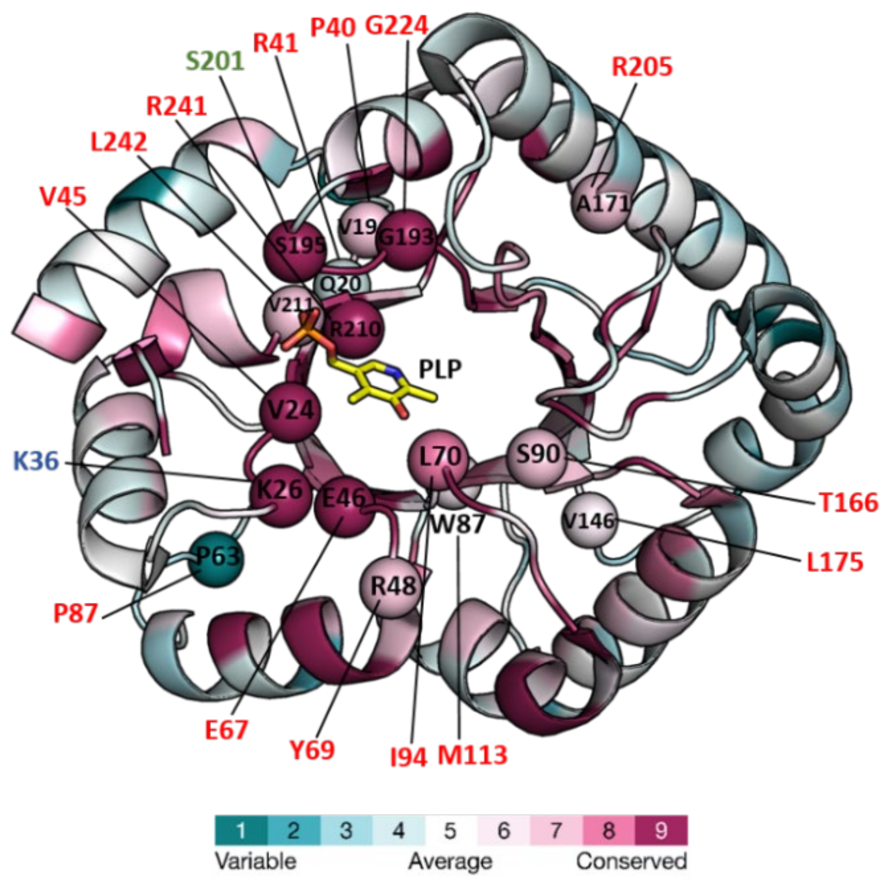
Figure 1. Structure of PipY from S. elongatus (PDB 5NM8) colored for the evolutionary conservation of residues among PLPBP homologs, and mapping therein, residues targeted by missense mutations. The structure is in a cartoon representation except for the PLP, which is in a stick representation with C, O, N, and P atoms in yellow, red, blue, and orange, respectively. Color-coding of the structure from cyan to magenta according to the residue conservation score (the higher, the more conserved) given by The ConSurf Server “URL https://consurf.tau.ac.il/consurf_index.php (accesed on 3 August 2022) when queried with chain A of the PDB 5NM8, with default parameters. Spheres mark the location in PipY of known human PLPHP mutations (see Table 2). Residue numbers are given in one letter code, in black for S. elongatus, and shown in red, green, and blue, the human mutations causing vitamin B6-dependent epilepsy, and the in vitro mutations obtained in the corresponding proteins of F. nucleatum, and E. coli, respectively.
Life 12 01622 g001
Figure 2. PipY structures with and without PLP illustrate the two positions of helix 9. Cartoon representation of PipY structure from S. elongatus complexed with PLP (PDB 5NM8) and PipY-Apo form (PBD 5NLC), with α-helix 9 shown in blue and green, respectively. Inset: Displacement of α-helix 9 observed in the PLP-containing form is highlighted by superimposing both protein forms. The PLP molecule is illustrated using stick representation, where C, O, N, and P atoms are colored yellow, red, blue, and orange, respectively.
Life 12 01622 g002
Figure 3. Roles of PipY and affected processes in cyanobacteria. PipY would function as a PLP storage and delivery module, and influence the activity of PLP-holoenzymes, which in turn affect the amino/keto acid pool and the processes responsive to (some of) these metabolites. Phenotypes dependent on PipY include the control of gene expression, PN sensitivity, survival in the presence of antibiotics targeting PLP-holoenzymes, and altered cell dimensions. Abbreviations: PN, pyridoxine; DCS, D-cycloserine.
Life 12 01622 g003
Table 1. Structures of COG0325/PLPB family proteins were determined and deposited in the Protein DataBank (PDB).
OrganismProteinPDB FileVitamerLigandsAmino Acid ChangesResolut.
(Å)
Deposition YearRef.
Escherichia coliYggS1W8GPLPIsocitrateNone2.002004
3SY1PLPMES
Acetate
L32V/G56S/N58H/H81N/
I83A/H102I/M165S/S202A/
M205Q/R221A hexamutant
1.472011
7UBQPNP *NoneNone2.602022
7UB4PLPNoneK36A/K38A/K233A/K2342.402022
7UAXNonePO4H3K36A/K38A2.072022
7U9HNoneSO4H2None2.002022
7UBPPLPSO4H2K36A/K137A2.302022
7UB8PLPButanediolK38A/K137A/K233A/K234A2.302022
7UAUPLPSO4H2K137A2.102022
7UATPLPPO4H3K36A2.002022
7U9CPLPPO4H3None2.102022
Bifidobacterium
adolescentis
YggS3CPGPLPAcetateSe-Met **1.712008
Agrobacterium tumefaciensYggS3R79PLPAcetate
Pr+3
Se-Met **1.902011
Synechococcus elongatusPipY5NLCNonePO4H3None1.902017[22]
5NM8PLPCa2+None1.932017[22]
Fusobacterium nucleatumYggS7F8ENoneSO4H2Se-Met **2.082021
6KZWNonePO4H3T5A/N202S, Se-Met **2.082019
7YGFStructure not released2.082022[24]
Saccharomyces cerevisiaeYBL036C1CT5PLPNoneSe-Met **2.001999[21]
1B54PLPNoneNone2.101999[21]
* Pyridoxine-5′-phosphate. ** Methionine replaced by selenomethionine—data not available.
Publisher’s Note: MDPI stays neutral with regard to jurisdictional claims in published maps and institutional affiliations.